STEM CELL DERIVED FACTORS FOR TREATING PATHOLOGIC CONDITIONS
US20110003743A1
2011-01-06
12/726,468
2010-03-18
Abstract:
A purified paracrine factor of a mesenchymal stem cell, such as a Secreted frizzled related protein (Sfrp) is useful to reduce cell death an/or tissue injury associated with ischemic condtions.
Interested in similar patents?
Get notified when new applications in this technology area are published.
Classification:
C12N5/10 » CPC further
Undifferentiated human, animal or plant cells, e.g. cell lines; Tissues; Cultivation or maintenance thereof; Culture media therefor Cells modified by introduction of foreign genetic material
C07K14/435 » CPC further
Peptides having more than 20 amino acids; Gastrins; Somatostatins; Melanotropins; Derivatives thereof from animals; from humans
A61K38/1709 » CPC main
Medicinal preparations containing peptides; Peptides having more than 20 amino acids; Gastrins; Somatostatins; Melanotropins; Derivatives thereof from animals; from humans from vertebrates from mammals
A61K9/0019 » CPC further
Medicinal preparations characterised by special physical form; Galenical forms characterised by the site of application Injectable compositions; Intramuscular, intravenous, arterial, subcutaneous administration; Compositions to be administered through the skin in an invasive manner
A61K9/0053 » CPC further
Medicinal preparations characterised by special physical form; Galenical forms characterised by the site of application Mouth and digestive tract, i.e. intraoral and peroral administration
A61K9/0085 » CPC further
Medicinal preparations characterised by special physical form; Galenical forms characterised by the site of application Brain, e.g. brain implants; Spinal cord
A61K38/17 » CPC further
Medicinal preparations containing peptides; Peptides having more than 20 amino acids; Gastrins; Somatostatins; Melanotropins; Derivatives thereof from animals; from humans
A61K45/06 » CPC further
Medicinal preparations containing active ingredients not provided for in groups - Mixtures of active ingredients without chemical characterisation, e.g. antiphlogistics and cardiaca
A61P9/00 » CPC further
Drugs for disorders of the cardiovascular system
C07K14/475 » CPC further
Peptides having more than 20 amino acids; Gastrins; Somatostatins; Melanotropins; Derivatives thereof from animals; from humans Growth factors; Growth regulators
C12N15/09 » CPC further
Mutation or genetic engineering; DNA or RNA concerning genetic engineering, vectors, e.g. plasmids, or their isolation, preparation or purification; Use of hosts therefor Recombinant DNA-technology
C12N15/63 » CPC further
Mutation or genetic engineering; DNA or RNA concerning genetic engineering, vectors, e.g. plasmids, or their isolation, preparation or purification; Use of hosts therefor; Recombinant DNA-technology Introduction of foreign genetic material using vectors; Vectors; Use of hosts therefor; Regulation of expression
A61K38/18 » CPC further
Medicinal preparations containing peptides; Peptides having more than 20 amino acids; Gastrins; Somatostatins; Melanotropins; Derivatives thereof from animals; from humans Growth factors; Growth regulators
A61P9/10 » CPC further
Drugs for disorders of the cardiovascular system for treating ischaemic or atherosclerotic diseases, e.g. antianginal drugs, coronary vasodilators, drugs for myocardial infarction, retinopathy, cerebrovascula insufficiency, renal arteriosclerosis
Description
RELATED APPLICATIONS
This application is a divisional application of U.S. Ser. No. 11/508,010, filed Aug. 21, 2006, now U.S. Pat. No. 7,638,128, issued Dec. 29, 2009, which claims priority to U.S. Ser. No. 60/710,028, filed Aug. 19, 2005, abandoned, and U.S. Ser. No. 60/711,287, filed Aug. 25, 2005, now abandoned, which are incorporated herein by reference in their entireties.
STATEMENT AS TO FEDERALLY SPONSORED RESEARCH
This invention was made with U.S. government support under National Institutes of Health grant number HL073219. The government has certain rights in the invention.
FIELD OF THE INVENTION
The invention relates to cardiac disorders.
BACKGROUND OF THE INVENTION
Patient mortality and morbidity is increased by cell/tissue damage or death resulting from acute and chronic injury or disease of the heart muscle, such as myocardial infarction, cardiac failure, stroke, degenerative neurological disease, spinal injury, musculoskeletal diseases, hypertension, and diabetes.
SUMMARY OF THE INVENTION
The invention is based upon the surprising discovery that paracrine factors secreted from mesenchymal stem cells (MSC) confer a therapeutic benefit to bodily tissues. Thus, stem cells serve as a factory of biologic products that are purified and administered to subjects.
The paracrine factors are useful in cellular and tissue protection, repair, and regeneration. Mesenchymal stem cells or progenitor cells that secrete cytoprotective paracrine factors preferably comprise an Akt gene (Akt-MSC). One or more secreted compounds (e.g., and isolated compound or a mixture of secreted compounds such as a MSC culture supernatant) confers a clinical benefit to a variety of injured, compromised, or disease tissues.
A method of reducing cell death or enhancing tissue repair is carried out by contacting an injured or diseased tissue with a composition comprising a paracrine factor of a mesenchymal stem cell (MSC). The composition is administered to healthy tissue that is determined to be at high risk of injury or to injured tissue following the occurrence of an injury. Preferably, the factor is a Secreted frizzled related protein (Sfrp). Optionally, the composition contains one or more paracrine factors, e.g., two, three, five, ten or more factors. The factors provide cell reparative benefits in a synergistic manner. For example, the composition contains one or more Sfrp, e.g., Sfrp-1, Sfrp-2, and Sfrp-3. In one embodiment, Sfrp-1 comprises an amino acid sequence of SEQ ID NO:5, a mature processed form of SEQ ID NO:5, or a fragment thereof; in another embodiment, Sfrp-2 comprises an amino acid sequence of SEQ ID NO:7, a mature processed form of SEQ ID NO:7, or a fragment thereof; and in yet another embodiment, Sfrp-3 comprises an amino acid sequence of SEQ ID NO:9, a mature processed form of SEQ ID NO:9, or a fragment thereof The amount of apoptotic cell death is reduced in the presence of a paracrine factor such as an Sfrp compared to in its absence.
Cytoprotective and cell reparative effects are conferred to many types of bodily tissues such as cardiac tissue. For example, in the case of a myocardial infarction, cardiac infarct size is reduced following contact of myocardial tissue with the paracrine factor.
Factors derived from Akt-MSCs, which have been genetically altered to contain a recombinant Akt gene sequence, confer a therapeutic benefit at each stage of a hypoxic cardiac event (early, middle, and late stage). Early on, factors confer a cell protective effect, followed by inotropy, angiogenesis, and cardiac remodeling.
The invention also features methods of inhibiting cell damage, inducing or enhancing cell repair or regeneration or inhibiting an ischemic or reperfusion related injury in a subject. Cell damage or injury is inhibited by administering to the subject or contacting a cell with a composition containing a purified cytoprotective compound such as a substantially pure polypeptide, or a mixture of substantially pure polypeptides such as the Sfrp proteins described above. Other purified proteins, e.g., h1, h5, h8, h12, and h13 are also useful to prevent or reduce cell damage. Accordingly, a method of reducing cell death is carried out by contacting an injured or diseased tissue with a composition comprising a purified paracrine factor of a mesenchymal stem cell selected from the group consisting of h1, h5, h8, h12 and h13 or fragment thereof For example, h12 comprises a fragment of SEQ ID NO:17.
Similarly, cell repair or regeneration is induced by administering to the subject or contacting a cell with a composition containing a purified cytoprotective compound. Polypeptides or other compounds described herein are said to be “substantially pure” when they are within preparations that are at least 60% by weight (dry weight) the compound of interest. Preferably, the preparation is at least 75%, more preferably at least 90%, and most preferably at least 99%, by weight the compound of interest. Purity is measured by any appropriate standard method, for example, by column chromatography, polyacrylaminde gel electrophoresis, or HPLC analysis. The polypeptide is purified from MSC culture media or recombinantly produced.
Cell or tissue damage is defined by a loss or diminution of cell function. Such loss or decrease in function leads to eventual cell death. The cell is a cardiac cell such as a cardiomyocyte, a kidney cell, a liver cell, a neurological (e.g., brain, spinal cord) cell, or a pancreatic cell. For example, a loss of cardiomyocyte function results in the loss of the contractile function of the cell. Cardiomyocytes that have lost their ability to contract form round cells rather that rod shaped cells when cultured. Ischemia causes irreversible cellular/tissue damage and cell death. Reperfusion exacerbates ischemic damage by activating inflammatory response and oxidative stress. Oxidative stress modifies membrane lipids, proteins and nucleic acids resulting in cellular/tissue damage or death, and depression of cardiac, endothelial and kidney function.
Also included in the invention are methods of regenerating an injured myocardial tissue by administered to the tissue a composition containing a cytoprotective compound. The cardiac muscle has been damaged by disease, such as a myocardial infarction. By regenerating an injured myocardial tissue is meant restoring ventricular function and/or decreasing infarct size. Ventricular function is measured by methods known in the art such as radionuclide angiography.
A cytoprotective compound is a compound, which is capable of inhibiting cell damage such as oxidative-stress induced cell death or apoptosis. In addition to Sfrps, cytoprotective compounds include for example adipsin, adrenomedullin, chemokine (C—C motif) ligand 2, cysteine rich protein 61, lysyl oxidase-like 2, or serine proteinase inhibitor.
The composition is administered to the subject prior to, at the time of, or shortly after (1, 5, 10, 15, 30, 60 minutes; 1.5, 2, 4, 6, 12, 18, 24, 48 hours) identification of cell damage or identification of a symptom of ischemia or reperfusion injury. For example the composition is administered to a subject prior to a cardiac event or ischemic-reperfusion injury. Such a subject is a risk candidate for an ischemic event or condition. Symptoms of a cardiac event include for example, chest pain, arm pain, fatigue and shortness of breath. For example, the composition is administered at the onset of symptoms, e.g., chest pain, associated with a cardiac event such as a myocardial infarction. The composition is administered systemically or locally. For example, the composition is administered directly, i.e., by myocardial injection to the cardiac tissue, or systemically, e.g., interperitoneally, orally, intravenously. In another example, administration of the composition is carried out by infusion into a coronary artery. Slow-release formulations, e.g., a dermal patch, in which diffusion of the composition from an excipient such as a polymeric carrier mediates drug delivery are also within the invention. Optionally, the subject is further administered VEGF or thyrosin beta 4.
The composition is administered at a dose sufficient to inhibit apoptotic death or oxidative stress-induced cell death. To determine whether the composition inhibits oxidative-stress induced cell death, the composition is tested by incubating the composition with a primary or immortalized cell such as a cardiomyocyte. A state of oxidative stress of the cells is induced (e.g., by incubating cells with H2O2), and cell viability is measured using standard methods. As a control, the cells are incubated in the absence of the composition and then a state of oxidative stress is induced. A decrease in cell death (or an increase in the number of viable cells) in the compound treated sample indicates that the composition inhibits oxidative-stress induced cell death. Alternatively, an increase in cell death (or an decrease in the number of viable cells) in the compound treated sample indicates that the composition does not inhibit oxidative-stress induced cell death. The test is repeated using different doses of the composition to determine the dose range in which the composition functions to inhibit oxidative-stress induced cell death.
A subject to be treated is suffering from or at risk of developing a condition characterized by aberrant cell damage such as oxidative-stress induced cell death (e.g., apoptotic cell death) or an ischemic or reperfusion related injury. A subject suffering from or at risk of developing such a condition is identified by the detection of a known risk factor, e.g., gender, age, high blood pressure, obesity, diabetes, prior history of smoking, stress, genetic or familial predisposition, attributed to the particular disorder, or previous cardiac event such as myocardial infarction or stroke.
Conditions characterized by aberrant cell damage or death include cardiac disorders (acute or chronic) such as stroke, myocardial infarction, chronic coronary ischemia, arteriosclerosis, congestive heart failure, dilated cardiomyopathy, restenosis, coronary artery disease, heart failure, arrhythmia, angina, atherosclerosis, hypertension, renal failure, kidney ischemia, ischemic hepatitis, hepatic vein thrombosis, cirrhosis, portal vein thrombosis, pancreatitis, ischemic colitis, or myocardial hypertrophy. Cardiac repair or regeneration is evaluated by detecting an improvement of symptoms such as chest pain or shortness of breath as well as by evaluation of heart function by standard methods such as cardiac magnetic resonance, echocardiography, and/or ventricular angiography.
Also within the invention is a cell culture or preservation media containing purified Sfrp2 and a method of maintaining inhibiting stem cell differentiation, e.g., inhibiting myogenesis, by contacting a population of isolated stem cells with purified Sfrp2. Isolated stem cells are selected from the group consisting of embryonic stem cells, mesenchymal stem cells, and hematopoetic stem cells. Stem cells are isolated from the tissue of origin by fractionation by cell surface markers or other distinguishing characteristics. Preferably, a population of isolated cells is at least 85% stem cells. More preferably, the population is 90, 95, 98, 99, 100% stem cells.
This factor is involved in the maintenance and self renewal of tissue specific and embryonic stem cells. For example, differentiation of stem cells, e.g., embryonic stem cells, is inhibited by Sfrp2. Myogenesis is inhibited by contacting stem cells with Sfrp2. In another example, bone marrow-derived hematopoetic stem are maintained in a stem cell state by contacting the cells with purified Sfrp2. Preservation of stem cells in this manner is useful in transport and storage of stem cells prior to transplantation into a subject for therapeutic purposes.
Unless otherwise defined, all technical and scientific terms used herein have the same meaning as commonly understood by one of ordinary skill in the art to which this invention belongs. Although methods and materials similar or equivalent to those described herein can be used in the practice or testing of the present invention, suitable methods and materials are described below. All publications, patent applications, patents, and other references mentioned herein are incorporated by reference in their entirety. In the case of conflict, the present specification, including definitions, will control. In addition, the materials, methods, and examples are illustrative only and not intended to be limiting.
Other features and advantages of the invention will be apparent from the following detailed description and claims.
BRIEF DESCRIPTION OF THE DRAWINGS
FIGS. 1A and 1B are bar graphs showing that Sfrps are expressed in mesenchymal stem cells. FIG. 1A shows levels of Sfrp1, Sfrp2 and Sfrp3 expression as estimated by microarray analysis and shows a nearly 10 fold upregulation of Sfrp2 in Akt-MSC compared to GFP-MSC. FIG. 1B shows a quantitative real-time RT-PCR validation of mRNA to expression levels that demonstrates a 100 fold upregulation of Sfrp2 gene expression in Akt-MSC compared to GFP-MSC.
FIG. 2A is a photograph of results of a Western Blotting assay for Sfrp2. The data demonstrates presence of Sfrp2 protein in conditioned medium collected from AKT or GFP MSCs and inhibition of its accumulation in the medium in the presence of Pi3K inhibitor
FIG. 2B is a bar graph showing relative reduction in mRNA levels of Sfrp2 in Akt-MSC following knockdown of Sfrp2 with siRNA.
FIG. 2C is a bar graph showing the effect of conditioned medium on apoptosis in ARVCs. Caspase activity of ARVCs after 24 hours of hypoxia under different culture conditions (control conditioned medium, Ctr CM; Akt conditioned medium, Akt CM; Akt conditioned medium following Sfrp2 knockdown, Akt CM minus Sfrp2) demonstrates reduction of caspase activity following Akt-CM treatment and attenuation of this effect following treatment with Akt CM minus Sfrp2. These data demonstrate that paracrine factors from Akt-MSCs mediate the survival signaling on cardiomycytes.
FIG. 3A is a bar graph showing the effect of Sfrp2 on caspase activity. Cleaved-caspase 3 activity as measured by a fluorometric assay demonstrated decreased caspase activity in hypoxic cardiomyocytes following Sfrp2 treatment in a dose dependent manner. The activity was calculated as fold changes with the same control.
FIG. 3B is a bar graph showing the number of round shaped cardiomyocytes that were counted in 6 random high power fields (40×) following 24 hour hypoxic exposure with/without Sfrp2 treatment. Data is expressed as a percentage of total number of cells present.
FIG. 3C is a series of representative high power field photographs demonstrating decreased number of round shaped cardiomyocytes following treatment with Sfrp2. Collectively, these data demonstrate that Sfrp2 decreases caspase 3 activity
FIG. 4 is a bar graph showing that Sfrp2 decreased cardiac infarct size. Above each bar, is a photograph of TIC staining showing bi-ventricular sections of similar thickness perpendicular to the long axis of the heart. The staining data deomonstrates decreased infarct size with Akt-CM and Sfrp2 and attenuation of reduction in infarct size with Akt-Sfrp2. Infarct size is expressed as a percentage of the total ventricular area. Rat hearts were treated with PBS as control, Akt-MSCs CM (Akt), CM form Akt-MSCs that did express reduced levels of Sfrp2 due to siRNA treatment (Akt-Sfrp2).
FIG. 5A is a photograph of an electrophoretic gel showing that Wnt3a mRNA expression as detected by RT-PCR is increased in hypoxic cardiomyocytes while expression of Wnt5 remains unchanged. The data indicate that hypoxic cardiomyocytes upregulate Wnt3a expression and that Sfrp2 blocks pro-apototic effects of Wnt3a.
FIG. 5B is a bar graph showing that Wnt3a (3 nM) increases caspase activity of cardiomyocytes undergoing hypoxia/reoxygenation injury; Sfrp2 at a similar concentration significantly attenuates Wnt3a induced caspase activity (* vs. normoxia, p<0.05; ** vs. wnt+hypoxia/reoxygenation, p<0.05, n=6/group).
FIG. 6A is a bar graph showing genes upregulated by Sfrp2 under hypoxia. Microarray analysis demonstrates Sfrp2 mediated upregulation of Birc1b gene expression in hypoxic cardiomyocytes.
FIG. 6B is a photograph of an electrophoretic gel showing the effect of Sfrp2 on mRNA levels on Birc1b. RT-PCR confirmed increased Birc1b expression in hypoxic cardiomyocytes following Sfrp2 treatment.
FIG. 6C is a photograph of results of a Western Blot showing that beta-catenin levels are increased by Sfrp2. Western blotting for nuclear and total f3catenin expression in ARVCs demonstrates a reduction of βcatenin following hypoxia and upregulation following treatment with Sfrp2.
FIG. 7 is a bar graph showing the effect of cytoprotective factor h12 compared to IGF-1 on myocyte apoptosis.
FIG. 8 is a line graph showing caspase inhibition in cardiomyocytes by h12.
FIG. 9 is a series of photographs electrophoretic gels showing that h12 phosphorylates/activates AKT in cardiomyocytes
FIG. 10 is a photograph showing inhibition of cytochrome C release by h12.
FIG. 11 is a photograph of an electrophoretic gel showing mitochondrial Bcl-2 protein stabilization by h12.
DETAILED DESCRIPTION
The present invention is based upon the unexpected discovery of that MSC-secreted products confer a therapeutic benefit to injured or compromised tissues. Disclosed herein is a Akt-MSC mediated paracrine mechanism of organ protection and repair. More particularly, the invention provides purified polypeptides such as Srfps isolated from Akt-MSCs or recombinantly or synthetically produced and methods of using these polypeptides to prevent or reduce myocardial damage and ventricular dysfunction.
Akt genes
Akt-MSCs are produced by introducing (e.g., by retrovirus-mediated transduction) into mesenchymal stem cells isolated from the bone marrow an Akt coding sequence or fragment, e.g., Akt-1, Ak-2 or Akt-3. The Akt nucleic acid is human, mouse, or rat.
Exemplary human Akt-1 polypeptides include GenBank Accession numbers NP—005154 and AAH00479. Exemplary human Akt-2 polypeptides includes for example GenBank Accession numbers P31751 and NP—001617. Exemplary human Akt-3 polypeptides includes for example GenBank Accession numbers Q9Y243 and NP—005456. Exemplary nucleic acids encoding Akt include human Akt-1 available at GENBANK™ Accession No. NM—005163 (SEQ ID NO:1), human Akt-2 available at GENBANK™ Accession No. NM—001626 (SEQ ID NO:2) and human Akt-3 available at GENBANK™ Accession No. AJ245709 (SEQ ID NO:3) (all of which are hereby incorporated by reference)or nucleic acids encoding the human Akt polypeptides described above. mRNA sequences and the corresponding coding region for human Akt are shown below.
| 1 | atcctgggac agggcacagg gccatctgtc accaggggct tagggaaggc cgagccagcc | |
| 61 | tgggtcaaag aagtcaaagg ggctgcctgg aggaggcagc ctgtcagctg gtgcatcaga | |
| 121 | ggctgtggcc aggccagctg ggctcgggga gcgccagcct gagaggagcg cgtgagcgtc | |
| 181 | gcgggagcct cgggcaccat gagcgacgtg gctattgtga aggagggttg gctgcacaaa | |
| 241 | cgaggggagt acatcaagac ctggcggcca cgctacttcc tcctcaagaa tgatggcacc | |
| 301 | ttgattggct acaaggagcg gccgcaggat gtggaccaac gtgaggctcc cctcaacaac | |
| 361 | ttctctgtgg cgcagtgcca gctgatgaag acggagcggc cccggcccaa caccttcatc | |
| 421 | atccgctgcc tgcagtggac cactgtcatc gaacgcacct tccatgtgga gactcctgag | |
| 481 | gagcgggagg agtggacaac cgccatccag actgtggctg acggcctcaa gaagcaggag | |
| 541 | gaggaggaga tggacttccg gtcgggctca cccagtgaca actcaggggc tgaagagatg | |
| 601 | gaggtgtccc tggccaagcc caagcaccgc gtgaccatga acgagtttga gtacctgaag | |
| 661 | ctgctgggca agggcacttt cggcaaggtg atcctggtga aggagaaggc cacaggccgc | |
| 721 | tactacgcca tgaagatcct caagaaggaa gtcatcgtgg ccaaggacga ggtggcccac | |
| 781 | acactcaccg agaaccgcgt cctgcagaac tccaggcacc ccttcctcac agccctgaag | |
| 841 | tactctttcc agacccacga ccgcctctgc tttgtcatgg agtacgccaa cgggggcgag | |
| 901 | ctgttcttcc acctgtcccg ggaacgtgtg ttctccgagg accgggcccg cttctatggc | |
| 961 | gctgagattg tgtcagccct ggactacctg cactcggaga agaacgtggt gtaccgggac | |
| 1021 | ctcaagctgg agaacctcat gctggacaag gacgggcaca ttaagatcac agacttcggg | |
| 1081 | ctgtgcaagg aggggatcaa ggacggtgcc accatgaaga ccttttgcgg cacacctgag | |
| 1141 | tacctggccc ccgaggtgct ggaggacaat gactacggcc gtgcagtgga ctggtggggg | |
| 1201 | ctgggcgtgg tcatgtacga gatgatgtgc ggtcgcctgc ccttctacaa ccaggaccat | |
| 1261 | gagaagcttt ttgagctcat cctcatggag gagatccgct tcccgcgcac gcttggtccc | |
| 1321 | gaggccaagt ccttgctttc agggctgctc aagaaggacc ccaagcagag gcttggcggg | |
| 1381 | ggctccgagg acgccaagga gatcatgcag catcgcttct ttgccggtat cgtgtggcag | |
| 1441 | cacgtgtacg agaagaagct cagcccaccc ttcaagcccc aggtcacgtc ggagactgac | |
| 1501 | accaggtatt ttgatgagga gttcacggcc cagatgatca ccatcacacc acctgaccaa | |
| 1561 | gatgacagca tggagtgtgt ggacagcgag cgcaggcccc acttccccca gttctcctac | |
| 1621 | tcggccagca gcacggcctg aggcggcggt ggactgcgct ggacgatagc ttggagggat | |
| 1681 | ggagaggcgg cctcgtgcca tgatctgtat ttaatggttt ttatttctcg ggtgcatttg | |
| 1741 | agagaagcca cgctgtcctc tcgagcccag atggaaagac gtttttgtgc tgtgggcagc | |
| 1801 | accctccccc gcagcggggt agggaagaaa actatcctgc gggttttaat ttatttcatc | |
| 1861 | cagtttgttc tccgggtgtg gcctcagccc tcagaacaat ccgattcacg tagggaaatg | |
| 1921 | ttaaggactt ctacagctat gcgcaatgtg gcattggggg gccgggcagg tcctgcccat | |
| 1981 | gtgtcccctc actctgtcag ccagccgccc tgggctgtct gtcaccagct atctgtcatc | |
| 2041 | tctctggggc cctgggcctc agttcaacct ggtggcacca gatgcaacct cactatggta | |
| 2101 | tgctggccag caccctctcc tgggggtggc aggcacacag cagcccccca gcactaaggc | |
| 2161 | cgtgtctctg aggacgtcat cggaggctgg gcccctggga tgggaccagg gatgggggat | |
| 2221 | gggccagggt ttacccagtg ggacagagga gcaaggttta aatttgttat tgtgtattat | |
| 2281 | gttgttcaaa tgcattttgg gggtttttaa tctttgtgac aggaaagccc tcccccttcc | |
| 2341 | ccttctgtgt cacagttctt ggtgactgtc ccaccggagc ctccccctca gatgatctct | |
| 2401 | ccacggtagc acttgacctt ttcgacgctt aacctttccg ctgtcgcccc aggccctccc | |
| 2461 | tgactccctg tgggggtggc catccctggg cccctccacg cctcctggcc agacgctgcc | |
| 2521 | gctgccgctg caccacggcg tttttttaca acattcaact ttagtatttt tactattata | |
| 2581 | atataatatg gaaccttccc tccaaattct |
| Coding sequence = nucleotide 199-1641. |
| 1 | gaattccagc ggcggcgccg ttgccgctgc cgggaaacac aaggaaaggg aaccagcgca | |
| 61 | gcgtggcgat gggcgggggt agagccccgc cggagaggct gggcggctgc cggtgacaga | |
| 121 | ctgtgccctg tccacggtgc ctcctgcatg tcctgctgcc ctgagctgtc ccgagctagg | |
| 181 | tgacagcgta ccacgctgcc accatgaatg aggtgtctgt catcaaagaa ggctggctcc | |
| 241 | acaagcgtgg tgaatacatc aagacctgga ggccacggta cttcctgctg aagagcgacg | |
| 301 | gctccttcat tgggtacaag gagaggcccg aggcccctga tcagactcta ccccccttaa | |
| 361 | acaacttctc cgtagcagaa tgccagctga tgaagaccga gaggccgcga cccaacacct | |
| 421 | ttgtcatacg ctgcctgcag tggaccacag tcatcgagag gaccttccac gtggattctc | |
| 481 | cagacgagag ggaggagtgg atgcgggcca tccagatggt cgccaacagc ctcaagcagc | |
| 541 | gggccccagg cgaggacccc atggactaca agtgtggctc ccccagtgac tcctccacga | |
| 601 | ctgaggagat ggaagtggcg gtcagcaagg cacgggctaa agtgaccatg aatgacttcg | |
| 661 | actatctcaa actccttggc aagggaacct ttggcaaagt catcctggtg cgggagaagg | |
| 721 | ccactggccg ctactacgcc atgaagatcc tgcgaaagga agtcatcatt gccaaggatg | |
| 781 | aagtcgctca cacagtcacc gagagccggg tcctccagaa caccaggcac ccgttcctca | |
| 841 | ctgcgctgaa gtatgccttc cagacccacg accgcctgtg ctttgtgatg gagtatgcca | |
| 901 | acgggggtga gctgttcttc cacctgtccc gggagcgtgt cttcacagag gagcgggccc | |
| 961 | ggttttatgg tgcagagatt gtctcggctc ttgagtactt gcactcgcgg gacgtggtat | |
| 1021 | accgcgacat caagctggaa aacctcatgc tggacaaaga tggccacatc aagatcactg | |
| 1081 | actttggcct ctgcaaagag ggcatcagtg acggggccac catgaaaacc ttctgtggga | |
| 1141 | ccccggagta cctggcgcct gaggtgctgg aggacaatga ctatggccgg gccgtggact | |
| 1201 | ggtgggggct gggtgtggtc atgtacgaga tgatgtgcgg ccgcctgccc ttctacaacc | |
| 1261 | aggaccacga gcgcctcttc gagctcatcc tcatggaaga gatccgcttc ccgcgcacgc | |
| 1321 | tcagccccga ggccaagtcc ctgcttgctg ggctgcttaa gaaggacccc aagcagaggc | |
| 1381 | ttggtggggg gcccagcgat gccaaggagg tcatggagca caggttcttc ctcagcatca | |
| 1441 | actggcagga cgtggtccag aagaagctcc tgccaccctt caaacctcag gtcacgtccg | |
| 1501 | aggtcgacac aaggtacttc gatgatgaat ttaccgccca gtccatcaca atcacacccc | |
| 1561 | ctgaccgcta tgacagcctg ggcttactgg agctggacca gcggacccac ttcccccagt | |
| 1621 | tctcctactc ggccagcatc cgcgagtgag cagtctgccc acgcagagga cgcacgctcg | |
| 1681 | ctgccatcac cgctgggtgg ttttttaccc ctgcc |
| Coding sequence = nucleotide 204-1649. |
| 1 | gggagtcatc atgagcgatg ttaccattgt gaaagaaggt tgggttcaga agaggggaga | |
| 61 | atatataaaa aactggaggc caagatactt ccttttgaag acagatggct cattcatagg | |
| 121 | atataaagag aaacctcaag atgtggattt accttatccc ctcaacaact tttcagtggc | |
| 181 | aaaatgccag ttaatgaaaa cagaacgacc aaagccaaac acatttataa tcagatgtct | |
| 241 | ccagtggact actgttatag agagaacatt tcatgtagat actccagagg aaagggaaga | |
| 301 | atggacagaa gctatccagg ctgtagcaga cagactgcag aggcaagaag aggagagaat | |
| 361 | gaattgtagt ccaacttcac aaattgataa tataggagag gaagagatgg atgcctctac | |
| 421 | aacccatcat aaaagaaaga caatgaatga ttttgactat ttgaaactac taggtaaagg | |
| 481 | cacttttggg aaagttattt tggttcgaga gaaggcaagt ggaaaatact atgctatgaa | |
| 541 | gattctgaag aaagaagtca ttattgcaaa ggatgaagtg gcacacactc taactgaaag | |
| 601 | cagagtatta aagaacacta gacatccctt tttaacatcc ttgaaatatt ccttccagac | |
| 661 | aaaagaccgt ttgtgttttg tgatggaata tgttaatggg ggcgagctgt ttttccattt | |
| 721 | gtcgagagag cgggtgttct ctgaggaccg cacacgtttc tatggtgcag aaattgtctc | |
| 781 | tgccttggac tatctacatt ccggaaagat tgtgtaccgt gatctcaagt tggagaatct | |
| 841 | aatgctggac aaagatggcc acataaaaat tacagatttt ggactttgca aagaagggat | |
| 901 | cacagatgca gccaccatga agacattctg tggcactcca gaatatctgg caccagaggt | |
| 961 | gttagaagat aatgactatg gccgagcagt agactggtgg ggcctagggg ttgtcatgta | |
| 1021 | tgaaatgatg tgtgggaggt tacctttcta caaccaggac catgagaaac tttttgaatt | |
| 1081 | aatattaatg gaagacatta aatttcctcg aacactctct tcagatgcaa aatcattgct | |
| 1141 | ttcagggctc ttgataaagg atccaaataa acgccttggt ggaggaccag atgatgcaaa | |
| 1201 | agaaattatg agacacagtt tcttctctgg agtaaactgg caagatgtat atgataaaaa | |
| 1261 | gcttgtacct ccttttaaac ctcaagtaac atctgagaca gatactagat attttgatga | |
| 1321 | agaatttaca gctcagacta ttacaataac accacctgaa aaatatgatg aggatggtat | |
| 1381 | ggactgcatg gacaatgaga ggcggccgca tttccctcaa ttttcctact ctgcaagtgg | |
| 1441 | acgagaataa gtctctttca ttctgctact tcactgtcat cttcaattta ttactgaaaa | |
| 1501 | tgattcctgg acatcaccag tcctagctct tacacatagc aggggca |
| Coding sequence = nucleotide 11-1450 |
Intramyocardial transplantation of adult stem cells has been proposed as a therapy to repair and regenerate damaged myocardium and to restore cardiac function after acute myocardial infarction (MI). Given their multipotency, low immunogenicity, amenability to ex vivo expansion and genetic modification, autologous bone marrow derived MSCs are suitable for this purpose. Injection of MSCs reduces post-infarction ventricular remodeling and in some cases improves left ventricular function. However prior to the invention, mechanism(s) underlying these therapeutic effects have not been clearly defined. In situ differentiation of the transplanted MSCs into cardiomyocytes and other constituent cardiac cell types has been suggested. Intramyocardial transplantation of MSCs transduced with a retroviral vector overexpressing the survival gene Akt markedly improves the therapeutic efficacy of MSCs in preventing ventricular remodeling and restoring cardiac function.
The data described herein shows that therapeutic effects seen with the administration of cells occur in less than 72 hours after infarction. These early dramatic effects cannot be readily attributed to myocardial regeneration or neoangiogenesis, but rather indicate that Akt-MSCs release biologically active factors that exert paracrine actions on the ischemic cardiomyocytes. Under hypoxic stimulation, genetically-modified bone marrow derived MSCs overexpressing the Akt gene release paracrine factors that exert cytoprotective effects on isolated cardiomyocytes. Intramyocardial injection of these substances reduces infarct size, prevents left ventricular dysfunction, and decreases in the number of apoptotic cardiomyocytes in vivo. In addition, no increase in microvessel density was observed in is the treated groups compared to controls 72 hours after the injection of the conditioned medium Thus, a significant portion of the salutary effects of Akt-MSCs transplantation is attributable to protection and functional recovery of ischemic myocardium, instead of, or in addition to, de novo cardiac repair and regeneration. The ability of bone marrow derived MSCs, especially Akt-MSCs, to produce factor(s) capable of protecting cardiomyocytes from cell death has not been previously demonstrated.
Secreted Frizzled-Related Proteins
The GENBANK™ Accession numbers of human Sfrps include BCO36503 (Sfrp1), BC008666 (Sfrp2), and NM001463 (Sfrp3), hereby incorporated by reference. The amino acid sequence of exemplary Sfrp polypeptides and nucleotides encoding the polypeptides (coding sequences) are described below. The Sfrp polypeptides, mature processed forms, and/or fragments thereof are used in the cardioprotective and repair methods described herein.
Human SFRP1 mRNA Sequence (SEQ ID NO:4)
| 1 | cctgcagcct ccggagtcag tgccgcgcgc ccgccgcccc gcgccttcct gctcgccgca | |
| 61 | cctccgggag ccggggcgca cccagcccgc agcgccgcct ccccgcccgc gccgcctccg | |
| 121 | accgcaggcc gagggccgcc actggccggg gggaccgggc agcagcttgc ggccgcggag | |
| 181 | ccgggcaacg ctggggactg cgccttttgt ccccggaggt ccctggaagt ttgcggcagg | |
| 241 | acgcgcgcgg ggaggcggcg gaggcagccc cgacgtcgcg gagaacaggg cgcagagccg | |
| 301 | gcatgggcat cgggcgcagc gaggggggcc gccgcggggc agccctgggc gtgctgctgg | |
| 361 | cgctgggcgc ggcgcttctg gccgtgggct cggccagcga gtacgactac gtgagcttcc | |
| 421 | agtcggacat cggcccgtac cagagcgggc gcttctacac caagccacct cagtgcgtgg | |
| 481 | acatccccgc ggacctgcgg ctgtgccaca acgtgggcta caagaagatg gtgctgccca | |
| 541 | acctgctgga gcacgagacc atggcggagg tgaagcagca ggccagcagc tgggtgcccc | |
| 601 | tgctcaacaa gaactgccac gccggcaccc aggtcttcct ctgctcgctc ttcgcgcccg | |
| 661 | tctgcctgga ccggcccatc tacccgtgtc gctggctctg cgaggccgtg cgcgactcgt | |
| 721 | gcgagccggt catgcagttc ttcggcttct actggcccga gatgcttaag tgtgacaagt | |
| 781 | tccccgaggg ggacgtctgc atcgccatga cgccgcccaa tgccaccgaa gcctccaagc | |
| 841 | cccaaggcac aacggtgtgt cctccctgtg acaacgagtt gaaatctgag gccatcattg | |
| 901 | aacatctctg tgccagcgag tttgcactga ggatgaaaat aaaagaagtg aaaaaagaaa | |
| 961 | atggcgacaa gaagattgtc cccaagaaga agaagcccct gaagttgggg cccatcaaga | |
| 1021 | agaaggacct gaagaagctt gtgctgtacc tgaagaatgg ggctgactgt ccctgccacc | |
| 1081 | agctggacaa cctcagccac cacttcctca tcatgggccg caaggtgaag agccagtact | |
| 1141 | tgctgacggc catccacaag tgggacaaga aaaacaagga gttcaaaaac ttcatgaaga | |
| 1201 | aaatgaaaaa ccatgagtgc cccacctttc agtccgtgtt taagtgattc tcccgggggc | |
| 1261 | agggtgggga gggagcctcg ggtggggtgg gagcgggggg gacagtgccc cgggaacccg | |
| 1321 | gtgggtcaca cacacgcact gcgcctgtca gtagtggaca ttgtaatcca gtcggcttgt | |
| 1381 | tcttgcagca ttcccgctcc cttccctcca tagccacgct ccaaacccca gggtagccat | |
| 1441 | ggccgggtaa agcaagggcc atttagatta ggaaggtttt taagatccgc aatgtggagc | |
| 1501 | agcagccact gcacaggagg aggtgacaaa ccatttccaa cagcaacaca gccactaaaa | |
| 1561 | cacaaaaagg gggattgggc ggaaagtgag agccagcagc aaaaactaca ttttgcaact | |
| 1621 | tgttggtgtg gatctattgg ctgatctatg cctttcaact agaaaattct aatgattggc | |
| 1681 | aagtcacgtt gttttcaggt ccagagtagt ttctttctgt ctgctttaaa tggaaacaga | |
| 1741 | ctcataccac acttacaatt aaggtcaagc ccagaaagtg ataagtgcag ggaggaaaag | |
| 1801 | tgcaagtcca ttatgtaata gtgacagcaa agggaccagg ggagaggcat tgccttctct | |
| 1861 | gcccacagtc tttccgtgtg attgtctttg aatctgaatc agccagtctc agatgcccca | |
| 1921 | aagtttcggt tcctatgagc ccggggcatg atctgatccc caagacatgt ggaggggcag | |
| 1981 | cctgtgcctg cctttgtgtc agaaaaagga aaccacagtg agcctgagag agacggcgat | |
| 2041 | tttcgggctg agaaggcagt agttttcaaa acacatagtt aaaaaagaaa caaatgaaaa | |
| 2101 | aaattttaga acagtccagc aaattgctag tcagggtgaa ttgtgaaatt gggtgaagag | |
| 2161 | cttaggattc taatctcatg ttttttcctt ttcacatttt taaaagaaca atgacaaaca | |
| 2221 | cccacttatt tttcaaggtt ttaaaacagt ctacattgag catttgaaag gtgtgctaga | |
| 2281 | acaaggtctc ctgatccgtc cgaggctgct tcccagagga gcagctctcc ccaggcattt | |
| 2341 | gccaagggag gcggatttcc ctggtagtgt agctgtgtgg ctttccttcc tgaagagtcc | |
| 2401 | gtggttgccc tagaacctaa caccccctag caaaactcac agagctttcc gtttttttct | |
| 2461 | ttcctgtaaa gaaacatttc ctttgaactt gattgcctat ggatcaaaga aattcagaac | |
| 2521 | agcctgcctg tccccccgca ctttttacat atatttgttt catttctgca gatggaaagt | |
| 2581 | tgacatgggt ggggtgtccc catccagcga gagagtttca aaagcaaaac atctctgcag | |
| 2641 | tttttcccaa gtaccctgag atacttccca aagcccttat gtttaatcag cgatgtatat | |
| 2701 | aagccagttc acttagacaa ctttaccctt cttgtccaat gtacaggaag tagttctaaa | |
| 2761 | aaaaatgcat attaatttct tcccccaaag ccggattctt aattctctgc aacactttga | |
| 2821 | ggacatttat gattgtccct ctgggccaat gcttataccc agtgaggatg ctgcagtgag | |
| 2881 | gctgtaaagt ggccccctgc ggccctagcc tgacccggag gaaaggatgg tagattctgt | |
| 2941 | taactcttga agactccagt atgaaaatca gcatgcccgc ctagttacct accggagagt | |
| 3001 | tatcctgata aattaacctc tcacagttag tgatcctgtc cttttaacac cttttttgtg | |
| 3061 | gggttctctc tgacctttca tcgtaaagtg ctggggacct taagtgattt gcctgtaatt | |
| 3121 | ttggatgatt aaaaaatgtg tatatatatt agctaattag aaatattcta cttctctgtt | |
| 3181 | gtcaaactga aattcagagc aagttcctga gtgcgtggat ctgggtctta gttctggttg | |
| 3241 | attcactcaa gagttcagtg ctcatacgta tctgctcatt ttgacaaagt gcctcatgca | |
| 3301 | accgggccct ctctctgcgg cagagtcctt agtggagggg tttacctgga acattagtag | |
| 3361 | ttaccacaga atacggaaga gcaggtgact gtgctgtgca gctctctaaa tgggaattct | |
| 3421 | caggtaggaa gcaacagctt cagaaagagc tcaaaataaa ttggaaatgt gaatcgcagc | |
| 3481 | tgtgggtttt accaccgtct gtctcagagt cccaggacct tgagtgtcat tagttacttt | |
| 3541 | attgaaggtt ttagacccat agcagctttg tctctgtcac atcagcaatt tcagaaccaa | |
| 3601 | aagggaggct ctctgtaggc acagagctgc actatcacga gcctttgttt ttctccacaa | |
| 3661 | agtatctaac aaaaccaatg tgcagactga ttggcctggt cattggtctc cgagagagga | |
| 3721 | ggtttgcctg tgatttccta attatcgcta gggccaaggt gggatttgta aagctttaca | |
| 3781 | ataatcattc tggatagagt cctgggaggt ccttggcaga actcagttaa atctttgaag | |
| 3841 | aatatttgta gttatcttag aagatagcat gggaggtgag gattccaaaa acattttatt | |
| 3901 | tttaaaatat cctgtgtaac acttggctct tggtacctgt gggttagcat caagttctcc | |
| 3961 | ccagggtaga attcaatcag agctccagtt tgcatttgga tgtgtaaatt acagtaatcc | |
| 4021 | catttcccaa acctaaaatc tgtttttctc atcagactct gagtaactgg ttgctgtgtc | |
| 4081 | ataacttcat agatgcagga ggctcaggtg atctgtttga ggagagcacc ctaggcagcc | |
| 4141 | tgcagggaat aacatactgg ccgttctgac ctgttgccag cagatacaca ggacatggat | |
| 4201 | gaaattcccg tttcctctag tttcttcctg tagtactcct cttttagatc ctaagtctct | |
| 4261 | tacaaaagct ttgaatactg tgaaaatgtt ttacattcca tttcatttgt gttgtttttt | |
| 4321 | taactgcatt ttaccagatg ttttgatgtt atcgcttatg ttaatagtaa ttcccgtacg | |
| 4381 | tgttcatttt attttcatgc tttttcagcc atgtatcaat attcacttga ctaaaatcac | |
| 4441 | tcaattaatc aatgaaaaaa aaaaa |
Human SFRP1 Protein Sequence (SEQ ID NO:5)
| MGIGRSEGGRRGAALGVLLALGAALLAVGSASEYDYVSFQSDIG |
| PYQSGRFYTKPPQCVDIPADLRLCHNVGYKKMVLPNLLEHETMAEVKQQA |
| SSWVPLLN |
| KNCHAGTQVFLCSLFAPVCLDRPIYPCRWLCEAVRDSCEPVMQFFGFYWP |
| EMLKCDKF |
| PEGDVCIAMTPPNATEASKPQGTTVCPPCDNELKSEAIIEHLCASEFALR |
| MKIKEVKKENGDKKIVPKKKKPLKLGPIKKKDLKKLVLYLKNGADCPCHQ |
| LDNLSHHFLIMGRKVK |
| SQYLLTAIHKWDKKNKEFKNFMKKMKNHECPTFQSVFK |
Human SFRP2 mRNA Sequence (SEQ ID NO:6)
| 1 | caacggctca ttctgctccc ccgggtcgga gccccccgga gctgcgcgcg ggcttgcagc | |
| 61 | gcctcgcccg cgctgtcctc ccggtgtccc gcttctccgc gccccagccg ccggctgcca | |
| 121 | gcttttcggg gccccgagtc gcacccagcg aagagagcgg gcccgggaca agctcgaact | |
| 181 | ccggccgcct cgcccttccc cggctccgct ccctctgccc cctcggggtc gcgcgcccac | |
| 241 | gatgctgcag ggccctggct cgctgctgct gctcttcctc gcctcgcact gctgcctggg | |
| 301 | ctcggcgcgc gggctcttcc tctttggcca gcccgacttc tcctacaagc gcagcaattg | |
| 361 | caagcccatc cctgccaacc tgcagctgtg ccacggcatc gaataccaga acatgcggct | |
| 421 | gcccaacctg ctgggccacg agaccatgaa ggaggtgctg gagcaggccg gcgcttggat | |
| 481 | cccgctggtc atgaagcagt gccacccgga caccaagaag ttcctgtgct cgctcttcgc | |
| 541 | ccccgtctgc ctcgatgacc tagacgagac catccagcca tgccactcgc tctgcgtgca | |
| 601 | ggtgaaggac cgctgcgccc cggtcatgtc cgccttcggc ttcccctggc ccgacatgct | |
| 661 | tgagtgcgac cgtttccccc aggacaacga cctttgcatc cccctcgcta gcagcgacca | |
| 721 | cctcctgcca gccaccgagg aagctccaaa ggtatgtgaa gcctgcaaaa ataaaaatga | |
| 781 | tgatgacaac gacataatgg aaacgctttg taaaaatgat tttgcactga aaataaaagt | |
| 841 | gaaggagata acctacatca accgagatac caaaatcatc ctggagacca agagcaagac | |
| 901 | catttacaag ctgaacggtg tgtccgaaag ggacctgaag aaatcggtgc tgtggctcaa | |
| 961 | agacagcttg cagtgcacct gtgaggagat gaacgacatc aacgcgccct atctggtcat | |
| 1021 | gggacagaaa cagggtgggg agctggtgat cacctcggtg aagcggtggc agaaggggca | |
| 1081 | gagagagttc aagcgcatct cccgcagcat ccgcaagctg cagtgctagt cccggcatcc | |
| 1141 | tgatggctcc gacaggcctg ctccagagca cggctgacca tttctgctcc gggatctcag | |
| 1201 | ctcccgttcc ccaagcacac tcctagctgc tccagtctca gcctgggcag cttccccctg | |
| 1261 | ccttttgcac gtttgcatcc ccagcatttc ctgagttata aggccacagg agtggatagc | |
| 1321 | tgttttcacc taaaggaaaa gcccacccga atcttgtaga aatattcaaa ctaataaaat | |
| 1381 | catgaatatt tttatgaagt ttaaaaatag ctcactttaa agctagtttt gaataggtgc | |
| 1441 | aactgtgact tgggtctggt tggttgttgt ttgttgtttt gagtcagctg attttcactt | |
| 1501 | cccactgagg ttgtcataac atgcaaattg cttcaatttt ctctgtggcc caaacttgtg | |
| 1561 | ggtcacaaac cctgttgaga taaagctggc tgttatctca acatcttcat cagctccaga | |
| 1621 | ctgagactca gtgtctaagt cttacaacaa ttcatcattt tataccttca atgggaactt | |
| 1681 | aaactgttac atgtatcaca ttccagctac aatacttcca tttattagaa gcacattaac | |
| 1741 | catttctata gcatgatttc ttcaagtaaa aggcaaaaga tataaatttt ataattgact | |
| 1801 | tgagtacttt aagccttgtt taaaacattt cttacttaac ttttgcaaat taaacccatt | |
| 1861 | gtagcttacc tgtaatatac atagtagttt acctttaaaa gttgtaaaaa tattgcttta | |
| 1921 | accaacactg taaatatttc agataaacat tatattcttg tatataaact ttacatcctg | |
| 1981 | ttttacctat aaaaaaaaaa aaaaa |
Human SFRP2 Protein Sequence (SEQ ID NO:7)
| MLQGPGSLLLLFLASHCCLGSARGLFLFGQPDFSYKRSNCKPIP |
| ANLQLCHGIEYQNMRLPNLLGHETMKEVLEQAGAWIPLVMKQCHPDTKKF |
| LCSLFAPV |
| CLDDLDETIQPCHSLCVQVKDRCAPVMSAFGFPWPDMLECDRFPQDNDLC |
| IPLASSDH |
| LLPATEEAPKVCEACKNKNDDDNDIMETLCKNDFALKIKVKEITYINRDT |
| KIILETKSKTIYKLNGVSERDLKKSVLWLKDSLQCTCEEMNDINAPYLVM |
| GQKQGGELVITSVKRW |
| QKGQREFKRISRSIRKLQC |
Human SFRP3 mRNA Sequence (SEQ ID NO:8)
| 1 | gttgggaaag agcagcctgg gcggcagggg cggtggctgg agctcggtaa agctcgtggg | |
| 61 | accccattgg gggaatttga tccaaggaag cggtgattgc cgggggagga gaagctccca | |
| 121 | gatccttgtg tccacttgca gcgggggagg cggagacggc ggagcgggcc ttttggcgtc | |
| 181 | cactgcgcgg ctgcaccctg ccccatcctg ccgggatcat ggtctgcggc agcccgggag | |
| 241 | ggatgctgct gctgcgggcc gggctgcttg ccctggctgc tctctgcctg ctccgggtgc | |
| 301 | ccggggctcg ggctgcagcc tgtgagcccg tccgcatccc cctgtgcaag tccctgccct | |
| 361 | ggaacatgac taagatgccc aaccacctgc accacagcac tcaggccaac gccatcctgg | |
| 421 | ccatcgagca gttcgaaggt ctgctgggca cccactgcag ccccgatctg ctcttcttcc | |
| 481 | tctgtgccat gtacgcgccc atctgcacca ttgacttcca gcacgagccc atcaagccct | |
| 541 | gtaagtctgt gtgcgagcgg gcccggcagg gctgtgagcc catactcatc aagtaccgcc | |
| 601 | actcgtggcc ggagaacctg gcctgcgagg agctgccagt gtacgacagg ggcgtgtgca | |
| 661 | tctctcccga ggccatcgtt actgcggacg gagctgattt tcctatggat tctagtaacg | |
| 721 | gaaactgtag aggggcaagc agtgaacgct gtaaatgtaa gcctattaga gctacacaga | |
| 781 | agacctattt ccggaacaat tacaactatg tcattcgggc taaagttaaa gagataaaga | |
| 841 | ctaagtgcca tgatgtgact gcagtagtgg aggtgaagga gattctaaag tcctctctgg | |
| 901 | taaacattcc acgggacact gtcaacctct ataccagctc tggctgcctc tgccctccac | |
| 961 | ttaatgttaa tgaggaatat atcatcatgg gctatgaaga tgaggaacgt tccagattac | |
| 1021 | tcttggtgga aggctctata gctgagaagt ggaaggatcg actcggtaaa aaagttaagc | |
| 1081 | gctgggatat gaagcttcgt catcttggac tcagtaaaag tgattctagc aatagtgatt | |
| 1141 | ccactcagag tcagaagtct ggcaggaact cgaacccccg gcaagcacgc aactaaatcc | |
| 1201 | cgaaatacaa aaagtaacac agtggacttc ctattaagac ttacttgcat tgctggacta | |
| 1261 | gcaaaggaaa attgcactat tgcacatcat attctattgt ttactataaa aatcatgtga | |
| 1321 | taactgatta ttacttctgt ttctcttttg gtttctgctt ctctcttctc tcaacccctt | |
| 1381 | tgtaatggtt tgggggcaga ctcttaagta tattgtgagt tttctatttc actaatcatg | |
| 1441 | agaaaaactg ttcttttgca ataataataa attaaacatg ctgttaccag agcctctttg | |
| 1501 | ctggagtctc cagatgttaa tttactttct gcaccccaat tgggaatgca atattggatg | |
| 1561 | aaaagagagg tttctggtat tcacagaaag ctagatatgc cttaaaacat actctgccga | |
| 1621 | tctaattaca gccttatttt tgtatgcctt ttgggcattc tcctcatgct tagaaagttc | |
| 1681 | caaatgttta taaaggtaaa atggcagttt gaagtcaaat gtcacatagg caaagcaatc | |
| 1741 | aagcaccagg aagtgtttat gaggaaacaa cacccaagat gaattatttt tgagactgtc | |
| 1801 | aggaagtaaa ataaatagga gcttaagaaa gaacattttg cctgattgag aagcacaact | |
| 1861 | gaaaccagta gccgctgggg tgttaatggt agcattcttc ttttggcaat acatttgatt | |
| 1921 | tgttcatgaa tatattaatc agcattagag aaatgaatta taactagaca tctgctgtta | |
| 1981 | tcaccatagt tttgtttaat ttgcttcctt ttaaataaac ccattggtga aagtcccaaa | |
| 2041 | aaaaaaaaaa aaaaaaaa |
Human SFRP3 Protein Sequence (SEQ ID NO:9)
| MVCGSPGGMLLLRAGLLALAALCLLRVPGARAAACEPVRIPLCK |
| SLPWNMTKMPNHLHHSTQANAILAIEQFEGLLGTHCSPDLLFFLCAMYAP |
| ICTIDFQHEPIKPCKSVCERARQGCEPILIKYRHSWPENLACEELPVYDR |
| GVCISPEAIVTADGADFPMDSSNGNCRGASSERCKCKPIRATQKTYFRNN |
| YNYVIRAKVKEIKTKCHDVTAVVE |
| VKEILKSSLVNIPRDTVNLYTSSGCLCPPLNVNEEYIIMGYEDEERSRLL |
| LVEGSIAEKWKDRLGKKVKRWDMKLRHLGLSKSDSSNSDSTQSQKSGRNS |
| NPRQARN |
Coronary Disorders
Many patients are either at risk for or have suffered from various types of heart failure, including myocardial infarction, symptomatic or unsymptomatic left ventricular dysfunction, or congestive heart failure (CHF). An estimated 4.9 million Americans are now diagnosed with CHF, with 400,000 new cases added annually. This year over 300,000 Americans will die from congestive heart failure. Without therapeutic invention, cardiac muscle does not normally have reparative potential. The ability to augment weakened cardiac muscle as described herein is a major advance in the treatment of cardiomyopathy and heart failure. Despite advances in the medical therapy of heart failure, the mortality due to this disorder remains high, where most patients die within one to five years after diagnosis.
Coronary disorders are categorized into at least two groups. Acute coronary disorders include myocardial infarction, and chronic coronary disorders include chronic coronary ischemia, arteriosclerosis, congestive heart failure, angina, atherosclerosis, and myocardial hypertrophy. Other coronary disorders include stroke, myocardial infarction, dilated cardiomyopathy, restenosis, coronary artery disease, heart failure, arrhythmia, angina, or hypertension.
Acute coronary disorders result in a sudden blockage of the blood supply to the heart which deprives the heart tissue of oxygen and nutrients, resulting in damage and death of the cardiac tissue. In contrast, chronic coronary disorders are characterized by a gradual decrease of oxygen and blood supply to the heart tissue overtime causing progressive damage and the eventual death of cardiac tissue.
Cytoprotective Compounds
A cytoprotective (i.e., cell protective or regenerative) compound is a compound that that is capable of inhibiting cell damage such as apoptosis induced or oxidative-stress induced cell death. Cytoprotective compounds also include compounds that induce cell repair and regeneration. A cytoprotective compound is a polypeptide or nucleic acid encoding the polypeptide, the expression of which is increased in MSC-Akt cells under hypoxic conditions as compared to normoxic condition. For example, a cytoprotective polypeptide includes Sfrp-1, 2, and/or 3, adipsin, adrenomedullin, chemokine (C—C motif) ligand 2, cysteine rich protein 61, lysyl oxidase-like 2, serine proteinase inhibitor or vascular endothelial growth factor or fragment thereof Other proteins/polypeptides with cytoprotective and regenerative properties include h1, 5, 8, 12, and 13. In some aspects the compound is a nucleic acid that increases expression of a nucleic acid that encodes a polypeptide or an agonist of a cytoprotective polypeptide.
Therapeutic Methods
The invention provides methods of inhibiting cell or tissue damage and ischemic or reperfusion related injuries. Also included are methods of regenerating injured myocardial tissue. The therapeutic methods include administering to a subject, or contacting a cell or tissue with a composition containing a cytoprotective compound.
Cell/tissue damage is characterized by a loss of one or more cellular functions characteristic of the cell type which can lead to eventual cell death. For example, cell damage to a cardiomyocyte results in the loss contractile function of the cell resulting in a loss of ventricular function of the heart tissue. An ischemic or reperfusion related injury results in tissue necrosis and scar formation.
Injured myocardial tissue is defined for example by necrosis, scarring or yellow softening of the myocardial tissue. Injured myocardial tissue leads to one or more of several mechanical complications of the heart, such as ventricular dysfunction, decrease forward cardiac output, as well as inflammation of the lining around the heart (i.e., pericarditis). Accordingly, regenerating injured myocardial tissue results in histological and functional restoration of the tissue.
The cell is any cell subject to apoptotic or oxidative stress induced cell death. For example, the cell is a cardiac cell such as a cardiomyocyte, a liver cell or a kidney cell. Tissues to be treated include a cardiac tissue, a pulmonary tissue, or a hepatic tissue. For example, the tissue is an muscle tissue such as heart muscle. The tissue has been damaged by disease or deprivation of oxygen.
Cells or tissues are directly contacted with a cytoprotective compound, e.g. by direct injection into the myocardium. Alternatively, the cytoprotective compound is administered systemically. The cytoprotective compounds are administered in an amount sufficient to decrease (e.g., inhibit) apoptosis induced or oxidative stress induced cell death as compared to untreated cells or tissues. Cells undergoing apoptosis are identified by detecting cell shrinkage, membrane blebbing, caspase activation, chromatin condensation and fragmentation as is well know in the art. Cell undergoing oxidative stress are identified by detecting an increase production of reactive oxygen species (ROS). A decrease in cell death (i.e., an increase in cell viability) is measured by using standard cell viability measurements such as BrdU incorporation assay and trypan blue exclusion.
The methods are useful to alleviate the symptoms of a variety disorders, such as disorders associated with aberrant cell damage, ischemic disorders, and reperfusion related disorders. For example, the methods are useful in alleviating a symptom of stroke, myocardial infarction, chronic coronary ischemia, arteriosclerosis, congestive heart failure, dilated cardiomyopathy, restenosis, coronary artery disease, heart failure, arrhythmia, angina, atherosclerosis, hypertension, renal failure, kidney ischemia or myocardial hypertrophy. The disorders are diagnosed and or monitored, typically by a physician using standard methodologies. Alleviation of one or more symptoms of the disorder indicates that the compound confers a clinical benefit, such as a reduction in one or more of the following symptoms: shortness of breath, fluid retention, headaches, dizzy spells, chest pain, left shoulder or arm pain, and ventricular dysfunction
Therapeutic Administration
The invention includes administering to a subject a composition comprising a cytoprotective compound (also referred to herein as a “therapeutic compound”).
An effective amount of a therapeutic compound administered systemically in the range of about 0.1 mg/kg to about 150 mg/kg. Proteins or peptides are administered directly into the heart by injection at a dose of 1-1000 μg. For example, 10, 20, 30, 40, 50, 60, 75, 100 μg are administered by myocardial injection. Effective doses vary, as recognized by those skilled in the art, depending on route of administration, excipient usage, and coadministration with other therapeutic treatments including use of other anti-apoptotic agents or therapeutic agents for treating, preventing or alleviating a symptom of a particular cardiac disorder. A therapeutic regimen is carried out by identifying a mammal, e.g., a human patient suffering from (or at risk of developing) an cardiac disorder, using standard methods.
The pharmaceutical compound is administered to such an individual using methods known in the art. Preferably, the compound is administered orally, nasally, topically or parenterally, e.g., subcutaneously, intraperitoneally, intramuscularly, and intravenously. The compound is administered prophylactically, or after the detection of an cardiac event such as a heart attack. The compound is optionally formulated as a component of a cocktail of therapeutic drugs to treat cardiac disorders. Examples of formulations suitable for parenteral administration include aqueous solutions of the active agent in an isotonic saline solution, a 5% glucose solution, or another standard pharmaceutically acceptable excipient. Standard solubilizing agents such as PVP or cyclodextrins are also utilized as pharmaceutical excipients for delivery of the therapeutic compounds.
The therapeutic compounds described herein are formulated into compositions for administration utilizing conventional methods. For example, cytoprotective compounds are formulated in a capsule or a tablet for oral administration. Capsules may contain any standard pharmaceutically acceptable materials such as gelatin or cellulose. Tablets are formulated in accordance with conventional procedures by compressing mixtures of a therapeutic compound with a solid carrier and a lubricant. Examples of solid carriers include starch and sugar bentonite. The compound is administered in the form of a hard shell tablet or a capsule containing a binder, e.g., lactose or mannitol, a conventional filler, and a tableting agent. Other formulations include an ointment, suppository, paste, spray, patch, cream, gel, resorbable sponge, or foam. Such formulations are produced using methods well known in the art.
Cytoprotective compounds are effective upon direct contact of the compound with the affected tissue, e.g. heart muscle. Alternatively, cytoprotective compounds are administered systemically. Additionally, compounds are administered by implanting (either directly into an organ such as the heart or subcutaneously) a solid or resorbable matrix which slowly releases the compound into adjacent and surrounding tissues of the subject. For example, the compound is delivered to the cardiac tissue (i.e., myocardium, pericardium, or endocardium) by direct intracoronary injection through the chest wall or using standard percutaneous catheter based methods under fluoroscopic guidance for direct injection into tissue such as the myocardium or infusion of an inhibitor from a stent or catheter which is inserted into a bodily lumen. Any variety of coronary catheter, or a perfusion catheter, is used to administer the compound. Alternatively, the compound is coated or impregnated on a stent that is placed in a coronary vessel.
The present invention is further illustrated, but not limited, by the following examples.
Example 1
The Family of Secreted Frizzled Related Proteins Mediate Akt-MSC Cardiac Protection and Repair Through Paracrine Mechanisms
Loss of myocardial tissue due to ischemia and infarction usually leads to inflammation, scarring and cardiac myocyte hypertrophy. However, since the myocardium has limited endogenous repair and regenerative capacity, these compensatory pathophysiological responses to myocardial damage are frequently inefficient to sustain cardiac function, resulting eventually in cardiac dilation and failure.
Cellular cardiomyoplasty has been proposed as a potential approach for reconstitution of infarcted myocardium and recuperation of cardiac function. Several cell-based strategies have evolved using a variety of alternatives, such as skeletal muscle myoblasts, embryonic stem cells, fetal cardiomyocytes, myocardial stem cells and marrow-derived mesenchymal stem cells (MSC). Among these methods, the use of MSCs has shown much promise for clinical applications.
Protection may result from differentiation of donor cells into cardiomyocytes, fusion of donor cells with host cardiomyocytes, or through enhanced myocardial perfusion. A significant mechanism by which cardiomyocyte survival/function is mediated by stem cells is through paracrine effects.
Intramyocardial transplantation of MSCs overexpressing the survival gene Akt (Akt-MSCs) resulted in reduced infarct size and volume, ventricular remodeling and cardiac dysfunction, 2 weeks after infarction, when compared to hearts transplanted with control MSCs alone. Moreover, conditioned medium from Akt-MSCs provided cytoprotection of cardiac myocytes exposed to hypoxia in vitro and once injected into infarcted hearts dramatically limited the infarct size and prevented ventricular dysfunction within 72 hours. Since this early effect cannot be readily explained by significant regeneration of cardiac myocytes from the donor cells or enhancement of angiogenesis, these data indicate that the observed effect is due to paracrine factors released by the Akt-MSCs that prevent myocyte loss. Although it has been reported that native MSCs can secrete angiogenic factors and cytokines, the ability of bone marrow derived MSCs, especially Akt-MSCs, to produce factor(s) capable of acutely protecting the cardiomyocytes from cell death has not been previously documented. Given that apoptosis is the principal cause of myocytes loss in the acute phase of MI, therapeutic methods that prevent or reduce apoptotic cell death are effective in reducing the severity and extent of myocardial infarction. Paracrine factor(s) secreted by MSCs were identified, and biological evidence of their therapeutic potential is described below.
A strategy was developed that allows large-scale identification and functional screening of secreted factors that are responsible for the enhanced cytoprotective effect of the Akt-MSCs. First, microarray analysis of Akt-MSC and control MSC under normoxia and 6 h of hypoxia was performed. Approximately 62 transcripts that were differentially regulated between the Akt-MSC and control MSC under normoxia or hypoxia encode for known secreted proteins based on their annotation. Included in this list were three members of the secreted Frizzled-related protein (Sfrp) family, Sfr1, Sfrp2 and Sfrp3. Sfrps bind to Wnt ligands or their frizzled receptors and modulate Wnt signaling. All three factors are associated with regulation of cell fate, differentiation, and cell death and cell growth. The data described herein provide evidence that Akt-MSCs exert an early protection to the injured heart by secreting Sfrps, which then modulate the X pathway in cardiac myocytes to prevent cell death.
The following material and methods were used to generate the data described below.
Purification of Mesenchymal Stem Cells and Retroviral Transduction.
Bone marrow cells from 8-10 week-old wild type male C57BL/6J mice (Jackson Laboratory), were collected in a-modified minimum essential media supplemented with 17% fetal bovine serum; 100 units/ml penicillin, 100 mg/ml streptomycin; amphotericin B 0.25 mg/ml. Mononuclear cells were then isolated from aspirates by Ficoll-Paque (Amersham Biosciences) gradient centrifugation. For the retroviral transduction, murine Akt1 cDNA tagged with a c-myc epitope by PCR-amplification was cloned into pMSCV-IRES-GFP vector. To overexpress Akt/GFP (Akt-MSC) or GFP alone (GFP-MSC), MSCs were infected with high-titer VSV-G pseudotyped retrovirus.
Gene Expression Profiling and RNA Validation
Eight micrograms of total RNA from mouse GFP-MSCs and Akt-MSCs (n=3 per group) under normoxia or hypoxia (6 hours) were used for microarray analysis. Affymetrix GeneChips of Mouse Genome 430 2.0 Arrays (Affymetrix. CA), which allows analysis of ˜45000 transcrips, was performed in triplicate, and analyzed with Affymetrix Microarray Suite (MAS 5.0). For further analysis various Dhip was used. All possible comparisons (Akt-MSC normoxia vs. GFP-MSC normoxia, Akt-MSC hypoxia vs. GFP-MSC hypoxia, GFP-MSC hypoxia vs. GFP-MSC normoxia and Akt-MSC hypoxia vs. Akt-MSC normoxia) were tested. The transcripts were then annotated using various databases to compile a list of potent secreted candidates.
Gene expression profiling was determined by quantitative real-time RT-PCR (QPCR) for selected genes with appropriate primer mixtures (TaqMan® Gene Expression Assays, No. 4331182) from Applied Biosystems (Sfrp1, Mm00489161; Sfrp2, Mm00485986; Sfrp3(Frzb), Mm00441378; Gapdh, Mm99999915).
Conditioned Media Collection and Concentration
Passage 3 to 5 GFP-MSCs and Akt-MSCs reached to 90% confluence in 10 cm dishes. The cells were then left either into a standard incubator or the hypoxic chamber in the medium (αMEM+0.2% FBS+ITS) for 6 hours. Plates with medium only were also left at the same conditions as control-conditioned medium. The medium was concentrated up to 50× using a Millipore system with membrane (Amicon Ultra-15).
Western Blotting
Proteins from conditioned medium from MSCs were separated by SDS page gel (Invitrogen) and transferred to nitrocellulose membranes (Bio-Rad). The blots were incubated with Sfrp2 primary antibody (Santa Cruz Biotechnology, Inc.) and then with appropriate secondary antibody conjugated with horseradish peroxidase (Amersham Biosciences). Complexes were detected by chemiluminescence (LumiGLO, Cell Signaling).
Suppression of Secreted Factor Effect by siRNA
GFP-MSCs or Akt-MSCs were incubated overnight with OptiMEM medium containing 1 μM siRNA for Sfrps (Sfrp1, sense (5′→3′): CGGAUUGUAAAGAACUGCATT (SEQ ID NO:10), antisens (5′→3′): UGCAGUUCUUUACAAUCCGTT (SEQ ID NO:11); Sfrp2, sense (5′→3′): GGACGACAACGACAUCAUGTT (SEQ ID NO:12), antisense (5′→3): CAUGAUGUCGUUGUCGUCCTC (SEQ ID NO:13); Sfrp3, sense (5′→3′): CCGUCAAUCUUUAUACCACTT (SEQ ID NO:14), antisense (5′→3′): GUGGUAUAAAGAUUGACGGTG (SEQ ID NO:15); Ambion). Rhodamine-labeled GFP siRNA (Qiagen) was used to assess the efficiency of transfection. Cells were incubated in normal medium for 48 hours, then exposed to a serum free medium (αMEM+ 0.2% FBS+ITS) at normoxia or hypoxia as described above. The medium was concentrated for further analysis. The efficiency of the siRNA-mediated reduction of Sfrps was assessed by QPCR using 18S as a control.
Adult Rat Ventricular Myocyte Isolation and Quantification of Apoptotic Cardiomyocytes
Adult rat ventricular myocytes (ARVMs) were isolated by enzymatic dissociation. 1×10⁶ cells were incubated in 10 cm dishes (Becton Dickinson) overnight with full 199 medium (0.2% albumin, 2 mM carnitine, 5 mM creatine, 5 mM taurine and 1 μg/ml of recombinant human insulin, Sigma). On the following day, the medium was replaced with optimal medium according to different assays. Hypoxic condition was created by incubating the cells at 37° C. into a hypoxia chamber with an atmosphere of 5% CO2/95% N2. Oxygen level into the chamber was controlled to 0.5%.
Apoptosis was determined by measuring the activity of cleaved-caspase 3 using a caspase-specific fluorogenic substrate according to the protocol for Caspase 3 assay kit (Sigma, St. Louis, Mo.). ARVMs were lysed after treatment with SFRPs for 24 hours under hypoxia. 5 ul of cell extract was incubated in reaction buffer at room temperature for 1 hour. The enzyme-catalyzed release of 7-amino-4-methyl coumarin (AMC) was measured by a fluorescence microplate reader. Fluorescent units were converted to pmole AMC/μl sample/min/μg protein, using a standard curve of AMC.
Quantitation of Morphologic Changes of ARVC Following Hypoxic Exposure
Isolated cardiomyocytes were seeded in multi-well plates (Becton Dickinson, Franklin Lakes, N.J., USA) precoated with laminin (1 μg/cm2) and left overnight in standard growth medium (M199). One day later, the medium was replaced by serum-free medium with different doses of Sfrp2. The ARVCs were then placed in the hypoxia chamber. The viability of the ARVCs was evaluated on the basis of their morphology using a phase contrast microscope, and rod-shaped cardiomyocytes were considered viable. The number of round shaped cardiomyocytes was counted in 6 random high power fields and expressed as a percentage of total number of cells.
Myocardial Infarction Model and Determination of Infarct Size
Ligation of the left anterior descending coronary artery was performed on 170 to 200 grams female Sprague Dawley rats (Harlan World Headquarters, Indianapolis, Ind.). A left thoracotomy was performed under anesthesia, and the vessel was ligated with a silk suture at midway between the left atrium and the apex of the heart. The infarction was then assessed by the change of color and kinesis of the apex and the anterior-lateral wall. Thirty minutes later 250 μl conditioned media was injected in 5 different sites around the border zone. An equivalent amount of PBS was injected in the control group. Then the wound was sutured immediately.
Infarct size was analyzed by staining the tissue 5 min at 37° C. with planar morphometry in triphenyl tetrazolium chloride (TTC, Sigma Chemicals) followed by fixation of 12 hours in 10% phosphate-buffered formalin, and expressed as a percentage of the total ventricular area.
Akt Regulated Expression of Sfrps in MSCs
Since the secreted frizzled-related sequence, protein 2 (Sfrp2) appeared to be expressed highly only in Akt-MSCs, and two other members of the same family (Sfrp1 and Sfrp3) were also upregulated in these cells, efforts were focused on these molecules. First, MSCs-Akt and control Gfp-MSCs were cultured under normoxia or 6 hours of hypoxia and the RNA was collected and used to confirm the microarray data by quantitative PCR (Q-PCR). The expression pattern of all genes Sfrp1, Sfrp2 and Sfrp3 was consistent with the microarray results. Although both Sfrp1 and Sfrp3 exhibited a consistent trend (P<0.1) of being expressed higher in Akt-MSCs, the most dramatic and significant differences were shown in the Sfrp2 levels (almost undetectable in control cells as opposed to high levels in Akt-MSCs). No significant changes were observed in the levels of all three genes in regard to hypoxia treatment in either control MSC or Akt-MSCs.
To further validate the observations at the protein level and to evaluate the effect of Akt on Sfrp2 expression, control mouse MSCs and Akt-MSCs were cultured under normoxic or hypoxic conditions for 6 hours with PI3K inhibitor (LY294002 50 mM) or vehicle. The conditioned medium was then collected and concentrated for protein analysis. Sfrp2 was highly expressed in the conditioned medium from the Akt-MSC cells at both normoxia and hypoxia. The levels of Sfrp2 were low or undetectable in the supernatant from GFP-MSCs under normoxia or hypoxia. Furthermore, the expression of Sfrp2 in the Akt-MSC cells was dependent to the PI3K pathway since inhibition of the PI3K, also abolished Sfrp2 expression from the medium. Sfrp1 and Sfrp3 showed similar patterns of protein expression.
Akt-MSCs Promote Cardiac Yyocyte Cell Survival After Injury Through Sfrp Mediated
STATEMENT AS TO FEDERALLY SPONSORED RESEARCH
This invention was made with U.S. government support under National Institutes of Health grant number HL073219. The government has certain rights in the invention. Paracrine Effects
To determine whether Sfrps are a key mediator of the early cytoprotective effect of the conditioned medium in vitro, the effects of conditioned medium from cultured Akt-MSCs (Akt CM) and Akt-MSCs that did not express Sfrp1, Sfrp2 or Sfrp3 due to siRNA treatment (Akt-Sfrp2 CM) on the viability of adult rat cardiac myocytes (ARVCs) subjected to hypoxia were assessed. The conditioned media (CM) from Akt-MSCs of Gfp-MSCs was collected and concentrated after 6 hours of exposure to either normoxia or hypoxia. The CM was then added to ARVCs that were exposed to 24 h of hypoxia. The experimental conditions included ARVCs that were incubated with either control conditioned medium (Ctr CM), conditioned medium from Akt-MSCs or Gfp-MSCs (Akt CM and Gfp CM) and conditioned medium Akt-MSCs or Gfp-MSCs that did not express Sfrp2 due to siRNA treatment (Akt minus Sfrp2 CM and Gfp minus Sfrp2 CM respectively). The data showed that ARVCs maintained under normoxic conditions for 24 hours were viable and exhibited their typical rod-shaped appearance. Exposure of ARVCs to 24 hours of hypoxia in control conditioned medium (Ctr CM) resulted in a 200% increase in cell death as indicated by caspase activity assay. Moreover, as expected the addition of Gfp CM had no effect whereas addition of Akt CM resulted in a reduction of caspase activity (64% as compared to Ctr CM) to levels similar to normoxic conditions. However, exposure of hypoxic myocytes to Akt minus Sfrp2 CM resulted in increased caspase activity compared to Ctr CM indicating higher cell death levels.
Finally, reduction of Sfrp2 expression in the Gfp CM did not have did not have any significant impact on its effect on hypoxic cardiac myocytes.
To examine the direct effect of Sfrps on ARVCs we also performed gain of function experiments in vitro. ARVCs were maintained in standard growth medium at normoxia or at 24 h hypoxia. Sfrp1, Sfrp2, Sfrp3 or vehicle was then added at various concentrations and apoptosis levels were assessed as before by measuring caspase activity. Treatment with as low as 0.1 ng/ml of Sfrp1 or Sfrp2 resulted in significant reduction in caspase activity (36% and 33% respectively). However, higher concentrations of Sfrp1 showed reduced protection. On the contrary, Sfrp2 mediated reduction of cell death was positive correlated to higher concentrations of the molecule and seemed to plateau around the concentration of 10 ng/ml (55% reduction in caspase activity). Sfrp3 treatment reduced caspase activity only in concentrations higher than 10 ng/ml and overall was less potent that the other molecules (54% reduction at 500 ng/ml).
Finally, to corroborate the results from the apoptosis assays, the relative number of healthy ARVCs after 24 hours of hypoxia was assessed based on their ATP synthesis levels. For this, again the cells were grown in normoxia or hypoxia with PBS or Akt CM, Akt-Sfrp2 CM, 10 ng/ml Sfrp2, or 500 ng/ml Sfrp3 for 24 h. Exposure of ARVCs to 24 h plus Akt CM increased cell viability by 35% whereas medium from Akt cells that did not express Sfrp2 increased cell viability only by 9%. Treatment with Sfrp2 and Sfrp3 resulted in 24% and 17% increase in viability respectively.
Sfrp Treatment Protects the Heart from Myocardial Injury
To elucidate the therapeutic potential of the Sfrps, we studied the direct effects of Sfrps on infarct size by intramyocardial injection of Sfrp1, Sfrp2 or Sfrp3 peptide. For this, 1 μg of Sfrp1, Sfrp2 or Sfrp3 were injected into 5 different sites in the heart at the infarct border zone 30 minutes after coronary artery occlusion. Additional groups included hearts injected with PBS as negative control, hearts injected with Akt CM as positive control and hearts injected with Akt minusSfrp2 CM to provide further evidence of Sfrp2 role in the protective Akt-MSC CM mediated cardiac protection in vivo. Hearts were isolated 72 hours later and infarct size was estimated by TTC staining. Injection of Sfrp2 had an effect of 69% reduction of infarct size, while injection of the same concentration of Sfrp1 resulted in 50% reduction in infarct size and the same dose of Sfrp3 did not have any effect on infarct size. Since Sfrp2have been shown to have the most potent effect from all the three Sfrps tested, its physiological significance in Akt-MSC mediated myocardial protection in vivo was also evaluated. Injection of Akt CM in infarcted hearts resulted in 71% reduction in the infarct size after MI within 72 hours, whereas injection Akt minus Sfrp2 CM did not show any significant protection. These results indicate that Sfrps secreted from Akt-MSCs protects the myocardium from injury.
Sfrps Mediate Cardioprotection
Despite vigorous efforts and the great potential of cell-based therapies for cardiac disease, the mechanisms underlying their therapeutic effect are still under debate. The data described herein indicates that MSCs exert an early protective effect in the injured myocardium by preventing myocyte cell death. This effect is enhanced by the overexpression of Akt and includes paracrine factors that regulate the Wnt signaling pathway in cardiac myocytes. Sfrp modulators of Wnt pathway protect from hypoxia cell death in vitro and result in reduction of infarct size in vivo.
Members of the Secreted frizzled-related protein (Sfrp) family act as modulators of the Wnt signaling pathway thereby influencing a range of biological processes, such as cell fate, cell adhesion, differentiation and survival. Sfrps are inhibitors of the Wnt signaling pathway. They act through binding of Wnts and altering their ability to bind to their frizzled receptors or by forming non functional complexes with the frizzled receptors themselves. However, some studies suggest that Sfrp1 at low concentrations may actually promote Wnt signaling. Furthermore, it has been reported that similar concentrations of Sfrp1 and Sfrp2 result in different cellular responses. For instance, Sfrp1 has been shown to sensitize cells to TNF induced apoptosis whereas Sfrp2 conferred resistance. A proposed explanation for these observations is that the Sfrp specific effects are closely dependent on the range of their Wnt partners present, the relative affinities of different Sfrps for Wnt or Frizzled receptors, tissues specific responses or biphasic responses to different concentrations of Sfrp. The present data support this mechanis, since the three different Sfrps tested confered variable degrees of protection to cardiac myocytes and these effect was dependent on their concentration levels.
Prior to the invention, little was known about the role of Sfrps in cardiac tissue. Sfrp1 has been associated with heart morphogeneiss, whereas Sfpr3 and Sfrp4 were found to be upregulated in volume overload induced hypertrophy. Evidence suggests that they are play a role during cardiac ischemia but again their role is diverse and not fully understood. For instance, overexpression of Sfrp1 seemed to protect the heart from injury in a model of coronary ligation but has been reported to alleviate it and reverse the benefit of preconditioning in a model of ischemia/reperfusion. Similarly few studies have been conducted in regard to the role of Wnt singaling in cardiac myocyte survival. The present data provides evidence that Sfrp activates/inhibits Wnt signaling.
The data do not exclude additional paracrine effects from other proteins secreted by the Akt-MSCs. Indeed, other secreted molecules are also expressed and are involved in different aspects of cardiac repair such as immunological responses, angiogenesis, recruitment/expansion of cardiac stem cells, regeneration and/or remodeling. For example, administration of vascular endothelial growth factor A (a growth factor with higher levels in Akt-MSCs) resulted in repaired myocardium by promoting angiogenesis and vascularization. Moreover, paracrine factors exert not only individual effects, but in some examples, one factor enhances the effect of another, i.e, a synergistic relationship is present between the different secreted factors expressed by the MSCs. In other examples, the presence of one factor inhibits the effects of one or more others.
Paracrine factors, e.g., Sfrps, contained in conditioned medium from Akt-MSCs are useful in therapeutic methods to prevent or reduce cell death, e.g., apoptotic cell death, of cardiac cells. The data indicates that simple administration of Sfrp2 alone or in combination with other molecules achieve cardioprotective results similar and in some cases better than those seen with stem cell based therapy. Methods that employ these paracrine factors have numerous advantages over cell based therapies. For example, many of the difficulties of stem cell based therapy such as availability of cells, laborious maintenance of cell lines, limited alternative administration methods as well as difficulties in successful delivery and survival of the cells can be avoided. Administration of a peptide or a cocktail of peptides to the injured myocardium is a simpler, delivery methods, and dosages are more easily modified to achieve higher efficiency with lower toxicity or side effects and does not involve any of the ethical concerns associated with cell therapy.
Example 2
Secreted Frizzled Related Protein 2 is the Key Stem Cell Paracrine Factor Mediating Myocardial Survival and Repair
Using a comprehensive functional genomic strategy, Sfrp2 was shown to be a key stem cell paracrine factor that mediates myocardial survival and repair following ischemic injury. Sfrp2 modulates Wnt signaling, and cardiomyocytes treated with secreted frizzled related protein increase cellular β-catenin and up-regulate expression of anti-apoptotic genes. These findings demonstrate the key role played by Sfrp2 in mediating the paracrine effects of Akt mesenchymal stem cells on tissue repair and identify modulation of Wnt signaling as a strategy for treating heart disease.
Microarray data confirmed by Western blot analysis demonstrated that one of the most prominently expressed and secreted protein by Akt-MSC compared to native MSC is the Sfrp2. Quantitative PCR showed 100 fold up regulation of Sfrp2 mRNA in Akt-MSC compared to control MSC. Sfrps are secreted glycoprotein molecules that structurally resemble cell surface frizzled receptors but lack the transmembrane domain. They have been increasingly recognized as potent regulators of cellular Wnt signaling and have been implicated in diverse cellular processes such as regulation of cell fate, differentiation, proliferation and cell death.
Sfrp2 was found to play a major role in mediating the survival signal of Akt-MSC on the ischemic myocardium. The data shows that Sfrp2 exerted survival effects on ischemic cardiomyocytes and that the pro-survival effects of Akt-MSC were markedly attenuated upon knockdown of Sfrp2 using siRNA. Sfrp2 increased total cellular and nuclear β-catenin in cardiomyocytes in vitro. Stabilization of β-catenin has been demonstrated to protect neonatal rat cardiomyocytes against hypoxia/re-oxygenation induced apoptosis. The canonical Wnt, Wnt3a, was found to be up-regulated in ischemic cardiomyocytes in vitro, and Wnt3a induced apoptosis of cardiomyocytes. Sfrp2 blocked the pro-apoptotic effect of Wnt3a. The data indicate that Sfrp2 is a major paracrine mediator of Akt-MSC myocardial survival and reparative effects and indicate that modulators of Wnt signaling such as Sfrp2 are useful as therapeutic agents in the management of myocardial injury.
Experiments were carried out as described above. Further profiling of secreted factors to identify cytoprotective proteins is described below.
Profiling of Secreted Factors Expressed in MSCs
To identify potential Akt-MSC secreted candidate paracrine factors mediating myocardial cell survival following ischemic injury, Affymetrix GeneChip® Mouse Genome 430 2.0 Arrays, which allows analysis of approximately 45,000 transcripts was used. Expression levels and quality analysis were carried out with the Affymetrix MAS 5.0 software. Further analysis was performed using the dChip software based on the following filtering criteria: a) Transcripts expressed (P call) in at least one of the sample compared, b) Fold change: at least 1.2×, (90% lower bound confidence). Approximately 650 transcripts were differentially regulated between the Akt-MSC and the GFP-MSC. Included in this list were 169 transcripts with unassigned function. The set of 650 transcripts was queried for transcripts coding for secreted proteins. This analysis revealed 62 transcripts encoding for 51 unique genes that contribute to the paracrine effects of the MSC cells (Table 1).
| TABLE 1 | ||||
| Fold change | Fold change Akt | |||
| Akt vs. Gfp at | vs. Gfp at | |||
| Probe set | Gene Title | Gene Symbol | normoxia | hypoxia |
| 1426858_at | inhibin beta-B | Inhbb | −2.27 | −4.34 |
| 1423635_at | bone morphogenetic protein | Bmp2 | −3.82 | −3.19 |
| 2 | ||||
| 1456404_at | a disintegrin-like and | Adamts5 | −1.22 | −3.08 |
| metalloprotease (reprolysin | ||||
| type) with thrombospondin | ||||
| type 1 motif, 5 | ||||
| (aggrecanase-2) | ||||
| 1426152_a_c | kit ligand/stem cell factor | Kitl | −1.64 | −2.78 |
| 1427760_s_at | Proliferin | Plf | −3.15 | −2.61 |
| 1431056_a_at | lipoprotein lipase | Lpl | −2 | −2.58 |
| 1450658_at | a disintegrin-like and | Adamts5 | −1.71 | −2.21 |
| metalloprotease (reprolysin | ||||
| type) with thrombospondin | ||||
| type 1 motif, 5 | ||||
| (aggrecanase-2) | ||||
| 1449528_at | c-fos induced growth factor | Figf | −2.27 | −2.14 |
| 1438953_at | c-fos induced growth factor | Figf | −3.02 | −2.09 |
| 1415904_at | lipoprotein lipase | Lpl | −1.55 | −2.08 |
| 1418450_at | immunoglobulin superfamily | Islr | −1.55 | −2.06 |
| containing leucine-rich | ||||
| repeat | ||||
| 1426951_at | cysteine-rich motor neuron | Crim1 | −2.41 | −2 |
| 1 | ||||
| 1437218_at | fibronectin 1 | Fn1 | −1.89 | −1.97 |
| 1438954_x_at | c-fos induced growth factor | Figf | −3.03 | −1.96 |
| 1435603_at | secreted protein SST3 | SST3 | −1.12 | −1.93 |
| 1422561_at | a disintegrin-like and | Adamts5 | −1.14 | −1.91 |
| metalloprotease (reprolysin | ||||
| type) with thrombospondin | ||||
| type 1 motif, 5 | ||||
| (aggrecanase-2) | ||||
| 1418061_at | latent transforming growth | Ltbp2 | −2.66 | −1.87 |
| factor beta binding protein 2 | ||||
| 1451243_at | arginyl aminopeptidase | Rnpep | −1.34 | −1.86 |
| (aminopeptidase B) | ||||
| 1460302_at | thrombospondin 1 | Thbs1 | 1.03 | −1.84 |
| 1417234_at | matrix metalloproteinase 11 | Mmp11 | −1.59 | −1.82 |
| 1438936_s_at | Angiogenin | Ang | 1.18 | −1.82 |
| 1447862_x_at | thrombospondin 2 | Thbs2 | −1.33 | −1.8 |
| 1425985_s_at | mannan-binding lectin | Masp1 | −1.72 | −1.79 |
| serine protease 1 | ||||
| 1448117_at | kit ligand | Kitl | −1.23 | −1.79 |
| 1438937_x_at | Angiogenin | Ang | −1.22 | −1.76 |
| 1416164_at | fibulin 5 | Fbln5 | −1.35 | −1.72 |
| 1448823_at | chemokine (C-X-C motif) | Cxcl12 | −1.1 | −1.62 |
| ligand 12 | ||||
| 1415949_at | carboxypeptidase E | Cpe | −1.33 | −1.6 |
| 1416953_at | connective tissue growth | Ctgf | −6.01 | −1.57 |
| factor | ||||
| 1449187_at | platelet derived growth | Pdgfa | −2.33 | −1.55 |
| factor, alpha | ||||
| 1423396_at | Angiotensinogen | Agt | −2.48 | −1.51 |
| 1421228_at | chemokine (C-C motif) | Ccl7 | −3.4 | −1.25 |
| ligand 7 | ||||
| 1438133_a_at | cysteine rich protein 61 | Cyr61 | −3.93 | −1.18 |
| 1419662_at | Osteoglycin | Ogn | 2.19 | −1.07 |
| 1420380_at | chemokine (C-C motif) | Ccl2 | −6.73 | 1.01 |
| ligand 2 | ||||
| 1416039_x_at | cysteine rich protein 61 | Cyr61 | −4.61 | 1.04 |
| 1417130_s_at | angiopoietin-like 4 | Angptl4 | −1.04 | 1.02 |
| 1421991_a_at | insulin-like growth factor | Igfbp4 | 2.32 | 1.19 |
| binding protein 4 | ||||
| 1416371_at | apolipoprotein D | Apod | 1.88 | 1.34 |
| 1423294_at | mesoderm specific | Mest | 2.21 | 1.34 |
| transcript | ||||
| 1416594_at | secreted frizzled-related | Sfrp1 | 2.23 | 1.42 |
| sequence protein 1 | ||||
| 1450325_at | angiopoietin 4 | Agpt4 | 2.43 | 1.6 |
| 1417634_at | superoxide dismutase 3, | Sod3 | 4.31 | 1.61 |
| extracellular | ||||
| 1417256_at | matrix metalloproteinase 13 | Mmp13 | 2.21 | 1.74 |
| 1417633_at | superoxide dismutase 3, | Sod3 | 3.23 | 1.78 |
| extracellular | ||||
| 1429348_at | sema domain, | Sema3c | 2.61 | 1.92 |
| immunoglobulin domain | ||||
| (Ig), short basic domain, | ||||
| secreted, (semaphorin) 3C | ||||
| 1451866_a_at | hepatocyte growth factor | Hgf | 2.32 | 2.26 |
| 1429679_at | leucine rich repeat | Lrrc17 | 2.36 | 2.35 |
| containing 17 | ||||
| 1452436_at | lysyl oxidase-like 2 | Loxl2 | 1.8 | 2.62 |
| 1431591_s_at | interferon, alpha-inducible | G1p2 | 4.75 | 2.71 |
| protein | ||||
| 1448424_at | secreted frizzled-related | Sfrp3 | 3.15 | 3.14 |
| sequence protein 3 | ||||
| 1419043_a_at | interferon-inducible GTPase | ligp1 | 3.97 | 3.15 |
| 1 | ||||
| 1419042_at | interferon-inducible GTPase | ligp1 | 4.61 | 3.55 |
| 1 | ||||
| 1451959_a_at | vascular endothelial growth | Vegfa | −1.07 | 3.64 |
| factor A | ||||
| 1447839_x_at | Adrenomedullin | Adm | −3.72 | 4.03 |
| 1417867_at | Adipsin | Adn | 3.5 | 4.15 |
| 1448254_at | Pleiotrophin | Ptn | 5.21 | 4.48 |
| 1416211_a_at | Pleiotrophin | Ptn | 5.68 | 4.79 |
| 1416077_at | Adrenomedullin | Adm | −2.78 | 8.36 |
| 1419149_at | serine (or cysteine) | Serpine1 | −6.34 | 10.35 |
| proteinase inhibitor, clade | ||||
| E, member 1 | ||||
| 1448201_at | secreted frizzled-related | Sfrp2 | 10.04 | 11.66 |
| sequence protein 2 | ||||
Among these upregulated genes, Sfrp2 was the most dramatically upregulated. Other cytokines such as Vegf, Hgf and FGF were not differentially expressed between Akt-MSC and FP-MSC under normoxic conditions. The expression of Sfrp2 was Akt pathway dependent. The expression of the other Sfrp family members were minimally altered in Akt-MSC (FIG. 1A).
Akt Regulated Expression of Sfrps in MSCs
The results of microarray analysis was confirmed by quantitative PCR (Q-PCR). RNA was collected from cultured Akt-MSC and GFP-MSC that were cultured in vitro. As shown in FIG. 1B, the expression pattern of Sfrp1, Sfrp2 and Sfrp3 was consistent with the microarray results. Neither Sfrp1 and Sfrp3 was significantly upregulated in Akt-MSC vs GFP-MSC, whereas Sfrp2 expression was 100 fold higher in Akt-MSC.
To further validate our observations at the protein level and to evaluate the effect of Akt on Sfrp2 expression, control mouse MSCs and Akt-MSCs were cultured for 6 hours with PI3Kinase inhibitor (LY294002 50 mM) or vehicle. The conditioned medium was then collected and concentrated for Western blot protein analysis. As shown in FIG. 2A, Sfrp2 was secreted at high levels into the conditioned medium from the Akt-MSC cells (lanes 1). The levels of Sfrp2 were low or undetectable in the conditioned medium of GFP-MSCs (lanes 2). Furthermore, the expression/release of Sfrp2 in the Akt-MSC cells was dependent on the PI3K pathway since inhibition of the PI3K, abolished Sfrp2 accumulation in the medium (lanes 3).
Akt-MSCs Promote Cardiomyocyte Cell Survival Through Paracrine Mechanisms mediated by Sfrp
To prove whether Sfrp2 is a key paracrine mediator of the survival signaling of Akt-MSC, the apoptotic response (caspase activity) of adult rat ventricular cardiomyocytes (ARVC) exposed to conditioned medium collected from Akt-MSC treated with siRNA (Akt-MSC minus Sfrp2 CM) against Sfrp2 was evaluated. ARVC were subjected to hypoxia for 24 hours in the presence of Akt-MSC CM, Akt-MSC minus Sfrp2 CM or standard growth medium (GM). (FIGS. 2B,C). ARVCs maintained in standard growth medium under normoxic conditions for 24 hours were viable and exhibited their typical rod-shaped appearance while ARVC grown in the same medium and subjected to 24 hour hypoxia exhibited a 82% increase in caspase activity (FIG. 2C). Compared to hypoxic ARVC maintained in standard growth medium, hypoxic ARVC exposed to Akt-MSC CM exhibited a 40% reduction in caspase activity (FIG. 3B). Moreover, exposure of hypoxic cardiomyocytes to Akt minus Sfrp2 CM resulted in a significant increase in caspase activity compared to hypoxic ARVC treated with Akt CM. A 33% increase in caspase activity was observed in hypoxic ARVC following knockdown of Sfrp2 expression in Akt-MSC. These observations demonstrate the key role played by Sfrp2 in mediating survival effects of Akt-MSC CM on cardiomyocytes.
To examine the direct effect of Sfrp2 on ARVCs, gain of function experiments were carried out. ARVCs were maintained in standard growth medium at normoxia or subjected to 24 h hypoxia. Sfrp2 or vehicle was then added at various concentrations and apoptosis levels were assessed by measuring caspase activity. Treatment with Sfrp2 resulted in significant reduction in caspase activity, and a dose dependent cytoprotective response was observed with increasing Sfrp2 concentrations up to 15 nM (FIG. 3A).
The cytoprotective effects of Sfrp2 on cardiomyocytes was confirmed by observing changes in cardiomyocyte cell morphology following exposure to hypoxia. ARVC following exposure to 24 hour hypoxia, lose their typical rod shaped morphology, become round in shape, subsequently detach and die. Hypoxia alone increased the number of round shaped cardiomyocytes by approximately 36% (FIG. 3B, C). However when ARVC were treated with Sfrp2 (3 nM), the number of round shaped cardiomyocytes was decreased by approximately 31% compared to untreated controls (FIG. 3B, C). The data strongly indicate that Sfrp2 promotes cardiomyocyte survival and protects cardiomyocytes from hypoxic injury.
Suppression of Sfrp2 Expression in Akt-MSCs Reduces the Paracrine Protection of Myocardial Injury In Vivo.
Experiments were carried out to evaluate the physiological significance of Sfrp2 in Akt-MSC mediated paracrine myocardial protection in vivo. To demonstrate the importance of Sfrp2 as a key paracrine factor mediating prosurvival effects of injected Akt-MSC, in vivo effects of conditioned medium collected from Akt-MSC treated with siRNA against Sfrp2 were compared with those of untreated Akt-MSC CM. Akt-MSC treated with siRNA against Sfrp2 had a 60% decrease in Sfrp2 mRNA expression following 48 hours of exposure to siRNA (FIG. 2B). The conditioned medium either from untreated or siRNA treated cells was collected, concentrated and then injected into 5 different sites at the infarct border zone 30 minutes after coronary artery ligation (a standard model for MI). Hearts were then isolated 72 hours later and infarct size was estimated by TTC staining. The results were analyzed by an investigator blinded to the treatment groups. As shown (FIGS. 4A, B) injection of Akt CM in infarcted hearts resulted in 71% reduction in the infarct size after MI within 72 hours, whereas injection of conditioned medium from siRNA treated Akt-MSC did not show any significant protection. Collectively, these results indicate that Sfrp2 possesses cell survival signaling properties and mediates myocardial protective effects following myocardial infarction.
Sfrp2 Leads to Upregulation of βcatenin in Hypoxic Cardiomyocytes
Sfrp2 is an antagonist of Wnt signaling. Unlike Sfrp1 which can potentiate Wnt signaling under certain conditions, Sfrp2 has not been known to activate Wnt signaling. However, evidence described herein indicates that Sfrp2 increases total cellular as well as nuclear β catenin mimicking canonical Wnt signaling. Using Western blotting, Sfrp2 was found to induce a dose dependent increase in nuclear as well as total cellular β catenin levels in cardiomyocytes exposed to hypoxia (FIG. 5C). Increased β catenin within cardiomyocytes is associated with increased cellular protection against ischemic injury in vitro. These data indicate that Sfrp2 promotes the survival of cardiomyocytes against hypoxia induced apoptosis via potentiation of canonical signaling. Experiments were then carried out to determine if Wnts are up-regulated in cardiomyocytes exposed to 24 hour hypoxia. The data indicated that Wnt3a was expressed at very low levels in normoxic cells but increased in hypoxic cells (FIG. 5A). Cardiomyocytes were incubated both under normoxia and hypoxia/reoxygenation with Wnt3a alone and in combination with Sfrp2. The data demonstrated that under normoxic conditions, as compared to control cardiomyocytes Wnt3a treatment resulted in a modest increase in caspase 3 activity which was attenuated by Sfrp2 treatment. Furthermore, under hypoxia/reoxygenation conditions, Wnt3a treatment resulted in a significant increase in caspase activity which was inhibited by the addition of Sfrp2 (FIG. 5B).
Sfrp2 Upregulates Expression of Anti-Apoptotic Gene Birc1b in Hypoxic Cardiomyocytes
To further investigate the molecular mechanism by which Sfrp2 protects cardiomyocytes from cell death, RNA from hypoxic cardiomyocytes following Sfrp2 treatment (10 ng/mL) was collected and expression of multiple genes involved in cell survival/death pathways was determined using microarray analysis. Using an oligo GE Array for rat signal transduction pathways, gene expression of 95 marker genes associated with 18 different signaling pathways was analyzed. In this analysis, 43 genes showed differential expression between the Sfrp2 treated and the control cardiomyocytes. Sfrp2 upregulated the expression of Birc1b, an anti-apoptotic gene belonging to the neuronal apoptosis inhibitory protein (NAIP) family. Expression of other cytoprotective genes such as Bcl2 were only minimally increased in hypoxic cardiomyocytes in the presence of Sfrp2 (FIGS. 6A, B).
Sfrp-Based Therapy for Cardiac Disorders
Sfrp2 was identified as an Akt-MSC secreted protein exerting prosurvival effects on the myocardium. Several lines of evidence support the role of Sfrp2 as a principal mediator of anti apoptotic effects exerted on the myocardium by Akt-MSC. First, Sfrp2 expression is dramatically upregulated (100×) in Akt-MSC compared to GFP-MSC and its expression/secretion is dependent on the PI3 kinase/Akt pathway. Secondly, Sfrp2 conferred prosurvival effects on hypoxic cardiomyocytes. Moreover, knockdown of Sfrp2 expression resulted in the attenuation of the prosurvival action of Akt-MSC conditioned medium both in vitro and in vivo.
Sfrp2 is a secreted glycoprotein molecule that structurally resembles cell surface Frizzled receptors but lacks the latter's transmembrane domains. Sfrps compete with the frizzled receptor for Wnt ligands by direct binding of Wnts thus preventing activation of Wnt signaling in the cell. The Wnt family currently comprises 19 different proteins. Wnts are known to regulate organogenesis during embryonic development and in mammals and in other species such as amphibians and birds have been implicated in cardiac morphogenesis as well. They regulate diverse cellular processes such as proliferation, differentiation and apoptosis, but the role of the Wnts in regulating such processes in the post natal heart was not known. Although various Wnts such as Wnt10b and several frizzled receptors are expressed in the human heart, it was not known whether they play a role in cardiac homeostasis. The data described herein indicates that Sfrp2, a known modulator of Wnt signaling exerts prosurvival action on cardiomyocytes. The data demonstrate that Sfrp2 increases as well as nuclear βcatenin within the hypoxic cardiomyocyte in a dose dependent manner. βcatenin when activated translocates to the nucleus and initiates transcription of a wide variety of genes; thus the nuclear fraction represents a more accurate measure of activated βcatenin. Sfrp1 has previously been shown to potentiate Wnt signaling by directly binding to Frizzled receptors. In hypoxic cardiomyocytes, Sfrp2 binds locally present Wnts and alters the balance of intracellular Wnt signaling within a cardiomyocyte to favor a canonical pathway. Wnt3a was found to be upregulated in hypoxic cardiomyocytes. Wnt3a increases cardiomyocyte apoptosis and Sfrp2 blocks this effect of Wnt3a. Sfrp2 may also bind directly to frizzled receptor on cardiomyocytes activating the canonical pathway. The data indicate that Sfrp2 by increasing cellular and nuclear βcatenin enhances the survival response of cardiomyocytes against hypoxia induced apoptosis. Sfrp2 also upregulated expression of Birc1b, an anti-apoptotic gene belonging to the NAIP family. Sfrp2 mediated increased βcatenin activates transcription of anti-apoptotic genes such as Birc1b in hypoxic cardiomyocytes. Indeed, pharmacologic inhibition of GSK3β, resulting in increased βcatenin has been found to upregulate expression of anti-apoptotic genes such as Bcl2. Sfrp2 is involved in regulating cardiomyocyte cell survival and preserving cardiac function following myocardial infarction. Sfrp2 also plays a role as an important paracrine factor mediating beneficial effects of stem cell therapy. Sfrp2 alters the local milieu around the infarct zone to favor cardiomyocyte cell survival. Simple administration of Sfrp2 protein or fragments that modulate the Wnt-βcatenin pathway achieve results similar to stem cell based cardiac therapy, and a protein based therapy has advantages over cell based cardiac therapy for acute myocardial infarction and other ischemic cardiac disorders.
Example 3
Sfrp2 Maintains Cells in a Stem Cell State
Sfrp2 was found to be strongly expressed by mouse embryonic stem cells (e.g., P19CL6 cell line which readily differentiates into cardiomyocytes under certain conditions). Sfrp2 was found to strongly inhibit differentiation of the murine embryonic P19C16 cell line. Overexpression of Sfrp2 or addition of recombinant Sfrp2 protein inhibited differentiation of these cells. This data indicates that Sfrp2, by inhibiting differentiation of stem cells and maintaining them in the undifferentiated state, plays a role in maintenance of a stem cell phenotype and self renewal of stem cells. When added to P19CL6 cells, purified Sfrp2 prevented these cells from differentiating into cardiomyocytes. This result indicates that Sfrp2 by inhibiting differentiation of embryonic stem cells and maintaining them in the undifferentiated state preserves a stem cell phenotype of such cells. Maintenance of sternness is a fundamental and essential property of stem cells. It is not only of essential biological importance but great clinical significance. For example, bone marrow transplantation involves selection and administration of hematopoietic stem cells. A composition, e.g., Sfrp2 or other paracrine factor, that maintain the sternness of embryonic and adult stem cells is useful to preserve and maintain stem cells for tissue repair and regeneration.
Example 4
Identification of Protective Factors Secreted by Akt-MSC
Microarray analysis of Akt-MSC and control MSC under normoxia or hypoxia was performed to identify transcripts that were differentially regulated between these conditions. Using this approach, 61 proteins of know paracrine function were identified, e.g., pleiotrophin, chemokine ligands 2 and 7 and various angiogenic factors such as VEGFa, angiopoietin 4 and HGF. Upregulated transcripts with unassigned function were subjected to genomic analysis using a combination of bioinformatic software programs that allows predictions of potential secreted peptides. Putative secreted proteins thus indentified were then screened using siRNA technologies in a high throughout cell-based assays to examine key physiological mechanisms involved in the cardioprotective effects of Akt-MSCs. Using this approach, secreted proteins were indentified that are overexpressed in Akt-MSCs. One of these was highly expressed in Akt-MSCs but nearly undetectable in control MSCs. Permanent clones of Akt-siRNA knock down were then established for each of these genes and conditioned medium from these cells was compared to conditioned medium from Akt-MSCs for its cytoprotective effect in cardiac myocytes in vitro by apoptosis and cell viability assays.
Subsequently, the open reading frames of these novel transcripts were cloned and expressed in E. coli as maltose binding protein (MBP) fusion proteins. Compared with MBP alone, one of the MBP-novel fusion proteins (Protein #12; “h12”) significantly reduced the H2O2-induced apoptosis in H9C2 myocytes. Protein 12 was re-cloned into pET vector to allow rapid purification as a 6× His tagged recombinant protein. Since Protein 12 is cysteine rich, purification was performed under denaturing condition and the protein was refolded by dialysis with a redox pair to promote disulfide bond formation. To test the cardioprotective effects of Protein 12, the effects of addition of this protein on H2O2-induced apoptosis in H9C2 myocytes was evaluated. Myocytes were treated with 100 μM H2O2 or vehicle and the levels of apoptosis was assessed by FACS analysis following Annexin V/PI staining. H2O2 induced high levels of early apoptosis, yielding approximately 30% Annexin V positive cells with less than 5% necrotic cells (PI positive). Pre-treatment of the cells with 10 nM of Protein 12 for 30 min reduced early apoptosis by nearly 50%. This protein significantly reduced H2O2 induced caspase 9 activity in adult rat cardiomyocytes by 38.5%, dramatically inhibited the mitochondrial release of cytochrome C and increased the total survival rate by 28%. The data indicate that this cysteine-rich Protein 12 possesses cardio-protective effects of Akt-MSCs.
A total of 5 transcripts with previously undefined function were found to account account for myocardial protection of AKT-MSCs. The open reading frame of these novel transcripts were subsequently cloned, expressed and purified from E. coli, as either fusion proteins of maltose binding protein-novel proteins or as 6× His tagged recombinant proteins. Protein No. 12, which is a cysteine-rich insoluble protein when expressed in E. coli., was then purified under denaturing condition and refolded by dialysis with a redox pair to promote disulfide bond formation. This No. 12 protein was used in various assays for oxidative stress induced apoptosis in cardiomyocytes and was found to have a strong cardio-protective effect.
For Human No. 12, the coding sequence without the predicted N-terminal signal region (1158 base pairs) were amplified and cloned in-frame of protein translation into pMal-C vector to generate a fusion protein of maltose binding protein-Human No. 12, designated as MBP-h12 (˜80 KDa). Expression was induced by IPTG in E. coli. and purification of MBP-h12 was done by standard affinity chromatography according to New England Biolab's instructions. MBP-h12 was further purified by FPLC system. Compared with control MBP alone, this MBP-h12 fusion protein significantly prevents H2O2-induced early apoptosis in H9C2 myocytes (˜30% reduction of apoptosis), by Annexin V/PI double staining with FACS analysis. To gain further insight of protein No. 12′ function in cardiovascular biology, same coding region (1158 base pairs) were re-amplified and cloned in-frame into pET 15b vector to generate 6× His-tagged recombinant protein, designated as His-h12 (˜40 KDa). Protein was first purified under denaturing condition and refolded by dialysis with gradually decreasing amount of dithiothreital. Oxidized and reduced of glutathione as the ‘redox pair’ was added in the final step to promote disulfide bond formation. Refolded His-h12 proteins were dialyzed extensively in PBS and were used in subsequent apoptosis assays.
FIG. 7 shows the results of Annexin V/PI staining with FACS analysis in H9C2 myocytes for early apoptosis. H9C2 myoctyes were seeded at 1×10⁴ per well in 6-well plate one day before experiment. Recombinant His-h12 proteins were added to the cells at different concentration for 30 min first and then the cells were challenged with 100 μM of H2O2 for 2 hours to induce apoptosis. The apoptotic cells were calculated as the percentage of Annexin V positive cells in total cells in FACS analysis. Recombinant human IGF-1 proteins were used as a positive control. Pre-incubation of this His-h12 recombinant protein dramatically reduced subsequent H2O2-induced early apoptosis in H9C2 myocytes, resulting in a ˜50% reduction in annexin V positive cells, P<0.001. The effect of Human No. 12 is equivalent to human recombinant IGF-1 protein at the same dose, 10 nM.
An assay was carried out to evaluate caspase inhibition by recombinant His-h12 protein in adult rat cardiomyocytes (FIG. 8). Adult rat cardiomyocytes were pre-incubated with 10 nM of recombinant His-h12 protein for 30 min and then challenged with 100 μM of H2O2 for different time points. Cell lysates were used for the measurement of relative amount of active caspase with Promega's Caspase-Glo kits. His-h12 protein significantly reduced caspase 9 activity starting from 5 hours onward, reaching highest inhibition (˜40% inhibition) at 9 hour, p<0.001. The absolute amount of active Caspase 3/7 is relatively lower than that of Caspase 9 in there cells, however, His-h12 protein also significantly reduced caspase 3/7 activity at 9 hours, p<0.01.
Survival signaling mechanism of His-h12 protein on cardiomyocytes was also evaluated. Experiments were carried out to determine whether His-h12 exerts its protective effect for H2O2-induced apoptosis of cardiomyocytes, in a paracrine fashion mainly through intracellular survival signaling transduction, which positively regulates the whole machinery of apoptosis network. The expression of apoptosis-related genes was studied in rat adult cardiomyocytes after incubation of His-h12 protein at 10 nM at various time points. Adult rat cardiomyocytes were incubated with recombinant His-h12 protein at 10 nM final concentration for 10 min, 30 min, 1 h, 2 h and 3 h. Whole cell lysates were separated on 10% SDS-PAGE gels and probed with phosphor-Akt antibodies, total Akt antibody and GSK-3β antibody (FIG. 9). Lane 1, vehicle PBS control treatment; Lane 2-6, 10 min, 30 min, 1 h, 2 h and 3 h incubation of cardiomyoctyes with His-h12 protein respectively. Compared with lane 1 vehicle PBS control treatment, incubation of His-h12 protein dramatically activates phosphor AktThr308 at 30 min, with the concurrent phosphorylation of Akt's substrate-GSK-3β, at the same time point. No significant changes were found in total Akt and β-tublin as loading controls.
FIG. 10 shows the results of an assay to evaluate cytochrome C release. Adult rat cardiomycytes were pre-incubated with recombinant His-h12 protein at 10 nM for 30 min, then challenged with 100 μM of H2O2 for 6 h to induce apoptosis. Cytosolic proteins were separated by 15% SDS-PAGE gel and probed with anti-cytochrome C antibodies. Lane 1-2, vehicle PBS control treatment; Lane 3-4, H2O2 treatment of cardiomyocytes for 6 h; Lane 5-8, cardiomyoctyes pre-incubated with His-h12 protein for 30 min and then challenged with H2O2 for 6 h. Compared with Lane 1-2 controls, H2O2 treatment of Lane 3-4 resulted in a dramatic release of cytochrome C into cytosolic compartment of cardiomyocytes. However, pre-incubation of His-h12 protein with cardiomyocytes for 30 min significantly prevented the release of cytochrome C.
FIG. 11 shows stabilization of mitochondrial Bcl-2 protein level by His-h12 protein during cardiomyocyte apoptosis. Adult rat cardiomycytes were pre-incubated with recombinant His-h12 protein at 10 nM for 30 min, then challenged with 100 μM of H2O2 for 6 h to induce apoptosis. Mitochondrial proteins were separated by 12.5% SDS-PAGE gel and probe with anti-Bcl-2 antibody. Lane 1, no treatment control; Lane 2, cardiomyocytes challenged with 100 μM of H2O2 for 6 h; Lane 3-6, cardiomyocytes pre-incubated with 10 nM of His-h12 for 30 min and then challenged with 100 μM of H2O2 for 6 h. Compared with Lane 1 control, H2O2 treatment of Lane 2 resulted in a modest decrease of mitochondrial Bcl-2. Pre-incubation of cardiomyocytes with His-h12 protein stabilized the mitochondrial Bcl-2 protein level.
Sequences and GenBank Accession Number of His-h12
No. 12 has a GenBank designation human BC037293. This gene product is also know as chromosome 3 open reading frame 58 (c3orf58). Mouse No. 12 homologous gene is currently unknown. The cDNA of human No. 12 clone was purchased from ATCC, coding region were amplified to make the expression construct, N-terminal signal deletion coding sequence of human No. 12 were amplified and clone into pMal-C vector for fusion protein expression and purification, which were used in the initial screening studies. Human No. 12 was further expressed as 6× His tagged recombinant protein for cardio-protection studies.
Human No. 12 Full-Length mRNA Sequence (h12; SEQ ID NO16)
| 1 | gccggagtcg gagggcgggg agctaggagg agggagctcg agagttgtgg agactagtga | |
| 61 | ctgggagaag tcgcagcccg ctcaggcccg cgccttcccg ctccccgtct tcctctctca | |
| 121 | cacacctact ccgccctccg ccccagcccg cgcgctagct ccttctctcg cccggggttc | |
| 181 | ctgccggtag ctctccgggt cttggcgcgg cgggggcgcc ccgggggtgc cctcgccctc | |
| 241 | ccgttgcggg cgggcgggcg gtatgtggcg cctggtgccc ccgaagctgg gccgcctgtc | |
| 301 | ccgctcgctg aagctggcgg cgctgggcag cctgttggtg ctgatggtgc tgcactcgcc | |
| 361 | gtcgctgctc gcctcttggc agcgcaacga actgaccgac cggcgcttcc tgcagctcaa | |
| 421 | taagtgcccg gcgtgcttcg gcacgagctg gtgccgccgc ttcctcaacg ggcaggtggt | |
| 481 | attcgaggcg tggggccgct tgcgcctgct ggacttcctc aacgtgaaga acgtgtactt | |
| 541 | cgcgcagtac ggcgagcccc gcgagggcgg ccgccgccga gtggtgctca agcgcctcgg | |
| 601 | ctcgcagcgc gagctggcgc agctcgacca gagcatctgc aagcgggcca ccggccggcc | |
| 661 | ccgctgcgac ctgctgcagg ccatgccccg gaccgagttc gcgcgcctca acggcgacgt | |
| 721 | gcgtctgctc acgcccgagg cggtggaggg ctggtcggac ctggtgcact gcccctcgca | |
| 781 | gcgccttctc gaccgcctgg tgcgccgcta cgcggagacc aaggactcgg gcagcttcct | |
| 841 | gcttcgcaac ctcaaggact cggagcgcat gcagctgctg ctgaccctgg ccttcaaccc | |
| 901 | cgagccgctg gtgctacaga gttttccgtc tgatgaaggt tggccatttg caaagtatct | |
| 961 | tggagcttgt ggaagaatgg tggctgtaaa ttatgttgga gaagaactgt ggagttactt | |
| 1021 | taatgcgcca tgggaaaaac gagttgacct cgcttggcaa ttaatggaaa tagcagaaca | |
| 1081 | gcttacaaac aatgactttg aatttgcact ctacctcctg gacgtcagct ttgacaattt | |
| 1141 | tgcagttggt cctagagatg ggaaggtaat cattgtggat gctgaaaatg ttttggttgc | |
| 1201 | tgacaaaaga ttaattagac aaaataaacc tgaaaattgg gatgtatggt atgaaagcaa | |
| 1261 | gtttgatgac tgtgataagg aggcttgctt atcattttca aaagaaattc tttgtgctcg | |
| 1321 | tgccactgtg gaccacaatt actatgctgt ttgtcagaac ctcttatcca gacatgccac | |
| 1381 | ctggcgtggc acttctggag gactccttca tgatccacca agtgaaattg ccaaagatgg | |
| 1441 | ccggctcgag gccttgctgg atgagtgtgc caacccaaag aagcgctatg gcagattcca | |
| 1501 | ggctgcaaaa gaactgcgtg aatacctagc acaattaagt aacaacgtga ggtagtctat | |
| 1561 | ggtgaacttt tctttttttc tccatttaaa cagcactggc taaaactaaa ccaccaaaaa | |
| 1621 | acgatctgaa aaaatgaaat ttggaagtgt tacattcaga ggatgataaa cttgcactga | |
| 1681 | tagatcttaa tgttaacatc catcaaaata agacattact tcaaaaatca catgatgctt | |
| 1741 | ctgcaaataa gtatgttctt atactttgga ggcttgagct gtcatcagct gctccccact | |
| 1801 | accccggaat gcttgagtgg attaatgaat attgttaagc tattggaaat gagtctgata | |
| 1861 | gtacattggc ttgtgtatca aagggtactt ggtacttagt ttgcatttac tatcatgatt | |
| 1921 | ttgtgaatct cttgcattta ctttgaatgt caagtcagat tggtctgttt tataggccgc | |
| 1981 | tttttccttc tgatgtgtag ggttttttcc cccttttttt ttttaattaa attttgaaaa | |
| 2041 | ttcaggttac tgtaggtgtt catttaaatt tttaatagtt gtcattcagt gctatttggt | |
| 2101 | acatatttac tgttagggca ggattcccag gtttactgtg tttttttttt ttttttttta | |
| 2161 | aagaaagcta aatattacat tatgtaaata cttcttttca ccaacttctg tagtttcacc | |
| 2221 | attgcatggt gtcatttcag gttatttaac agttatatcc ctctatgcca ataattagaa | |
| 2281 | gtgtacacta aacatgaagt ttggcatatg ttgcaaaatg tcattttatc tttctaaagg | |
| 2341 | ctttaagaag aatatactag aatctatata ttgatgttaa ttttgattca gaaaaaaaat | |
| 2401 | acaacccagt atctaaaaag tgttaactag tccaagatag taatgcatat gccaaagaaa | |
| 2461 | tattacacct aatctcatgt ttagaattta aaatagaatt ggtcagctac ttattcttac | |
| 2521 | caccctactt ccagtatttt agctctgtca ttattaaatt cagatcttcc tgattatttt | |
| 2581 | ttctgttgaa agttaaacta ctgctttcaa gtaatttaaa gttatcctac cttttattca | |
| 2641 | tgggtagttt tgcaaaatta acatggtagc cattgtttga atttaatcgg gcatcataac | |
| 2701 | ttttcattta ttgaggaact aatcattatt actataaagc atacaaatta gccagtcagc | |
| 2761 | acactttggt cttctttacc taagggttaa acatcagaac atcaaattta attatttgca | |
| 2821 | tagaaatgtg tgggctcttt atataagttg actatcacta acaggtaata tttttctgtt | |
| 2881 | tgaagttgtt acttttgttt acagcaaagt ttgatgtagt gtgcagtagt gagctctaga | |
| 2941 | ctgatctttt tctaaatcag aaagtgatta aagtatgcac aaccaaaggc aggtttttct | |
| 3001 | ttttcattta ttcagcaact atttattaag catcaactct gtgccaggca cgttactagc | |
| 3061 | tgctacatac tgtctgaaca tgacatacgg ttaagtaact ttacaattat tatcaaatac | |
| 3121 | ttcaatgtag atatttctta agttgaaata gcattaacta ggataatgct ttcatgttat | |
| 3181 | tttattgtct tgtgatagaa attcaacttg taccatctaa aactaggttg ctataaaaat | |
| 3241 | aggaggatga agtcaataaa gtttatgcca gtttaaaaac tggaaggaaa aggtaagagc | |
| 3301 | tctccattat aaaatagttg cattcggtta atttttacac attagtgcat tgcgtatatc | |
| 3361 | aactggccct caatgaagca tttaagtgct tggaatttta ctaaactgac ttttttgcaa | |
| 3421 | ctttgggaga tttttgaggg gagtgttgaa aattgccaaa cactcacctc ttactcaaaa | |
| 3481 | cttcaaataa aatacacatt ttcaagaggg agcacctttt atatttgata agttttcatt | |
| 3541 | ataaacctta taataccagt cacaaagagg ttgtctgtct atggtttagc aaacatttgc | |
| 3601 | ttttcttttt ggaagtgtga ttgcaattgc agaacagaaa gtgagaaaac actgccagcg | |
| 3661 | gtgattgcta cttgaggtag ttttttacaa ctaccatttc ccctccatga aattatgtga | |
| 3721 | aatttatttt atctttggga aaagttgaga agatagtaaa agaattagga atttaaaatt | |
| 3781 | acagggaaaa atatgtaagt gaaaagcaat aaatattttg ttcactttgc tatcaagatg | |
| 3841 | ttcactatca gatatttatt atatggcagc aatttatatt tttaatcatt gcccattaat | |
| 3901 | agacgcagta aaatattttt gaatcagaca tttggggttt gtatgtgcat taaaattgtc | |
| 3961 | ttttgtactg taagttactg ttaatttgaa tattttattg aactgtctcc ctgtgccttt | |
| 4021 | ataatataaa gttgtttcta caacttttaa tgatcttaat aaagaatact ttaggaaaaa | |
| 4081 | aaaaaaaaaa a |
Human No. 12 Protein Sequence (Sequence in Underlined Type Was Used to Generate Recombinant His-h12 Protein) (h12; SEQ ID NO:17)
| MWRLVPPKLGRLSRSLKLAALGSLLVLMVLHSPSLLASWQRNEL |
| TDRRFLQLNKCPACFGTSWCRRFLNGQVVFEAWGRLRLLDFLNVKNVYFA |
| QYGEPREGGRRRVVLKRLGSQRELAQLDQSICKRATGRPRCDLLQAMPRT |
| EFARLNGDVRLLTPEAVEGWSDLVHCPSQRLLDRLVRRYAETKDSGSFLL |
| RNLKDSERMQLLLTLAFNPEPLVLQSFPSDEGWPFAKYLGACGRMVAVNY |
| VGEELWSYFNAPWEKRVDLAWQLMEIAEQLTNNDFEFALYLLDVSFDNFA |
| VGPRDGKVIIVDAENVLVADKRLIRQNKPENWDVWYESKFDDCDKEACLS |
| FSKEILCARATVDHNYYAVCQNLLSRHATWRGTSGGLLHDPPSEIAKDGR |
| LEALLDECANPKKRYGRFQAAKELREYLAQLSNNVR |
Other genes and gene products, the function and activity of which have previously not been known, have now been identified as having cardioprotective activity. The nucleic acid and amino acid sequences of these factors are described below.
Human No. 1 mRNA Sequence (h1: SEQ ID NO:18)
| 1 | gcatcttggc agggtccggg gacgtggact atttcgcaca ccacaccacg gggagggatt | |
| 61 | tttttctatt ttccctacga aaaacagatc tttttaagga tggtgctgct ccactggtgc | |
| 121 | ctgctgtggc tcctgtttcc actcagctca aggacccaga agttacccac ccgggatgag | |
| 181 | gaactttttc agatgcagat ccgggacaag gcattttttc atgattcgtc agtaattcca | |
| 241 | gatggagctg aaattagcag ttatctcttt agagatacac ctaaaaggta tttctttgtg | |
| 301 | gttgaagaag acaatactcc attatcagtc acagtgacgc cctgtgatgc gcctttggag | |
| 361 | tggaagctga gcctccagga gctgccagag gacaggagcg gggaaggctc aggtgatctg | |
| 421 | gaacctcttg agcagcagaa gcagcagatc attaatgagg aaggcactga gttattctcc | |
| 481 | tacaaaggca atgatgttga gtattttata tcgtctagtt ccccatccgg tttgtatcag | |
| 541 | ttggatcttc tttcaacaga gaaagacaca catttcaaag tatatgccac cacaactcca | |
| 601 | gaatctgatc agccataccc tgagttaccc tatgacccaa gagtagatgt gacctcactg | |
| 661 | gggcgcacca cggtcacttt ggcctggaaa ccaagcccca ctgcctcttt gctgaaacaa | |
| 721 | cccattcagt actgtgtggt catcaacaaa gagcacaatt tcaaaagtct ctgtgcagtg | |
| 781 | gaagcaaaac tgagtgcaga tgatgctttt atgatggcac cgaaacctgg tctggacttc | |
| 841 | agcccctttg actttgccca ctttggattt ccttctgata attcaggtaa agaacgcagt | |
| 901 | ttccaggcaa agccttctcc aaaactgggg cgtcatgtct actccaggcc caaggttgat | |
| 961 | attcagaaaa tctgcatagg aaacaagaac atcttcaccg tctctgatct gaaacccgac | |
| 1021 | acgcagtact actttgatgt atttgtggtc aacatcaaca gcaacatgag caccgcttat | |
| 1081 | gtaggtacct ttgccaggac caaggaagaa gccaaacaga agacagtcga gctaaaagat | |
| 1141 | gggaagataa cagatgtatt tgttaaaagg aagggagcaa agtttctacg gtttgctcca | |
| 1201 | gtctcttctc accaaaaagt caccttcttt attcactctt gtctggatgc tgtccaaatc | |
| 1261 | caagtgagaa gagatgggaa acttcttctg tctcagaatg tggaaggcat tcagcagttt | |
| 1321 | cagcttagag gaaaacctaa agctaaatac ctcgttcgac tgaaaggaaa caagaaagga | |
| 1381 | gcatctatgt tgaaaattct agctaccaca aggcctacta agcagtcatt tccctctctt | |
| 1441 | cctgaagaca caagaatcaa agcctttgac aagctccgta cctgttcctc ggccaccgtg | |
| 1501 | gcttggctag gcactcagga aaggaacaag ttttgcatct acaaaaaaga agtggatgat | |
| 1561 | aactacaatg aagaccagaa gaaaagagag caaaaccaat gtctaggacc agatataagg | |
| 1621 | aagaagtcag aaaaggtcct ctgtaaatat ttccacagtc aaaacttgca gaaagcagtg | |
| 1681 | accacagaaa caattaaagg tcttcagcct ggcaaatctt acctgctgga tgtttatgtc | |
| 1741 | ataggacatg gggggcactc tgtaaagtat cagagtaagg ttgtgaaaac tagaaagttc | |
| 1801 | tgttagttac cttcttatag agatatatta tgtagaactc caggagggac attaaatcac | |
| 1861 | tttaagtata aactgactac tcccacagtt gagagaagtt gtgacctgta cttgtactat | |
| 1921 | ggaaggaagg atatcaacgt gtgtatattg atgtttatat aagtaactct tgaaggagac | |
| 1981 | ttgttctagc gtgccccatg gtacctagtg tgtgtctgat gccggttggt gtcaaagata | |
| 2041 | gagggcttct tgaaggaact tgccattcct tgctttgacc actgcatgaa ctgcttctaa | |
| 2101 | attattttat tacctaaaaa tttaaaatat gccattcatt gcacacaccc acaaatgcaa | |
| 2161 | atcattcctc tctatagatg ctaggatata tataaattat tttataaatt cttgttttaa | |
| 2221 | atgtcagtgt ttctatgatt gtaaactatt aaattctttt cctattaaag tacagatcta | |
| 2281 | atctaagtat tattaagttg atagccctct agtcagttat attgctattg taaattcttg | |
| 2341 | tttgttgagt aaaatgttta aatactatat gtatctcatg tacaaagttg acatacatta | |
| 2401 | tattcatgta cataaaatta aagagattag attataa |
Human No. 1 Protein Sequence (h1: SEQ ID NO:19)
| MVLLHWCLLWLLFPLSSRTQKLPTRDEELFQMQIRDKAFFHDSS |
| VIPDGAEISSYLFRDTPKRYFFVVEEDNTPLSVTVTPCDAPLEWKLSLQE |
| LPEDRSGEGSGDLEPLEQQKQQIINEEGTELFSYKGNDVEYFISSSSPSG |
| LYQLDLLSTEKDTHFKVYATTTPESDQPYPELPYDPRVDVTSLGRTTVTL |
| AWKPSPTASLLKQPIQYCVVINKEHNFKSLCAVEAKLSADDAFMMAPKPG |
| LDFSPFDFAHFGFPSDNSGKERSFQAKPSPKLGRHVYSRPKVDIQKICIG |
| NKNIFTVSDLKPDTQYYFDVFVVNINSNMSTAYVGTFARTKEEAKQKTVE |
| LKDGKITDVFVKRKGAKFLRFAPVSSHQKVTFFIHSCLDAVQIQVRRDGK |
| LLLSQNVEGIQQFQLRGKPKAKYLVRLKGNKKGASMLKILATTRPTKQSF |
| PSLPEDTRIKAFDKLRTCSSATVAWLGTQERNKFCIYKKEVDDNYNEDQK |
| KREQNQCLGPDIRKKSEKVLCKYFHSQNLQKAVTTETIKGLQPGKSYLLD |
| VYVIGHGGHSVKYQSKVVKTRKFC |
Human No. 5 mRNA Sequence (h5; SEQ ID NO:20
| 1 | agcgggatag cccgcggccg cgcctgcccg ctcgcacccc tctcccgcgc ccggttctcc | |
| 61 | ctcgcagcac ctcgaagtgc gcccctcgcc ctcctgctcg cgccccgccg ccatggctgc | |
| 121 | ctcccccgcg cggcctgctg tcctggccct gaccgggctg gcgctgctcc tgctcctgtg | |
| 181 | ctggggccca ggtggcataa gtggaaataa actcaagctg atgcttcaaa aacgagaagc | |
| 241 | acctgttcca actaagacta aagtggccgt tgatgagaat aaagccaaag aattccttgg | |
| 301 | cagcctgaag cgccagaagc ggcagctgtg ggaccggact cggcccgagg tgcagcagtg | |
| 361 | gtaccagcag tttctctaca tgggctttga cgaagcgaaa tttgaagatg acatcaccta | |
| 421 | ttggcttaac agagatcgaa atggacatga atactatggc gattactacc aacgtcacta | |
| 481 | tgatgaagac tctgcaattg gtccccggag cccctacggc tttaggcatg gagccagcgt | |
| 541 | caactacgat gactactaac catgacttgc cacacgctgt acaagaagca aatagcgatt | |
| 601 | ctcttcatgt atctcctaat gccttacact acttggtttc tgatttgctc tatttcagca | |
| 661 | gatcttttct acctactttg tgtgatcaaa aaagaagagt taaaacaaca catgtaaatg | |
| 721 | ccttttgata tttcatggga atgcctctca tttaaaaata gaaataaagc attttgttaa | |
| 781 | aaagaaaaaa aaaaaaaaaa |
Human No. 5 Protein Sequence (h5; SEQ ID NO:21)
| MAASPARPAVLALTGLALLLLLCWGPGGISGNKLKLMLQKREAP |
| VPTKTKVAVDENKAKEFLGSLKRQKRQLWDRTRPEVQQWYQQFLYMGFDE |
| AKFEDDITYWLNRDRNGHEYYGDYYQRHYDEDSAIGPRSPYGFRHGASVN |
| YDDY |
Human No. 8 mRNA Sequence (h8; SEQ ID NO:22)
| 1 | cactgggaga cagtccactt aaatgcagct ccagggttgc gaggcaccca ccagcatcat | |
| 61 | tccccatgcg aggtggcaaa tgcaacatgc tctccagttt ggggtgtcta cttctctgtg | |
| 121 | gaagtattac actagccctg ggaaatgcac agaaattgcc aaaaggtaaa aggccaaacc | |
| 181 | tcaaagtcca catcaatacc acaagtgact ccatcctctt gaagttcttg cgtccaagtc | |
| 241 | caaatgtaaa gcttgaaggt cttctcctgg gatatggcag caatgtatca ccaaaccagt | |
| 301 | acttccctct tcccgctgaa gggaaattca cagaagctat agttgatgca gagccgaaat | |
| 361 | atctgatagt tgtgcgacct gctccacctc caagtcaaaa gaagtcatgt tcaggtaaaa | |
| 421 | ctcgttctcg caaacctctg cagctggtgg ttggcactct gacaccgagc tcagtcttcc | |
| 481 | tgtcctgggg tttcctcatc aacccacacc atgactggac attgccaagt cactgtccca | |
| 541 | atgacagatt ttatacaatt cgctatcgag aaaaggataa agaaaagaag tggatttttc | |
| 601 | aaatctgtcc agccactgaa acaattgtgg aaaacctaaa gcccaacaca gtttatgaat | |
| 661 | ttggagtgaa agacaatgtg gaaggtggaa tttggagtaa gattttcaat cacaagactg | |
| 721 | ttgttggaag taaaaaagta aatgggaaaa tccaaagtac ctatgaccaa gaccacacag | |
| 781 | tgccagcata tgtcccaagg aaactaatcc caataacaat catcaagcaa gtgattcaga | |
| 841 | atgttactca caaggattca gctaaatccc cagaaaaagc tccactggga ggagtgatac | |
| 901 | tagtccacct tattattcca ggtcttaatg aaactactgt aaaacttcct gcatccctaa | |
| 961 | tgtttgagat ttcagatgca ctcaagacac aattagctaa gaatgaaacc ttggcattac | |
| 1021 | ctgccgaatc taaaacacca gaggttgaaa aaatctcagc acgacccaca acagtgactc | |
| 1081 | ctgaaacagt tccaagaagc actaaaccca ctacgtctag tgcattagat gtttcagaaa | |
| 1141 | caacactggc ttcaagtgaa aagccatgga ttgtgcctac agctaaaata tctgaagatt | |
| 1201 | ccaaagttct gcagcctcaa actgcaactt atgatgtttt ctcaagccct acaacatcag | |
| 1261 | atgagcctga gatatcagat tcctacacag caacaagtga tcgtattctg gattctatcc | |
| 1321 | cacctaaaac ttctagaact cttgaacagc caagggcaac actggctcca agtgaaacac | |
| 1381 | catttgttcc tcaaaaactg gaaatcttta ccagtccaga aatgcagcct acgacacctg | |
| 1441 | ctccccagca aactacatct atcccttcta cacctaaacg acgcccccgg cccaaaccgc | |
| 1501 | caagaaccaa acctgaaaga accacaagtg ccggaacaat tacacctaaa atttctaaaa | |
| 1561 | gccctgaacc tacatggaca acaccggctc ccggtaaaac acaatttatt tctctgaaac | |
| 1621 | ctaaaatccc tctcagccca gaagtgacac acaccaaacc tgctcccaag cagacaccac | |
| 1681 | gtgctcctcc taagccaaaa acatcaccac gcccaagaat cccacaaaca caaccagttc | |
| 1741 | ctaaggtgcc ccagcgtgtt actgcaaaac caaaaacgtc accaagtcca gaagtgtcat | |
| 1801 | acaccacacc tgctccaaaa gatgtgctcc ttcctcataa accataccct gaggtctctc | |
| 1861 | agagcgaacc tgctcctcta gagacacgag gcatcccttt tatacccatg atttccccaa | |
| 1921 | gtcctagtca agaggaacta cagaccactc tggaagaaac agaccaatcc acccaagaac | |
| 1981 | ctttcacaac taagattcca cgaacaactg aactagcaaa gacaactcag gcgccacaca | |
| 2041 | gattttatac tactgtgagg cccagaacat ctgacaagcc acacatcaga cctggggtca | |
| 2101 | agcaagcacc caggccatca ggtgctgata gaaatgtatc agtggactct acccacccca | |
| 2161 | ctaaaaagcc agggactcgc cgcccaccct tgccacccag acctacacac ccacgaagaa | |
| 2221 | aacctttacc accaaataat gtcactggaa agccaggaag tgcaggaatc atttcatcag | |
| 2281 | gcccaataac tacaccaccc ctgaggtcaa cacccaggcc tactggaact cccttggaga | |
| 2341 | gaatagagac agatataaag caaccaacag ttcctgcctc tggagaagaa ctggaaaata | |
| 2401 | taactgactt tagctcaagc ccaacaagag aaactgatcc tcttgggaag ccaagattca | |
| 2461 | aaggacctca tgtgcgatac atccaaaagc ctgacaacag tccctgctcc attactgact | |
| 2521 | ctgtcaaacg gttccccaaa gaggaggcca cagaggggaa tgccaccagc ccaccacaga | |
| 2581 | acccacccac caacctcact gtggtcaccg tggaagggtg cccctcattt gtcatcttgg | |
| 2641 | actgggaaaa gccactaaat gacactgtca ctgaatatga agttatatcc agagaaaatg | |
| 2701 | ggtcattcag tgggaagaac aagtccattc aaatgacaaa tcagacattt tccacagtag | |
| 2761 | aaaatctgaa accaaacacg agttatgaat tccaggtgaa acccaaaaac ccgcttggtg | |
| 2821 | aaggcccggt cagcaacaca gtggcattca gtactgaatc agcggaccca agagtgagtg | |
| 2881 | agccagtttc tgcaggaaga gatgccatct ggactgaaag accctttaat tcagactctt | |
| 2941 | actcagagtg taagggcaaa caatatgtca aaaggacatg gtataaaaaa tttgtaggag | |
| 3001 | tgcagctgtg caactctctc agatacaaga tttacttgag cgactccctc acaggaaaat | |
| 3061 | tttataacat aggtgatcag aggggccatg gagaagatca ctgccagttt gtggattcat | |
| 3121 | ttttagatgg acgcactggg cagcaactca cttctgacca gttaccaatc aaagaaggtt | |
| 3181 | atttcagagc agttcgccag gaacctgtcc aatttggaga aataggtggt cacacccaaa | |
| 3241 | tcaattatgt tcagtggtat gaatgtggga ctacaattcc tggaaaatgg tagatgctgc | |
| 3301 | acaaagttac cttctgtttc atcattgcaa acaaaaatca ttgaaaatac tatgccgcat | |
| 3361 | tcatttaaag ctattttgtt tactatgtat aaaagtctac aatctaatta atagcaatac | |
| 3421 | tagatgttta ttattagaaa agattgctga gagtatttat caggttttac aaagtcattt | |
| 3481 | taagaaagca agatactgat gttaacagaa taacattttt ggggaagctg gctccctatt | |
| 3541 | catggtattt taagagatca tttgtatatt atttatcaca ctgttgtaat gatgttttga | |
| 3601 | gatactttta taacaaaatt aacatcaaaa aggtatatac tttttaaaaa aaatttactt | |
| 3661 | ttattgatgt gtactcttcc tattgatgag ttaattccat aaatctctac ttagtttaac | |
| 3721 | ttattggatc aaattatctt cagcatgtat atctggggaa aaaaggtccg aattttcaca | |
| 3781 | tttatattta aacttcaatt ttttatattt aaacttcaat tttttagcaa cagctgaata | |
| 3841 | gctttgcgga ggagtttaat agttacacat tcatgctaat atacatttcc tttaaacatc | |
| 3901 | cacaaattct taaaaagatt gaatcagtaa atttcatttc agctaaaaat ggagtctaat | |
| 3961 | atattgtttc aaaagataca tttttaccca ccataaatgt tacaatatct gaatatgctt | |
| 4021 | tgtcaaacta tccctttatg caatcgtctt catattgttt ttatgattct aatcaagctg | |
| 4081 | tatgtagaga ctgaatgtga agtcaagtct gagcacaaaa agataatgca caatgagatt | |
| 4141 | gcctaccatt ttataggata tttactatgt atttatacgt taagacctct atgaatgaat | |
| 4201 | gtatcagaga atgtctttgt aactaactgt ttaattcaat ctgtaataaa aatctaacta | |
| 4261 | actaactcat ttatttctat taaaaaggta ttgtccttta ggcggggaat gggaatcctt | |
| 4321 | gctgcactgt tgcagtcatt ctgaaaggac ctttccctgt acttaccttt caacatgctt | |
| 4381 | caatcttatc aacgctacat tttgtatttt tcaaacaggt ataaattctg caataaagag | |
| 4441 | atgtagtttt tttttaaaaa aaaaaaaaaa aaaaaaaaaa aaaaaaaaaa aaaaaaaaaa | |
| 4501 | aaaaaaaaaa aaaaaaaaaa aaaaaaaaaa aaa |
Human No. 8 Protein Sequence (h8; SEQ ID NO:23)
| MRGGKCNMLSSLGCLLLCGSITLALGNAQKLPKGKRPNLKVHIN |
| TTSDSILLKFLRPSPNVKLEGLLLGYGSNVSPNQYFPLPAEGKFTEAIVD |
| AEPKYLIVVRPAPPPSQKKSCSGKTRSRKPLQLVVGTLTPSSVFLSWGFL |
| INPHHDWTLPSHCPNDRFYTIRYREKDKEKKWIFQICPATETIVENLKPN |
| TVYEFGVKDNVEGGIWSKIFNHKTVVGSKKVNGKIQSTYDQDHTVPAYVP |
| RKLIPITIIKQVIQNVTHKDSAKSPEKAPLGGVILVHLIIPGLNETTVKL |
| PASLMFEISDALKTQLAKNETLALPAESKTPEVEKISARPTTVTPETVPR |
| STKPTTSSALDVSETTLASSEKPWIVPTAKISEDSKVLQPQTATYDVFSS |
| PTTSDEPEISDSYTATSDRILDSIPPKTSRTLEQPRATLAPSETPFVPQK |
| LEIFTSPEMQPTTPAPQQTTSIPSTPKRRPRPKPPRTKPERTTSAGTITP |
| KISKSPEPTWTTPAPGKTQFISLKPKIPLSPEVTHTKPAPKQTPRAPPKP |
| KTSPRPRIPQTQPVPKVPQRVTAKPKTSPSPEVSYTTPAPKDVLLPHKPY |
| PEVSQSEPAPLETRGIPFIPMISPSPSQEELQTTLEETDQSTQEPFTTKI |
| PRTTELAKTTQAPHRFYTTVRPRTSDKPHIRPGVKQAPRPSGADRNVSVD |
| STHPTKKPGTRRPPLPPRPTHPRRKPLPPNNVTGKPGSAGIISSGPITTP |
| PLRSTPRPTGTPLERIETDIKQPTVPASGEELENITDFSSSPTRETDPLG |
| KPRFKGPHVRYIQKPDNSPCSITDSVKREPKEEATEGNATSPPQNPPTNL |
| TVVTVEGCPSFVILDWEKPLNDTVTEYEVISRENGSFSGKNKSIQMTNQT |
| FSTVENLKPNTSYEFQVKPKNPLGEGPVSNTVAFSTESADPRVSEPVSAG |
| RDAIWTERPFNSDSYSECKGKQYVKRTWYKKEVGVQLCNSLRYKIYLSDS |
| LTGKFYNIGDQRGHGEDHCQFVDSFLDGRTGQQLTSDQLPIKEGYFRAVR |
| QEPVQFGEIGGHTQINYVQWYECGTTIPGKW |
Human No. 13 mRNA Sequence (h13; SEQ ID NO:24)
| 1 | ctccggtgag ttttgtggcg ggaagcttct gcgctggtgc ttagtaaccg actttcctcc | |
| 61 | ggactcctgc acgacctgct cctacagccg gcgatccact cccggctgtt cccccggagg | |
| 121 | gtccagaggc ctttcagaag gagaaggcag ctctgtttct ctgcagagga gtagggtcct | |
| 181 | ttcagccatg aagcatgtgt tgaacctcta cctgttaggt gtggtactga ccctactctc | |
| 241 | catcttcgtt agagtgatgg agtccctaga gggcttacta gagagcccat cgcctgggac | |
| 301 | ctcctggacc accagaagcc aactagccaa cacagagccc accaagggcc ttccagacca | |
| 361 | tccatccaga agcatgtgat aagacctcct tccatactgg ccatattttg gaacactgac | |
| 421 | ctagacatgt ccagatggga gtcccattcc tagcagacaa gctgagcacc gttgtaacca | |
| 481 | gagaactatt actaggcctt gaagaacctg tctaactgga tgctcattgc ctgggcaagg | |
| 541 | cctgtttagg ccggttgcgg tggctcatgc ctgtaatcct agcactttgg gaggctgagg | |
| 601 | tgggtggatc acctgaggtc aggagttcga gaccagcctc gccaacatgg cgaaacccca | |
| 661 | tctctactaa aaatacaaaa gttagctggg tgtggtggca gaggcctgta atcccagctc | |
| 721 | cttgggaggc tgaggcggga gaattgcttg aacccgggga cggaggttgc agtgagccga | |
| 781 | gatcgcactg ctgtacccag cctgggccac agtgcaagac tccatctcaa aaaaaaaaaa | |
| 841 | aaaaaaaaaa aaaaaaaaa |
Human No. 13 Protein Sequence (h13; SEQ ID NO: 25)
| MKHVLNLYLLGVVLTLLSIFVRVMESLEGLLESPSPGTSWTTRSQLANTE |
| PTKGLPDHPSRSM |
Other Embodiments
Although particular embodiments have been disclosed herein in detail, this has been done by way of example for purposes of illustration only, and is not intended to be limiting with respect to the scope of the appended claims, which follow. In particular, it is contemplated by the inventors that various substitutions, alterations, and modifications may be made to the invention without departing from the spirit and scope of the invention as defined by the claims. The choice of nucleic acid starting material, clone of interest, or library type is believed to be a matter of routine for a person of ordinary skill in the art with knowledge of the embodiments described herein.
Other aspects, advantages, and modifications considered to be within the scope of the following claims.
Claims
What is claimed is:1. A method of reducing cell death, comprising contacting an injured or diseased tissue with a composition comprising a purified paracrine factor of a mesenchymal stem cell (MSC), wherein said factor comprises SEQ ID NO:17 or a fragment thereof.
2. The method of claim 1, wherein said composition comprises a mixture of at least two paracrine factors.
3. The method of claim 1, wherein said tissue is characterized by ischemic or reperfusion injury.
4. The method of claim 1, wherein said composition further comprises a compound selected from the group consisting of Sfrp-1, Sfrp-2, and Sfrp-3.
5. The method of claim 4, wherein said Sfrp-1 comprises an amino acid sequence of SEQ ID NO:5, a mature processed form of SEQ ID NO:5, or a fragment thereof.
6. The method of claim 4, wherein said Sfrp-2 comprises an amino acid sequence of SEQ ID NO:7, a mature processed form of SEQ ID NO:7, or a fragment thereof.
7. The method of claim 4, wherein said Sfrp-3 comprises an amino acid sequence of SEQ ID NO:9, a mature processed form of SEQ ID NO:9, or a fragment thereof.
8. The method of claim 1, wherein said tissue is cardiac tissue.
9. The method of claim 1, wherein the amount of apoptotic cell death is reduced in the presence of said factor compared to in its absence.
10. The method of claim 8, wherein said cardiac tissue has been damaged by myocardial infarction.
11. The method of claim 10, wherein cardiac infarct size is reduced following contact of myocardial tissue with said paracrine factor.
12. The method of claim 1, wherein said injured or diseased tissue is associated with a disorder selected from the group consisting of ischemic disorders, reperfusion related disorders, stroke, myocardial infarction, chronic coronary ischemia, arteriosclerosis, congestive heart failure, dilated cardiomyopathy, restenosis, coronary artery disease, heart failure, arrhythmia, angina, atherosclerosis, hypertension, renal failure, kidney ischemia and myocardial hypertrophy.
13. The method of claim 1, wherein said composition comprises a slow-release formulation.
14. The method of claim 1, wherein said composition is systemically administered.
15. The method of claim 1, wherein said composition is locally administered to said tissue.
16. The method of claim 1, wherein said composition is administered to said tissue prior to an ischemic event or ischemia-reperfusion injury.
17. The method of claim 1, wherein said composition is administered at the at the time of ischemia or ischemia or reperfusion injury.
18. The method of claim 1, wherein said composition is administered after an ischemic event or ischemia or reperfusion injury.
19. The method of claim 1, wherein said composition further comprises a factor comprising the amino acid sequence of SEQ ID NO:19, SEQ ID NO:21, SEQ ID NO:23, or SEQ ID NO:25.
20. The method of claim 1, wherein said composition is administered in an amount that reduces apoptotic cell death.
Images & Drawings included:
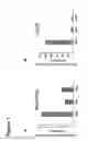
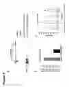
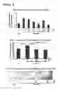

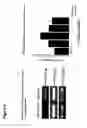
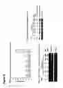
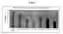
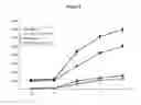


Sources:
- United States Patent and Trademark Office - verify current appl. status at the USPTO↗
Similar patent applications:
Recent applications in this class:
- » 20250170212 2025-05-29
COMPOSITIONS AND METHODS OF TREATING AMYOTROPHIC LATERAL SCLEROSIS (ALS) AND RELATED DISORDERS - » 20250161407 2025-05-22
Medical application and pharmaceutical composition of UbV.E4B protein - » 20250161406 2025-05-22
PREVENTING ALU SINES-MEDIATED PATHOLOGIES - » 20250152666 2025-05-15
COMPLEXES FOR TREATING SENSITIVITY - » 20250152665 2025-05-15
Mutant Peptides And Methods Of Treating Subjects Using The Same - » 20250152664 2025-05-15
COMPOSITIONS AND METHODS FOR TREATING METABOLIC DISORDERS - » 20250152663 2025-05-15
COMPOSITION FOR PREVENTION OR TREATMENT OF DRY EYE DISEASE COMPRISING HAPLN1 - » 20250134954 2025-05-01
THERAPEUTIC PEPTIDES - » 20250127852 2025-04-24
POLYPEPTIDES FOR TREATMENT OF CANCER - » 20250127851 2025-04-24
CYTOKINE MODULATION