KAOLIN-BASED HEMOSTATIC GAUZE AND PREPARATION METHOD THEREOF
US20240100217A1
2024-03-28
18/472,258
2023-09-22
Smart Summary: A new type of gauze is made from kaolin, a clay mineral, and is designed to stop bleeding. It uses a special mixture that includes kaolin, cerium (Ce), and iron oxide (α-Fe2O3) to enhance its effectiveness. The preparation process involves creating a suspension of this mixture and soaking a medical fabric in it. After soaking, the fabric is pressed and dried to form the final hemostatic gauze. This gauze is safe to use, works well to stop bleeding, and has antibacterial properties. 🚀 TL;DR
Abstract:
A kaolin-based hemostatic gauze comprises: a medical non-woven fabric as a carrier; and a kaolin-containing composite hemostatic material loaded on the medical non-woven fabric, wherein kaolin serves as a carrier in the kaolin-containing composite hemostatic material, the kaolin is doped with Ce and α-Fe2O3 is loaded on the Kaolin. A method for preparing the kaolin-based hemostatic gauze, includes: S1: preparing the kaolin-containing composite hemostatic material; S2: preparing the kaolin-containing composite hemostatic material into a suspension; S3: impregnating the medical non-woven fabric with the suspension thoroughly stirred, wherein upper and lower sides of the medical non-woven fabric each are impregnated once; and S4: pressing and oven-drying an impregnated medical non-woven fabric to obtain the kaolin-based hemostatic gauze. In the method, the medical non-woven fabric is combined with the powdery hemostatic material Ce-α-Fe2O3/Kaol through impregnation to prepare the medical hemostatic gauze product with excellent hemostatic performance, prominent biocompatibility, high safety, and antibacterial activity.
Inventors:
- Huaming YANG 7 🇨🇳 Wuhan, China
- Juan LIAO 3 🇨🇳 Wuhan, China
- Qiying ZHENG 2 🇨🇳 Wuhan, China
- Yunyang LIU 1 🇨🇳 Wuhan, China
Assignee:
- CHINA UNIVERSITY OF GEOSCIENCES (WUHAN) 38 🇨🇳 Wuhan, China
Applicant:
Interested in similar patents?
Get notified when new applications in this technology area are published.
Classification:
C04B33/131 » CPC further
Clay-wares; Preparing or treating the raw materials individually or as batches; Compounding ingredients Inorganic additives
C04B33/326 » CPC further
Clay-wares; Burning methods under pressure
A61L2300/102 » CPC further
Biologically active materials used in bandages, wound dressings, absorbent pads or medical devices containing or releasing inorganic materials Metals or metal compounds, e.g. salts such as bicarbonates, carbonates, oxides, zeolites, silicates
A61L2300/404 » CPC further
Biologically active materials used in bandages, wound dressings, absorbent pads or medical devices characterised by a specific therapeutic activity or mode of action Biocides, antimicrobial agents, antiseptic agents
A61L2300/418 » CPC further
Biologically active materials used in bandages, wound dressings, absorbent pads or medical devices characterised by a specific therapeutic activity or mode of action Agents promoting blood coagulation, blood-clotting agents, embolising agents
C04B2235/349 » CPC further
Aspects relating to ceramic starting mixtures or sintered ceramic products; Composition of constituents of the starting material or of secondary phases of the final product; Constituents and secondary phases not being of a fibrous nature; Non-metal oxides, non-metal mixed oxides, or salts thereof that form the non-metal oxides upon heating, e.g. carbonates, nitrates, (oxy)hydroxides, chlorides Clays, e.g. bentonites, smectites such as montmorillonite, vermiculites or kaolines, e.g. illite, talc or sepiolite
A61L15/44 » CPC main
Chemical aspects of, or use of materials for, bandages, dressings or absorbent pads; Bandages, dressings or absorbent pads for physiological fluids such as urine or blood, e.g. sanitary towels, tampons; Use of materials characterised by their function or physical properties Medicaments
A61L15/18 » CPC further
Chemical aspects of, or use of materials for, bandages, dressings or absorbent pads; Bandages, dressings or absorbent pads for physiological fluids such as urine or blood, e.g. sanitary towels, tampons containing inorganic materials
C04B33/04 » CPC further
Clay-wares; Preparing or treating the raw materials individually or as batches Clay; Kaolin
C04B33/13 IPC
Clay-wares; Preparing or treating the raw materials individually or as batches Compounding ingredients
C04B33/32 IPC
Clay-wares Burning methods
Description
CROSS-REFERENCE TO RELATED APPLICATIONS
This application claims priority to Chinese Patent Application No. 202211182898.X with a filing date of Sep. 27, 2022. The content of the aforementioned application, including any intervening amendments thereto, is incorporated herein by reference.
TECHNICAL FIELD
The present disclosure relates to the technical field of hemostatic gauzes, and in particular to a kaolin-based hemostatic gauze and a preparation method thereof.
BACKGROUND
The hemostatic products currently available on the market mainly include adhesive bandages, medical hemostatic gauzes, medical hemostatic bandages, etc. Common hemostatic dressings are spun from cotton, non-woven fabrics, etc. These hemostatic dressings themselves have certain hemostatic effect. However, the hemostatic effect is limited. It is a hot spot in the field of biological hemostasis to combine a hemostatic material with a gauze to prepare a multi-functional hemostatic dressing. The current hemostatic dressings such as chitosan-based dressings on the market are cost-ineffective. Inorganic materials such as natural clay have been reported to have excellent hemostatic effects. For example, in a Quikclot kaolinite-based hemostatic bandage produced by Z-Medica in the United States, a kaolinite powder is successfully loaded on a gauze, and the bandage has been confirmed to have excellent hemostatic performance. However, commercially-available hemostatic agents still have shortcomings such as single components and limited functions, which restrict the development and application of high-performance hemostatic products.
SUMMARY OF PRESENT INVENTION
An objective of the present disclosure is to provide a kaolin-based hemostatic gauze with excellent hemostatic performance, prominent biocompatibility, high safety, and antibacterial activity. A preparation method thereof is further provided.
The kaolin-containing composite hemostatic material comprises a medical non-woven fabric as a carrier; and a kaolin-containing composite hemostatic material load on the medical non-woven fabric. Kaolin serves as a carrier in the kaolin-containing composite hemostatic material, the Kaolin is doped with Ce, and α-Fe2O3 is loaded on the Kaolin.
In one embodiment, the kaolin includes flaky kaolinite and tubular halloysite.
In one embodiment, the kaolin is a raw ore.
In one embodiment, a loading rate of the α-Fe2O3 is 50% to 70%.
In one embodiment, the kaolin-containing composite hemostatic material is prepared according to the following steps:
-
- S1: mechanically mixing the kaolin with a polyhydroxyferric ion solution and a Ce salt solution; and
- S2: calcining a resulting mixture at a specified temperature to obtain a hemostatic material α-Fe2O3/Kaol with high biocompatibility, namely, the kaolin-containing composite hemostatic material.
In one embodiment, the calcination is conducted at 500° C. to 600° C.
In one embodiment, the calcination is conducted at 550° C.
In one embodiment, the polyhydroxyferric ion solution has a concentration of 0.4 mol/L, and a mass-to-volume ratio of the kaolin to the polyhydroxyferric ion solution is 1:50 g/mL; the Ce salt solution is a Ce(NO3)3 solution; and the Ce(NO3)3 solution has a concentration of 0.1 mol/L to 1.0 mol/L.
The method for preparing the kaolin-based hemostatic gauze, includes the following steps:
-
- S1: preparing the kaolin-containing composite hemostatic material;
- S2: preparing the kaolin-containing composite hemostatic material into a suspension;
- S3: impregnating the medical non-woven fabric with the suspension thoroughly stirred, wherein upper and lower sides of the medical non-woven fabric each are impregnated once; and
- S4: pressing and oven-drying an impregnated medical non-woven fabric to obtain the kaolin-based hemostatic gauze.
In one embodiment, the suspension of the kaolin-containing composite hemostatic material has a concentration of 0.001 g/mL to 0.05 g/mL.
In the kaolin-containing composite hemostatic material according to the present disclosure, α-Fe2O3 is loaded and Ce is doped to enhance a hemostatic effect; and the prepared Ce-α-Fe2O3/Kaol hemostatic material has no obvious cytotoxicity, no hemolysis, excellent biocompatibility, high safety, and antibacterial activity. Moreover, the preparation method involves simple steps and easy operations, and is conducive to large-scale production.
According to embodiments of the present disclosure, the medical non-woven fabric is combined with the powdery hemostatic material Ce-α-Fe2O3/Kaol through impregnation to prepare a medical hemostatic gauze product with excellent hemostatic performance, prominent biocompatibility, high safety, and antibacterial activity. The preparation method thereof is simple and is conducive to industrial production.
BRIEF DESCRIPTION OF THE DRAWINGS
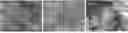
FIG. 1 shows morphological images of flaky kaolinite, tubular halloysite, and kaolin;
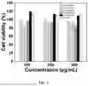
FIG. 2 shows test results of cytotoxicity of the α-Fe2O3/Kaol composite hemostatic material and α-Fe2O3 prepared in each of Examples 1 to 5 of the present disclosure;
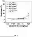
FIG. 3 shows test results of hemolysis of the α-Fe2O3/Kaol composite hemostatic material and α-Fe2O3 prepared in each of Examples 1 to 5 of the present disclosure;
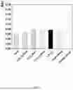
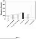
FIG. 4 shows test results of in vitro procoagulant effects of kaolin and different iron oxide/Kaol composite hemostatic materials;
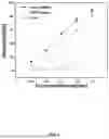
FIG. 5 shows test results of in vitro bleeding times of the Ce-α-Fe2O3/Kaol composite hemostatic materials prepared in Examples 7 to 9, a raw ore, the α-Fe2O3/Kaol prepared in Example 6, and the control group;
FIG. 6 shows test results of hemolysis of flaky kaolinite, tubular halloysite, and kaolin;
FIG. 7 is a schematic diagram showing the method for preparing the hemostatic gauze according to one embodiment of the present disclosure;
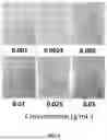
FIG. 8 shows pictures of 6 gauze products with different loads obtained through impregnation with hemostatic material suspensions of different concentrations (Examples 10 to 15);
FIG. 9 shows experimental results of liver hemostasis in animals with the Ce-α-Fe2O3/Kaol hemostatic gauze prepared in an embodiment of the present disclosure, an ordinary gauze, a Quikclot gauze (with kaolinite as a main component), and a Ce-α-Fe2O3/Kaol hemostatic powder; and
FIG. 10 shows experimental results of tail vein hemostasis in animals with the Ce-α-Fe2O3/Kaol hemostatic gauze prepared in an embodiment of the present disclosure, an ordinary gauze, a Quikclot gauze (with kaolinite as a main component), and a Ce-α-Fe2O3/Kaol hemostatic powder.
DETAILED DESCRIPTION OF THE EMBODIMENTS
The technical solutions of the present disclosure are described in further detail below with reference to the specific embodiments and accompanying drawings, but the present disclosure is not limited thereto.
The term “kaolin” in the disclosure has a chemical formula of Al2O3·2SiO2·2H2O; and in some cases, the kaolin includes about 45.31% of silica, about 37.21% of alumina, and about 14.1% of water.
It has been found through quantitative mineralogical analysis by X-ray diffraction (XRD) that the kaolin used in the embodiments of the disclosure includes halloysite and kaolinite; and it is found that the halloysite is tubular and the kaolinite is flaky according to scanning electron microscopy (SEM) analysis results shown in FIG. 1.
Preparation of Kaolin
A method for preparing a kaolin sample includes the following steps: crushing kaolin raw ores with a crusher to obtain a powder, mixing the powder thoroughly by a nine-square grid beneficiation method, and collecting a material in a middle of a nine-square grid and further grinding with a three-head grinder to obtain the sample.
Preparation of a Polyhydroxyferric Ion Solution
A polyhydroxyferric ion solution with a concentration of 0.4 mol/L is prepared with FeCl3·6H2O and NaOH.
Preparation of a Ce(NO3)3 Solution
A 0.12 mol/L Ce(NO3)3 solution is prepared with Ce(NO3)3·6H2O and water.
Preparation of α-Fe2O3/Kaol Composite Products with Different Loading Rates (Examples 1 to 6)
Example 1
In this example, a preparation method of a α-Fe2O3/Kaol1 composite hemostatic material with a α-Fe2O3 content of 50.41% was provided. The preparation method included the following steps: 5 g of the Kaolin sample and 250 mL of the polyhydroxyferric ion solution were mixed, a pH of a resulting mixture was adjusted with a 5 mol/L NaOH solution to about 3, a resulting system was stirred at 60° C. for 5 h and then centrifuged at 8,000 rpm, and a resulting precipitate was washed 3 times and dried to obtain a FeOOH/Kaol kaolin composite. The FeOOH/Kaol kaolin composite was ground and then calcined (including calcining at 250° C. for 1 h, calcining at 350° C. for 1 h, and calcining at 550° C. for 4 h) to obtain the α-Fe2O3/Kaol1 composite hemostatic material with a α-Fe2O3 content of 50.41%.
Example 2
In this example, a preparation method of a α-Fe2O3/Kaol2 composite hemostatic material with a α-Fe2O3 content of 34.52% was provided. The preparation method included the following steps: 10 g of the Kaolin sample and 250 mL of the polyhydroxyferric ion solution were mixed, a pH of a resulting mixture was adjusted with a 5 mol/L NaOH solution to about 3, a resulting system was stirred at 60° C. for 5 h and then centrifuged at 8,000 rpm, and a resulting precipitate was washed 3 times and dried to obtain a FeOOH/Kaol kaolin composite. The FeOOH/Kaol kaolin composite was ground and then calcined (including calcining at 250° C. for 1 h, calcining at 350° C. for 1 h, and calcining at 550° C. for 4 h) to obtain the α-Fe2O3/Kaol2 composite hemostatic material with a α-Fe2O3 content of 34.52%.
Example 3
In this example, a preparation method of a α-Fe2O3/Kaol4 composite hemostatic material with a α-Fe2O3 content of 22.29% was provided. The preparation method included the following steps: 20 g of the Kaolin sample and 250 mL of the polyhydroxyferric ion solution were mixed, a pH of a resulting mixture was adjusted with a 5 mol/L NaOH solution to about 3, a resulting system was stirred at 60° C. for 5 h and then centrifuged at 8,000 rpm, and a resulting precipitate was washed 3 times and dried to obtain a FeOOH/Kaol kaolin composite. The FeOOH/Kaol kaolin composite was ground and then calcined (including calcining at 250° C. for 1 h, calcining at 350° C. for 1 h, and calcining at 550° C. for 4 h) to obtain the α-Fe2O3/Kaol4 composite hemostatic material with a α-Fe2O3 content of 22.29%.
Example 4
In this example, a preparation method of a α-Fe2O3/Kaol8 composite hemostatic material with a α-Fe2O3 content of 6.26% was provided. The preparation method included the following steps: 40 g of the Kaolin sample and 250 mL of the polyhydroxyferric ion solution were mixed, a pH of a resulting mixture was adjusted with a 5 mol/L NaOH solution to about 3, a resulting system was stirred at 60° C. for 5 h and then centrifuged at 8,000 rpm, and a resulting precipitate was washed 3 times and dried to obtain a FeOOH/Kaol kaolin composite. The FeOOH/Kaol kaolin composite was ground and then calcined (including calcining at 250° C. for 1 h, calcining at 350° C. for 1 h, and calcining at 550° C. for 4 h) to obtain the α-Fe2O3/Kaol8 composite hemostatic material with a α-Fe2O3 content of 6.26%.
Example 5
In this example, a preparation method of a α-Fe2O3/Kaol10 composite hemostatic material with a α-Fe2O3 content of 7.45% was provided. The preparation method included the following steps: 50 g of the Kaolin sample and 250 mL of the polyhydroxyferric ion solution were mixed, a pH of a resulting mixture was adjusted with a 5 mol/L NaOH solution to about 3, a resulting system was stirred at 60° C. for 5 h and then centrifuged at 8,000 rpm, and a resulting precipitate was washed 3 times and dried to obtain a FeOOH/Kaol kaolin composite. The FeOOH/Kaol kaolin composite was ground and then calcined (including calcining at 250° C. for 1 h, calcining at 350° C. for 1 h, and calcining at 550° C. for 4 h) to obtain the α-Fe2O3/Kaol10 composite hemostatic material with a α-Fe2O3 content of 7.45%.
Example 6
In this example, a preparation method of a α-Fe2O3/Kaol composite hemostatic material with a α-Fe2O3 content of 62.19% was provided. The preparation method included the following steps: 30 g of the Kaolin sample and 1500 mL of the polyhydroxyferric ion solution were mixed, a pH of a resulting mixture was adjusted with a 5 mol/L NaOH solution to about 3, a resulting system was stirred at 60° C. for 5 h and then centrifuged at 8,000 rpm, and a resulting precipitate was washed 3 times and dried to obtain a FeOOH/Kaol kaolin composite. The FeOOH/Kaol kaolin composite was ground and then calcined (including calcining at 250° C. for 1 h, calcining at 350° C. for 1 h, and calcining at 550° C. for 4 h) to obtain the α-Fe2O3/Kaol composite hemostatic material with a α-Fe2O3 content of 62.19%.
The α-Fe2O3/Kaol1, α-Fe2O3/Kaol2, α-Fe2O3/Kaol4, α-Fe2O3/Kaol8, and α-Fe2O3/Kaol10 composite hemostatic materials and α-Fe2O3 prepared in Examples 1 to 5 each were subjected to a cytotoxicity test (FIG. 2) and a hemolysis test (FIG. 3). Test results showed that α-Fe2O3 itself has high cell viability and low hemolysis, and the α-Fe2O3/Kaol hemostatic material with a α-Fe2O3 loading rate of greater than 50% exhibits better biocompatibility than hemostatic materials with α-Fe2O3 loading rates of less than 50%.
Preparation of Ce-α-Fe2O3/Kaol Composite Hemostatic Materials
Example 7
In this example, a preparation method of a Ce-α-Fe2O3/Kaol composite hemostatic material was provided. The preparation method included the following steps: 5 g of the kaolin sample and 250 mL of the polyhydroxyferric ion solution were mixed and stirred, 20 mL of the Ce(NO3)3 solution was added dropwise, and a pH of a resulting mixed solution was adjusted with a 5 mol/L NaOH solution to about 3; a resulting system was stirred at 60° C. for 5 h and then centrifuged at 8,000 rpm, and a resulting precipitate was washed 3 times and dried to obtain a Ce—FeOOH/Kaol composite. The Ce—FeOOH/Kaol composite was ground and then calcined (including calcining at 250° C. for 1 h, calcining at 350° C. for 1 h, and calcining at 550° C. for 4 h) to obtain the Ce-α-Fe2O3/Kaol composite hemostatic material. The Ce-α-Fe2O3/Kaol composite hemostatic material was subjected to composition analysis by X-ray fluorescence (XRF) spectrometry (Table 1).
| TABLE 1 |
| Analysis results of oxide contents in the Ce-α-Fe2O3/ |
| Kaol composite hemostatic material |
| Component | Content (wt %) | |
| Fe2O3 | 41.54 | |
| SiO2 | 41.29 | |
| Al2O3 | 15.07 | |
| K2O | 1.37 | |
| CeO2 | 0.0566 | |
It can be seen from Table 1 that, in the Ce-α-Fe2O3/Kaol, a load of Fe2O3 is 41.54%, and a small amount of Ce is successfully doped.
Example 8
In this example, a preparation method of a α-Fe2O3—Ce/Kaol-Aladdin composite hemostatic material was provided. The preparation method included the following steps: 5 g of flaky kaolinite (Kaol-Aladdin; Aladdin, CAS:1332-58-7) and 250 mL of the polyhydroxyferric ion solution were mixed and stirred, 20 mL of the Ce(NO3)3 solution was added dropwise, and a pH of a resulting mixed solution was adjusted with a 5 mol/L NaOH solution to about 3; a resulting system was stirred at 60° C. for 5 h and then centrifuged at 8,000 rpm, and a resulting precipitate was washed 3 times and dried to obtain a Ce—FeOOH/Kaol-Aladdin composite. The Ce—FeOOH/Kaol-Aladdin composite was ground and then calcined (including calcining at 250° C. for 1 h, calcining at 350° C. for 1 h, and calcining at 550° C. for 4 h) to obtain the Ce-α-Fe2O3/Kaol-Aladdin composite hemostatic material.
Example 9
In this example, a preparation method of a Ce-α-Fe2O3/HNTs-Sigma composite hemostatic material was provided. The preparation method included the following steps: 5 g of tubular halloysite (HNTs-Sigma; Sigma, CAS: 12298-43-0) and 250 mL of the polyhydroxyferric ion solution were mixed and stirred, 20 mL of the Ce(NO3)3 solution was added dropwise, and a pH of a resulting mixed solution was adjusted with a 5 mol/L NaOH solution to about 3; a resulting system was stirred at 60° C. for 5 h and then centrifuged at 8,000 rpm, and a resulting precipitate was washed 3 times and dried to obtain a Ce—FeOOH/HNTs-Sigma composite. The Ce—FeOOH/HNTs-Sigma composite was ground and then calcined (including calcining at 250° C. for 1 h, calcining at 350° C. for 1 h, and calcining at 550° C. for 4 h) to obtain the Ce-α-Fe2O3/HNTs-Sigma composite hemostatic material.
The above hemostatic materials each were subjected to an in vitro bleeding time test (FIG. 5). Test results showed that hemostatic effects of the Ce-α-Fe2O3/Kaol-Aladdin composite hemostatic material (Example 8) and the Ce-α-Fe2O3/HNTs-Sigma composite hemostatic material (Example 9) were comparable to a hemostatic effect of the Ce-α-Fe2O3/Kaol composite hemostatic material (Example 7), and are more significant than a hemostatic effect of the α-Fe2O3/Kaol (Example 6), indicating that the Ce-doped composite hemostatic materials exhibited significantly-improved hemostatic effects.
Preparation of α-Fe2O3/Kaol, Fe3O4/Kaol, γ-Fe2O3/Kaol, and FeOOH/Kaol Composite Hemostatic Materials
A preparation method of the α-Fe2O3/Kaol, Fe3O4/Kaol, γ-Fe2O3/Kaol, and FeOOH/Kaol composite hemostatic materials was provided. The preparation method included the following steps: 5 g of a kaolin sample and 250 mL of the polyhydroxyferric ion solution were mixed, a pH of a resulting mixture was adjusted with a 5 mol/L NaOH solution to about 3, a resulting system was stirred at 60° C. for 5 h and then centrifuged at 8,000 rpm, and a resulting precipitate was washed 3 times and dried to obtain a FeOOH/Kaol composite; the FeOOH/Kaol composite was ground and then calcined (including calcining at 250° C. for 1 h, calcining at 350° C. for 1 h, and calcining at 550° C. for 4 h) to obtain the α-Fe2O3/Kaol composite hemostatic material; the α-Fe2O3/Kaol composite obtained after calcination was calcined for 1 h at 450° C. in a H2/Ar (volume ratio: 1:9) atmosphere to obtain the Fe3O4/Kaol. The Fe3O4/Kaol was calcined for 2 h at 250° C. in an air atmosphere to obtain the γ-Fe2O3/Kaol.
Given that different oxide types may have different impacts on improving a hemostatic effect of kaolin, the prepared α-Fe2O3/Kaol, Fe3O4/Kaol, γ-Fe2O3/Kaol, and FeOOH/Kaol were evaluated through an in vivo procoagulant test (FIG. 4), and evaluation results showed that α-Fe2O3/Kaol exhibited the optimal procoagulant performance.
Hemolysis Experiment:
Preparation of a 2% red blood cell (RBC) suspension: 1 mL of fresh anticoagulant rabbit blood was collected and centrifuged at 2,500 rpm for 5 min, a resulting supernatant was removed, and a resulting precipitate was washed 3 times with phosphate-buffered saline (PBS) and then suspended; and 500 μL of a resulting suspension was taken and added to a 50 mL centrifuge tube, and PBS was added to 50 mL.
Hemolysis experiment: Kaolin, flaky kaolinite, and tubular halloysite each were prepared with PBS into solutions with concentrations of 0.125 mg/mL, 0.25 mg/mL, 0.5 mg/mL, 1.0 mg/mL, and 2.0 mg/mL, respectively, which each were of 3 mL. 500 μL of the solution of each concentration was taken and thoroughly mixed with 500 μL of a prepared 2% RBC suspension. A positive control group was set as follows: 500 μL of deionized water was mixed with 500 μL of the 2% RBC suspension; and a negative control group was set as follows: 500 μL of PBS was mixed with 500 μL of the 2% RBC suspension. 3 replicates were set per group. A sample was incubated in a 37° C. water bath for 1 h and then centrifuged at 2,500 rpm, a resulting supernatant was collected, and the absorbance of the supernatant was measured by a microplate reader (414 nm).
Hemolysis rate (%)=(absorbance of a sample−absorbance of negative control)/(absorbance of positive control−absorbance of negative control)×100%.
The lower the hemolysis, the higher the biocompatibility. When a hemolysis rate is lower than 5%, it is considered that there is no hemolysis.
Hemolysis test results were shown in FIG. 6. It can be seen from FIG. 6 that Kaol leads to a lower hemolysis rate than Kaol-Aladdin and HNTs-Sigma, indicating that Kaol has optimal biosafety.
In Vitro Procoagulant Experiment:
100 μL of anticoagulant whole blood was added dropwise to a 6-well plate, and 10 μL of a 0.2 mol/L CaCl2 solution was quickly added dropwise to the whole blood to recalcify the whole blood; 10 mg of a material was added to the whole blood by a dropper with a rubber head removed, and no material was added in a blank control group; the plate was incubated in a 37° C. water bath for 9 min, and then 10 mL of deionized water was slowly added dropwise around blood droplets, where coagulated blood was prevented from being impacted; and after the dropwise addition was completed, 1 mL of a resulting aqueous solution was immediately taken and centrifuged at 1,000 rpm, and then the absorbance (540 nm) was measured by a microplate reader.
10 mg of each of kaolin and the prepared different iron oxide/Kaol composite hemostatic materials was weighed to prepare experimental groups, and no material was added in the blank control group; and 3 replicates were set per group.
In vitro procoagulant test results were shown in FIG. 4. It can be seen from FIG. 4 that α-Fe2O3/Kaol has lower absorbance than Fe3O4/Kaol, γ-Fe2O3/Kaol, and FeOOH/Kaol, indicating that α-Fe2O3/Kaol has optimal procoagulant performance.
In Vitro Bleeding Time Experiment:
10 mg of each of Kaol, Ce-α-Fe2O3/Kaol-Aladdin, Ce-α-Fe2O3/HNTs-Sigma, α-Fe2O3/Kaol, and Ce-α-Fe2O3/Kaol composite hemostatic materials was weighed, added to a 2 mL centrifuge tube, and pre-warmed in a 37° C. water bath for 3 min; 200 μL of anticoagulant whole blood of a New Zealand white rabbit was added dropwise to a sample powder at a bottom of the centrifuge tube, and then 10 μL of a 0.2 mol/L CaCl2) solution was quickly added dropwise to a resulting mixed system to calcify the blood to trigger blood coagulation; and a resulting mixed system was immediately incubated in a 37° C. water bath, during which the centrifuge tube was shaken every 15 s, the flow of blood in the tube was observed until the blood was coagulated, and a hemostasis time was recorded. 3 replicates were set for each material.
In vitro bleeding time test results were shown in FIG. 5. It can be seen from FIG. 5 that, compared with the α-Fe2O3/Kaol (Example 6), a bleeding time of the Ce-α-Fe2O3/Kaol composite hemostatic material (Example 7) is shortened, indicating that the Ce doping can improve a hemostatic effect of the composite hemostatic material.
In Vivo Hemostasis Experiment:
6-week-old female Kunming mice were selected and randomly grouped according to a body weight, with 5 mice in each group. Each mouse was fixed with a tail exposed, and a 1 cm wound was cut with a scalpel blade on a tail vein of the mouse to allow bleeding. Then a corresponding material powder was quickly applied to the wound, the wound was immediately covered with a hemostatic gauze and gently pressed to stop the bleeding until the bleeding was completely stopped, and a hemostasis time was recorded by a timer. Blood flowing out from the wound was dipped by a gauze and weighed, and a bleeding amount was calculated. Hemostasis time and bleeding amount results were shown in Table 2.
| TABLE 2 |
| Bleeding times and bleeding amounts of Ce-α-Fe2O3/Kaol |
| composite hemostatic materials |
| Hemostasis | Bleeding | ||
| Group | Material | time (s) | amount (mg) |
| Blank control | No material is added | 179.8 ± 36.6 | 132.7 ± 75.9 |
| group | |||
| Raw ore | Kaol | 153.2 ± 33.7 | 104.8 ± 50.5 |
| Example 6 | α-Fe2O3/Kaol | 136 ± 34.7 | 96.4 ± 42.6 |
| Example 7 | Ce-α-Fe2O3/Kaol | 112.6 ± 23.4 | 84.81 ± 40.4 |
| Positive | Yunnan Baiyao | 136.6 ± 40.9 | 53.1 ± 32.5 |
| control group | |||
It can be seen from Table 2 that the doping of Ce in α-Fe2O3/Kaol can effectively improve a hemostasis rate and reduce a bleeding amount.
6 Gauze Products with Different Loads Obtained Through Impregnation with Hemostatic Material Suspensions of Different Concentrations (Examples 10 to 15)
Preparation of a Ce-α-Fe2O3/Kaol Composite Hemostatic Agent
5 g of kaolin and 250 mL of the polyhydroxyferric ion solution were mixed and stirred, 20 mL of the Ce(NO3)3 solution was added dropwise, and a pH of a resulting mixed solution was adjusted with a 5 mol/L NaOH solution to about 3; a resulting system was stirred at 60° C. for 5 h and then centrifuged at 8,000 rpm, and a resulting precipitate was washed 3 times and dried to obtain a Ce—FeOOH/Kaol composite. The Ce—FeOOH/Kaol composite was ground and then calcined (including calcining at 250° C. for 1 h, calcining at 350° C. for 1 h, and calcining at 550° C. for 4 h) to obtain the Ce-α-Fe2O3/Kaol composite hemostatic material.
The Ce-α-Fe2O3/Kaol composite hemostatic material prepared above was adopted in each of Examples 10 to 15.
Example 10
As shown in FIG. 7, 0.2 g of the Ce-α-Fe2O3/Kaol hemostatic material was added to 200 mL of water, and a resulting mixture was thoroughly stirred to obtain a homogeneous suspension; a piece of a non-woven fabric (area: 10*9.5 cm2) was cut and directly impregnated with the homogeneous suspension such that the material adhered to a surface of the non-woven fabric, where upper and lower sides of the non-woven fabric each were impregnated once, with a total impregnation time of about 2 s; an impregnated non-woven fabric was placed on a rolling machine and pressed once with a distance of 0.05 mm between upper and lower rollers of the rolling machine to strengthen an adhesion degree between the powdery material and the non-woven fabric; and finally, the pressed non-woven fabric was hung by dovetail clips in an oven at 60° C. and blow-dried.
Example 11
0.5 g of the Ce-α-Fe2O3/Kaol hemostatic material was added to 200 mL of water, and a resulting mixture was thoroughly stirred to obtain a homogeneous suspension; a piece of a non-woven fabric (area: 10*9.5 cm2) was cut and directly impregnated with the homogeneous suspension such that the material adhered to a surface of the non-woven fabric, where upper and lower sides of the non-woven fabric each were impregnated once, with a total impregnation time of about 2 s; an impregnated non-woven fabric was placed on a rolling machine and pressed once with a distance of 0.05 mm between upper and lower rollers of the rolling machine to strengthen an adhesion degree between the powdery material and the non-woven fabric; and finally, the pressed non-woven fabric was hung by dovetail clips in an oven at 60° C. and blow-dried.
Example 12
1.0 g of the Ce-α-Fe2O3/Kaol hemostatic material was added to 200 mL of water, and a resulting mixture was thoroughly stirred to obtain a homogeneous suspension; a piece of a non-woven fabric (area: 10*9.5 cm2) was cut and directly impregnated with the homogeneous suspension such that the material adhered to a surface of the non-woven fabric, where upper and lower sides of the non-woven fabric each were impregnated once, with a total impregnation time of about 2 s; an impregnated non-woven fabric was placed on a rolling machine and pressed once with a distance of 0.05 mm between upper and lower rollers of the rolling machine to strengthen an adhesion degree between the powdery material and the non-woven fabric; and finally, the pressed non-woven fabric was hung by dovetail clips in an oven at 60° C. and blow-dried.
Example 13
2.0 g of the Ce-α-Fe2O3/Kaol hemostatic material was added to 200 mL of water, and a resulting mixture was thoroughly stirred to obtain a homogeneous suspension; a piece of a non-woven fabric (area: 10*9.5 cm2) was cut and directly impregnated with the homogeneous suspension such that the material adhered to a surface of the non-woven fabric, where upper and lower sides of the non-woven fabric each were impregnated once, with a total impregnation time of about 2 s; an impregnated non-woven fabric was placed on a rolling machine and pressed once with a distance of 0.05 mm between upper and lower rollers of the rolling machine to strengthen an adhesion degree between the powdery material and the non-woven fabric; and finally, the pressed non-woven fabric was hung by dovetail clips in an oven at 60° C. and blow-dried.
Example 14
5.0 g of the Ce-α-Fe2O3/Kaol hemostatic material was added to 200 mL of water, and a resulting mixture was thoroughly stirred to obtain a homogeneous suspension; a piece of a non-woven fabric (area: 10*9.5 cm2) was cut and directly impregnated with the homogeneous suspension such that the material adhered to a surface of the non-woven fabric, where upper and lower sides of the non-woven fabric each were impregnated once, with a total impregnation time of about 2 s; an impregnated non-woven fabric was placed on a rolling machine and pressed once with a distance of 0.05 mm between upper and lower rollers of the rolling machine to strengthen an adhesion degree between the powdery material and the non-woven fabric; and finally, the pressed non-woven fabric was hung by dovetail clips in an oven at 60° C. and blow-dried.
Example 15
10.0 g of the Ce-α-Fe2O3/Kaol hemostatic material was added to 200 mL of water, and a resulting mixture was thoroughly stirred to obtain a homogeneous suspension; a piece of a non-woven fabric (area: 10*9.5 cm2) was cut and directly impregnated with the homogeneous suspension such that the material adhered to a surface of the non-woven fabric, where upper and lower sides of the non-woven fabric each were impregnated once, with a total impregnation time of about 2 s; an impregnated non-woven fabric was placed on a rolling machine and pressed once with a distance of 0.05 mm between upper and lower rollers of the rolling machine to strengthen an adhesion degree between the powdery material and the non-woven fabric; and finally, the pressed non-woven fabric was hung by dovetail clips in an oven at 60° C. and blow-dried.
FIG. 8 shows pictures of 6 gauze products with different loads obtained through impregnation with hemostatic material suspensions of different concentrations; and as shown in FIG. 8, the Ce-α-Fe2O3/Kaol hemostatic material is successfully loaded on the gauze.
Loads of the hemostatic materials in the gauze products obtained in Examples 10 to 15 were shown in Table 3.
| TABLE 3 |
| Loads of hemostatic materials in Ce-α-Fe2O3/Kaol gauze products |
| Mass of a powder | ||||||
| in 200 mL of | Concentration | Mass of a nude | Mass after | Load | Load | |
| Example | water (g) | (g/mL) | fabric (g) | oven-drying (g) | (g/cm2) | (g) |
| Example 10 | 0.2 | 0.0010 | 0.5039 | 0.5056 | 0.000000000 | 0.0017 |
| Example 11 | 0.5 | 0.0025 | 0.5165 | 0.5310 | 0.000152632 | 0.0145 |
| Example 12 | 1.0 | 0.0050 | 0.5068 | 0.5517 | 0.000472632 | 0.0449 |
| Example 13 | 2.0 | 0.0100 | 0.4966 | 0.5756 | 0.000831579 | 0.0790 |
| Example 14 | 5.0 | 0.0250 | 0.5016 | 0.7448 | 0.002560000 | 0.2432 |
| Example 15 | 10.0 | 0.0500 | 0.5188 | 0.8391 | 0.003371579 | 0.3203 |
In Vivo Hemostasis Experiment:
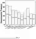
Liver Hemostasis Experiment:
6-week-old female Kunming mice were selected and randomly grouped according to a body weight, with 5 mice in each group. Each mouse was anesthetized and fixed, an abdominal cavity of the mouse was opened, and a wound of about 1 cm was cut with a scalpel on a left lobe tissue of the liver; the bleeding left lobe of the liver was covered with a gauze sample (area: 47.5 cm2) (or a powdery sample); and a hemostasis time was recorded, and a mass change of the gauze sample was measured to calculate a bleeding amount. Hemostasis time and bleeding amount results were shown in Table 4 and FIG. 9.
| TABLE 4 |
| Bleeding times and bleeding amounts of Ce-α-Fe2O3/ |
| Kaol hemostatic gauzes |
| Group | Material | Hemostasis time (s) | Bleeding amount (g) |
| Control group | No material is added | 88.60 ± 13.00 | 0.2686 ± 0.1892 |
| Blank gauze | Additive-free gauze | 84.25 ± 7.01 | 0.1262 ± 0.0506 |
| Example 10 | Ce-α-Fe2O3/KaolYGT1-loaded gauze | 66.75 ± 11.34 | 0.1711 ± 0.0377 |
| Example 11 | Ce-α-Fe2O3/KaolYGT2.5-loaded gauze | 65.00 ± 6.16 | 0.1038 ± 0.0215 |
| Example 12 | Ce-α-Fe2O3/KaolYGT5-loaded gauze | 55.75 ± 9.62 | 0.1522 ± 0.0793 |
| Example 13 | Ce-α-Fe2O3/KaolYGT10-loaded gauze | 45.50 ± 10.78 | 0.1161 ± 0.0353 |
| Example 14 | Ce-α-Fe2O3/KaolYGT25-loaded gauze | 57.00 ± 8.15 | 0.2188 ± 0.0884 |
| Example 15 | Ce-α-Fe2O3/KaolYGT50-loaded gauze | 74.75 ± 20.11 | 0.1998 ± 0.1214 |
| Quikclot | Quikclot gauze | 50.75 ± 8.58 | 0.2266 ± 0.1769 |
| Powdery sample | Ce-α-Fe2O3/KaolYGT | 46.75 ± 6.49 | 0.0733 ± 0.0338 |
Tail Vein Hemostasis Experiment:
6-week-old female Kunming mice were selected and randomly grouped according to a body weight, with 5 mice in each group. Each mouse was fixed with a tail exposed, and a 1 cm wound was cut with a scalpel blade on a tail vein of the mouse to allow bleeding; then a corresponding material powder was quickly applied to the wound, the wound was immediately covered with a hemostatic gauze and gently pressed to stop the bleeding until the bleeding was completely stopped, and a hemostasis time was recorded by a timer; and blood flowing out from the wound was dipped by a gauze and weighed, and a bleeding amount was calculated. Hemostasis time and bleeding amount results were shown in Table 5 and FIG. 10.
| TABLE 5 |
| Bleeding times and bleeding amounts of Ce-α-Fe2O3/ |
| Kaol hemostatic gauzes |
| Group | Material | Hemostasis time (s) | Bleeding amount (g) |
| Control group | No material is added | 67.40 ± 8.8 | 0.00934 ± 0.00588 |
| Blank gauze | Additive-free gauze | 146.00 ± 57.5 | 0.03402 ± 0.02575 |
| Example 10 | Ce-α-Fe2O3/KaolYGT1-loaded gauze | 82.60 ± 14.2 | 0.03584 ± 0.02089 |
| Example 11 | Ce-α-Fe2O3/KaolYGT2.5-loaded gauze | 69.60 ± 13.9 | 0.03970 ± 0.01297 |
| Example 12 | Ce-α-Fe2O3/KaolYGT5-loaded gauze | 69.20 ± 16.12 | 0.02690 ± 0.01991 |
| Example 13 | Ce-α-Fe2O3/KaolYGT10-loaded gauze | 60.00 ± 24.37 | 0.01326 ± 0.00807 |
| Example 14 | Ce-α-Fe2O3/KaolYGT25-loaded gauze | 74.00 ± 60.64 | 0.03230 ± 0.01342 |
| Example 15 | Ce-α-Fe2O3/KaolYGT50-loaded gauze | 47.00 ± 26.12 | 0.01500 ± 0.01487 |
| Quikclot | Quikclot gauze | 48.80 ± 13.30 | 0.00806 ± 0.00560 |
| Powdery sample | Ce-α-Fe2O3/KaolYGT | 65.17 ± 7.62 | 0.01737 ± 0.00870 |
It can be seen from Table 4, Table 5, FIG. 9, and FIG. 10 that, compared with the existing hemostatic gauzes on the market, the Ce-α-Fe2O3/Kaol hemostatic gauze can improve a hemostasis rate and reduce a bleeding amount to some degree.
What is not mentioned above can be acquired in the prior art.
Although some specific embodiments of the present disclosure have been described in detail by way of examples, those skilled in the art will appreciate that the above examples are provided for illustration only and not for limiting the scope of the present disclosure. A person skilled in the art can make various modifications or supplements to the specific embodiments described or replace them in a similar manner, but it may not depart from the direction of the present disclosure or the scope defined by the appended claims. Those skilled in the art should understand that any modification, equivalent replacement, and improvement that are made to the above embodiments according to the technical essence of the present disclosure shall be included in the protection scope of the present disclosure.
Claims
1. A kaolin-based hemostatic gauze, comprising:
a medical non-woven fabric as a carrier; and
a kaolin-containing composite hemostatic material loaded on the medical non-woven fabric;
wherein kaolin serves as a carrier in the kaolin-containing composite hemostatic material, the Kaolin is doped with Ce, and α-Fe2O3 is loaded on the Kaolin; the kaolin comprises flaky kaolinite and tubular halloysite;
the kaolin-containing composite hemostatic material is prepared according to the following steps:
S1: mechanically mixing the kaolin with a polyhydroxyferric ion solution and a Ce salt solution; and
S2: calcining a resulting mixture at a specified temperature to obtain a hemostatic material α-Fe2O3/Kaol with high biocompatibility, wherein the hemostatic material α-Fe2O3/Kaol is the kaolin-containing composite hemostatic material.
2. (canceled)
3. The kaolin-based hemostatic gauze according to claim 1, wherein the kaolin is a raw ore.
4. The kaolin-based hemostatic gauze according to claim 1, wherein a loading rate of the α-Fe2O3 is 50% to 70%.
5. (canceled)
6. The kaolin-based hemostatic gauze according to claim 1, wherein the specified temperature is 500° C. to 600° C.
7. The kaolin-based hemostatic gauze according to claim 1, wherein the specified temperature is 550° C.
8. The kaolin-based hemostatic gauze according to claim 1, wherein the polyhydroxyferric ion solution has a concentration of 0.4 mol/L, and a mass-to-volume ratio of the kaolin to the polyhydroxyferric ion solution is 1:50 g/mL; the Ce salt solution is a Ce(NO3)3 solution; and the Ce(NO3)3 solution has a concentration of 0.1 mol/L to 1.0 mol/L.
9. A method for preparing the kaolin-based hemostatic gauze according to claim 1, comprising the following steps:
S1: preparing the kaolin-containing composite hemostatic material;
S2: preparing the kaolin-containing composite hemostatic material into a suspension;
S3: impregnating the medical non-woven fabric with the suspension thoroughly stirred, wherein upper and lower sides of the medical non-woven fabric each are impregnated once; and
S4: pressing and oven-drying an impregnated medical non-woven fabric to obtain the kaolin-based hemostatic gauze.
10. The method according to claim 9, wherein the suspension of the kaolin-containing composite hemostatic material has a concentration of 0.001 g/mL to 0.05 g/mL.
Images & Drawings included:


Sources:
- United States Patent and Trademark Office - verify current appl. status at the USPTO↗
Recent applications in this class:
- » 20250108141 2025-04-03
TOPICAL FORMULATIONS FOR TARGETED DELIVERY OF THERAPEUTICS - » 20250090717 2025-03-20
THERAPEUTIC USE OF FIBROBLASTS FOR ADHESIVE BANDAGE WOUND HEALING - » 20250090716 2025-03-20
Thimble Bank - » 20250041474 2025-02-06
MEDICAL DEVICE HAVING A PHOTOSENSITIZER AND RELATED METHODS - » 20240382646 2024-11-21
WOUND DRESSING APPARATUSES AND METHODS FOR NITRIC OXIDE DELIVERY - » 20240374780 2024-11-14
ANTIMICROBIAL COMPONENT FOR A WOUND DRESSING - » 20240358884 2024-10-31
FERROCENE-CONTAINING POLYELECTROLYTE COMPLEX AND WOUND DRESSING - » 20240293595 2024-09-05
MULTILAYER DEVICE FOR SUPPLYING NITRIC OXIDE - » 20240261459 2024-08-08
Bioactive polymeric dressing for accelerated wound closure - » 20240181121 2024-06-06
A WOUND CARE PRODUCT COMPRISING AN ANTIMICROBIAL COATING
Recent applications for this Assignee:
- » 20250171320 2025-05-29
HIGH-PURITY KAOLIN AND PREPARATION METHOD THEREOF - » 20250115720 2025-04-10
HYPERBRANCHED POLYCAPROLACTONE-POLYLACTIC ACID COPOLYMER (PCL-PLA), PREPARATION METHOD AND SOLID ELECTROLYTE APPLICATION THEREOF - » 20250099610 2025-03-27
MAGNETITE-BASED MICRO/NANOROBOT AND PREPARATION METHOD AND USE THEREOF - » 20250090577 2025-03-20
CLAY MINERAL BIOMATERIAL, AND PREPARATION METHOD AND USE THEREOF - » 20250022151 2025-01-16
METHOD AND DEVICE FOR ALIGNING LASER POINT CLOUD AND IMAGE BASED ON DEEP LEARNING - » 20250020055 2025-01-16
METHOD AND DEVICE FOR PROCESSING FUSED "UNDERGROUND+GROUND" DATA IN DEEP FORMATION EXPLORATION - » 20250013962 2025-01-09
METHODS AND SYSTEMS FOR OPTIMIZING OPEN-PIT MINE PRODUCTION PLANS BASED ON TIME-SERIES PREDICTION OF COPPER PRICE - » 20250003741 2025-01-02
Method and device for determining reasonable sampling interval with three-dimensional (3D) laser scanning, and storage medium - » 20240374784 2024-11-14
HALLOYSITE-BASED COMPOSITE HEMOSTATIC MATERIAL AND PREPARATION METHOD AND USE THEREOF - » 20240345289 2024-10-17
OCEAN-ONTO-LAND DROUGHT (OTLD) IDENTIFICATION AND PROPAGATION MECHANISM ANALYSIS METHOD AND SYSTEM