Assay systems, kits and methods for detecting microorganisms
US20050123933A1
2005-06-09
10/729,980
2003-12-09
✅ Patent granted
US 7,575,862 B2
2009-08-18
-
-
Rodney P. Swartz
2023-12-09
Abstract:
The present invention relates to an assay system, kits and methods for detecting microorgansims (especially for M. tuberculosis) of suspected patient. The present invention also relates to an apparatus for performing the integration of thermal and magnetic control in the same apparatus to largely reduce the whole process of M. tuberculosis detection is less than 5 hours.
Inventors:
- George Chin-Sheng Chou 8 🇹🇼 Tainan Hsien, Taiwan
- Chang-Yi Huang 2 🇹🇼 Tainan Hsien, Taiwan
Assignee:
- AsiaGen Corporation 2 🇹🇼 Hsin-Shi, Taiwan
Interested in similar patents?
Get notified when new applications in this technology area are published.
Classification:
C07H21/02 IPC
Compounds containing two or more mononucleotide units having separate phosphate or polyphosphate groups linked by saccharide radicals of nucleoside groups, e.g. nucleic acids with ribosyl as saccharide radical
C12Q1/689 » CPC main
Measuring or testing processes involving enzymes, nucleic acids or microorganisms ; Compositions therefor; Processes of preparing such compositions involving nucleic acids; Nucleic acid products used in the analysis of nucleic acids, e.g. primers or probes for detection or identification of organisms for bacteria
B01L7/52 » CPC further
Heating or cooling apparatus ; Heat insulating devices with provision for submitting samples to a predetermined sequence of different temperatures, e.g. for treating nucleic acid samples
G01N33/5695 » CPC further
Investigating or analysing materials by specific methods not covered by groups -; Biological material, e.g. blood, urine ; Haemocytometers; Chemical analysis of biological material, e.g. blood, urine; Testing involving biospecific ligand binding methods; Immunological testing; Immunoassay; Biospecific binding assay; Materials therefor for microorganisms, e.g. protozoa, bacteria, viruses; Bacteria Mycobacteria
B01L7/02 » CPC further
Heating or cooling apparatus ; Heat insulating devices Water baths; Sand baths; Air baths
B01L2200/0668 » CPC further
Solutions for specific problems relating to chemical or physical laboratory apparatus; Fluid handling related problems; Handling flowable solids, e.g. microscopic beads, cells, particles Trapping microscopic beads
C12Q1/68 IPC
Measuring or testing processes involving enzymes, nucleic acids or microorganisms ; Compositions therefor; Processes of preparing such compositions involving nucleic acids
C07H21/04 IPC
Compounds containing two or more mononucleotide units having separate phosphate or polyphosphate groups linked by saccharide radicals of nucleoside groups, e.g. nucleic acids with deoxyribosyl as saccharide radical
Description
FIELD OF THE INVENTIONThe present invention relates to an assay system, kits and methods for detecting microorganisms (especially M. tuberculosis) from samples of suspected patient. The present invention also relates to an apparatus that integrates thermal and magnetic control in the same apparatus to largely reduce the hybridization time.
BACKGROUND OF THE INVENTIONTuberculosis (TB) is the leading infectious killer of youth and adults and the first most common infectious disease worldwide. One third of the world's population is currently infected and 20 million of those infected are active cases; TB will kill 30 million people this decade. More than 50 million people may already be infected with multidrug-resistant (MDR) strains of TB. Drug resistance has been brought about because of complacency in the public health sector and poorly managed TB control programs. Prior to MDR tuberculosis, the success rate of drug combination treatment was greater than 90%, even in AIDS patients. MDR tuberculosis, however, is not only highly infectious but also essentially incurable with a mortality of 50%. TB is now becoming the leading cause of death among HIV positive people where it kills much more rapidly with a fatality of 80%.
Tuberculosis is caused by infection with Mycobacterium tuberculosis, a bacillus bacterium. It is spread by aerosol droplets and causes irreversible lung destruction. If it escapes the lung it may cause systemic disease affecting many organs including bones, joints, liver, spleen, gastrointestinal tract and brain. 50% of people exposed to M. tuberculosis are infected with the bacteria and 15% of those infected develop disease. Poverty, malnutrition and overpopulation contribute dramatically to the perseverance and wild spread of tuberculosis.
Past means of controlling TB have involved the use of combinations of antibiotics. Recently, because of complications due to multidrug-resistant strains, the number and combination of antibiotics administered must be individually tailored depending on the strain the patient is harboring. In extreme cases, surgical removal of the infected portion of the lung is required.
Traditionally, the diagnosis of TB has been made on the basis of clinical findings and chest radiographs and confirmed by sputum or tissue smears that show TB bacilli. These methods remain the “gold standard” for diagnosis, but development of DNA probes, polymerase chain reaction (PCR) assays, and liquid media now allow more sensitive and rapid diagnosis. Unfortunately, increased sensitivity of rapid techniques is not always associated with increased specificity.
Skin testing should be used in conjunction with other clinical findings and is not a sensitive or specific test for establishing the diagnosis when the patient had been vaccinated with BCG or infected by Mycobacterium other than tuberculosis. In extrapulmonary TB, site-specific tissue or fluid samples or both are submitted for smear, culture, and histological analysis. Typically, histological features of a tuberculous lesion include caseating and noncaseating granulomata with giant cells. Rapid laboratory testing to identify and determine the drug susceptibility of M. tuberculosis isolates is vital to effective diagnosis, treatment, and control of TB in the community.
Clearly, expedient diagnosis is important in controlling the spread of tuberculosis. Sputum samples, evaluated first by direct microscopic evaluation (smear), are visualized with either the easily detected acid-fast fluorochrome dye auramine O, or the more specific Ziehl-Neelsen stain. Specimens are cultured on either solid media (Lowenstein-Jensen slant), or are grown in a liquid medium, such as the BACTEC automated radiometric system. Next, biochemical or nucleic acid probe testing is used to identify various strains. Isolates are tested for resistance to commonly used antituberculosis drugs, often by using the new method of susceptibility testing in liquid broth rather than the traditional agar dilution method.
The Ziehl-Neelsen carbolfuchsin or Kinyoun carbolfuchsin stains have been essential in TB diagnosis for nearly 100 years. Although less sensitive than culture, the acid-fast smear is a rapid and inexpensive test that can be performed with a minimum of equipment and is very specific for mycobacteria. Depending on the bacterial load, a single sputum smear has sensitivity between 22% and 80%, but the yield is improved when multiple sputum specimens are examined.
Most laboratories in the United States use fluorochrome stains, such as auramine-rhodamine stain. With these techniques, mycobacteria fluoresce with a bright orange color and can be easily seen on low-power microscopy, increasing the sensitivity of the smears.
The Amplified Mycobacterium Tuberculosis Direct Test (Gen-Probe) targets mycobacterial ribosomal RNA by transcription-mediated amplification. The test uses DNA probes that are highly specific for M tuberculosis species. It is best used (and only approved for use) in patients in whom acid-fast bacilli smears are positive and cultures are in process. Since specificity is less than 100%, even in patients with positive smears, occasional false-positive results do occur, usually in patients with nontuberculous mycobacterial infections.
This technique amplifies even very small portions of a predetermined target region of M tuberculosis-complex DNA. The test uses an automated system that can rapidly detect as few as one organism from sputum, bronchoalveolar lavage, blood, cerebrospinal fluid, pleural fluid, or other fluid and tissue samples and has shown sensitivity and specificity of nearly 90% in pulmonary disease.
The Mantoux test is the preferred and standard skin test for detecting TB. It involves injection of 5 TU of purified protein derivative (PPD, tuberculin), usually 0.1 mL, intradermally. Induration is then assessed at 48 to 72 hours. The extent of induration (not erythema) should be measured across two diameters at right angles and the two measurements then averaged. Interobserver variability can be decreased through the use of a ballpoint pen carefully brought from outside the zone of induration toward the center. As the area of induration is reached, resistance increases, and marking should stop at the outer edge of induration. However, about 20% of patients with active TB may have negative skin tests, and some populations have an even higher incidence of false-negative results. For example, false-negative rates up to 50% have been reported in patients with advanced HIV infection. Alternately, false-positive results may occur in patients infected by other nontuberculous mycobacteria (e.g., Mycobacterium avium complex). Therefore, a negative skin test never rules out TB, and a positive skin test alone does not establish the diagnosis.
The US Centers for Disease Control and Prevention and the World Health Organization recommend initial susceptibility testing for all M tuberculosis isolates because of the emergence of drug resistance worldwide.
This ingenious assay uses the fluorescent capabilities of fireflies genetically implanted in M tuberculosis. The procedure offers the possibility of testing mycobacterial drug susceptibility in hours. It is in the development stages but may become widely available in the next few years.
Given the above, current available assay cannot quickly and completely detect M tuberculosis. It requires a quick assay with high specificity and sensitivity to detect M tuberculosis from available samples.
SUMMARY OF THE INVENTIONThe present invention relates to an assay system, kits and methods for detecting microorganisms (especially M. tuberculosis) from the samples of suspected patient.
The present invention also relates to an apparatus for performing the dissociation of nucleic acid double strands, hybridization, washing, the separation of magnetic beads and thermal control in the same apparatus.
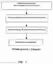
BRIEF DESCRIPTION OF THE DRAWINGSFIG. 1 shows the outline of the detection method of the invention.
FIG. 2 shows M. tuberculosis genomic DNA detected by the kit of the invention.
FIG. 3 shows the identification of M. tuberculosis by the assay of the invention.
FIG. 4 shows the specificity of the assay of the invention.
FIG. 5 shows the specificity and sensitivity of the kit of the invention.
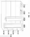
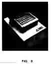
FIG. 6 shows the apparatus of the invention.
DETAILED DESCRIPTION OF THE INVENTIONThe present invention provides a method for detecting microorganism DNA comprising:
-
- (a) hybridizing the microorganism cDNA with microorganism-specific probes in hybridization tube wherein the probe is linked to magnetic bead;
- (b) transferring hybridization tubes to magnetic wells for washing;
- (c) adding blocking solution into the tubes;
- (d) adding avidin enzyme complex or streptavidin enzyme complex into the tubes;
- (e) performing washing reaction to remove interfering material by the aid of magnetic field;
- (f) suspending magnetic beads; and
- (g) detecting the luminescent or color change after adding substrate of enzyme.
The method of the present invention can be applied in any microorganisms including virus, bacteria, fungi etc. The method of the present invention is particularly suitable for detecting Mycobacterium tuberculosis.
In general, any body fluid such as CSF, serum, blood, sputum, pleural effusion, throat swab and stools can be used in the clinical tests. The preferred samples for M. tuberculosis are from CSF, serum, blood, sputum, pleural effusion, throat swab.
Polymerase Chain Reaction (PCR) PCR is described in Saiki et al. (1985), Science, 230 1350. PCR consists of repeated cycles of DNA polymerase generated primer extension reactions. The target DNA is heat denatured and two oligonucleotides, which bracket the target sequence on opposite strands of the DNA to be amplified, are hybridized. These oligonucleotides become primers for use with DNA polymerase. The DNA is copied by primer extension to make a second copy of both strands. By repeating the cycle of heat denaturation, primer hybridization and extension, the target DNA can be amplified a million fold or more in about two to four hours. PCR is a molecular biology tool which must be used in conjunction with a detection technique to determine the results of amplification. In the present invention, biotinylated primer pairs are used in the PCR amplification.
As used herein, a “probe” is a substance, e.g., a molecule, that can be specifically recognized by a particular target. Generally, probes will be linked to solid support to facilitate the separation of DNA. In the invention, the probes linked to magnetic beads (MagProbe) are preferred. The sequence of the probe in MagProbe is amine-TAACCGGCTGTGGGTAGCAG.
Commonly employed labels include, but are not limited to, biotin, fluorescent molecules, radioactive molecules, chromogenic substrates, chemi-luminescence, and the like. The methods for biotinylating nucleic acids are well known in the art, as are methods for introducing fluorescent molecules and radioactive molecules into oligonucleotides and nucleotides.
When biotin is employed, it is detected by avidin, streptavidin or the like, which is conjugated to a detectable marker, such as an enzyme (e.g., horseradish peroxidase). Enzyme conjugates are commercially available from, for example, Vector Laboratories (Burlingame, Calif.). Steptavidin binds with high affinity to biotin, unbound streptavidin is washed away, and the presence of horseradish peroxidase enzyme is then detected using a luminescence-emission substrate in the presence of peroxide and appropriate buffers. The product may be detected using a Berthold Luminometer (Pforzheim, Germany).
Detection methods are well known for fluorescent, radioactive, chemiluminescent, chromogenic labels, as well as other commonly used labels. Briefly, chemiluminescence can be identified and quantitated most directly by their emission wavelengths and intensity.
To achieve the integration of thermal and magnetic control in one device facilitating the operation, the present invention also provides an apparatus for performing the dissociation of nucleic acid double strands, hybridization, washing, the separation of magnetic beads and thermal control in the same apparatus, comprising:
-
- (a) the means for fitting reaction containers;
- (b) the means for controlling the temperature of the containers; and
- (c) the means for controlling the magnetic force of the containers,
- wherein the means for controlling the temperature of the containers are connected to the means for fitting reaction containers, and the means for controlling the magnetic force of the containers are connected to the means for fitting reaction containers.
In particular, the means for controlling the temperature of the containers to heat the containers to perform the dissociation of nucleic acid double strands according to temperature change. The thermal controllers are easily bought from the device market. Because most of operating procedure after hybridization involves MagProbe, the means for controlling the magnetic force of the containers are integrated with the thermal controller to make the apparatus of the invention. In particular, the means for controlling the magnetic force of the containers perform the magnetic change of magnetic bead to facilitate hybridization, washing and the separation of magnetic beads in the containers.
The present invention also provides a diagnostic kit for detecting microorganism cDNA comprising:
-
- (a) a probe linked to magnetic bead;
- (b) bioactive primers;
- (c) avidin enzyme complex or streptavidin enzyme complex; and
- (d) enzyme substrate.
In the kit, the bioactive primers are made by reacting DNA labeling reagent with the primers. The DNA labeling reagent is one reagent labeling DNA. The preferred reagent is not limited but the compound having the formula:
Fu-BE-D
Wherein Fu represents a furocoumarin derivative selected from the group consisting of angelicin derivatives and psoralen derivatives; Wherein BE represents none or a binding enhancer selected from the group consisting of C4-12 alkyl, alkyenyl, polyalkylamine and polyethylene glycol; and Wherein D represents a detectable group selected from the group consisting of: biotin, fluorescence, acridinium ester and acridinium-9-carboxamide. The most preferred DNA labeling reagent is 9-(4″-(Aminomethyl)-4′,5′-Dimethyl-angelicin) acridinium carboxamide.
An assay system for detecting microorganisms, the system comprising:
-
- (i) diagnostic kit for detecting microorganism cDNA comprising:
- (a) a probe linked to magnetic bead;
- (b) bioactive primers;
- (c) avidin enzyme complex or streptavidin enzyme complex; and
- (d) enzyme substrate
- (ii) an apparatus for performing the dissociation of nucleic acid double strands, hybridization, washing, the separation of magnetic beads and thermal control in the same apparatus, comprising:
- (a) the means for fitting reaction containers;
- (b) the means for controlling the temperature of the containers; and
- (c) the means for controlling the magnetic force of the containers,
- wherein the means for controlling the temperature of the containers are connected to the means for fitting reaction containers, and the means for controlling the magnetic force of the containers are connected to the means for fitting reaction containers;
- (iii) a magnetic rack to bind the magnetic bead on the wall of the containers; and
- (iv) a detector.
- (i) diagnostic kit for detecting microorganism cDNA comprising:
In the assay system of the invention, the kit further comprises hybridization buffer, washing buffer and blocking buffer. These buffers are easily purchased from commercial products such as those of Pierce, Biolab, Qiagen etc. In general, the assay system of the invention can reduce the whole process of M. tuberculosis detection to less than 5 hours.
EXAMPLESThe examples below are non-limiting and are merely representative of various aspects and features of the present invention.
Material and Methods
Major Kit I:
- (1) Lysis Buffer I (5 ml)
- (2) Lysis Buffer II (4 ml)
- (3) Hybridization Buffer (5 ml)
- (4) Wash Buffer (60 ml)
- (5) Lysis tubes (1.8 ml, 25 tubes)
- (6) Hybridization tubes (12×75 mm, 50 tubes)
- (7) Extension buffer (3 ml, stored in −20° C. after arriving)
Major Kit-II: (50 reactions/kit, store in 4° C.)
- (1) MagProbe (450 μl stored in 4° C. after arriving)
Detection kit-I: (250 reactions/kit, store in 4° C.)
- (1) Blocking buffer (0.5%, 60 ml, stored in 4° C.)
- (2) Substrate A (7.5 ml, stored in 4° C.)
- (3) Substrate B (7.5 ml, stored in 4° C.)
Detection kit-II: (250 reactions/kit, store in −20° C.)
- (1) Bioactive catalyst (BC; 1 mg/ml, 15 μl, stored in −20° C.)
Other Material and Equipments:
- (1) Magnetic Rack
- (2) NALC (N-acetyl-L-cysteine)
- (3) 4% NaOH solution
- (4) 2.94% sodium citrate solution
- (5) PBS, pH 7.0
- (6) 0.1% PBST.(PBS with 0.1% tween-20)
- (7) 0.5% PBST (PBS with 0.5% tween-20)
- (8) Magnetic Dry Bath
- (9) Berthol Luminometer with PC connection
Procedures:
I. Decontamination of Clinical Samples (Performed in P3 Level Laboratory by Each Medical Center)
- (1) Collect and keep clinical samples in 4° C. refrigerator.
- (2) Dissolve 1 g of NALC into 100 ml of sterile 4% NaOH and 100 ml of 2.94% sodium citrate solution (Daily prepared).
- (3) Add equal volume of NaOH-citrate-NALC into each clinical sample.
- (4) Vortex for 30 second and invert sample tube for several times. Keep in room temperature (RT) for 15 minutes.
- (5) Add PBS to 50 ml level of sample tube, then centrifuge at 3000 rpm for 20 minutes.
- (6) Remove supernatant and use 1 ml of PBS to resuspend precipitate.
II. Lysis of Precipitate (Can be Performed in P2 Laboratory)
- (1) Mix 10 ml ddH2O with 1 ml of resuspended precipitate. Vortex 20 sec, then centrifuge at 3,800 rpm for 15 min.
(2) Remove supernatant; add 150 μl of Lysis buffer I and vortex for 1 min. Keep at RT for 10 min.
- (3) Keep Lysis tube in 100° C. water bath for 20 min and then add 125 μl of Lysis buffer II.
- (4) Centrifuge at 10,000 rpm for 2 min, collect DNA lysate and store it in −20° C. freezer.
III. Target Amplification: Two Steps
Step I:
(1) Set up a new 0.2 ml microfuge tube by adding up the following reagent
| Reagent | Volume | |
| DNA | 1 μl | |
| Reaction mixture* | 49 μl | |
*The reaction mixture contains the following cocktail: |
| Reagent | Volume |
| 10X extension buffe | 5 | μl | |
| #4 primer(TGAGGGCACGAGGTGGCA) | 5 | μl | |
| #5 primer(CGTAGGCGTCGGTCACAA) | 5 | μl | |
| dNTP | 1 | μl | |
| Taq DNA polymerase (2U/μl) | 0.5 | μl | |
| ddH2O | 32.5 | μl |
- 1. Initiate the following program with heated lid enabled
Extension Program:
| Temperature | Time | Number of cycles | |
| 1 | 94° C. | 5 min | 1 cycle |
| 2 | 94° C. | 30 sec | 30 cycles |
| 62.5° C. | 15 sec | ||
| 72° C. | 15 sec | ||
| 3 | 72° C. | 10 min | 1 cycle |
| 4 | 4° C. | Hold | — |
Step II:
1. Set up a new 0.2 ml microfuge tube by adding up the following
| Reagent | Volume | |
| PCR product from step I | 15 μl | |
| Reaction mixture* | 35 μl | |
*The reaction mixture contains the following cocktail: |
| Reagent | Volume |
| 10X extension buffe | 5 | μl | |
| #6 primer(GATGCACCGTCGAACGGC) | 5 | μl | |
| #7 primer(CCACGTAGGCGAACCCT) | 5 | μl | |
| dNTP | 1 | μl | |
| Taq DNA polymerase (2U/μl) | 0.5 | μl | |
| ddH2O | 18.5 | μl |
- 2. Initiate the extension program.
- Extension program is the same as step I.
IV. Hybridization
- (1) In a hybridization tube, mix 125 μl of ddH2O, 15 μl of MagProbe, 150 μl of hybridization buffer and 10 μl of each amplified DNA sample together.
- (2) Keep hybridization tubes at 100° C. dry bath for 5 min.
- (3) Transfer hybridization tubes to a 60° C. dry bath and hold for 20 min.
- (4) Transfer hybridization tubes to magnetic wells of a magnetic dry bath and hold for 5 min.
- (5) Remove hybridization buffer by aspiration.
- (6) Add 1 ml of pre-heated 60° C. Wash buffer to each tube, vortex and put tubes back to magnetic wells and hold for 5 min.
- (7) Remove hybridization buffer by aspiration.
- (8) Repeat Steps 6-7.
- (9) Keep hybridization tubes at RT.
V. Detection
- (1) Add 200 μl of blocking solution into each tube, vortex.
- (2) Add 5 μl of freshly prepared BC (99 μl 0.1% PBST+1 μl BC stock), vortex and disperse evenly. Sit at RT for 20 min. Avoid light.
- (3) Put hybridization tubes into magnetic rack and sit for 5 min. Then remove solution by aspiration.
- (4) Add 1 ml of 0.5% PBST, vortex and put tubes back to magnetic rack. Sit for 5 min then remove solution by aspiration. Repeat once.
- (5) Use 200 μl of PBS each tube to resuspend magnetic beads by vortexing.
- (6) Take 20 μl of resuspend solution from step 5.
- (7) Add 50 μl of mixed substrate to each tube (25 μl substrate A+25 μl substrate B).
- (8) Read luminescence by Luminometer.
VI. Interpretation of Results
- (1) ≧100,000 RLU :Positive for M. tb complex
- (2) ≧25,000 RLU :Negative for M. tb complex
- (3) 25,000-100,000 RLU : Probable M. tb complex positive;
- Retest to verify results.
- (4) Retest value≧25,000 RLU: Positive for M. tb complex.
- (5) Retest value≧25,000 RLU: Negative for M. tb complex.
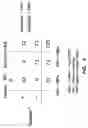
Following the above procedures, ten fentogram (10 fg) of mycobacterial genomic DNA from both M. tb and M. avium were analyzed. It is clearly indicated in FIG. 2 that M. tb could be differentiated from M. avium, suggesting that this detection kit could detect as low as 1 to 20 copies of M. tb.
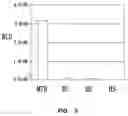
Example 2As indicated in FIG. 3, MTB assay specifically detected M. tuberculosis (MTB) in contrast to E. coli (EC), human DNA (HU) or Herring Sperm DNA (HS).
Example 3Different samples were assayed by the assay system of the invention. It is clearly shown in FIG. 4 that MTB assay specifically detected M. tuberculosis but not MOTT (Mycobacterium Other Than Tuberculosis) including M. matinum (Mma), M. avium (Mav) and M. intracellulare (Mint) from clinical samples.
Example 4Clinical samples, both positive and negative cases determined by smear test, culture confirmation and Nested-PCR, were examined by the invention. The results in FIG. 5 were identical to that mentioned prior. These results had shown that the M. tuberculosis detection kits of the invention achieved extremely high sensitivity and specificity.
While the invention has been described and exemplified in sufficient detail for those skilled in this art to produce and use it, various alternatives, modifications, and improvements should be apparent without departing from the spirit and scope of the invention.
One skilled in the art readily appreciates that the present invention is well adapted to carry out the objects and obtain the ends and advantages mentioned, as well as those inherent therein. The cell lines, embryos, animals, and processes and methods for producing them are representative of preferred embodiments, are exemplary, and are not intended as limitations on the scope of the invention. Modifications therein and other uses will occur to those skilled in the art. These modifications are encompassed within the spirit of the invention and are defined by the scope of the claims.
It will be readily apparent to a person skilled in the art that varying substitutions and modifications may be made to the invention disclosed herein without departing from the scope and spirit of the invention.
All patents and publications mentioned in the specification are indicative of the levels of those of ordinary skill in the art to which the invention pertains. All patents and publications are herein incorporated by reference to the same extent as if each individual publication was specifically and individually indicated to be incorporated by reference.
The invention illustratively described herein suitably may be practiced in the absence of any element or elements, limitation or limitations, which are not specifically disclosed herein. The terms and expressions which have been employed are used as terms of description and not of limitation, and there is no intention that in the use of such terms and expressions of excluding any equivalents of the features shown and described or portions thereof, but it is recognized that various modifications are possible within the scope of the invention claimed. Thus, it should be understood that although the present invention has been specifically disclosed by preferred embodiments and optional features, modification and variation of the concepts herein disclosed may be resorted to by those skilled in the art, and that such modifications and variations are considered to be within the scope of this invention as defined by the appended claims.
Other embodiments are set forth within the following claims.
Claims
1. A method for detecting microorganism DNA comprising:
(a)hybridizing the microorganism cDNA with microorganism-specific probes in hybridization tube wherein the probe linked to magnetic bead;
(b) transferring hybridization tubes to magnetic wells for washing;
(c) adding blocking solution into the tubes;
(d) adding avidin enzyme complex or streptavidin enzyme complex into the tubes
(e) performing washing reaction to remove interfering material by the aid of magnetic field;
(f) suspending magnetic beads; and
(g)detecting the luminescent or color change after adding substrate of enzyme.
2. The method of claim 1, wherein the microorganism is Mycobacterium tuberculosis.]
3. The method of claim 1, wherein the microorganism cDNA are obtained from the PCR amplification mediated by bioactive primers.
4. The method of claim 1, wherein the streptavidin enzyme complex in the step (d) is streptavidin horseradish peroxidase (SA-HRP).
5. The method of claim 1, wherein the step (f) suspending magnetic beads is performed by vortexing the tube.
6. The method of claim 1, wherein the detection of the step (g) is performed by luminometer or spectrophotometer.
7. The method of claim 1, wherein the steps (a)-(g) are performed in the same tube.
8. An apparatus for performing the dissociation of nucleic acid double strands, hybridization, washing, the separation of magnetic beads and thermal control in the same apparatus, comprising:
(a) the means for fitting reaction containers;
(b) the means for controlling the temperature of the containers; and
(c) the means for controlling the magnetic force of the containers,
wherein the means for controlling the temperature of the containers are connected to the means for fitting reaction containers, and the means for controlling the magnetic force of the containers are connected to the means for fitting reaction containers.
9. The apparatus of claim 8, wherein the means for controlling the temperature of the containers to heat the containers to perform the dissociation of nucleic acid double strands according to temperature change.
10. The apparatus of claim 8, wherein the means for controlling the magnetic force of the containers to perform the magnetic change of magnetic bead to facilitate hybridization, washing and the separation of magnetic beads in the containers.
11. A diagnostic kit for detecting microorganism cDNA comprising:
(a) a probe linked to magnetic bead;
(b) bioactive primers;
(c) avidin enzyme complex or streptavidin enzyme complex; and
(d) nzyme substrate.
12. The kit of claim 11, wherein the bioactive primers are made by reacting DNA labeling reagent with the primers.
13. The kit of claim 12, wherein the DNA labeling reagent is the compound having the formula:
Fu-BE-D
wherein Fu represents a furocoumarin derivative selected from the group consisting of angelicin derivatives and psoralen derivatives;
wherein BE represents none or a binding enhancer selected from the group consisting of C4-12 alkyl, alkyenyl, polyalkylamine and polyethylene glycol; and Wherein D represents a detectable group selected from the group consisting of: biotin, fluorescence, acridinium ester and acridinium-9-carboxamide.
14. The kit of claim 12, wherein the DNA labeling reagent is 9-(4″-(Aminomethyl)-4′, 5′-Dimethyl-angelicin) acridinium carboxamide.
15. An assay system for detecting microorganisms, the system comprising:
(i) diagnostic kit for detecting microorganism cDNA comprising:
(a) a probe linked to magnetic bead;
(b) bioactive primers;
(c) avidin enzyme complex or streptavidin enzyme complex; and
(d) enzyme substrate
(ii) an apparatus for performing the dissociation of nucleic acid double strands, hybridization, washing, the separation of magnetic beads and thermal control in the same apparatus, comprising:
(a) the means for fitting reaction containers;
(b) the means for controlling the temperature of the containers; and
(c) the means for controlling the magnetic force of the containers,
wherein the means for controlling the temperature of the containers are connected to the means for fitting reaction containers, and the means for controlling the magnetic force of the containers are connected to the means for fitting reaction containers;
(iii) a magnetic rack to bind the magnetic bead on the wall of the containers; and
(iv) a detector.
16. The assay system of claim 15, wherein the bioactive primers are made by reacting DNA labeling reagent with the primers.
17. The assay system of claim 15, wherein the streptavidin enzyme complex in the kit is streptavidin horseradish peroxidase (SA-HRP).
18. The assay system of claim 15, which can differentiate M. tuberculosis from M. marinum, M. avium and M. intracellulare19. The assay system of claim 15, wherein the detector is luminometer or spectrophotometer.
20. The assay system of claim 15 The kit of claim 15, wherein the DNA labeling reagent in the kit is 9-(4″-(Aminomethyl)-4′,5′-Dimethyl-angelicin) acridinium carboxamide.
Images & Drawings included:
Sources:
- United States Patent and Trademark Office - verify current appl. status at the USPTO↗
Similar patent applications:
Recent applications in this class:
- » 20250290159 2025-09-18
METHOD FOR THE DETECTION OF LEGIONELLA - » 20250290158 2025-09-18
METHOD FOR ASSESSING SKIN - » 20250290157 2025-09-18
PATHOGEN TESTING DEVICE - » 20250277276 2025-09-04
AMPLIFICATION PRIMER KIT, A METHOD FOR DETECTING A SEXUALLY TRANSMITTED BACTERIAL INFECTION, AND A KIT FOR DETECTING THE INFECTION - » 20250277275 2025-09-04
SET OF PRIMERS, COMPOSITION OF REAGENTS AND METHOD OF DETECTING ATYPICAL BACTERIA - » 20250277274 2025-09-04
MICROBIOME SIGNATURE - » 20250270658 2025-08-28
COMPOSITION FOR DIAGNOSING PERIODONTAL DISEASES BY USING BACTERIAL CLUSTERS IN GINGIVAL CREVICULAR FLUID, AND USE THEREOF - » 20250270657 2025-08-28
Detection of Antibiotic Resistance Genes - » 20250257412 2025-08-14
METHODS FOR DETECTING BORDETELLA - » 20250236919 2025-07-24
DNA SEPARATION
Recent applications for this Assignee:
- » 20080268425 2008-10-30
Methods and kits for detecting classical swine fever virus