Methods for delivering extracellular target into cells
US20070173470A1
2007-07-26
11/337,109
2006-01-23
Abstract:
The invention relates to a method for intracellular delivery of substances, comprising the steps of suspending the substance in aqueous solution and providing sufficient speed to enable the suspended substance to penetrate the cell surface and become incorporated into the cell. This method does not require the accompaniment or aid of any particle, and both the substance and the cell retain their biological activity after the entry of substance into the cell.
Interested in similar patents?
Get notified when new applications in this technology area are published.
Classification:
C12N13/00 » CPC main
Treatment of microorganisms or enzymes with electrical or wave energy, e.g. magnetism, sonic waves
C12N15/895 » CPC further
Mutation or genetic engineering; DNA or RNA concerning genetic engineering, vectors, e.g. plasmids, or their isolation, preparation or purification; Use of hosts therefor; Recombinant DNA-technology; Introduction of foreign genetic material using processes not otherwise provided for, e.g. co-transformation using microinjection using biolistic methods
A61K48/00 IPC
Medicinal preparations containing genetic material which is inserted into cells of the living body to treat genetic diseases; Gene therapy
C12N15/09 IPC
Mutation or genetic engineering; DNA or RNA concerning genetic engineering, vectors, e.g. plasmids, or their isolation, preparation or purification; Use of hosts therefor Recombinant DNA-technology
C12N15/74 IPC
Mutation or genetic engineering; DNA or RNA concerning genetic engineering, vectors, e.g. plasmids, or their isolation, preparation or purification; Use of hosts therefor; Recombinant DNA-technology; Introduction of foreign genetic material using vectors; Vectors; Use of hosts therefor; Regulation of expression Vectors or expression systems specially adapted for prokaryotic hosts other than E. coli, e.g. Lactobacillus, Micromonospora
C12N15/82 IPC
Mutation or genetic engineering; DNA or RNA concerning genetic engineering, vectors, e.g. plasmids, or their isolation, preparation or purification; Use of hosts therefor; Recombinant DNA-technology; Introduction of foreign genetic material using vectors; Vectors; Use of hosts therefor; Regulation of expression; Vectors or expression systems specially adapted for eukaryotic hosts for plant cells, e.g. plant artificial chromosomes (PACs)
C12N15/87 » CPC further
Mutation or genetic engineering; DNA or RNA concerning genetic engineering, vectors, e.g. plasmids, or their isolation, preparation or purification; Use of hosts therefor; Recombinant DNA-technology Introduction of foreign genetic material using processes not otherwise provided for, e.g. co-transformation
Description
FIELD OF THE INVENTIONThis invention relates to methods for delivery a target into cells.
DESCRIPTION OF PRIOR ARTDelivery of bioactive molecules to intact living cells or tissues can be achieved by a variety of chemical or physical methods. Some of the commonly used fluorescence dyes, such as Hoechst 33258 and Lucifer Yellow, cannot directly cross the plasma membrane by themselves; these probes are typically microinjected, or “permeabilized” into cells that are lightly extracted by mild detergent treatments (Arndt-Jovin and Jovin, 1989, In “Methods in Cell Biology”, Ch. 16, pp. 417-448, Academic Press, New York; Swanson et al., 1987, J. Cell. Biol., 104: 1217-1222). Introduction of bioactive molecules into cells can also be achieved by chemical means. In the conventional “transfection” procedure, DNA molecules can be effectively delivered to cultured cells by using calcium phosphate precipitation, or by coating with cationic lipid or being included in the liposomes (Bergan et al., 2000, Pharm. Res., 17(8): 967-973 ; Hsiung et al., 1982, Mol. Cell. Biol. 2(4): 401-411); however, such process has not been successfully applied to the delivery of (exogenously made) recombinant proteins. Moreover, certain types of cells (such as neuronal cells or functionally well-differentiated cells) have been shown to be more resistant to such transfection protocols than others. In fact, gene deliveries to primary cultured neurons and/or polarized epithelial cells (such as MDCK and Hep G2 cells) are notoriously difficult. DNA delivery can also be accomplished by biological means. Recent progress has made possible packing and delivering biomolecules into living cells both in vitro and in vivo by adenovirus, retrovirus, and papillomavirus (Fujita et al., 1995, J. Virol., 69(10): 6180-6190; Kalpana, 1999, Semin. Liver Dis., 19(1): 27-37; Sverdrup et al., 1999, Gene Therapy, 6: 1317-1321); again, such protocols do not allow delivery of recombinant proteins.
In another approach, bioactive molecules (such as DNA or proteins) can be linked or physically absorbed onto a solid substrate carrier (such as microparticles). The microparticle carriers can then be accelerated to a very high speed in the medium such that they can penetrate through the plasma membrane without significantly damaging the cell, as has been described in the so-called “gene gun” method (Sanford et al., 1987, Partic. Sci. Technol., 5: 27-37). Although this method possesses the advantage in penetration depth especially when it is used in plant cells/tissues or animal skins, the presence of the deliver particles within the cytosol may unequivocally cause some adverse effects of the resident cell. Note also that the coating and absorption of bioactive molecules on the deliver particles by conventional gene gun protocols are better developed for DNA instead of proteins.
However, using gene gun for delivering DNA into cells still has some problems. The gene gun is a particle delivery way, the “bullet” is essential. DNA has to be coated on microparticle to penetrate the cell membrane. The microparticle commonly used is golden particle or tungsten particle. Preparation of bullet includes pre-treating the dried powder of microparticle by glycerol, coating the DNA on the pre-treated microparticle, spreading the coated microparticle on plastic tube, and cutting the plastic tube to pieces to be the bullet. The process has disadvantages including complicated steps, time-consuming and high tech threshold. Further, the pre-treated microparticle has limited preservation period and using the golden and tungsten particle is high cost.
The methods of present invention can overcome these disadvantages and provide more easy and effective process.
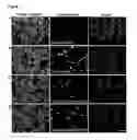
BRIEF DESCRIPTION OF THE DRAWINGSFIG. 1 illustrates delivery effects of chemical molecules in different sizes and properties. Cell morphology is observed under phase contrast microscope, and fluorescence signals are observed under fluorescence microscope. CHO cells delivered with Hoechst 33258 are shown in (A), the result is detected immediately after PBS wash. Signals of Lucifer yellow are shown in (B). Results of delivered dextrans (MW:70K) conjugated with TRITC and dextrans (MW:500K) conjugated with FITC are shown in (C) and (D). Scale bar indicates 20 um.
FIG. 2 illustrates delivery of chemical molecules in different cell types. Hep G2 cells are used in chemical molecules delivery assay. (A) Hoechst 33258, (B) Lucifer yellow, (C) dextrans (MW:70K) conjugated with TRITC and (D) dextrans (MW:500K) conjugated with FITC are observed under phase contrast and fluorescence microscopy. Scale bar indicates 20 um.
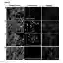
FIG. 3 illustrates delivery and expression of plasmid DNA in different cell types. EYFP fused subcellular localization plasmids are used for delivery and expression; results are recorded by confocal microscopy with fluorescence and DIC images. (A) Expressed EYFP-actin shows submembrane pattern and the apical domain can be found (arrow). (B) Filamentous structure can be easily observed in Hep G2 cells with expression of delivered EYFP-tubulin plasmids. (C) Expression of EYFP-nuclei shows nuclear morphology and condensed signal in nucleolus (arrows). The nuclear identification can be also found in DIC images. (D) Membrane localization by expressing EYFP-membrane plasmid can be found in delivered Hep G2 cells (arrows). (E) Expression of EYFP-mitochondria plasmids reveals mitochondria localization in Hep G2 cells. Scale bar indicates 10 um in (A)˜(E). (F) EYFP-tubulin plasmids are delivered into retina explants of goldfish and observed under microscope after 24 hr. Neuronal axon which expressed EYFP-tubulin can be found under fluorescence microscopy (arrows). Bar=200 um.
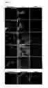
FIG. 4 shows delivery of fluorescence labeled actin and tubulin monomers into cells. Rhodamine (Rh)-labeled actin and tubulin monomers are used. (A) CHO cells are fixed and stained with Rd-Ph. Rh-labled monomeric actin assembles into actin filaments (arrows). (B) Fish keratocytes are subjected to the monomer delivery and observed under fluorescence microscope. Fixed and stained with actin antibodies shows the assembled actin filaments after incubation and colocalization in several stress fibers can be easily found (arrows). (C) Lived fish keratocytes delivered with Rd-F-actin are observed under fluorescence microscope and recorded with time-lapsed imaging system. (D) The same experimental setup is performed in Rd-F-tubulin delivery in fish keratocyte. Scale bar indicates 10 um
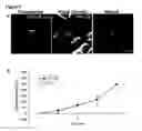
FIG. 5 shows delivery and replication of E. coli in cells. EYFP-E.coli is delivered into Hep G2 cells and gentamycin is supplement with cell culture medium for further incubation. (A) After delivery, Hep G2 cells are treated with gentamycin for 1 hr and observed under fluorescence and phase contrast microscope. E. coli inside of cells are found (arrow). (B) Cells treated with gentamycin for 1 hr and lysed for colony formation assay are determined as time point 0. In different time point, gentamycin is supplemented with cell culture medium until cell lysed to inhibit the bacteria growth. The growth curve of delivered E.coli is recorded (filled square). Negative control is performed with the same amount of E.coli and put into cell culture medium with Hep G2 cells with gentamycin (open circle). The result is collected from four independent experiments. Mean±SD is shown.
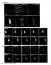
FIG. 6 shows direct and indirect mechanisms to deliver molecules. Different molecules are delivered into CHO cells. In direct delivery assay, fluorescence probes are put on the membrane for direct delivery. Cells are washed immediately with PBS (left). In indirect delivery assays, same volume of the H2O is put on the membrane to replaced fluorescence probes. Probes are mixed with cultured medium with CHO cells. After shocking, cells are washed immediately (middle) or further incubated for 5 min and washed twice with PBS (right). Cells are observed under phase contrast and fluorescence microscope. (A) Hoechst 33258, (B) lucifer yellow, (C) TRITC labeled (MW:3K), (D) (MW:40K), (E) (MW:70K) dextrans, and (F) FITC labeled dextrans (MW:500K).
SUMMARY OF THE INVENTIONThis invention provides a method for delivering a target into cells comprising: (a) mixing the target with a solution to form a mixture, and (b) delivering the mixture into a solution containing the cells by high pressure force.
This invention also provides a cell used for the method described as above, which facilitates the absorption of exogenous target into the cell based on the condition of the membrane of the cells by high pressure force.
This invention further provides a method for indirectly delivering a target into cells, comprising: (a) mixing the target and the cells, and (b) changing the condition of the membranes of the cells by high pressure force.
DETAILED DESCRIPTION OF THE INVENTIONTerm Definition
“Target” used in this invention means the substance delivered into cells.
Macromolecule: A very large molecule, such as a polymer or protein, consisting of many smaller structural units linked together.
This invention provides a method for delivering a target into cells comprising: (a) mixing the target with a solution to form a mixture, and (b) delivering the mixture into a solution containing the cells by high pressure force. The method herein is characterized as non-particle delivery.
The target delivered into cells in this invention is selected from the group consisting of chemical, fluorescent compound, molecule exhibiting bio-activity, micromolecule, macromolecule and microorganism. After the delivery, the target can still maintain function or activity in the cell.
In one embodiment, the chemical is dye, fluorescent dye, polysaccharide, or pharmaceutical compound. In one embodiment, the molecule exhibiting bio-activity is enzyme, drug, vaccine, antigen or antibody. In one embodiment, the macromolecule is protein, polypeptide, RNA, DNA, or other oligonucleotide. Further, the oligonucleotide is promoter, enhancer, siRNA, morpholino or other regulatory sequences.
The target can also be a cell in smaller size than the delivered cell. In one embodiment, the target is microorganism. In a preferred embodiment, the microorganism is bacterium or yeast. In a more preferred embodiment, the microorganism is transformed bacteria or yeast. Further, the microorganism maintains life cycle and propagation capability in the cell after delivery.
The cell delivered with exogenous targets herein is prokaryotic cell or eukaryotic cell. The cell is selected from the group consisting of animal, insect, plant, fungus, bacterium, rickettsia and Chlamydia.
The solution used in this method is charged or uncharged. In a preferred embodiment, the solution is water, phosphate saline buffer or media.
The high pressure force is produced by gene gun. The gene gun can be available from commercial product. The conditions of gene gun used in this method are set according to the experimental situation such as cell type, target type or other factors.
The method of the invention can be applied in various fields, for example but not limitation, the field of gene therapy and the field of evolutionary study.
The present invention also provides a cell used for the method described as above, which facilitates the absorption of exogenous target into the cell based on the condition of the membrane of the cells by high pressure force. The target is selected from the group consisting of dye, fluorescent dye, polysaccharide, pharmaceutical compound, fluorescent compound, enzyme, drug, vaccine, antigen, antibody, protein, polypeptide, RNA, DNA, promoter, enhancer, siRNA, morpholino, regulatory sequences and microorganism.
The invention further provides a method for indirectly delivering a target into cells, comprising: (a) mixing the target and the cells, and (b) changing the condition of the membranes of the cells by high pressure force. This method is characterized as non-particle delivery. More detailed, high pressure force is used in this method to alter the strength of cell membrane and to make the membrane loosen. The pressure force also causes the mixture containing cells and targets shaking to increase the opportunities of collision between cells and targets. Based on the above reasons, the target can pass through the cell membrane and be absorbed by cells.
In a more preferred embodiment, the high pressure force is produced by gene gun. The target is selected from one group consisting of dye, fluorescent dye, polysaccharide, pharmaceutical compound, fluorescent compound, enzyme, drug, vaccine, antigen, antibody, protein, polypeptide, RNA, DNA, promoter, enhancer, siRNA, morpholino, other regulatory sequences, bacterium and yeast. The cell is selected from the group consisting of animal, insect, plant, fungus, bacterium, rickettsia and Chlamydia.
This method can be applied in various fields, for example but not limitation, the field of gene therapy and the field of evolutionary study.
The methods provided by this invention also can be applied in producing transgenic plant or transgenic animal. The transgenic plant includes corn, rice, soybean, potato, rape, cotton, mushroom, orchid, crops and garden plants. The transgenic animal includes pets, domestic animals, domestic fowls, experimental animals or even protein-producing animal.
The methods of this invention can be applied in gene therapy, for example but not limited, DNA vaccine or cancer therapy. Further, the methods of this invention can delivery not only DNA but also chemical and bioactive compound. This enlarges the field of gene therapy.
For the evolutionary study, it is believed that extremely conditions such as unstable atmosphere, continued thunder and lighten, volcanic eruption, and earthquake had appeared in youth of the Earth. The first life style is speculated to appear on Earth in the age. The primitive cell first appeared in the world is speculated to absorb other molecules such as RNA, perhaps active or passive, during evolution. The force of absorbing is speculated from the extremely environment providing such as thunder and lighten. This invention provides a method to give the cell a high pressure force. The method can be used for mimic the conditions described as above and applied to study biological evolution.
EXAMPLEMaterials and Methods
Cell Lines
CHO (Chinese Hamster Ovary) and Hep G2 cell lines were used in this investigation. Standard cell culture protocols were followed. Cells were typically cultured with DMEM (GIBCO BRL) supplemented with 10% fetal calf serum, 2 mM L-glutamine, and 5% non-essential amino acids (GIBCO.BRL) and incubated at 37° C. in the presence of 5% CO2.
Primary Cells
Isolation of retinal explants and keratocytes, goldfish was anesthetized with 0.05% ethyl 3-aminobenzoate (Sigma). Optic nerve crushes were administered by crushing the exposed nerve behind the orbit. Nerve crushed fish were stored in water tank for 7 days in room temperature. Before retina isolation, the fish was dark-adapted for 30 min and then anesthetized. Eyes were removed and put into the Hanks' buffer (Sigma). Retina was isolated from eyecup, and chopped into 0.4 mm square pieces. Isolated explants were washed with Hanks' buffer. Retina explants were plated onto poly-L-lysine coated coverslips for plasmid delivery.
Fish keratocytes were isolate from fish scales. In brief, fish scales were extracted with tweezers, place on dry coverslips, until it almost dry. Hepes-DMEM (GIBCO Life Technology) supplemented with 10% fetal bovine serum, and 0.1% gentamicin solution was used to culture over night and allow the keratocyte migrate out of the fish scales. Migrated keratocyte were trypsinized by Trypsin EDTA. Collected keratocytes were put on the sterilized coverslips for molecule delivery.
Chemicals, Polysaccharides, Plasmids and Proteins
All fluorescence marker and fluorescence labeled dextrans were purchased from Molecular probes (Molecular probes, Eugene, Oreg.). Each shot, Hoechst 33258 was used in 3 ug, lucifer yellow was used in 15 ug. TRITC labelled dextrans (MW:70K) were used in 60 ug, and FITC labelled dextrans (MW:500K) were used in 60 ug. EYFP fused subcellular localization plasmids were purchased from Clontech. Plasmids were used in 6 ug for each experiment. Rhodamine conjugated actin and tubulin (Cytoskeleton, Denver) were used in 40 ug every shot.
Transformation
Competent cells (strain BL21-DE) for bacteria transformation were prepared by calcium chloride. The optimal optical density (OD600) range for competent cell preparation was 0.15-0.45. Conventional transformation was performed. In brief, 50 μl competent cells with 1 μg/μl plasmid DNA (pTriEx-3 Vector from Novagen) were mixed, incubated for 30 min at 37° C., heat shocked at 42° C. for 90 s, transfered to ice for 2 min, added with 100 μl liquid LB medium, and then recovered at 37° C. for 45 min. Transformed bacteria was spread on the LB plate with ampicillin at 37° C. over night.
Colony Formation Assay
Hep G2 cells were cultured on 10-cm dishes for 24 hr for bacteria delivery assay. Cells were washed with PBS twice immediately after bacteria delivery, and divided into five 35-mm dishes by trypsinization. Cell culture medium with gentamycin (100 μg per ml) supplement was used to inhibit extracellular bacteria activity. In the period of time indicated in experiments, cells were lysed with NET buffer (150 mM NaCl, 0.5% NP-40, 50 mM Tris, 1 mM EDTA and 1% Triton X-100, supplemented with protease inhibitors) for 30 min at 4° C. Cell pellets were collected after 12,000×g centrifugation at 4° C. for 30 min, and spread on the LB plates with ampicillin (40 μg/ml). Cultured overnight at 37° C. incubator and formed colony were first confirmed by immunofluorescence microscope and counted for statistics. Colony formation number was averaged from 4 independent experiments and standard deviation was provided.
Solution Accelerated Method
The target for delivery was mixed with a solution and accelerated by high-pressured pure helium gas. PDS-1000/He system (Bio-Rad) was used to supply the high velocity. The bombarded volume is 6 ul in every experiment. The target cells were put on the target shelf and the compositions for delivery were loaded on the microcarrier membrane. Then, the rapture disk was assembled to the rapture disk retaining cap and the microcarrier was assembled to the macrocarrier cover as described in the user guide (Bio-Rad). The door of the bombardment chamber was closed; the suction was turned on to vacuum the chamber. When the degree of the vacuity was enough, the helium tank was turned on to supply the high velocity. The high pressure forced the target penetrating into the cells. After every shot, cells were immediately wash with PBS twice and fill with fresh medium to avoid biological endocytosis. Cells were then observed under fluorescence microscope (Leica GmbH, Heidelberg, Germany) or further incubated if need.
Imaging Study
Every sample was observed immediately after bombarded, except for plasmid delivery and rhodamine conjugated actins and tubulins. Plasmids delivered cells were further incubated for extra 48 hrs at 37° C. Fluorescece labeled proteins were incubated at 37° C. for 30 min after delivering. All images were taken by SIT camera (Hamammatsu 2400) and analyzed by Metamorph (Universal Imaging Corporation, West Chester, Pa.), or by confocal microscope (Leica GmbH, Heidelberg, Germany).
Statistics
Cells with positive signals under microscope were calculated field by field and versus total cell number. Finally, total cell number was normalized into 1000 in terms of comparison.
Immunostaining
For immunofluorescence staining, cells were typically cultured on a 22×22 mm square coverslip which was pretreated with 6N HCl and 95% ethanol, and coated with 200 g/ml poly-L-lysine (MW 70-150 KDa, Sigma) as previously described (Lian et al., 1999, Hepatology, 30(3): 748-760; Lin and Forscher, 1993, J. Cell Biol., 121(6): 1369-1383). Cells were fixed with 4% paraformaldehyde/2 mM EGTA/400 mM sucrose/PBS at RT for 15 min, then permeabilized with 0.5% Triton X-100 in the fix solution for 5 min. The samples were then incubated with 5 mg/ml BSA/PBS, then with primary antibodies at RT for 1 hr. The concentrations of primary antibodies utilized were 1:100 anti-beta-tubulin antibody (Sigma). After extensive PBS washes, fluorophore-conjugated secondary antibodies (Jackson Immuno Research, West Grove, Pa.) were added at the concentrations recommended by the manufacturer at room temperature for 1 hr. About 1 unit/ml FITC-Ph was used for F-actin staining. The stained samples were mounted using an anti-photobleaching medium containing 20 mM n-propyl-gallate (Sigma) in 80% glycerol/20% PBS All images were recorded in a digital platform for data analysis and image processing.
EXAMPLE 1Delivery of Fluorescent Compounds and Polysaccharides in Different Sizes into Different Cell-types
To demonstrate the molecules delivery through solution accelerating methods, CHO (Chinese Hamster Ovary) cells were first used. The bisbenzimide dyes-Hoechst 33258 is a fluorescence chemical compound, which can bind to the minor groove of double-helix DNA. The molecular weight of Hoechst is 623.96. Hoechst 33258 is used as a nucleic marker due to its fluorescence characteristic and high affinity to DNA. Usually, Hoechst 33258 can only be used in fixed cells or detergent-permeable cells. Although, Hoechst 33258 has been reported with slightly lower permeability efficiency than Hoechst 33342 (Arndt-Jovin and Jovin, 1989, In “Methods in Cell Biology”, Ch. 16, pp. 417-448, Academic Press, New York). Invisible signals were found while cells were incubated with Hoechst 33258 for 5 minutes. Solution accelerating method provided an extremely short period of time for contacts between molecules and cell membrane. To discriminate the results from endocytosis, cells were washed twice with PBS and changed with new medium for further incubation every shot. Compared with results from incubating cells with soluble molecules, solution accelerating method performed a quick and harmful-less delivery route.
The result of Hoechst 33258 was shown in FIG. 1A. The concentration of Hoechst 33258 was diluted into 1/10. Delivery rate was reduced after dilution (Table 1). Hence, it was found that molecule using solution accelerating to deliver showed a dose dependent manner.
N-(2-aminoethyl)-4-amino-3,6-disulfo-1,8-naphthalimide, dipotassium salt (lucifer yellow ethylenediamine) has the molecular weight in 491.57 (Stewart, 1981, Nature, 292 (2): 17-21). Lucifer yellow CH (LY-CH or LY) was used to determine whether the polar trace molecule can be delivered through this unique solution accelerating method in present invention. LY is an impermeable fluorescence dye, which is often used as a microinjection marker and can not be transported through gap junction (Powley and Berthoud, 1991, J. Neuro. Methods, 36: 9-15). LY showed the similar results, which can be delivered through solution accelerating method and showed the dose dependent manner (See Table 1). In FIG. 1B, fluorescence signal from LY was found in entire cell.
Dextrans is a hydrophilic polysaccharide synthesized by Leuconostoc bacteria. Dextrans has been widely used in various biological applications due to their high water solubility and low toxicity. Dextrans is available in different sizes and different fluorochrome-labeled forms. Dextrans (MW:70K) is used as an endocytosis marker (Makarow, 1985, EMBO J., 4(7): 1861-1866). Here both TRITC labeled dextran (MW:70K) and FITC labeled dextrans (MW:500K) were used to demonstrate the delivery manners in different molecule sizes. It was found that dextrans (MW:70K) can be transfered into cells by the method herein. To avoid the possibility that the cytosolic signal may come from endocytosis, soluble dextrans (MW:70K) were incubated in the same concentration for 5 minutes in 37° C. No significant signals in cells were found.
Therefore, large molecule delivery by dextrans (MW:500K) was further tested. In FIGS. 1C and D, both dextrans (MW:70K) and dextrans (MW:500K) were successfully introduced into cells as small molecules, demonstrating that the method of the present invention can deliver not only small molecules but also large molecules in liquid phase. Both dextrans (MW:70K) and dextrans (MW:500K) showed dose dependent manner (Table 1). Interestingly, the dose-dependent effect was more obvious in dextrans (MW:500K) (reduced from 39.1 to 8.28% while the concentration is half) than in dextrans (MW:70K) (reduced from 38.16 to 25.44% while the concentration id half) dextrans. The result supported that the method of the present invention may play more than one role in molecules delivery, or this method is restricted in large molecules with lower concentration.
| TABLE 1 |
| Molecule delievery efficiency by the solution accellerating method |
| Delivery | |||
| effciency# | |||
| Target | (targeted cells/ | ||
| cells | Molecules delivered | Concentrations* | 500 cells) |
| CHO | Hoechst 33258 | 1 | mg/ml | 65.48 ± 18.81 |
| (M.W. = 623.96) | 0.5 | mg/ml | 57.7 ± 25.56 | |
| 0.1 | mg/ml | 27.25 ± 7.22 | ||
| 2.5 | mg/ml | 35.47 ± 8 | ||
| Lucifer yellow | 1.25 | mg/ml | 19.9 ± 2.64 | |
| (M.W. = 491.57) | 0.5 | mg/ml | 11.95 ± 3.36 | |
| 10 | mg/ml | 38.16 ± 8.19 | ||
| 70 kD dextran | 5 | mg/ml | 25.44 ± 9.46 | |
| 2.5 | mg/ml | 19.04 ± 4.61 | ||
| 10 | mg/ml | 39.1 ± 8.79 | ||
| 500 kD dextran | 5 | mg/ml | 8.28 ± 2.19 | |
| 2.5 | mg/ml | 4.35 ± 2.66 | ||
| CHO | DNA (GFP-tubulin gene) | 1 | mg/ml | 34.82 ± 5.07 |
| Hep G2 | DNA (GFP-tubulin gene) | 1 | mg/ml | 9.06 ± 1.85 |
| CHO | Actin proteins | 3 | mg/ml | 67.15 ± 13.04 |
| CHO | Tubulin proteins | 3 | mg/ml | 62.36 ± 5.89 |
*Concentrations of the solutions used for delivery are shown; delivery volume = 6 ml. |
||||
#Mean +/− STDEV is shown. |
To rule out the possibility that using solution accelerating method may be discriminated by distinct cell models, the same experiments were carried out in human liver blastoma cell line, Hep G2 cells, to see whether the delivery could be achieved or not. In FIG. 2A-B, Hoechst 33258 and LY were successfully delivered into Hep G2 cells by solution accelerating method. The delivered efficiency was not affected by different cell types from statistic results in CHO cells (Table 1) and Hep G2 cells (Table 2, probe direct bombardment). Both dextrans (MW:70K) and dextrans (MW:500K) were delivered to Hep G2 cells (FIG. 2C-D). Efficiency comparison can be found from Table 1 and Table 2, showing that the molecules delivery through solution accelerating method is not cell type specific manner.
EXAMPLE 2Delivery and Expression of Plasmid DNA in Different Cell Types
Particle carrier method (gene gun) has been widely used for DNA delivery (Johnston and Tang, 1994, in “Methods in Cell Biology”, vol. 43, ch. 17, pp. 353-365, Academic Press). Similar to the works with high-pressured helium which accelerates plasmid DNA, accelerating soluble plasmid DNA (EYFP fused subcellular localization plasmids) alone without any particle carriers was tested herein. After delivery, Hep G2 cells were further incubated for 24 hr and observed under confocal microscope. Interestingly, accelerated soluble plasmids were delivered and expressed in cells without particle carriers. Hep G2 cells are polarized hepatocytes. Membranes in polarized Hep G2 cells were distinguishable from basolateral domain and apical domain. Apical domain in Hep G2 cells was full of microvilli and actin accumulated signals were detectable by F-actin staining (Lian et al., 1999, Hepatology, 30(3): 748-760).
Expression of EYFP-actin by solution accelerating methods was found to be located on both submembrane and apical domain (FIG. 3A, arrows) in Hep G2 cells. EYFP signals in microtubule filaments can be found in Hep G2 (FIG. 3B). EYFP-nuclei showed nuclear localization, strong EYFP signal was observed in nucleolus (FIG. 3C, arrows). Nuclear and nucleolus morphology were confirmed by DIC images (FIG. 3C). EYFP signal can be found on cell membrane by the expression of EYFP-membrane plasmid (FIG. 3D, arrows). The expression of EYFP-mitochondria represented the mitochondria localization in Hep G2 cells (FIG. 3E). These results indicated that without particles as carriers, plasmid can be delivered and express in cells properly. Different cell line were also used for plasmid delivery experiments (Table 1), different cell type affected the delivery efficiency.
Tissue explants are difficult to be delivered with plasmid for expression. Goldfish retina explants were used to test whether the plasmid delivery can be used by solution accelerating method or not. It was shown that EYFP signal can be observed in neuronal cells inside of retina explant (FIG. 3F, arrows). This result demonstrated that DNA delivery without particle carriers can be performed both on cells and tissues.
EXAMPLE 3Delivery of Proteins and Maintenance of its Activity in Different Cell Types
Delivery of proteins is also difficult to achieve in cellular level, especially in large peptides. In previous studies, researchers have tried different methods to accomplish this issue, such as using a short peptide vector which contains a hydrophobic region as a carrier (Hawiger, 1997, Curr. Opin. Immunol., 9:189-194; Morris et al., 1997, Nucle. Acids Res., 25(4): 2730-2736), or incorporating a palmitoyl-lysine residue into the N- or C-terminal end to deliver the short hydrophilic peptides (Loing et al., 1996, Peptide Research, 9(5): 229-232). Moreover, Johnson et. al., have used saponin to produce the transient cell membrane permeabilization in neonatal cardiac myocytes, which showed they successfully introduced 125 I-labeled calmodulin and a 20 KDa protein kinase C epsilon fragment into the cells (Johnson et al., 1996, Circulation Research, 79: 1086-1099). Recently, Pep-1, a cell penetrated peptide was reported from Morris et. al, (Morris et al., 2001, Nature Biotechnology, 19: 1173-1176). It showed a powerful delivery function in varies of peptides and proteins.
In the present invention, whether proteins can be delivered into cells through soluble acceleration method or not was also examined. Rhodamine labeled actin and tubulin were tested in solution phase. After proteins were delivered, cells were washed with PBS twice immediately to avoid further uptake the soluble exogenous proteins in medium. After washing out the non-delivered proteins, cells were further incubated for 30 minutes in cell culture incubator to examine whether exogenous proteins can be assembled or not. CHO cells and fish keratocytes were used in protein delivery experiments. In order to confirm the F-actin or microtubule filaments, immuno-fluorescence was also performed by FITC-phalloidin and anti-tubulin antibodies. Distribution pattern from rhodamine and FITC showed almost co-localization, which represented that delivered rhodamine-actin can not only entry cell membrane successfully but also incorporated into actin assembly (FIG. 4A-B, arrows). The increasing cytosolic rhodamine signals in delivered cells were also found, probably due to the free form rhodamine labeled actin. Rhodamine labeled tubulin was examined and found to be incorporated into microtubule assembling through our solution acceleration method (Table 1). From statistic results as shown in Table 1, protein delivery efficiency in cellular level is about 67%.
Function of delivered proteins was also tested by kinetic experiments in fish keratocytes. Fish keratocytes delivered rodamine labeled actin and tubulin were observed under time-lapsed fluorescence microscope. Cell migration and the incorporation of exogenous proteins were recorded. In FIG. 4C, although freely formed rhodamine actin affected the observation of actin filament, stress fibers still can be observed during cell migration (arrows). Condensed rhodamine signals in membrane ruffling also indicate the actin localization (arrow head). Rhodamine labeled tubulin showed filamentous structure and dynamic activities during cell migration (FIG. 4D). It was demonstrated that delivered exogenous protein can incorporated and assembled with actin and tubulin, and participated to the cytoskeletal function in cell migration.
Hence, it was successfully proved that the method of the invention can delivered larger proteins as actin (43 KDa) or tubulin (55 KDa), and the biological function is still maintained.
EXAMPLE 4Delivery and Replication of Bacteria
Pathogenic bacteria invade to cells through several mechanisms. Listeria and Shigella has been reported the invasive pathway and molecule interaction with cell membrane proteins and cytoskeleton for invasion and intracellular transport (Gouin et al., 2005, Curr. Opin. Microbiol., 8: 35-45). Different invasive mechanism involve different molecules on cell membrane, however, initiation of internalization caused bacteria rounded by vacuole. It is essential to escape from vacuole to avoid it (Higley and Way, 1997, Curr. Opin. Cell Biol., 9: 62-69). The method of the invention provides the possibility to dissect whether the internalization and vacuole formation is essential for bacteria invasion and survival or not. First, E. coli, strain BL21-DE was transformed with the bacteria expression GFP vector. 2 ml O.D. 0.2 (OD=600) EGFP E.coli was harvested in LB broth, and concentrated to 20 ul by centrifugation. 6 ul was used for each shot or add into cell culture medium as the control. Gentamycin was supplement with culture medium at least one hour to inhibit bacteria activities after PBS wash twice. For further incubation, gentamycin was always supplemented with cell culture medium. After 1 hr incubation with gentamycin, Hep G2 cells were washed with complete medium and observed under fluorescence microscope. EGFP E.coli survived in Hep G2 cells was recorded under microscope (FIG. 5A).
To further demonstrate the survival and bio-function in delivered EGFP E.coli, replication was used as the activity assay for delivered EGFP E.coli. Cells were lysed and pellets subject to colony formation assay were collected in different time point as indicate in FIG. 5B (closed square). Co-culture EGFP E.coli with Hep G2 cells supplemented with gentamycin was collected in different time point for colony formation assay as the negative control (FIG. 5B, open circle). EGFP E.coli was used in equal amount under solution accelerating methods in control experiments. From statistic results, control experiments indicated that bacteria activities were successfully inhibited by gentamycin during extended culture time. In FIG. 5B (closed square), the delivered EGFP E.coli showed increasing colony number following further incubation, indicating that the EGFP E.coli can still replicate after the delivery method herein. To sum up, the survival and biological function of delivered bacteria were still maintained by the method of the present invention.
EXAMPLE 5Delivery of the Targets can be Achieved via Both Direct and Indirect Ways
Previous studies have demonstrated that accelerated helium generated shock wave with the solution (ex. culture medium) into target cells. Shock wave was found to affect cell permeability and was used as the delivery tool (Delius and Adams, 1999, Cancer Research, 59: 5227-5232; Kodama et al., 2002, Biochimica et Biophysica Acta, 1542: 186-194; Lauer et al., 1997, Gene Therapy, 4: 710-715).
Here the effect of shock wave in the method of the invention was determined. H2O was used to replace fluorescence probes to generate shock wave. Fluorescence probes were added into culture medium with the same concentration as previous experiments. Results by using CHO cells as the cell model was shown in FIG. 6 and Table 2. After H2O delivery, cells were washed immediately (“H2O” in FIG. 6 and Table 2) or washed after 5 min incubation in room temperature. Results were compared with using fluorescence probes as the delivery molecules (“probes” in FIG. 6 and Table 2) and the control without H2O delivery (“incubate”, Table 2). Fluorescence probes added in the culture medium before H2O delivery did not show significant signals (“incubate”, Table 2). Statistic results were calculated from recorded images and more than 1000 cells were counted from 10-15 images.
It was found that Hoechst 33258 showed highly increase by H2O delivery with further incubate for 5 min compare with wash immediately (FIG. 6A, Table 2), the efficiency of Hoechst 33258 positive cells with 5 min incubation showed equilibrium as direct bombardment (Table 2). Lucifer yellow showed the similar results as Hoechst 33258 (FIG. 6B, Table 2).
These results demonstrated that the accelerating H2O generated shock wave to cells, which can be used to deliver soluble molecules. To dissect whether such delivery by shock wave is size dependent or not, different sizes of fluorescence labeled dextrans were used. In 3 KDa and 40 KDa dextrans, H2O delivery with immediately wash reduced the probe delivery efficiency in consequences of direct bombardment (FIG. 6C-D). However, 5 min further incubation rescued the delivery efficiency (Table 2).
Together, it was found that the delivery by H2O bombardment is size dependent. Similar results were also found in Hep G2 cells. In conclusion, molecules can be delivered by using the novel solution accelerating methods herein, which involve direct and indirect mechanisms.
| TABLE 2 |
| Molecules delivery with direct and indirect force |
| Probe direct | H2O and further | |||
| bombardment | H2O | incubate 5 min | Incubate | |
| Hoechst 33258 | 51.23 ± 19.68 | 15.53 ± 8.04 | 49.33 ± 21.97 | 0 |
| (0.5 mg/ml) | ||||
| Lucifer Yellow | 36.7 ± 10.74 | 19.66 ± 6.45 | 37.94 ± 13.45 | 0 |
| (2.5 mg/ml) | ||||
| Dextran (MW: 3K) | 25.19 ± 8.2 | 12.23 ± 5.54 | 34.21 ± 8.75 | 0 |
| (10 mg/ml) | ||||
| Dextran (MW: 40K) | 22.23 ± 3.44 | 10.02 ± 5.47 | 22.43 ± 8.01 | 0 |
| (10 mg/ml) | ||||
| Dextran | 26.92 ± 19.64 | 4.01 ± 1.35 | 16.29 ± 5.54 | 0 |
| (MW: 70K)(10 mg/ml) | ||||
| Dextran (MW: 500K) | 40.84 ± 17.89 | 1.498 ± 1.382 | 1.66 ± 1.53 | 0 |
| (10 mg/ml) | ||||
*Concentrations of the solutions used for delivery are shown; delivery volume = 6 ml. |
||||
#Mean +/− STDEV were shown. |
One skilled in the art readily appreciates that the present invention is well adapted to carry out the objects and obtain the ends and advantages mentioned, as well as those inherent therein. The cell lines, animals, and processes and methods for producing them are representative of preferred embodiments, are exemplary, and are not intended as limitations on the scope of the invention. Modifications therein and other uses will occur to those skilled in the art. These modifications are encompassed within the spirit of the invention and are defined by the scope of the claims.
It will be readily apparent to a person skilled in the art that varying substitutions and modifications may be made to the invention disclosed herein without departing from the scope and spirit of the invention.
All patents and publications mentioned in the specification are indicative of the levels of those of ordinary skill in the art to which the invention pertains. All patents and publications are herein incorporated by reference to the same extent as if each individual publication was specifically and individually indicated to be incorporated by reference.
The invention illustratively described herein suitably may be practiced in the absence of any element or elements, limitation or limitations, which are not specifically disclosed herein. The terms and expressions which have been employed are used as terms of description and not of limitation, and there is no intention that in the use of such terms and expressions of excluding any equivalents of the features shown and described or portions thereof, but it is recognized that various modifications are possible within the scope of the invention claimed. Thus, it should be understood that although the present invention has been specifically disclosed by preferred embodiments and optional features, modification and variation of the concepts herein disclosed may be resorted to by those skilled in the art, and that such modifications and variations are considered to be within the scope of this invention as defined by the appended claims.
Other embodiments are set forth within the following claims.
Claims
1. A method for delivering a target into cells comprising:
(a) mixing the target with a solution to form a mixture, and
(b) delivering the mixture into a solution containing the cells by high pressure force.
2. The method of claim 1, which is characterized as non-particle delivery.
3. The method of claim 1, wherein the target is selected from a group consisting of chemicals, fluorescent compounds, molecules exhibiting bio-activity, micromolecules, macromolecules and microorganisms.
4. The method of claim 3, wherein the target is a chemical selected from a group consisting of dyes, fluorescent dyes, polysaccharides and pharmaceutical compounds.
5. The method of claim 3, wherein the target is a molecule exhibiting bio-activity selected from a group consisting of enzymes, drugs, vaccines, antigens and antibodies.
6. The method of claim 3, wherein the target is a macromolecule selected from a group consisting of proteins, polypeptides, RNA, DNA, and oligonucleotides.
7. The method of claim 6, wherein the macromolecule is an oligonucleotide selected from a group consisting of promoters, enhancers, siRNA, morpholino and regulatory sequences.
8. The method of claim 3, wherein the target is a microorganism selected from a group consisting of transformed bacteria and yeast.
9. The method of claim 1, wherein the cell is selected from a group consisting of animal cells, insect cells, plant cells, fungus cells, bacteria, rickettsia and Chlamydia.
10. The method of claim 1, wherein the solution is charged or uncharged.
11. The method of claim 10, wherein the solution is water, phosphate saline buffer or media.
12. The method of claim 1, wherein the high pressure force is produced by gene gun.
13. The method of claim 1, which can be applied in the field of gene therapy.
14. The method of claim 1, which can be applied in the field of evolutionary study.
15. A cell used for the method of claim 1, which facilitates the absorption of exogenous target into the cell based on the condition of the membrane of the cells by high pressure force.
16. The cell of claim 15, wherein the target is selected from a group consisting of dyes, fluorescent dyes, polysaccharides, pharmaceutical compounds, fluorescent compounds, enzymes, drugs, vaccines, antigens, antibodies, proteins, polypeptides, RNA, DNA, promoters, enhancers, siRNA, morpholino, regulatory sequences and microorganisms.
17. The cell of claim 16, wherein the microorganism maintains life cycle and propagation capability.
18. A method for indirectly delivering a target into cells, comprising:
mixing the target and the cells, and
changing the condition of the membranes of the cells by high pressure force.
19. The method of claim 18, wherein the high pressure force is produced by gene gun.
20. The method of claim 18, which can be applied in the field of gene therapy.
21. The method of claim 18, which can be applied in the field of evolutionary study.
22. The method of claim 18, wherein the target is selected from one group consisting of dye, fluorescent dye, polysaccharide, pharmaceutical compound, fluorescent compound, enzyme, drug, vaccine, antigen, antibody, protein, polypeptide, RNA, DNA, promoter, enhancer, siRNA, morpholino, other regulatory sequences, bacterium and yeast.
23. The method of claim 18, wherein the cell is selected from the group consisting of animal, insect, plant, fungus, bacterium, rickettsia or Chlamydia.
Images & Drawings included:
Sources:
- United States Patent and Trademark Office - verify current appl. status at the USPTO↗
Similar patent applications:
- » 20180100158
Rationally-designed synthetic peptide shuttle agents for delivering polypeptide cargos from an extracellular space to the cytosol and/or nucleus of a target eukaryotic cell, uses thereof, methods and kits relating to same - » 20200270307
Rationally-designed synthetic peptide shuttle agents for delivering polypeptide cargos from an extracellular space to the cytosol and/or nucleus of a target eukaryotic cell, uses thereof, methods and kits relating to same - » 20230348537
Rationally-designed synthetic peptide shuttle agents for delivering polypeptide cargos from an extracellular space to the cytosol and/or nucleus of a target eukaryotic cell, uses thereof, methods and kits relating to same
Recent applications in this class:
- » 20250101406 2025-03-27
3D MODELS FOR PREDICTING TREATMENT RESPONSES TO ALTERNATING ELECTRIC FIELDS - » 20250043268 2025-02-06
PLATE MAGNET - » 20250002895 2025-01-02
ELECTROPORATION APPARATUSES AND THEIR METHOD OF USE - » 20240344049 2024-10-17
BIOASSEMBLY METHOD FOR SYNTHESIS USING MULTIWAVELENGTH FARADAY WAVES AND USE THEREOF - » 20240309354 2024-09-19
Systems and methods for bleaching microbial cells - » 20240301389 2024-09-12
Methods and Systems for Generation, Use, and Delivery of Activated Stem Cells - » 20240294897 2024-09-05
APPARATUS AND METHOD FOR IMMUNOMAGNETIC CELL SEPARATION - » 20240287494 2024-08-29
IMMUNE CELL TREATED BY MEANS OF MAGNETIC FIELD AND USE THEREOF - » 20240279636 2024-08-22
METHOD AND SYSTEM FOR THE IDENTIFICATION OF OPTIMIZED TREATMENT CONDITIONS FOR TREATING CELLS WITH ELECTRIC PULSES - » 20240279635 2024-08-22
BIOFILM ACTIVITY ADJUSTING METHOD