Medical device for determining the position of intracorporeal implants
US20080146926A1
2008-06-19
11/952,230
2007-12-07
✅ Patent granted
US 8,386,018 B2
2013-02-26
-
-
James Kish
Bachman & LaPointe, P.C.
2031-05-05
Abstract:
A medical device for determining the position of intracorporeal implants such as fixation systems, plates or the like, comprises at least one impedance-measuring device and an ultrasound device which are both connected to at least one intracorporeal probe, the impedance-measuring device being additionally equipped with at least one connector for connection to the implant. The device is suitable for determining the position of pedicle screws and the like in a spinal column fixation system with metal rods as stabilizers.
Assignee:
- WITTENSTEIN AG 49 🇩🇪 Igersheim, Germany
Applicant:
Interested in similar patents?
Get notified when new applications in this technology area are published.
Classification:
A61B5/05 IPC
Measuring for diagnostic purposes ; Identification of persons Detecting, measuring or recording for diagnosis by means of electric currents or magnetic fields; Measuring using microwaves or radio waves
A61B5/103 » CPC main
Measuring for diagnostic purposes ; Identification of persons Detecting, measuring or recording devices for testing the shape, pattern, colour, size or movement of the body or parts thereof, for diagnostic purposes
A61B5/063 » CPC further
Measuring for diagnostic purposes ; Identification of persons; Devices, other than using radiation, for detecting or locating foreign bodies ; determining position of probes within or on the body of the patient; Determining position of a probe within the body employing means separate from the probe, e.g. sensing internal probe position employing impedance electrodes on the surface of the body using impedance measurements
A61B5/4504 » CPC further
Measuring for diagnostic purposes ; Identification of persons; For evaluating or diagnosing the musculoskeletal system or teeth Bones
A61B5/053 » CPC further
Measuring for diagnostic purposes ; Identification of persons; Detecting, measuring or recording for diagnosis by means of electric currents or magnetic fields; Measuring using microwaves or radio waves Measuring electrical impedance or conductance of a portion of the body
A61B8/0875 » CPC further
Diagnosis using ultrasonic, sonic or infrasonic waves; Detecting organic movements or changes, e.g. tumours, cysts, swellings for diagnosis of bone
A61B8/12 » CPC further
Diagnosis using ultrasonic, sonic or infrasonic waves in body cavities or body tracts, e.g. by using catheters
A61B17/70 » CPC further
Surgical instruments, devices or methods, e.g. tourniquets; Surgical instruments or methods for treatment of bones or joints; Devices specially adapted therefor for osteosynthesis, e.g. bone plates, screws, setting implements or the like; Internal fixation devices, including fasteners and spinal fixators, even if a part thereof projects from the skin Spinal positioners or stabilisers ; Bone stabilisers comprising fluid filler in an implant
A61B34/20 » CPC further
Computer-aided surgery; Manipulators or robots specially adapted for use in surgery Surgical navigation systems; Devices for tracking or guiding surgical instruments, e.g. for frameless stereotaxis
A61B2018/0044 » CPC further
Surgical instruments, devices or methods for transferring non-mechanical forms of energy to or from the body for treatment of particular body parts; Neural system Spinal cord
A61B2034/2063 » CPC further
Computer-aided surgery; Manipulators or robots specially adapted for use in surgery; Surgical navigation systems; Devices for tracking or guiding surgical instruments, e.g. for frameless stereotaxis; Tracking techniques Acoustic tracking systems, e.g. using ultrasound
A61B2090/3929 » CPC further
Instruments, implements or accessories specially adapted for surgery or diagnosis and not covered by any of the groups - , e.g. for luxation treatment or for protecting wound edges; Markers, e.g. radio-opaque or breast lesions markers ultrasonic Active markers
A61B2090/397 » CPC further
Instruments, implements or accessories specially adapted for surgery or diagnosis and not covered by any of the groups - , e.g. for luxation treatment or for protecting wound edges; Markers, e.g. radio-opaque or breast lesions markers electromagnetic other than visible, e.g. microwave
A61B8/00 IPC
Diagnosis using ultrasonic, sonic or infrasonic waves
A61B8/14 IPC
Diagnosis using ultrasonic, sonic or infrasonic waves; Tomography Echo-tomography
Description
BACKGROUND OF THE INVENTION
The invention relates to a medical device for determining the position of intracorporeal implants such as fixation systems, plates or the like.
In the medical treatment of spinal fractures, for example, which can be caused by falls, osteoporosis, tumors, etc., various kinds of fixation systems are used in which screws with an eyelet, referred to as pedicle screws, are used, the fixation rods being inserted through their respective eyelets.
In the conventional operating procedure, the pedicle screws and fixation rods are introduced by detaching large areas of the muscles from the spinal column, the pedicle screws being screwed into the vertebral body of the spinal column on both sides of the vertebral canal, and the fixation rods being inserted from the top downward through the eyelets of the pedicle screws.
In young patients, the fixation rods are removed after about six months, since otherwise there is a danger of the screws/rods breaking with the increasing mobility of the patient.
In older patients, for example with osteoporotic damage (fractures, sintering) of the spinal column, permanent fusion is often generally carried out by treatment with a rod system (internal fixator, optionally in combination with bone cement) or a plate system according to the fixator principle. However, there is a danger here of the screws breaking through into the intervertebral disc space because of the reduced bone strength in osteoporosis.
By contrast, in the so-called minimally invasive method, the spinal column is not exposed, and instead the treatment is performed from the outside, only through small incisions in the skin. This method includes, for example, vertebroplasty, in which bone cement is injected into the vertebral bodies without these being straightened beforehand, or kyphoplasty, in which collapsed vertebrae are straightened with the aid of an inflatable balloon and then with injected biological cement. By contrast, the introduction of fixation rods in the treatment of fractures of the vertebral bodies has not hitherto been possible by a minimally invasive procedure.
The object of the invention is to make it possible to determine the position of intracorporeal implants in a simple way, without the need for major surgical procedures.
SUMMARY OF THE INVENTION
According to the invention, this object is achieved by using a medical device comprising an impedance-measuring device and an ultrasound device which are both connected to at least one intracorporeal probe, the impedance-measuring device additionally being equipped with at least one connector for electrically conductive connection to the implant.
According to the invention, the measurement parameter used in the impedance measurement for determining the position is the phase displacement between current and voltage that occurs as a function of the distance between the intracorporeal probe and the implant, i.e. if it is small, this means a small distance, and if it is large, this means a large distance. As a result, the size of the impedance is a function of the distance between probe and implant.
The combination of impedance measurement and ultrasound has the advantage that the impedance-measuring device can be used for a rough determination of position and the ultrasound device can be used for a precise determination of position, where a rough determination of position is to be understood as the first approach of the probe to the implant, whereas the remaining path of the probe as far as the implant, or even into the implant, is effected by precise determination of position with the aid of the ultrasound device.
In other words, taking the analogy of the lighting of a vehicle: “full beam” for the rough positioning by approach via the impedance measurement, where the tissue composition can also be measured, i.e. which tissue is passed and what vitality it has. As soon as a relative approach to the implant is achieved, a switch is made to “dimmed beam” or ultrasound for the more precise positioning.
If several implants are present one behind another for example, it is expedient for the impedance-measuring device and the ultrasound device to be switched on alternately, i.e. the rough determination of position and the precise determination of position take place anew for each consecutive implant.
It is also possible for the impedance-measuring device and the ultrasound device to be switched in such a way that they are simultaneously active, which improves the guiding of the probe.
The main advantage of the medical device according to the invention lies in the use in minimally invasive surgery, since extensive areas of tissue and/or muscle no longer have to be detached, thus resulting in reduced blood loss, less damage to the muscle nerves, preservation of the proprioreception, less scar formation on the skin and muscles, and therefore shorter periods of confinement and earlier mobilization of the patients. Finally, the X-ray burden for physician and patient during intraoperative application can also be reduced by the use of ultrasound technology.
The device according to the invention is suitable in particular for determining the position in a spinal column fixation system with rods as stabilizers and with so-called pedicle screws in minimally invasive surgery, the rods serving specifically as intracorporeal probes with which the impedance-measuring device and the ultrasound device are connected, while the additional connector of the impedance-measuring device is electrically conductively connected to the pedicle screws.
It is expedient for the intracorporeal probe to have a rod-shaped design and, at its free hand end, essentially to have an ultrasound head and a measurement transducer, these being secured to the probe by a thread, a bayonet catch or the like, such that they can be removed again at the end of an operation. At the same time, however, it must be ensured that this threaded connection cannot come loose during manipulation in an operation, which in particular must not be allowed to happen upon rotation of the probe to the left and right, for which reason a suitable fine thread is to be preferred here. Moreover, cut grooves in the rod are necessary for focusing the ultrasound, and these can be located also in the handpiece.
The device according to the invention is operated as follows: first, during its intracorporeal introduction, the probe is used for a rough determination of position, specifically with the aid of the impedance-measuring device measuring the phase displacement between current and voltage as a function of the distance between the probe and the implant, whereas, shortly before the implant is reached by the probe, a precise determination of position is carried out with the aid of the ultrasound device.
BRIEF DESCRIPTION OF THE DRAWINGS
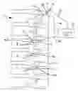
Further features and advantages of the present invention will become clear from the following description of an illustrative embodiment in which reference is made to the drawing, which only shows a FIG. 1 in which the side view of a portion of a spinal column is indicated schematically, where three vertebral bodies can be connected to one another in accordance with the invention, i.e. can be stabilized.
DETAILED DESCRIPTION
In detail, FIG. 1 shows a lateral portion of a spinal column 1 in three vertebral bodies 2-4 of which so-called pedicle screws 5-7 are screwed, for which purpose the skin designated by 8 has relatively small incisions, indicated by 9-11. The individual pedicle screws 5-7 are composed mainly of, taking the example of the first screw 5, an eyelet 5.1 and a threaded shaft 5.2 and are made of titanium, it also being possible to use chromium/cobalt/nickel alloys.
The three vertebral bodies 2-4 are intended to be connected to one another, i.e. stabilized, for which purpose, according to the minimally invasive method, a fixation rod 12 is used which at its head end has an ultrasound head 13 and a measurement transducer 14, which is connected via an electrical lead 15 to an impedance-measuring device 16 with an optical and/or acoustic display 17. At the same time, the impedance-measuring device 16 is connected via the lead 18 to the fixation rod 12 and via a connector 19 to the eyelet 5.1 of the pedicle screw 5.
The minimally invasive placement of the pedicle screws 5-7 and of the fixation rod 12 takes place as follows. In FIG. 1 only a half of the screw arrangement is shown, since in practice a screw is applied on both sides of the vertebral canal of the vertebral bodies 2-4 concerned, with the result that two rows of screws are present, each with a fixation rod 12, of which, however, only one row can be seen in FIG. 1.
To insert the pedicle screws 5-7, suitably short incisions 9-11 are made in the skin 8, and the pedicle screws 5-7 are screwed, after preliminary drilling, with their respective threaded shaft (example: 5-5.2) into the vertebral bodies 2-4 (example: 2) in such a way that their eyelets (example: 5.1) are horizontal. It should be noted here that the screws 5-7 are placed through the respective muscle, i.e. for this purpose the muscle should if possible be divided longitudinally or pushed aside.
For minimally invasive introduction of the fixation rod 12 through the eyelets of the pedicle screws 5-7, the rod should also if possible be pushed through the muscle. At the start, the skin 8 must be penetrated by an incision (not shown).
To start with, a targeting aid for the fixation rod 12 lies in the impedance measurement in which the measurement parameter used is the phase displacement between current and voltage that can be read off via a display 17, in particular of an optical and/or acoustic nature, i.e. the smaller this phase displacement, the closer the tip of the fixation rod 12 is to the eyelet 5.1. When this “Δ small” has reached a defined near area, the ultrasound head 13 is switched to, since with the latter the fixation rod 12 can be guided with greater precision and thus more easily through the eyelet 5.1.
The continued guiding of the fixation rod 12 through the eyelets of the two further pedicle screws 6 and 7 takes place analogously, and the lead 19 then has to be connected to the eyelet of each of these screws. Two possibilities can be selected here, specifically, in the first case, the impedance method as rough orientation and then, for more precise approach and guiding through the eyelet, ultrasound or both.
The ultrasound head 13 is secured to the measurement transducer 14 on the fixation rod 12 as probe by means of a fine thread, bayonet catch or the like, specifically such that this unit can be removed again at the end of the intervention.
Moreover, the remaining portion of the fixation rod 12 in the spinal column 1 has a notch 20 on its circumference, such that this portion can be removed again in due course with the aid of a suitable extraction instrument.
Overall, therefore, the present invention represents a further milestone in minimally invasive surgery to the benefit of patients.
Claims
1. Medical device for determining the position of an intracorporeal implant (5) comprises at least one impedance-measuring device (16) and an ultrasound device (13) which are both connected to at least one intracorporeal probe (12), the impedance-measuring device (16) having at least one connector (19) for connection to the implant (5).
2. Medical device according to claim 1, wherein the impedance measuring device comprises means for determining the position of an implant (5) by measuring the phase displacement between current and voltage which occurs as a function of distance between the intracorporeal probe (12) and the implant (5).
3. Medical device according to claim 2, wherein the impedance-measuring device (16) is used for a rough determination of position and the ultrasound device (13) is used for a precise determination of position.
4. Medical device according to claim 3, including means for switching the impedance-measuring device (16) and the ultrasound device (13) on alternately.
5. Medical device according to claim 3, including means for switching the impedance-measuring device (16) and the ultrasound device (13) in such a way that they are simultaneously active.
6. Medical device according to claims 1, wherein the intracorporeal probe (12) has a rod-shaped design and at a free hand end has an ultrasound head (13) and a measurement transducer (14).
7. Medical device according to claim 6, wherein the ultrasound head (13) with the measurement transducer (14) is secured to the probe (12) by connection means.
8. Medical device according to claim 6, wherein a portion of the rod-shaped probe (12) remaining on the spinal column (1) has, at least at one end, a notch (20) for attachment of an extraction instrument.
9. Medical device for determining the position of pedicle screws in a spinal column fixation system with rod-shaped probes as stabilizers comprises an intracorporeal probe (12) connected to an impedance-measuring device (16) and to an ultrasound device (13), and an additional connector (19) of the impedance-measuring device (16) is electrically conductively connected to the respective pedicle screws (5-7).
10. Method for determining the position of intracorporeal implants with the aid of an device comprising at least one impedance-measuring device (16) and an ultrasound device (13) which are both connected to at least one intracorporeal probe (12), the impedance-measuring device (16) additionally being equipped with at least one connector (19) for connection to the implant (5), comprising the steps of using the probe (12), upon introduction into a body, to carry out a rough determination of position of the implant by means of the impedance-measuring device (16) measuring the phase displacement between current and voltage as a function of the distance between the probe (12) and the implant (5), whereas, shortly before the implant (5) is reached by the probe (12), a precise determination of position takes place with the ultrasound device (13).
11. Method according to claim 10, wherein the rough determination of position and thereafter the precise determination of position are carried out successively on several consecutively arranged implants.
12. Method according to claim 11, wherein the determination of position in one or more consecutively arranged implants is carried out simultaneously by the impedance-measuring device (16) and the ultrasound device (13).
Images & Drawings included:
Sources:
- United States Patent and Trademark Office - verify current appl. status at the USPTO↗
Similar patent applications:
Recent applications in this class:
- » 20250127420 2025-04-24
WIRELESS DATA COMMUNICATION AND POWER TRANSMISSION ATHLETIC APPAREL MODULE - » 20250114016 2025-04-10
FOOT-MOUNTED SENSOR SYSTEMS FOR TRACKING BODY MOVEMENT - » 20240415410 2024-12-19
SYSTEMS AND METHODS FOR MONITORING A PHYSIOLOGICAL PARAMETER OF PERSONS ENGAGED IN PHYSICAL ACTIVITY - » 20240065577 2024-02-29
KINETIC ASSESSMENT AND ALIGNMENT OF THE MUSCULAR-SKELETAL SYSTEM AND METHOD THEREFOR - » 20230355133 2023-11-09
Wireless data communication and power transmission athletic apparel module - » 20230346258 2023-11-02
SYSTEM FOR DETERMINING CHANGE IN POSITION OF AN IMPLANTED MEDICAL DEVICE WITHIN AN IMPLANT POCKET - » 20230165484 2023-06-01
SYSTEM AND METHOD FOR ANALYZING FORCE SENSOR DATA - » 20220079471 2022-03-17
Systems and methods for monitoring a physiological parameter of persons engaged in physical activity - » 20220022774 2022-01-27
Kinetic assessment and alignment of the muscular-skeletal system and method therefor - » 20210059563 2021-03-04
Systems and methods for anatomical alignment
Recent applications for this Assignee:
- » 20170058933 2017-03-02
Fastening system for a machine element - » 20160298749 2016-10-13
Drive and method for operating a drive - » 20160298748 2016-10-13
Gearing - » 20160298747 2016-10-13
Gearing - » 20160231288 2016-08-11
METHOD AND APPARATUS FOR MONITORING A DEVICE OF A DRIVE SYSTEM - » 20160058483 2016-03-03
Medullary pin - » 20150043990 2015-02-12
Fastening system with eccentric - » 20140239749 2014-08-28
Rotor for electric machine - » 20140000395 2014-01-02
Spindle drive - » 20130255421 2013-10-03
Transmission