Prosthetic socket stabilization apparatus and technique
US20110118853A1
2011-05-19
12/949,421
2010-11-18
✅ Patent granted
US 8,911,505 B2
2014-12-16
-
-
Marcia Watkins
Trop, Pruner & Hu, P.C.
2030-12-13
Abstract:
A portable device, with method, advantageously applies dynamic stimulation of enclosed muscle tissue to stabilize a prosthetic socket on a residual limb. Dynamic stimulation is in response to physical conditions such as prosthesis motion, position and/or internal pressures. Tissue volume contained within the socket may be stabilized by varying average stimulation levels in response to internal socket pressure.
Assignee:
- Articulate Labs Inc. 9 🇺🇸 Austin, TX, United States
Applicant:
Interested in similar patents?
Get notified when new applications in this technology area are published.
Classification:
A61N1/3603 » CPC further
Electrotherapy; Circuits therefor; Applying electric currents by contact electrodes alternating or intermittent currents for stimulation; External stimulators, e.g. with patch electrodes Control systems
A61N1/00 IPC
Electrotherapy; Circuits therefor
A61B5/1038 » CPC further
Measuring for diagnostic purposes ; Identification of persons; Detecting, measuring or recording devices for testing the shape, pattern, colour, size or movement of the body or parts thereof, for diagnostic purposes; Measuring load distribution, e.g. podologic studies Measuring plantar pressure during gait
A61B5/6811 » CPC further
Measuring for diagnostic purposes ; Identification of persons; Arrangements of detecting, measuring or recording means, e.g. sensors, in relation to patient specially adapted to be attached to or worn on the body surface; Sensor mounted on worn items External prosthesis
A61F2/7812 » CPC further
Filters implantable into blood vessels; Prostheses, i.e. artificial substitutes or replacements for parts of the body; Appliances for connecting them with the body; Devices providing patency to, or preventing collapsing of, tubular structures of the body, e.g. stents; Prostheses not implantable in the body; Means for protecting prostheses or for attaching them to the body, e.g. bandages, harnesses, straps, or stockings for the limb stump Interface cushioning members placed between the limb stump and the socket, e.g. bandages or stockings for the limb stump
A61N1/0452 » CPC further
Electrotherapy; Circuits therefor; Details; Electrodes for external use; Use-related aspects Specially adapted for transcutaneous muscle stimulation [TMS]
A61N1/0472 » CPC further
Electrotherapy; Circuits therefor; Details; Electrodes for external use Structure-related aspects
A61N1/0492 » CPC further
Electrotherapy; Circuits therefor; Details; Electrodes for external use; Structure-related aspects Patch electrodes
A61N1/08 » CPC further
Electrotherapy; Circuits therefor; Details Arrangements or circuits for monitoring, protecting, controlling or indicating
A61N1/36003 » CPC further
Electrotherapy; Circuits therefor; Applying electric currents by contact electrodes alternating or intermittent currents for stimulation of motor muscles, e.g. for walking assistance
A61N1/36014 » CPC further
Electrotherapy; Circuits therefor; Applying electric currents by contact electrodes alternating or intermittent currents for stimulation External stimulators, e.g. with patch electrodes
A61B5/0048 » CPC further
Measuring for diagnostic purposes ; Identification of persons Detecting, measuring or recording by applying mechanical forces or stimuli
A61B5/1114 » CPC further
Measuring for diagnostic purposes ; Identification of persons; Detecting, measuring or recording devices for testing the shape, pattern, colour, size or movement of the body or parts thereof, for diagnostic purposes; Measuring movement of the entire body or parts thereof, e.g. head or hand tremor, mobility of a limb; Local tracking of patients, e.g. in a hospital or private home Tracking parts of the body
A61B5/6828 » CPC further
Measuring for diagnostic purposes ; Identification of persons; Arrangements of detecting, measuring or recording means, e.g. sensors, in relation to patient specially adapted to be attached to or worn on the body surface; Specially adapted to be attached to a specific body part Leg
A61B5/6829 » CPC further
Measuring for diagnostic purposes ; Identification of persons; Arrangements of detecting, measuring or recording means, e.g. sensors, in relation to patient specially adapted to be attached to or worn on the body surface; Specially adapted to be attached to a specific body part Foot or ankle
A61B5/6843 » CPC further
Measuring for diagnostic purposes ; Identification of persons; Arrangements of detecting, measuring or recording means, e.g. sensors, in relation to patient specially adapted to be attached to or worn on the body surface Monitoring or controlling sensor contact pressure
A61B2562/0219 » CPC further
Details of sensors; Constructional details of sensor housings or probes; Accessories for sensors; Details of sensors specially adapted for in-vivo measurements Inertial sensors, e.g. accelerometers, gyroscopes, tilt switches
A61F2002/5026 » CPC further
Filters implantable into blood vessels; Prostheses, i.e. artificial substitutes or replacements for parts of the body; Appliances for connecting them with the body; Devices providing patency to, or preventing collapsing of, tubular structures of the body, e.g. stents; Prostheses not implantable in the body adjustable for adjusting a diameter
A61F2002/5027 » CPC further
Filters implantable into blood vessels; Prostheses, i.e. artificial substitutes or replacements for parts of the body; Appliances for connecting them with the body; Devices providing patency to, or preventing collapsing of, tubular structures of the body, e.g. stents; Prostheses not implantable in the body adjustable for adjusting cross-section
A61F2002/5036 » CPC further
Filters implantable into blood vessels; Prostheses, i.e. artificial substitutes or replacements for parts of the body; Appliances for connecting them with the body; Devices providing patency to, or preventing collapsing of, tubular structures of the body, e.g. stents; Prostheses not implantable in the body adjustable self-adjustable, e.g. self-learning
A61F2002/762 » CPC further
Filters implantable into blood vessels; Prostheses, i.e. artificial substitutes or replacements for parts of the body; Appliances for connecting them with the body; Devices providing patency to, or preventing collapsing of, tubular structures of the body, e.g. stents; Prostheses not implantable in the body; Means for assembling, fitting or testing prostheses, e.g. for measuring or balancing, e.g. alignment means; Measuring means for measuring dimensions, e.g. a distance
A61F2002/7635 » CPC further
Filters implantable into blood vessels; Prostheses, i.e. artificial substitutes or replacements for parts of the body; Appliances for connecting them with the body; Devices providing patency to, or preventing collapsing of, tubular structures of the body, e.g. stents; Prostheses not implantable in the body; Means for assembling, fitting or testing prostheses, e.g. for measuring or balancing, e.g. alignment means; Measuring means for measuring force, pressure or mechanical tension
A61F2002/764 » CPC further
Filters implantable into blood vessels; Prostheses, i.e. artificial substitutes or replacements for parts of the body; Appliances for connecting them with the body; Devices providing patency to, or preventing collapsing of, tubular structures of the body, e.g. stents; Prostheses not implantable in the body; Means for assembling, fitting or testing prostheses, e.g. for measuring or balancing, e.g. alignment means; Measuring means for measuring acceleration
A61F2002/7655 » CPC further
Filters implantable into blood vessels; Prostheses, i.e. artificial substitutes or replacements for parts of the body; Appliances for connecting them with the body; Devices providing patency to, or preventing collapsing of, tubular structures of the body, e.g. stents; Prostheses not implantable in the body; Means for assembling, fitting or testing prostheses, e.g. for measuring or balancing, e.g. alignment means; Measuring means for measuring fluid pressure
A61F2002/802 » CPC further
Filters implantable into blood vessels; Prostheses, i.e. artificial substitutes or replacements for parts of the body; Appliances for connecting them with the body; Devices providing patency to, or preventing collapsing of, tubular structures of the body, e.g. stents; Prostheses not implantable in the body; Means for protecting prostheses or for attaching them to the body, e.g. bandages, harnesses, straps, or stockings for the limb stump; Sockets, e.g. of suction type Suction sockets, i.e. utilizing differential air pressure to retain the prosthesis on the stump
A61N1/0476 » CPC further
Electrotherapy; Circuits therefor; Details; Electrodes for external use; Structure-related aspects Array electrodes (including any electrode arrangement with more than one electrode for at least one of the polarities)
A61F2/80 » CPC main
Filters implantable into blood vessels; Prostheses, i.e. artificial substitutes or replacements for parts of the body; Appliances for connecting them with the body; Devices providing patency to, or preventing collapsing of, tubular structures of the body, e.g. stents; Prostheses not implantable in the body; Means for protecting prostheses or for attaching them to the body, e.g. bandages, harnesses, straps, or stockings for the limb stump Sockets, e.g. of suction type
A61F2/76 » CPC further
Filters implantable into blood vessels; Prostheses, i.e. artificial substitutes or replacements for parts of the body; Appliances for connecting them with the body; Devices providing patency to, or preventing collapsing of, tubular structures of the body, e.g. stents; Prostheses not implantable in the body Means for assembling, fitting or testing prostheses, e.g. for measuring or balancing, e.g. alignment means
A61B5/103 IPC
Measuring for diagnostic purposes ; Identification of persons Detecting, measuring or recording devices for testing the shape, pattern, colour, size or movement of the body or parts thereof, for diagnostic purposes
A61B5/00 IPC
Measuring for diagnostic purposes ; Identification of persons
A61F2/78 IPC
Filters implantable into blood vessels; Prostheses, i.e. artificial substitutes or replacements for parts of the body; Appliances for connecting them with the body; Devices providing patency to, or preventing collapsing of, tubular structures of the body, e.g. stents; Prostheses not implantable in the body Means for protecting prostheses or for attaching them to the body, e.g. bandages, harnesses, straps, or stockings for the limb stump
A61N1/04 IPC
Electrotherapy; Circuits therefor; Details Electrodes
A61N1/36 IPC
Electrotherapy; Circuits therefor; Applying electric currents by contact electrodes alternating or intermittent currents for stimulation
A61B5/11 IPC
Measuring for diagnostic purposes ; Identification of persons; Detecting, measuring or recording devices for testing the shape, pattern, colour, size or movement of the body or parts thereof, for diagnostic purposes Measuring movement of the entire body or parts thereof, e.g. head or hand tremor, mobility of a limb
A61F2/50 IPC
Filters implantable into blood vessels; Prostheses, i.e. artificial substitutes or replacements for parts of the body; Appliances for connecting them with the body; Devices providing patency to, or preventing collapsing of, tubular structures of the body, e.g. stents Prostheses not implantable in the body
Description
REFERENCE TO RELATED APPLICATION
This application claims priority to U.S. Provisional Patent Application Ser. No. 61/262,733, filed Nov. 19, 2009, the entire content of which is incorporated herein by reference.
FIELD OF THE INVENTION
This invention relates generally to medical devices, and particularly to apparatus and methods to stabilize a prosthesis through dynamic stimulation of enclosed tissue.
BACKGROUND OF THE INVENTION
Prosthetic devices which replace biological limbs usually interface through a hard cup-shaped shell, referred to as a socket, which encloses a residual limb. In order to transfer the necessary forces, sockets are typically fabricated with composite materials, such as carbon fiber. Compliant materials such as urethane, silicone, and/or cotton or wool fabrics typically are used between the residual limb and socket to cushion and distribute forces within the socket.
Suction is the present preferred method to affix and stabilize the socket to the residual limb. Active regulated vacuum pumps, unidirectional air valves, neoprene sleeves, and silicone suction liners with distal tension pins are among the approaches commonly used to achieve sufficient vacuum to hold sockets to residual limbs. Although usually more effective than earlier mechanical fixation techniques using belts or straps, several factors are not addressed by extant vacuum attachment approaches.
To provide proper force distribution and obtain adequate vacuum for socket stability, minimal clearance inside the socket is required. Residual limbs confined within sockets, however, often undergo changes in volume and sometimes shape as well. Non-contiguous socket shells have demonstrated greater tolerance of volume changes, but attachment remains problematic. To accommodate volume differentials experienced between the residual limb and conventional sockets, one or more fabric layers, or socks, are commonly worn between the limb and socket. Imposition of porous fabric, which does not retain vacuum, has prompted the use of either neoprene sleeves overlaid to seal the juncture between the open socket end and proximal limb, or elastomer skin-contact liners with integral distal tension pins which lock within the socket. Neoprene sleeves used to seal sockets to residual limbs quickly develop pinhole leaks as the edge of the socket is bumped into any non-compliant surface, compromising suction and thus socket stability. Socket liners with distal tension pins stretch longitudinally and thus fail to distribute distal tensile force over the entire residual limb, often creating localized tissue disruption.
Muscles remaining from amputation within a residual limb usually lose the skeletal connection necessary for their original function, so naturally atrophy. In an effort to stabilize residual limb volume, patients are furthermore routinely encouraged to avoid contraction of viable residual muscle. Compliance from the resultant fatty tissue surrounding the bone of a residual limb degrades proprioception, and often impairs prosthetic positional control. This flaccid tissue as well provides little protection for painful distal neuromas, which often form at nerve resection sites. Disuse of residual muscle compounds circulatory issues imposed by amputation, in that muscle activity in biologically intact limbs normally pumps fluids through the body. This results in intolerance to cold temperatures for many amputees, and can exacerbate phantom pain.
Extrinsic muscle stimulation, particularly if applied during contraction, has been repeatedly shown to increase both size and strength of muscle tissue. For this reason, functional muscle stimulation is commonly used to allay atrophy or improve muscle function. This use, however, has been limited to largely pre-programmed stimulation patterns in clinical settings.
A need exists whereby a prosthetic socket may be definitively secured to a residual limb during normal activities, with minimal repercussion on the residual physiology.
SUMMARY OF THE INVENTION
The present invention resides in apparatus and methods for actively stabilizing a prosthesis on a biological limb through dynamic stimulation of residual limb muscle in response to physical conditions of the prosthesis. In addition to biologically enhanced movement and positional control, methods include optional maintenance of residual limb muscle at a relatively constant average volume through stimulation control.
BRIEF DESCRIPTION OF THE DRAWINGS
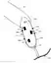
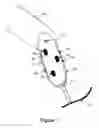
FIG. 1 shows a block diagram of an exemplary embodiment of the present invention.
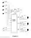
FIG. 2 shows a block diagram of a computational architecture demonstrating use of the present invention within the exemplary embodiment of FIG. 1.
DETAILED DESCRIPTION OF THE INVENTION
Referring now to FIG. 1, Prosthetic Socket 102 accepts the residual limb of Human Leg 101. Prosthetic Foot 105 is distally attached to Socket 102, per common practice. Position Sensors 103 and 104 are affixed to Socket 102, and measure spatial orientation and movement. Said Sensors 103 and 104 may be accelerometers, inclinometers, magnetometers, or other means of spacial position or motion sensing, as is known in the art. Force Sensors 106, 107, 108, and 109 are affixed to or are integral with Socket 102, with active sensing surfaces on the interior of said Socket 102. Alternately, one or more of said Pressure Sensors 106, 107, 108, and 109 may be implemented as position sensors sensitive to the relative position of the residual limb of Leg 101 within Socket 102. Controller 113 receives input from said Sensors 103. 104, 106, 107, 108, and 109; and, in response to said sensor inputs, emits high-voltage stimulation pulses to one or more of Stimulation Pads 110, 111, and 112. Pads 110, 111, and 112 are in intimate contact with Leg 101; and may be positioned within or integral to a prosthetic liner, or directly upon the interior surface of Socket 102. Modulation methods for energy to be applied to said Stimulation Pads 110, 111, and 112 may include one or more of amplitude, phase, pulse position, pulse width, frequency, or any other scheme known to the art. Dynamic selection of relative energy to be applied to one or more of said Stimulation Pads 110, 111, and 112 may be determined heuristically by Controller 113 and/or through control design with predefinition of muscle locations to be stimulated within Leg 101. Note that in the absence of a liner, the interior of said Socket 102 may be lined with a compliant material, such as silicone, before pad installation.
Referring now to FIG. 2, Pressure Sensors 206, 207, 208, and 209 correspond to Pressure Sensors of Sensors 106, 107, 108, and 109 of FIG. 1, respectively. Position Sensors 203 and 204 correspond to Sensors 103 and 104; and Stimulation Pads 210, 211, and 212 correspond to Pads 110, 111, and 112, all of FIG. 1.
Pressure Sensors 206, 207, 208, and 209 provide internal socket pressure indications to Summer 201, and internal socket pressure and/or relative limb position, as noted above, to Positional Control 205. Position Sensors 203 and 204 provide input to Position Control 205 only. Summer 201 provides a signal to Integrator 202 which is representative of the composite force applied to the interior of Socket 102 of FIG. 1. The output of Integrator 202 is inverted by Amplifier 219. Amplifier 219 thus provides an output signal which is inversely proportional to the average composite force within Socket 102, presumably over a period of days or weeks. The output of Amplifier 219 is supplied as common input to Multipliers 213, 214, and 215.
Position Control 205 comprises a positional control scheme, preferably embodied as analog circuitry and/or software executed by a control device, such as a microcontroller or digital signal processor. Under stimulation of said Sensors 206, 207, 208, 209, 203, and/or 204, said Controller 205, through any of control schemes known to the art, provides variable Stimulation Signals 220, 221, and 222 as input to Multipliers 213, 214, and 215, respectively. The outputs of Multipliers 213, 214, and 215 are supplied as input to High-Voltage Drivers 216, 217, and 218, respectively, which in turn provide high-voltage pulses to Stimulation Pads 210, 211, and 212, respectively. In that the common inputs of Multipliers 213, 214, and 215 are provided by Amplifier 219, Stimulation Signals 220, 221, and 222 provided by Position Control 205 are modulated indirectly by the average composite force indicated by Sensors 206, 207, 208, and 209.
Stimulation energy supplied to Pads 210, 211, and 212 is therefore inversely proportional to the average force within Socket 102 of FIG. 1. Stimulation Pads 210, 211, and 212, being in intimate contact with Leg 101 of FIG. 1, induce variable contractions of various leg muscles within Socket 102 of FIG. 1. Being localized, these contractions therefore create vectored forces directly upon Socket 102. It is noted that switching amplification, preferably controlled current or power, may advantageously be used in Drivers 216, 217, and 218.
Under control of said Sensors 206, 207, 208, 209, 203, and/or 204, Position Control 205 calculates appropriate stimulation outputs for application to Pads 210, 211, and 212 which serve to stabilize Socket 102 upon Leg 101 as it is used in normal activities. In that the relative positions of all devices of Socket 102 are fixed, standard control techniques, such as proportional-integral-derivative loops, may determine differential outputs for said Pads 210, 211, and 212. Alternatively, software models of the biological components within Socket 102 may be interposed in the architecture of Position Control 205 between sensor inputs and stimulation output control loops, so as to improve predictive behavior. It is assumed that a state-machine software architecture may be applied to algorithms executed in Position Control 205, selectively using historical data to determine present and future states.
Although depicted separately for the purpose of explanation, integration of the composite functions of Summer 201, Integrator 202, Amplifier 219, and Multipliers 213, 214, and 215 is anticipated with the functions described of Position Control 205, in any of the various possible implementations described above. Use of the current invention can as well be seen to be independent of the type of socket used, specific function of a prosthesis, socket liner use or type, and type of muscle stimulation employed.
It is assumed that Socket 102 of FIG. 1 employs at least one region of negative draft angle, wherein constriction increases with proximal direction. This is necessary to provide leverage upon Socket 102 by the muscles of Leg 101.
By the above discussion, it can be seen that control algorithms or circuitry, using positional and/or force data, may dynamically stabilize a prosthetic socket upon an appendage through stimulation of the contained muscle. In that muscle growth is known to result from stimulated contraction, and average pressure within the socket is roughly proportional to contained volume; it can as well be seen that muscle volume may be stabilized within a prosthetic socket through inclusion of average internal pressure in the control algorithm. Finally, muscle stimulation of the present invention can be seen to inherently follow movement, more closely replicating intact biological activity. Such activity has been shown to reduce phantom sensations, arguably through integrated sensory stimulation, and improve fluid circulation.
Claims
1. A system for stabilizing a prosthesis affixed to a biological feature comprising:
a shell to contain a biological feature for interface to an attached prosthesis;
means to measure at least one spacial attribute or force of said shell or attached prosthesis;
means to stimulate at least one area of muscle tissue contained within said shell; and
means to control stimulation applied to at least one said area of muscle tissue in response to at least one said spacial attribute or force.
2. The apparatus of claim 1, further comprising:
means to measure average pressure within said shell; and
means to regulate stimulation of at least one said area of muscle tissue in response to said average pressure.
3. The apparatus of claim 1, wherein said means to control stimulation comprises analog circuitry.
4. The apparatus of claim 1, wherein said means to control stimulation comprises digital circuitry and/or software executed by a processor.
5. The apparatus of claim 1, wherein said means to measure at least one spacial attribute or force comprises an accelerometer.
6. The apparatus of claim 1, wherein said means to stimulate at least one area of muscle tissue applies electrical stimulation.
7. The apparatus of claim 1, wherein said means to stimulate at least one area of muscle tissue applies magnetic stimulation.
8. The apparatus of claim 1, wherein an elastomeric liner is worn within said shell.
9. The apparatus of claim 1, wherein said shell includes at least one area of negative draft angle.
10. The apparatus of claim 1, wherein said means to stimulate at least one area of muscle tissue employs switching amplification.
11. A method for stabilizing a prosthesis to a biological feature comprising the steps of:
measuring at least one spacial attribute or force of a shell for prosthesis attachment or prosthesis attached to said shell;
stimulating at least one area of muscle tissue contained within said shell for prosthesis attachment; and
controlling stimulation of at least one said area of muscle tissue in response to measurement of at least one said spacial attribute or force.
12. The method of claim 11, further comprising the steps of:
measuring average pressure within said shell for prosthesis attachment; and
regulating stimulation of at least one said area of muscle tissue in response to said average pressure.
13. The method of claim 11, wherein said step of controlling stimulation employs mathematical integration or derivation of one or more terms.
14. The method of claim 11, wherein stimulation control employs the output of a model depicting one or more biological components contained within said shell for prosthesis attachment, as excited by at least one said spacial attribute or force.
15. The method of claim 11, wherein stimulation control is effected through amplitude modulation.
16. The method of claim 11, wherein stimulation control is effected through pulse width modulation.
17. The method of claim 11, wherein a state machine control structure is used.
18. The method of claim 11, wherein switching amplification is used to drive tissue stimulation.
Images & Drawings included:
Sources:
- United States Patent and Trademark Office - verify current appl. status at the USPTO↗
Recent applications in this class:
- » 20250288435 2025-09-18
PROSTHETIC SUSPENSION SYSTEM WITH COMBINATION OF SUSPENSION VALVE AND EXPULSION VALVE - » 20250262071 2025-08-21
PROSTHESIS SOCKET AND METHOD FOR PRODUCING SAME - » 20250248828 2025-08-07
METHOD FOR MANUFACTURING A NEW PROSTHESIS - » 20250228676 2025-07-17
SELF-ADJUSTING SOCKET FOR LOWER LIMB PROSTHESIS - » 20250152385 2025-05-15
EXTERNAL FIXATION ASSEMBLY FOR POST-AMPUTATION PROSTHESIS - » 20250114221 2025-04-10
EVOLVED PROSTHETIC SOCKET FOR UPPER LIMB PROSTHESES - » 20250099272 2025-03-27
PROSTHETIC SOCKET - » 20250090352 2025-03-20
INFLATABLE LIMB PROSTHESIS - » 20250032279 2025-01-30
METHOD FOR PRODUCING AN ORTHOPEDIC DEVICE AND ORTHOPEDIC DEVICE - » 20240382323 2024-11-21
PROSTHETIC SYSTEM
Recent applications for this Assignee:
- » 20210228393 2021-07-29
Orthotic support and stimulus systems and methods - » 20170337344 2017-11-23
Orthotic support and stimulus systems and methods - » 20150142129 2015-05-21
Adaptive Stimulation Apparatus and Technique - » 20140148873 2014-05-29
Joint rehabilitation apparatus and technique - » 20130253607 2013-09-26
Adaptive muscle stimulation technique - » 20130246036 2013-09-19
Orthotic support and stimulus systems and methods - » 20100327845 2010-12-30
Apparatus and technique to drive a variable load via transformer secondary winding - » 20100262042 2010-10-14
Acoustic myography system and methods