NANOPARTICLE-LOADED CELLS
US20120087868A1
2012-04-12
13/267,374
2011-10-06
Abstract:
The invention provides nanoparticle-loaded cells and compositions useful for improved imaging and therapy, for example radio-therapy. The invention also provides methods of manufacture of nanoparticle-loaded cells, methods of administering the nanoparticle-loaded cells, and methods for treatment and/or imaging.
Inventors:
- Alla Danilkovitch 91 🇺🇸 Columbia, MD, United States
- Gabriele Todd 1 🇺🇸 Elkridge, MD, United States
Interested in similar patents?
Get notified when new applications in this technology area are published.
Classification:
C12N5/0663 » CPC main
Undifferentiated human, animal or plant cells, e.g. cell lines; Tissues; Cultivation or maintenance thereof; Culture media therefor; Animal cells or tissues; Human cells or tissues; Vertebrate cells; Cells of skeletal and connective tissues; Mesenchyme; Stem cells Bone marrow mesenchymal stem cells (BM-MSC)
A61K35/28 » CPC further
Medicinal preparations containing materials or reaction products thereof with undetermined constitution; Materials from mammals; Compositions comprising non-specified tissues or cells; Compositions comprising non-embryonic stem cells; Genetically modified cells Bone marrow; Haematopoietic stem cells; Mesenchymal stem cells of any origin, e.g. adipose-derived stem cells
A61K41/0038 » CPC further
Medicinal preparations obtained by treating materials with wave energy or particle radiation ; Therapies using these preparations Radiosensitizing, i.e. administration of pharmaceutical agents that enhance the effect of radiotherapy
A61K41/0052 » CPC further
Medicinal preparations obtained by treating materials with wave energy or particle radiation ; Therapies using these preparations Thermotherapy; Hyperthermia; Magnetic induction; Induction heating therapy
A61K47/6901 » CPC further
Medicinal preparations characterised by the non-active ingredients used, e.g. carriers or inert additives; Targeting or modifying agents chemically bound to the active ingredient the non-active ingredient being chemically bound to the active ingredient, e.g. polymer-drug conjugates the conjugate being characterised by physical or galenical forms, e.g. emulsion, particle, inclusion complex, stent or kit Conjugates being cells, cell fragments, viruses, ghosts, red blood cells or viral vectors
A61K49/0423 » CPC further
Preparations for testing; X-ray contrast preparations; Physical forms of mixtures of two different X-ray contrast-enhancing agents, containing at least one X-ray contrast-enhancing agent which is not a halogenated organic compound; Particles, beads, capsules or spheres Nanoparticles, nanobeads, nanospheres, nanocapsules, i.e. having a size or diameter smaller than 1 micrometer
A61K49/1827 » CPC further
Preparations for testing; Nuclear magnetic resonance [NMR] contrast preparations; Magnetic resonance imaging [MRI] contrast preparations characterised by a special physical form, e.g. emulsions, microcapsules, liposomes particles, e.g. uncoated or non-functionalised microparticles or nanoparticles coated or functionalised microparticles or nanoparticles coated or functionalised nanoparticles having a (super)(para)magnetic core, being a solid MRI-active material, e.g. magnetite, or composed of a plurality of MRI-active, organic agents, e.g. Gd-chelates, or nuclei, e.g. Eu3+, encapsulated or entrapped in the core of the coated or functionalised nanoparticle
A61P1/00 » CPC further
Drugs for disorders of the alimentary tract or the digestive system
A61P5/14 » CPC further
Drugs for disorders of the endocrine system of the thyroid hormones, e.g. T3, T4
A61P19/02 » CPC further
Drugs for skeletal disorders for joint disorders, e.g. arthritis, arthrosis
A61P37/06 » CPC further
Drugs for immunological or allergic disorders; Immunomodulators Immunosuppressants, e.g. drugs for graft rejection
A61K2035/122 » CPC further
Medicinal preparations containing materials or reaction products thereof with undetermined constitution; Materials from mammals; Compositions comprising non-specified tissues or cells; Compositions comprising non-embryonic stem cells; Genetically modified cells for inducing tolerance or supression of immune responses
C12N2511/00 » CPC further
Cells for large scale production
A61K49/06 IPC
Preparations for testing Nuclear magnetic resonance [NMR] contrast preparations; Magnetic resonance imaging [MRI] contrast preparations
A61K9/14 IPC
Medicinal preparations characterised by special physical form Particulate form, e.g. powders, Processes for size reducing of pure drugs or the resulting products, Pure drug nanoparticles
A61P35/00 » CPC further
Antineoplastic agents
A61M37/00 » CPC further
Other apparatus for introducing media into the body ; Percutany, i.e. introducing medicines into the body by diffusion through the skin
A61K49/04 IPC
Preparations for testing X-ray contrast preparations
A61K35/12 IPC
Medicinal preparations containing materials or reaction products thereof with undetermined constitution Materials from mammals; Compositions comprising non-specified tissues or cells; Compositions comprising non-embryonic stem cells; Genetically modified cells
B82Y5/00 » CPC further
Nanobiotechnology or nanomedicine, e.g. protein engineering or drug delivery
Description
RELATED APPLICATIONS
This application claims priority to U.S. Provisional Application No. 61/391,482 entitled “Nanoparticle-loaded Cells”, filed on Oct. 8, 2010 bearing Attorney Docket No. 23738US01, and U.S. Provisional Application No. 61/391,452 entitled “Enhanced MSC Preparations”, filed on Oct. 8, 2010 bearing Attorney Docket No. 23734US01, the contents of which are hereby incorporated by reference in their entireties.
This application is being co-filed on Oct. 6, 2011 with, and incorporates by reference: International Patent Application entitled “Nanoparticle-loaded Cells” bearing Attorney Docket No. 23738WO01, U.S. Non-provisional Application entitled “Enhanced MSC Preparations” bearing Attorney Docket No. 23734U502, and International Patent Application entitled “Enhanced MSC Preparations” bearing Attorney Docket No. 23734WO01.
FEDERALLY SPONSORED RESEARCH OR DEVELOPMENT
[Not Applicable]
MICROFICHE/COPYRIGHT REFERENCE
[Not Applicable]
BACKGROUND OF THE INVENTION
The present invention relates to cells loaded with nanoparticles and methods of using nanoparticles for, for example, therapeutic purposes and diagnostic/analysis purposes such as imaging.
Electromagnetic radiation (e.g., X-rays) with very high-energy photon particles has traditionally been used for the therapeutic treatment of certain diseases such as cancer. The high-energy radiation beam can be focused to a specific location, even deep within the body, to destroy the targeted cells. However, normal cells in the beam's path, at any depth, are also killed. Consequently, there is always a conflict between the dosages that will effectively kill the disease cells while maintaining a sufficient amount of normal cells for patient recovery.
Radiation enhancers have been developed which enhance the radiation dose to nearby tissues. Radiation enhancers include elements with high atomic numbers which interact directly with the radiation beam to cause more tissue damage, for example, by scattering the radiation dose to nearby soft tissue that would otherwise be relatively transparent to the radiation beam.
Hainfeld (U.S. Pat. No. 6,645,464) describes the loading of metal particles into cells or membrane vesicles by placing metal seed particles into the cells or vesicles, then chemically depositing additional metal on the metal seed particles. Hainfeld further describes the use of metal particles to improve imaging and therapy by their interaction with externally applied energy. Hainfeld does not teach cells such as mesenchymal stem cells (MSCs) loaded with nanoparticles.
Bulte et al. (“Feridex-Labeled Mesenchymal Stem Cells: Cellular Differentiation and MR Assessment in a Canine Myocardial Infarction Model”; Academic Radiology, Vol 12, Suppl 1, May 2005) describe MRI tracking Feridex-labeled MSCs. Bulte et al. do not teach gold nanoparticle-loaded MSCs and do not teach radiotherapy using nanoparticle-loaded cells (e.g., nanoparticle-loaded MSCs).
Hainfeld et al. (Phys Med. Biol., 2004, 49:309) describe the preferential uptake of gold particles by tumor tissues, which allows selective killing of tumors by x-ray therapy. Hainfeld et al. do not teach the in-vitro loading of nucleated cells with nanoparticles and do not teach that gold particles have utility in radiation enhancement when loaded into cells before administration.
Bikram (US 2010/0003197) describes MSCs transfected with superparamagnetic iron oxide nanoparticles carrying an anti-tumor gene. The MSCs are administered to induce a pro-inflammatory response against the metastatic cancer cells while the superparamagnetic iron oxide nanoparticles are used as MR contrast agents. Bikram does not teach nanoparticles comprising a substantial amount of gold or nanoparticles comprising gold cores.
Despite several advances in therapeutic technology, promising laboratory data has not translated into clinical results. For example, patients with cancer such as advanced stage lung cancer still exhibit a high 1-year mortality rate. What is needed in the art are nanoparticles compositions and methods with a high therapeutic potential, that is, the ability to effectively treat diseases such as cancer and inflammation while sparing non-diseased tissue.
BRIEF SUMMARY OF THE INVENTION
The invention, in general, provides for one or more carrier cells, carrier cell compositions, and methods of using such cells and/or compositions (e.g., mesenchymal stem cells) having nanoparticles (“nanoparticle-loaded cells”) which are able to interact with electromagnetic radiation or magnetic fields. According to the practice of the present invention, it is believed that (while not being bound by any particular theory, however) the interaction of nanoparticles with electromagnetic radiation or magnetic fields enhances energy deposition to local environments. Preferably, the nanoparticles utilized in the practice of the present invention comprise a high-Z material. Additionally or alternatively, the interaction of those nanoparticles and nanoparticle-loaded cells with one or more electromagnetic radiations or magnetic fields provides opportunities for imaging of tissues and/or detection of various diseases, diseased cells, disease states and the like.
The invention also provides at least one composition comprising a plurality of nanoparticle-loaded cells (e.g., nanoparticle-loaded MSCs), for example, gold nanoparticle-loaded cells. Optionally, the composition comprises at least about 100,000 nanoparticle-loaded cells (e.g., gold nanoparticle-loaded MSCs). Optionally, at least about 10% of the cells in the composition are loaded with one or more nanoparticles. Optionally, at least about 10% of the nanoparticle-loaded cells, e.g., mesenchymal stem cells (MSCs), in the composition comprise at least about 100 ng of nanoparticles per cell. Optionally, as a further alternative, at least about 20%, about 30%, about 40%, about 50%, about 70%, or about 80% of the cells are viable after hypothermic storage or a freeze-thaw cycle.
The invention also provides one or more methods of loading cells with the nanoparticles of the present invention comprising the steps of providing at least one mixture of the cells and the nanoparticles and incubating the mixture, whereby the MSCs become loaded with the nanoparticles. Optionally, the method comprises the further step of using one or more transfection agents (e.g., protamine sulfate (Sigma Aldrich, Allentown, Pa.), Bioporter® QuikEase™ (Genesee Scientific, San Diego, Calif.) Lipodin-Pro™ (Abbiotech™, San Diego, Calif.), PULSIN™ (Polyplus-Transfection™, New York, N.Y.), Proteo-Juice™ (EMD chemicals, Gibbstown, N.J.), Pierce Imject® (Thermo Scientific, Rockford, Ill.), Pierce Pro-Ject™ (Thermo Scientific, Rockford, Ill.), TransPass™ P (New England Bio-Labs®, Ipswich, Mass.)) and/or nanoparticle-carrier conjugates (e.g., (Arginine)9 (Arg9), Tat protein, transferrin receptor, polyethylene glycol (PEG)). Optionally, the method can also comprise the step of poration (e.g., electroporation) or viral infection.
The invention also provides at least one method of detection of any tissue (e.g., normal tissue, diseased tissue) comprising the step of administering nanoparticle-loaded cells (e.g., gold particle-loaded MSCs) to a subject and imaging the subject, or a portion thereof. Optionally, the subject or portion thereof comprises a diseased tissue. Optionally, the method comprises the step of imaging the diseased tissue. Optionally, the diseased tissue releases MSC chemo-attractants. Optionally, the nanoparticle-loaded cells preferentially accumulate at or “home” to the diseased tissue, for example, a diseased tissue which releases chemo-attractants such as MSC chemo-attractants (e.g., a cancer or inflammation).
The invention also provides at least one method of treating a subject having one or more diseased tissues or diseased tissue types/cells comprising the step of administering nanoparticle-loaded cells (e.g., gold particle-loaded MSCs) to a subject, and irradiating the diseased tissue with electromagnetic (e.g., X-ray) radiation or applying an alternating magnetic field. Optionally, the nanoparticle-loaded cells preferentially accumulate at or “home” to the diseased tissue. Optionally, the diseased tissue releases chemo-attractants (e.g., MSC chemo-attractants). Optionally, the method further comprises a first incubation step, wherein the first incubation step is performed subsequent to the administration step and prior to any irradiation step, whereby a therapeutically effective amount of the administered cells accumulates at the diseased tissue during the first incubation step. By way of example, a therapeutically effective amount of administered cells is one that delivers at least about 0.1% or at least about 0.5% or at least about 1.0% or at least about 5.0% nanoparticles per gram of tumor.
Optionally, the method of treating a subject further comprises a second incubation step, wherein the second incubation step is performed prior to repeating the first administration and first irradiation steps, whereby a substantial portion of the accumulated cells dissipates from the diseased tissue during the second incubation step. Optionally, the method further comprises further repeating the administration and irradiation steps, sequentially, serially or otherwise.
In at least one embodiment, the method of treating a subject further comprises the step of detecting the location of the administered nanoparticle-loaded cells during the first incubation step, optionally, wherein detecting the location of the administered cells during the first incubation step comprises detecting said therapeutically effective amount of the administered cells accumulated at the diseased tissue. Additionally or alternatively, the method may further comprise the step of detecting the location of the administered cells during the second incubation step. Optionally, detecting the location of the administered cells during the second incubation step comprises detecting said dissipation of the substantial portion of the accumulated cells from the diseased tissue.
Nanoparticles of the present invention are high-Z material nanoparticles such as a lipid-based nanoparticle (liposome), silica nanoparticles, carbon nanoparticles, nanoparticles containing a high-Z element (e.g. gold), or combinations thereof.
Optionally, the high-Z material comprises radioenhancers and/or contrast enhancers having a metal with an atomic number of at least about 27 in a majority amount of the total radioenhancer and/or contrast enhancing content. Optionally, the heavy metal with an atomic number of at least about 27 is gold.
In one embodiment, the nanoparticles are gold nanoparticles. Optionally, the nanoparticles comprise a gold shell. Optionally, the nanoparticles comprise a gold core. Optionally, the nanoparticles comprise a majority of gold, or at least any of about 1%, about 5%, about 10%, or about 25% gold. Optionally, the nanoparticles comprises a diameter of less than about 10 nm, for example, between about 1 nm to about 5 nm, between about 1.4 nm to about 2.5 nm, or are preferably about 1.9 nm (e.g., Aurovist™ particles, Nanoprobes, Yaphank, N.Y.).
In at least one embodiment, the nanoparticles comprise a semiconductor such as a quantum dot. Optionally, the quantum dot comprises cadmium.
In at least one embodiment, the nanoparticles are magnetic, paramagnetic, or superparamagnetic particles. Optionally, the nanoparticles comprise a metal oxide. Optionally, the metal oxide is an iron oxide. Optionally, the superparamagnetic particles comprise a ferumoxide (e.g., ferumoxides (Feridex)).
In at least one embodiment, the cells are tumor-homing or inflammation-homing cells, for example, cells which preferentially accumulate at diseased tissue which releases cell chemo-attractants. Optionally, the cells are mesenchymal stem cells (MSCs), fibroblasts, or other stem cells.
In at least one embodiment, the cells are MSCs (e.g., hMSCs being “human mesenchymal stem cells). Optionally, the MSCs are bone marrow derived MSCs.
In at least one embodiment MSCs are isogenic MSCs. In at least one embodiment MSCs are allogeneic MSCs. Optionally, the MSCs are administered autologously.
In at least one embodiment, the carrier cells are additionally or alternatively loaded with an active agent (e.g., cancer therapeutic). Optionally, the active agent is a protein such as a cytokine.
Optionally, the nanoparticle-loaded cells are labeled with a targeting moiety. Optionally, the targeting moiety targets tumor cells. Optionally, the targeting moiety is an antibody.
Nanoparticle-loaded cells can accumulate in or identify any diseased tissue. In one embodiment, the diseased tissue is a cancer. Optionally, the cancer is lung cancer. Optionally, the cancer (e.g., lung cancer) is advanced stage lung cancer. Optionally, the cancer (e.g., lung cancer) is a small cell carcinoma. Optionally, the cancer (e.g., lung cancer) is a non-small cell cancer. Optionally, the cancer is breast cancer or prostate cancer.
In at least one embodiment, the diseased tissue is a tissue with inflammation.
Nanoparticle-loaded cells can be administered to any subject. In one embodiment, the subject has a tumor or inflammatory disease.
In at least one embodiment, the step of administering the nanoparticle-loaded cells comprises systemic administration, for example, infusion (e.g., intravenous (IV) infusion).
In other embodiments, the step of administering the nanoparticle-loaded cells comprises additional routes, including, for example, subcutaneous administration, intramuscular administration, or intraperitoneal administration.
In at least one embodiment, the step of administering the nanoparticle-loaded cells comprises direct injection, for example, injection into tissues, diseased tissues, cancer tissues, solid tumors, or the heart.
In other embodiments, the step of administering the nanoparticle-loaded cells comprises additional routes, including, for example, subcutaneous administration, intramuscular administration, or intraperitoneal administration.
In at least one embodiment, nanoparticle-loaded cells are administered to a subject and detected using an imaging step. Optionally, the imaging step comprises irradiating the diseased tissue with non-therapeutic electromagnetic radiation. Optionally, the imaging step comprises magnetic resonance imaging (MRI).
In at least one embodiment, nanoparticle-loaded cells are administered to a subject having diseased tissue and the diseased tissue is irradiated with electromagnetic radiation. Optionally, the electromagnetic radiation is x-ray radiation, for example, kilovoltage or megavoltage radiation.
In at least one embodiment, the step of irradiating comprises whole-body irradiation, irradiation of a diseased organ, or irradiation of a tumor site or inflammation site.
BRIEF DESCRIPTION OF SEVERAL VIEWS OF THE DRAWINGS
FIG. 1 depicts a method of imaging in an animal model where the animal is injected with gold nanoparticles.
FIG. 2 depicts a method of imaging in an animal model where the animal is injected with nanoparticle-loaded cells.
FIG. 3 depicts a method of radiation treatment in an animal model when radiotherapy is enhanced with nanoparticle-loaded cells.
FIG. 4 depicts fluorescence activated cell sorting (FACS) analysis of MSCs transfected with FluoroNanogold™ (Nanoprobes, Yaphank, N.Y.) using BioPORTER® QuikEase™ Reagent (Genesee Scientific, San Diego, Calif.).
FIG. 5 depicts FACS analysis of hMSCs transfected with FluoroNanogold™ using Lipodin-Pro™ Transfection Reagents (Abbiotech, San Diego, Calif.).
FIG. 6 depicts FACS analysis of hMSCs transfected with FluoroNanogold™ using PULSin™ Delivery Reagent (Genesee Scientific, San Diego, Calif.).
FIG. 7 depicts: a) Fluorescece microscopy of hMSCs transfected with 1:1 FluoroNanogold™ using about 50 ug/ml protamine sulfate; b) FACS analysis of hMSCs transfected with FluoroNanogold™ using protamine sulfate; and c) FACS analysis of unmodified hMSCs (negative control).
FIG. 8 depicts FACS analysis of hMSCs transfected with FluoroNanogold™ using BioPORTER® QuikEase™ Reagent (Genesee Scientific) and protamine sulfate.
FIG. 9 depicts FACS analysis of hMSCs transfected with FluoroNanogold™ using Lipodin-Pro™ Transfection Reagents (Abbiotech) and protamine sulfate.
FIG. 10 depicts FACS analysis of hMSCs transfected with FluoroNanogold™ using PULSin™ Delivery Reagent (Genesee Scientific) and protamine sulfate.
FIG. 11 depicts results of a chemotaxis assay (10× magnification) performed using a) unmodified hMSCs (positive control); and b) gold nanoparticle-loaded hMSCs.
FIG. 12 depicts results of a chemotaxis assay (20× magnification) performed using gold nanoparticle-loaded hMSCs.
FIG. 13 depicts population density of replated nanoparticle-loaded cells. Upper left: Unmodified hMSCs. Upper right: hMSCs loaded with 50 μg/cm2 Aurovist™, about 50 μg/ml protamine sulfate. Lower left: hMSCs modified with 50 μg/cm2 Aurovist™, 20 μg/ml protamine sulfate. Lower right: hMSCs modified with 100 μg/cm2 Aurovist™, 20 μg/ml protamine sulfate.
FIG. 14 depicts a method of generation, isolation and cleaning of nanoparticle-loaded cells.
FIG. 15 shows results of hypothermic storage of gold-nanoparticle loaded MSCs.
FIG. 16 depicts fluorescence microscopy and FACS analysis of hMSCs transfected with BSA Alexa Fluor® 488 (Invitrogen™, Carlsbad, Calif.) conjugate.
DETAILED DESCRIPTION OF THE INVENTION
As used herein, the following definitions and abbreviations apply:
“Carrier cells” (or “cells”) means any cells which can be loaded with nanoparticles of the instant invention. Examples of useful cells according to the present invention are mesenchymal stem cells (MSCs), fibroblasts, and other stem cells, e.g., hematopoietic stem cells (HSCs), or embryonic stem cells.
“Core”, as is pertains to nanoparticles, refers to the area at the center of the particle that is covered by at least one surface material. The core can make up any size portion of the entire nanoparticle as long as it is covered by at least one surface material. For example, the core may comprise the center ⅕ (by diameter) of the particle while the remaining portion of the nanoparticle is made up of at least one surface material covering the core. Single and multi-compartment nanoparticles have a core as used herein.
“Exemplary” (or “e.g.,” or “by example”) means a non-limiting example.
“Freeze-thaw cycle” or “cryoprotective freeze-thaw cycle” means cryogenic freezing followed by thawing and in vitro culturing under conditions to preserve viability, especially as taught herein according to the practice of the present invention.
“Heavy metals” or “high-Z elements” as used herein refer to metal elements with an atomic number of at least about 22, including, for example, gold (Z=79), silver (Z=47), platinum (Z=78), palladium (Z=46), cobalt (Z=27), iron (Z=26), copper (Z=29), tin (Z=50), tantalum (Z=73), vanadium (Z=23), molybdenum, tungsten (Z=74), osmium (Z=76), iridium (Z=77), rhenium (Z=75), hafnium (Z=72), thallium (Z=81), lead (Z=82), bismuth (Z=83), gadolinium (Z=64), dysprosium (Z=66), holmium (Z=67), and uranium (Z=92).
“hMSCs” means human MSCs.
“HSCs” means hematopoietic stem cells.
“Imaging effective amount” means the amount of radiation (or the amount of nanoparticles) required for imaging a subject administered with nanoparticle-loaded carrier cells of the present invention. An imaging effective amount can be determined by the skilled artisan based upon the radiation type and estimated dose enhancement based on the empirical absorption coefficients at different energies measured for tissue, gold and other metals. Specifically, attenuation in a material is given by: I/I o=exp(−μρx) where I is the transmitted intensity, Io is the initial intensity, μ is the mass attenuation coefficient, p is the density of the material and x is the thickness. Generally, but not always, an imaging effective amount is less than a “therapeutically effective amount” of radiation.
“Majority” means any amount more than half. In the absence of a unit description (expressly or impliedly), the unit is to be considered by weight (i.e. weight/weight).
“Nanoparticle-loaded cell” means a cell which is physically associated or complexed with nanoparticles. For example, a nanoparticle-loaded cell may be a cell which comprises one or more nanoparticles localized intracellularly (e.g., cytoplasmic or associated with a subcellular organelle or subcellular membrane). Other examples of nanoparticle-loaded cells include one or more cells which comprise transmembrane nanoparticles or nanoparticles otherwise associated with the plasma membrane. Such cell membrane association may be through electrostatic interaction with a membrane lipid, membrane protein, or a macromolecule (e.g., an antibody) conjugated to the plasma membrane.
“Selectively sparing” or (“selectively spared”) means the therapeutic benefit that results from a preferential destruction of “unhealthy cells” (e.g., cells, the destruction/death of which can have a therapeutic effect). Selective sparing can be demonstrated by a destruction of a higher percentage of unhealthy cells than the percentage of healthy cells destroyed in the same treated portion. Selective sparing is compared with a similar treatment without administrating the nanoparticle-loaded carrier cells.
“Therapeutically effective amount” means the amount of radiation (or the amount of nanoparticles) required for a therapeutic treatment of a subject administered with nanoparticle-loaded carrier cells of the present invention. Therapeutically effective amount can be determined by the skilled artisan based upon well-understood parameters including the radiation type and estimated dose enhancement based on the empirical absorption coefficients at different energies measured for tissue, gold and other metals.
Nanoparticles
Nanoparticles useful according to the practice of the present invention comprise a material which acts as a radioenhancer and/or contrast agent. A number of elements, alloys, and compounds are known to be useful radioenhancers, thermotherapeutic agents, and/or contrast agents. Each of the embodiments contemplated herein can optionally comprise a nanoparticle comprising gold or a nanoparticle with a gold core (unless otherwise expressly excluded).
In at least one embodiment, the nanoparticles are radioenhancing nanoparticles, i.e., contain a radioenhancer.
In at least one embodiment, the nanoparticles are contrast-enhancing nanoparticles, i.e., contain at least one contrast agent.
In at least one embodiment, the nanoparticles are dual radioenhancer-contrast agents.
In at least one embodiment, the nanoparticles of the present invention are high-Z material nanoparticles selected from lipid-based nanoparticle (liposome), silica nanoparticles, carbon nanoparticles, nanoparticles containing a high-Z element (e.g. gold), or combinations thereof.
In at least one embodiment, the nanoparticles comprise a semiconductor, e.g., a quantum dot. Optionally, the quantum dot comprises cadmium.
In at least one embodiment, the nanoparticles comprise a magnetic material.
In at least one embodiment, the nanoparticles comprise a diameter of between about 0.8 nm and about 400 nm. Optionally, the diameter is less than about 10 nm, for example, between about 1 nm to about 5 nm, between about 1.4 nm to about 2.5 nm, or preferably about 1.9 nm (e.g., Aurovist™ particles). Optionally, the diameter is about 0.8 nm to about 20 nm in diameter; or about 0.8 nm to about 3 nm in diameter. Optionally, the diameter is the diameter of a high-Z core portion of the nanoparticle. Optionally, the diameter is the diameter of the high-Z containing portion of the nanoparticle.
Nanoparticles may be provided with any organization or architecture. In one embodiment, the nanoparticles comprise nanostars, nanoshells, or nanorods. The nanoparticles can optionally form linear, branched, cyclic, or combinations thereof of self-assembled nanostructures.
Exemplary useful high-Z materials are high-Z elements in an amount sufficient to provide contrast- and/or radio-enhancement. A high-Z material can be a heavy metal in elemental form or complexes of heavy metals such as metal oxides and polyanions. Optionally, the high-Z element has an atomic number of at least about 27. Optionally, the high-Z material contains an element with an atomic number of at least about 27 and is present in a majority amount of the total amount of high-Z material present in the nanoparticle. Optionally, the high-Z material is a heavy metal (e.g., gold). Other examples of high-Z materials are well known in the art.
Useful magnetic materials according to the present invention include ferromagnetic, ferrimagnetic, paramagnetic, and superparamagnetic materials. Optionally, the magnetic particles comprise a metal oxide. Optionally, the metal oxide is an iron oxide. Optionally, the paramagnetic or superparamagnetic particles comprise a ferumoxide (e.g., Feridex). Useful paramagnetic and superparamagnetic metal oxide nanoparticles are described, for example, in US2010/0003197.
In one embodiment, the nanoparticles comprise gold. Optionally, the nanoparticle comprise a gold core. Optionally, the nanoparticles comprise gold as the primary metal, i.e. gold is the most abundant metal by weight. Optionally, the nanoparticles comprise gold in a majority amount of the total radioenhancer and/or contrast enhancing content. Optionally, the nanoparticles comprise gold in a substantial amount of the total radioenhancer and/or contrast enhancing content. Optionally, the nanoparticles comprise gold in an amount greater than an inert shell layer-amount, for example in an amount greater than nanoparticles described in US 2010/0003197. According to the present invention, gold nanoparticles loaded in carrier cells have a number of features that are desirable for in vivo therapeutic use. For example, gold nanoparticles may have a high solubility, accumulate specifically in the tumor and reside in tumors longer than in the blood or muscle. In addition, gold nanoparticles may be substantially non-toxic, may have very low liver accumulation and may be eliminated from the body predominantly through the kidney. Gold has the ability to form a range of sizes in the nanometer range, and is relatively inert and substantially non-toxic.
Gold nanoparticles offer several advantages in the present invention. Carrier cells such as MSCs which are loaded with gold nanoparticles taught herein have one or more of the following unexpected properties:
-
- viability;
- capacity to migrate
- capacity to preferentially accumulate or “home” to tumors and/or inflammation;
- capacity to proliferate;
- capacity to differentiate;
- any of the above properties in-vivo;
- any of the above properties after a freeze-thaw cycle;
- any of the above properties after hypothermic storage.
Other surprising properties of gold nanoparticles (e.g., when loaded in carrier cells) include a capability of cells to maintain the loading of a majority (e.g., more than half) of the nanoparticles in cells for at least about 1 day, at least about 2 days, or at least about 5 days.
Another property of gold nanoparticles (e.g., when loaded in carrier cells) according to the present invention include the low level of immunogenicity, the lack of an increase in immunogenicity, or total non-immunogenic nature.
In at least one embodiment, the nanoparticles comprise a high-z element in the core. Examples of such nanoparticles are solid (single compartment) nanoparticles and nanoparticles with a core and a shell containing another material. Optionally, the metal core is a solid metal (e.g. gold) core. Optionally, the nanoparticles comprise a heavy metal core and a surface or shell layer of another material. Optionally, the metal core consists primarily of one metal such as gold, silver, iron, platinum, palladium, iridium, tungsten and others listed above. In another embodiment, the metal core is a mixture or an ordered, concentric layering of such metals, or a combination of mixtures and layers of such metals. For example, alloys can be formed during synthesis by having two or more metal sources available for reduction. Alternatively, the metal core can be composed of two or more concentric shells of different metals. These are produced by forming the central metal particle, then depositing on it an additional layer of a different metal by electroless plating. Electroless plating, or autometallography, or metal enhancement, is performed by combining the starting metal particles with a source of ions of the same or a different metal and a reducing agent. The starting metal particles act catalytically to accelerate metal deposition from the solution, as opposed to extraneous metal deposition caused by autonucleation. By supplying only limited amounts of reducing agent or metal ions, the thickness of the metal coating can be controlled. Varying the time of the reaction is another way to control the deposited amount.
In one embodiment, the nanoparticles comprise a metal, metal alloy, or layers of metals. Alloyed or layered metal particles optionally have a number of advantages over nanoparticles of one metal. For example, alloyed or layered metal particles may have better pharmacokinetic characteristics. The toxicity of a more toxic metal can be controlled by coating or alloying with another metal that is non-toxic. For example, lead nanoparticles can be coated with a chemically inert and non-toxic layer of gold. In additional, because various metals interact with radiation differently, a wider range of choices for enhancement of dose is available with alloyed or layered metal particles. Moreover, other metals may be less expensive than gold, making some choices more commercially attractive.
In accordance with the present invention, non-metal elements can also be present in a metal core, such as silicon, oxygen, and phosphorus. An example is a metal heteropolytungstate. By way of further example, a metal heteropolytungstate can have the formula, W12O42Si.
The metal may be surrounded by a surface or shell layer of another material that is either covalently bound to the core or held to the core by non-covalent forces such as charge, hydrophobic forces, van der Waals interactions, or a combination thereof.
Surface layer materials suitable for use in accordance with the practice of the present invention include molecules containing, for example, sulfur, phosphorus or amines (e.g., phosphines, phenanthrolines, silanes and organo-thiols) since sulfur, phosphorus and amines can form bonds with surface metal atoms. The thiol group can be linked to a sugar compound, such as glucose, a sugar oligomer or polymer.
Other surface layer materials suitable for use in accordance with the present invention include synthetic polymers, proteins, antibodies, antibody fragments, peptides, nucleic acids, carbohydrates, lipids, drugs, and other compounds, which can bind to the metal core by non-covalent interactions such as charge, hydrophobic or van der Waals interactions, or bind to the metal core by covalent interactions.
The surface layer material may be present during the reduction process or pre-attached to metal atoms, either becoming incorporated into the shell in situ or being added after the metal particle is formed. Alternatively, a metal nanoparticle with a first surface layer material is formed, which then exchanges some or all of the surface material with a second surface material. This exchange process may in some cases be hastened by heating in the presence of excess second shell material. Metal particles with the original shell material or the second shell material can be linked via chemical reactions to virtually any other molecule desired, be it a lipid, antibody, carbohydrate, nucleic acid, peptide polypeptide, drug or synthetic molecule.
A shell layer may be provided, for example, to contribute to the particle's properties, such as solubility, toxicity, affinity, and pharmacokinetics (biodistribution in animals as a function of time). For example, it is known that gadolinium ions are highly toxic, but when complexed with an organic shell of DTPA (diethylenetriaminepentaacetate), they are non-toxic, and commonly used as a MRI contrast agent.
In at least one embodiment, gold nanoparticles are provided which are about 1 nm to about 3 nm in diameter (e.g., 1.9 nm) and optionally comprise thioglucose molecules as an organic shell material. The gold nanoparticles are useful, for example, as radioenhancers and/or contrast agents.
In at least one embodiment, the nanoparticles comprise polyanions of metals complexed with quaternary ammonium salts or covalently coated with an appropriate surface layer material for use in radiation enhancement. Polyanions are nanoparticle structures or metal-oxygen clusters formed by metals such as tungsten, vanadium, and molybdenum in an aqueous solution, which are characterized by metal-oxygen bonds rather than metal-metal bonds typical of nanoparticles of gold, silver, platinum, and palladium. Such polyanion particles are also referred to as heteropolyanions where a mixture of elements is present. An example of heteropolyanions is M12O42Xn—, where M=V, Mo, or W, and X=Si, P, V, Co, or B, and n>1. Other larger stable clusters are known such as ones containing M18 and M30. Heteropolyanions may be complexed with quaternary ammonium salts to provide stable forms that are tolerated in vivo, and are therefore useful and safe for use in enhancing the effects of radiation therapy. Forming a complex with quaternary ammonium salts can shield such high charge and thus reduce the toxicity of heteropolyanions.
Nanoparticles comprising a metal (e.g., metal core) can be made using techniques known in the art, e.g., those described in U.S. Pat. No. 5,521,289 and U.S. Pat. No. 6,369,206, the teachings of which are incorporated herein by reference. For example, gold particles may be formed by reducing a gold ion source with a reducing agent such as phosphorus, borane, citrate, sodium borohydride, ionizing radiation, alcohol, aldehyde, or other reducing agent.
The size of metal cores can be controlled by using a certain type of reducing agent, including additional components in the reduction reaction that affect particle size, or altering the amounts and concentration of component reagents. Alternatively, the size of metal cores can be controlled by taking a small completed nanoparticle and depositing additional metal by autometallography.
Enhancement of Radiation
According to the present invention, nanoparticles may be provided which comprise a radioenhancer and interact with radiation to enhance energy deposition to local tissue.
Useful radioenhancers include elements or other materials which exhibit a high degree of interaction with (e.g., absorption and/or scattering of) therapeutic radiation (e.g., relative to soft tissue) and enhance local energy deposition, for example, to surrounding soft tissue. Nanoparticles interact with radiation and scatter energy, for example, by the photoelectric effect, compton scattering, and pair production, although the photoelectric effect generally dominates. The interaction of radioenhancers with radiation enhances local energy deposition by the production of secondary electrons, alpha particles, Auger electrons, ionizations, fluorescent photons, and free radicals, for example. Examples of radioenhancers include high-Z materials and are well known in the art.
Useful radioenhancers include those agents known in the art as contrast agents and include elements or other materials which exhibit a high degree of interaction with imaging radiation (e.g., radiopaque materials) and/or are susceptible to magnetic fields used in MRI (e.g., relative to soft tissue), wherein the interaction is detectable via imaging. Examples of contrast agents include magnetic materials such as paramagnetic and superparamagnetic materials. Other examples are well known in the art.
In many cases, nanoparticles of the present invention are useful as both radioenhancers and contrast agents due to interaction of, for example, many heavy metals with both radiation and magnetic fields. Examples of dual radioenhancer-contrast agents are well known in the art.
Carrier Cells
According to the present invention, any cell type may be loaded with nanoparticles.
In at least one embodiment, the cells are homing cells that preferentially accumulate at diseased tissue, for example, inflammation and cancer tissue. Examples of homing cells include mesenchymal stem cells (MSCs), hematopoietic stem cells (HSCs), and fibroblasts.
In at least one embodiment, the cells comprise MSCs. Optionally, the MSCs are bone marrow-derived (BM) MSCs. MSCs and BM-MSCs are described, for example, in U.S. Pat. No. 6,863,900, US2007/0253931, U.S. Pat. No. 6,030,836, U.S. Pat. No. 6,387,367, U.S. Pat. No. 6,875,430, US2009/0214493, U.S. Pat. No. 5,908,782, U.S. Pat. No. 7,029,666, U.S. Pat. No. 5,486,359, WO/2008/042174, and WO/2010/019886.
Optionally, the MSCs are from preparations according to U.S. Provisional Pat App No. 61/391,452 entitled “Enhanced MSC Preparations”; filed 8 Oct. 2010 and “Enhanced MSC Preparations”; filed as a U.S. patent application and a P.C.T. application claiming priority to 61/391,452 and being filed on or about 8 Oct. 2011, each of which is incorporated herein by reference.
MSCs are especially useful cells according to the present invention. MSCs may demonstrate homing to cancerous (e.g., lung cancer) and injured (e.g., inflamed) tissue and have reduced immunogenicity. Without being bound by theory, it is believed that MSCs migrate to tumor tissues and/or injured tissue through chemo-attractants, i.e., factors secreted by the diseased tissue, such as growth factors, cytokines, and chemokines.
Fibroblasts can be used as carrier cells and are described, for example, in U.S. Pat. No. 7,491,388. Fibroblasts may exhibit disease-homing properties similar to MSCs. Accordingly, fibroblasts may be provided, for example, as an alternative to MSCs in any embodiment taught herein.
HSCs can be used as carrier cells. HSCs are described, for example, in U.S. Pat. No. 6,030,836, US 2007/0134208, and US 2005/0054097. HSCs are optionally CD34 positive. HSCs may exhibit disease-homing properties similar to MSCs. Accordingly, HSCs may be provided, for example, as an alternative to MSCs in any embodiment taught herein.
Surprisingly, cells such as MSCs provide superior vehicles for nanoparticles, for example, to enhance radiation therapy and/or imaging contrast.
Carrier cells such as MSCs have one or more of the following unexpected properties when loaded with nanoparticles of the present invention:
-
- viability;
- capacity to migrate;
- capacity to preferentially accumulate or “home” to tumors and/or inflammation;
- capacity to proliferate;
- capacity to differentiate;
- any of the above properties in-vivo;
- any of the above properties after a freeze-thaw cycle; and
- any of the above properties after hypothermic storage.
Other surprising properties of carrier cells (e.g., MSCs) loaded with nanoparticles according to the present invention include the capacity of the cells to maintain the loading of a majority (e.g., more than half) of the nanoparticles in a cell for at least about 1 day, at least about 2 days, or at least about 5 days.
Another property of carrier cells (e.g., MSCs) loaded with nanoparticles according to the present invention include the low level of immunogenicity, the lack of an increase in immunogenicity, or total non-immunogenic nature.
Carrier cells, when loaded with the nanoparticles of the present invention are optionally viable at a level at least about 20%, about 30%, about 40%, about 50%, about 70%, or about 80% for at least about 24 hours.
Cell Loading
According to the present invention, cells are loaded with radio-enhancing and/or contrast-enhancing nanoparticles. Cells may be loaded with nanoparticles by any method known in the art for loading cells with agents. Loading may be accomplished, for example, by endocytosis, diffusion, active transport, injection, transfection agent, and/or bombardment.
In general, loading may be accomplished by providing a mixture of cells (e.g., MSCs) and nanoparticles (e.g., gold nanoparticles) and incubating the mixture, whereby cells become loaded with the nanoparticles.
Exemplary methods of nanoparticle cell loading include membrane permeabilization, transfection agent mediated loading, conjugation or complexation to a carrier molecule, direct injection, and bombardment.
In one embodiment, cell loading comprises the use of a transfection agent. Optionally, the transfection agent comprises a lipid (e.g., Lipodin-Pro™ reagent), liposome, or polymeric transfection agent.
In one embodiment, a transfection agent is an ionic (e.g., cationic) transfection agent. Optionally, an ionic transfection agent is a cationic peptide (e.g., protamine sulfate), cationic lipid (e.g., BioPORTER® Protein Delivery Reagent), or cationic amphiphile (e.g., PULSin™ Delivery Reagent). Cationic agents such as poly-L-lysine work, for example, by coating the nanoparticles through electrostatic interactions and bind to the cell membrane, while inducing membrane bending, following which the nanoparticle is endocytosed, for example, as described in Bulte et al. (“Feridex-Labeled Mesenchymal Stem Cells: Cellular Differentiation and MR Assessment in a Canine Myocardial Infarction Model”; Academic Radiology, Vol 12, Suppl 1, May 2005). Other methods involving the use of transfection agents are described, for example, in US2010/0003197. Commercially available transfection agents include, for example, Proteo-Juice™, Pierce Imject®, or Pierce Pro-Ject™, and TransPass™ P.
In one embodiment, cell loading comprises conjugating or complexing a nanoparticle to a cell-penetrating carrier, for example, a cell penetrating peptide or other molecule known to carry conjugated agents across membranes. Exemplary cell-penetrating carriers include Arg9, Tat, transferrin, protamine sulfate, and PEG. Useful Tat peptides and other carriers are described, for example, in US 2002/0151004.
In one embodiment, cell loading comprises a step of cell membrane permeabilization. Optionally, cell loading comprises poration, or causing cell membranes to temporarily become porous. Exemplary poration methods include electroporation, sonoporation, and the like. Sonoporation is described, for example, by Miller, et al., 1998, Ultrasonics, 36: 947-952. Electroporation can be performed, for example, by mixing cell (e.g., MSCs) with nanoparticles and placing the mixture between electrodes such as parallel plates. Then, the electrodes are activated to apply an electrical field to the cell/nanoparticle mixture. The electric field generated between the electrodes causes the cell membranes to temporarily become porous, whereupon nanoparticles enter the cells.
In at least one embodiment, cell loading comprises viral infection. Optionally the nanoparticles are bound to the protein coat of the virus. Optionally the nanoparticles are incapsidated by the viral protein shell. With the teachings provided herein, the skilled artisan is now able to apply relevant incapsidation technologies as described, for example, by Loo et al. (J Am Chem. Soc. 2006 Apr. 12; 128(14):4502) and viral conjugation methods of Taeng et al. (See Nat. Nanotechnol. 2006 October; 1(1):72-7).
In at least one embodiment, cell loading comprises particle bombardment. Particle bombardment entails, for example, coating gold particles with the nanoparticles, dusting the particles onto a 22 caliber bullet, and firing the bullet into a restraining shield made of a bulletproof material and having a hole smaller than the diameter of the bullet, such that the gold particles continue in motion toward cells in vitro and, upon contacting these cells, perforate them and deliver the payload nanoparticles to the cell cytoplasm. In an alternative example, the nanoparticles themselves (e.g., gold nanoparticles) are directly dusted on the bullet.
Carrier cells can also be loaded with nanoparticles as described in US2010/0003197.
Carrier cells can be loaded on a per cell basis with at least any of about 0.05 atto grams (“a.g.”), about 0.5 a.g., about 5 a.g., about 50 a.g., or about 500 a.g. of nanoparticles.
Carrier cells can be loaded on a per cell basis with at least any of about 1, about 10, about 100, about 1,000, or about 10,000 nanoparticles per carrier cell.
A preparation of cells, according to the present invention, can comprise carrier cells wherein at least any of about 5%, about 10%, about 25%, or about 50% of the cells are carrier cells loaded as taught herein (i.e., per cell basis with any of at least about 1, about 10, about 100, about 1,000, or about 10,000 nanoparticles per carrier cell.).
Administration
One of ordinary skill in the art can readily ascertain a wide variety of methods of administration of nanoparticle-loaded cells. Nanoparticle-loaded cells may be administered, for example, systemically or locally.
In one embodiment, the cells are administered by intravenous (IV) injection. IV injection is well suited to delivery of nanoparticle-loaded cells to the vascular system of animals such as humans, primates, mammals, or other non-human animals, and is especially useful where the target tissue is a tumor or an inflamed tissue.
In one embodiment, the cells are administered by intratumoral or direct tissue injection. Such direct administration may be used in order to reduce the concentration of cells in other tissues and achieve a high concentration in, for example, tumor or inflamed tissue.
Diseases
In one embodiment, nanoparticle-loaded cells are administered in conjunction with radiotherapy, thermotherapy, and/or imaging of a subject having a disease. Any disease may be treated and/or imaged using nanoparticle-loaded cells of the invention.
Diseases that can usefully be treated and/or imaged include, for example, cancers, inflammatory diseases, cardiac diseases, neurological diseases, and other conditions with an inflammatory component.
In one embodiment, the disease is a cancer. Optionally, the cancer comprises a solid tumor. Optionally, the cancer comprises a soluble tumor. Optionally, the tumor is a primary tumor. Optionally, the tumor is a secondary tumor. Optionally, the cancer is metastatic. Optionally, the cancer is advanced stage cancer. Optionally, the cancer is lung cancer. Optionally, the cancer is a small cell cancer. Optionally, the cancer is a non-small cell cancer. Other cancers that can usefully be treated and/or imaged by the present invention include hematological cancers.
In one embodiment, the disease that is treated and/or imaged is a lung cancer. Optionally, the lung cancer is small cell lung cancer. Optionally, the lung cancer is non-small cell lung cancer (NSCLC). Optionally, the lung cancer is advanced stage lung cancer. Optionally, the lung cancer is a primary lung cancer.
Exemplary inflammatory diseases that can usefully be treated and/or imaged by the present invention include, for example, Acne vulgaris, Asthma, Autoimmune diseases, Chronic prostatitis, Glomerulonephritis, Hypersensitivities, Inflammatory bowel diseases, Pelvic inflammatory disease, Reperfusion injury, Rheumatoid arthritis, Sarcoidosis, Transplant rejection, Vasculitis, Interstitial cystitis. Any other acute and chronic diseases and conditions, which characterized by the presence of an inflammatory component, can be treated and/or imaged by the present invention. Such conditions include but are not limited to acute trauma.
Exemplary Inflammatory bowel diseases that can usefully be treated and/or imaged by the present invention include, for example, Crohn's Disease and Inflammatory Bowel Disease.
Methods of treating cancer (e.g., lung cancer, or advanced stage lung cancer) using nanoparticle-loaded cells of the present invention unexpectedly provide one or more of the following results, for example, compared to prior art treatments such as high-Z material-enhanced radiotherapy:
-
- substantially reduce the mortality rate (e.g., one-year mortality rate) of patients, for example, any of:
- to less than about 80% for about 1 year after diagnosis, and about 94% for about 5 year after diagnosis considering all types of lung cancer at all stages;
- the 1 year mortality rate to less than about 100% for stage IV recurrent NSCLC, to less than about 65% to about 70% for stage IIIb-IV NSCLC.
- the 5 year mortality rate to less than about 70% to about 85% for stage IIIa, to less than about 50% to about 60% for stage II NSCLC, and to less than about 30% to about 40% for stage I NSCLC.
- selectively kill diseased cells, i.e., substantially reduce the death of healthy cells compared to diseased cells;
- substantially reduce the number of treatment sessions and/or treatment time required for therapeutic results;
- substantially reduce recovery time;
- substantially reduce the volume of tumors, for example, solid tumors and/or soluble tumors;
- substantially reduce the amount of radioenhancer required for therapeutic results; and
- substantially reduce chemical toxicity (i.e., non radiation-based) toxicity.
- substantially reduce the mortality rate (e.g., one-year mortality rate) of patients, for example, any of:
When used to treat cancers or other conditions involving an inflammatory response, the nanoparticle-loaded cells of the present invention unexpectedly provide remarkable homing to the inflammation and induce only minimal or no deleterious immunogenic response.
Therapy
Radiation Therapy
Nanoparticle-loaded cells of the present invention may be used to enhance the local dose of therapeutic radiation. The radiation source may be any known in the art to be useful for treating diseased tissue.
The therapeutic radiation used in conjunction with the nanoparticle-loaded cells of the present technology may comprise, for example, the same therapeutic radiation used in conventional therapies that lack the nanoparticle-loaded cells of the present technology.
The radiation may comprise, for example, x-rays, visible light, lasers, infrared, microwave, radio frequencies, ultraviolet radiation, and other electromagnetic radiation at various frequencies. Various other sources may be employed, for example, electrons, protons, ion beams, and neutrons.
The radiation may comprise photo-thermal therapy with infrared or near infrared absorption by the nanoparticle-loaded cells of the present invention.
The use of radioenhancers in radiotherapy is known in the art. Gold nanoparticles, for example, have been shown to accumulate in the tumor area, where they enhance local energy deposition of therapeutic radiation. However, this accumulation may only be marginal relative to the levels of gold nanoparticles distributed through the other tissues in the body. Surprisingly, however, nanoparticle-loaded cells of the present invention provide superior ablation of diseased tissue. Without being bound by theory, the present inventors believe that nanoparticle-homing cells (e.g., MSCs) preferentially accumulate at diseased tissue such as cancer or inflammation, thereby targeting contrast-enhancing nanoparticles to the microenvironment of the diseased tissue. The superior properties of the instant nanoparticle-loaded cells are surprising, for example, because the cell membrane provides a barrier to any inherent property of nanoparticles to be taken up by diseased tissue such as cancer. Furthermore, although cells such as MSCs have been known to accumulate under certain conditions in some diseased tissue, it is surprising that such ability to accumulate persists or is even enhanced in nanoparticle-loaded cells.
It is further surprising that MSCs take up nanoparticles which are smaller than about 2 nm. Cancer cells are not known for the capacity to take up nanoparticles of a size, for example, of less than about 2 nm. Thus, the methods of the present invention provide a means to enhance radiation therapy. In situations where the carrier cells die (e.g., with time, post administration), the nanoparticles can be released in proximity to the cancer cells and continue to enhance radiation therapy.
In one embodiment, the radiation source comprises x-rays. Optionally, the x-rays comprise kilovoltage or megavoltage radiation. Optionally, the radiation source is a low energy x-ray, for example, of less than about 400 keV. Optionally, the radiation source is a high-energy x-ray of at least about 400 KeV, for example, up to about 25 MeV.
A number of interactions occur when a high Z material is subjected to x-rays. The primary beam may interact with the nucleus or electrons of the nanoparticle atoms or molecules (e.g., heavy metals with high Z). The interactions can be in the form of, for example, Compton scattering, elastic (e.g., Rayleigh) scattering, pair production, the photoelectric effect, or a combination thereof.
The choice of the radiation energy can be determined taking into consideration various factors including, e.g., the type and location of the target tissue. In the presence of low energy x-rays (e.g., less than 100 keV), the photoelectric effect is the predominant form of interaction, and the interactions with high Z material (e.g., heavy metal) nanoparticles are substantially stronger as compared to those with tissue (e.g., soft tissue) materials which typically have a low Z number. With higher energy x-rays, the differential effects of the radiation (i.e., high Z nanoparticles v. tissue) may be less significant; yet such higher energy sources provide energy which may permit electrons, ejected from the K or L shell of the high Z element, to traverse adjacent cells and impart a damaging effect.
In one embodiment, x-rays of about 250 kVp (where “p” stands for the peak, or greatest photon energy) used in conjunction with gold nanoparticles have stronger killing effects on tumor cells than a radiation source of about 100 kVp. However, for x-rays with energy levels far above the K or L shell excitation energy (e.g., >about 400 keV), the cross-section for creating a photoelectron may diminish.
Optionally, such high-energy x-rays are particularly useful for treating a target tissue deep (e.g., about 8 to about 11 cm) below the body surface. Traditionally, such high-energy x-rays are not a desirable option in implementing high Z dose enhancement radiation therapy, because the absorption coefficient differences between a high Z nanoparticle and tissue are believed to be much smaller than for low energy photons. However, a higher energy photon beam may degrade as it progresses into tissue, and may result in a lower energy component and secondary low energy particles generated from the tissue, including secondary electrons, fluorescent photons, Auger electrons, and the like. The low energy components and particles can then interact in a more favorable way with the high Z nanoparticle, giving a greater differential effect to the high Z material vs. tissue.
In still another embodiment, microbeam arrays of x-rays, now typically produced at synchrotrons, are used in practicing the methods of the present invention. Microbeam arrays or “microbeams” are beams of radiation that have been segmented into stacked sheets with no incident radiation between them. This is usually accomplished by taking a collimated source and passing it through a multislit collimator consisting of alternating transparent and opaque lines. However, the width of the microbeams is typically about 20 to about 80 microns wide, and the “dead space” between them is typically about 50 to about 800 microns wide. This form of radiation has been shown to spare normal tissue while being damaging to tumors. Having a nanoparticle-loaded cell proximal to the tumor would accentuate the microbeam effect.
In another embodiment, a radioactive isotope is used as the radiation source in conjunction with nanoparticle-loaded cells in cancer treatment or in other applications of tissue ablation. Optionally, the radioactive isotope is an isotope of iodine (e.g., I-125, t1/2=60.1 days), palladium (e.g., palladium-103, t1/2=17 days), iridium (e.g., iridium-192), or cesium (e.g., cesium-137). Optionally, the radioactive isotope is a high dose rate isotope (e.g., iridium-192). Optionally, the radioactive isotope is a non-high dose rate isotope. Treatment times for high dose rate isotopes may be, for example, in the range of several minutes, as compared to non-high dose rate isotopes which may have a treatment time of substantially longer.
A method of irradiating with a radioactive isotope may include, for example, packaging radioactive isotopes into small metal tubes or “seeds” (typically about 5×0.5 mm) and implanting the seeds in or proximal to lung, brain, prostate or other tumors. These implants provide radiation locally over a period related to the isotope's half-life. This implant approach is also referred to as “brachytherapy”. In an alternative example, radioactive isotopes are fed through catheters, which are placed in and/or around a tumor.
Other Therapeutic Methods
Other methods of delivering energy to nanoparticles are also contemplated. For example, in any embodiment of the present invention, the step of irradiating nanoparticles with radiation can be replaced with a step of applying an alternating magnetic field. The use of such an alternating magnetic field is known as thermotherapy. Thermotherapy involves applying an alternating magnetic field to provide energy to reorient the magnetic moment of nanoparticles such as paramagnetic nanoparticles. This magnetic energy, when dissipated, is converted to thermal energy, which results in destruction of nearby diseased tissue. In addition to causing changes in the magnetic moments, this energy can force the nanoparticles to physically rotate, producing additional heat. Frictional heating, however, generally contributes much less than magnetic heating to the particles' total heat generation. Particles with diameters of, for example, about 10 nm or less, typically demonstrate superparamagnetic properties. The magnetic moments of superparamagnetic nanoparticles are randomly reoriented by the thermal energy of their environment and do not display magnetism in the absence of a magnetic field.
Imaging
According to the present invention, nanoparticle-loaded cells of the present invention are useful in methods of imaging. Useful methods of imaging include x-ray imaging and magnetic resonance (MR) imaging.
The use of contrast agents in imaging techniques is known in the art. Surprisingly, however, nanoparticle-loaded cells of the present invention provide superior detection and/or imaging of diseased tissue. Without being bound by theory, the present inventors believe that the superior properties of the instant nanoparticle-loaded cells to preferentially accumulate at diseased tissue, such as cancer or inflammation, provide a basis for unexpected specificity in contrast-enhancement of the diseased tissue.
In one embodiment, imaging comprises magnetic resonance imaging (MRI). To obtain an image of an organ or tissue using MRI, a subject is placed in a strong external magnetic field and the effect of this field on the magnetic properties of the protons (hydrogen nuclei) contained in and surrounding the organ or tissue is observed. The proton relaxation times, termed T1 and T2 are of primary importance. T1 (also called the spin-lattice or longitudinal relaxation time) and T2 (also called the spin-spin or transverse relaxation time) depend on the chemical and physical environment of organ or tissue protons and are measured using the Rf pulsing technique. This information is then analyzed as a function of distance by a computer, which uses it to generate an image.
In order to achieve effective contrast between MR images of the different tissue types in a subject, it has long been known to administer to the subject MR contrast agents (e.g., paramagnetic metal species) which effect relaxation times of the MR imaging nuclei in the zones in which they are administered or at which they aggregate. Contrast enhancement has also been achieved by utilizing the “Overhauser effect” in which an electron spin resonance (ESR) transition in an administered paramagnetic species (hereinafter an OMRI contrast agent) is coupled to the nuclear spin system of the imaging nuclei. The Overhauser effect (also known as dynamic nuclear polarization) can significantly increase the population difference between excited and ground nuclear spin states of selected nuclei and thereby amplify the MR signal intensity by a factor of a hundred or more allowing OMRI images to be generated rapidly and with relatively low primary magnetic fields. Most of the OMRI contrast agents disclosed to date are radicals which are used to effect polarization of imaging nuclei in vivo.
In one embodiment, imaging comprises radio-imaging, for example, x-ray imaging. Briefly, transmitted radiation is used to produce a radiograph based upon overall tissue attenuation characteristics. Radiation (e.g., x-rays) passes through various tissues and is attenuated by scattering, i.e., reflection, refraction or energy absorption. However, certain body organs, vessels and anatomical sites exhibit so little absorption of radiation that radiographs of these body portions are difficult to obtain. To overcome this problem, radiologists routinely introduce an radiation absorbing medium containing a contrast agent into such body organs, vessels and anatomical sites.
In one embodiment, X-ray imaging comprises Computed Tomography (CT). CT, also known as computed axial tomography or computer-assisted tomography (CAT) and body section roentgenography, is a medical imaging method employing tomography where digital processing is used to generate a three-dimensional image of the internals of an object (or subject) from a large series of two-dimensional X-ray images taken around a single axis of rotation.
Cryopreservation and Hypothermic Storage
Nanoparticle-loaded carrier cells of the present invention may be used fresh or may be preserved for a period of time. By way of example, preservation methods are demonstrated in Example 20 (i.e., by cryopreservation) and Example 21 (i.e., by hypothermic storage).
In one embodiment, nanoparticle-loaded carrier cells are cryopreserved by freezing (e.g., at about −80° C. or colder). Freezing may comprise storage in a cryopreservation medium such as DMSO, glycerol, sericin, sugars, or mixtures thereof. Freezing may comprise, for example, incubating the loaded cells at about 4° C. for about 30 minutes to about 60 minutes, and then incubating at about −80° C. or colder until use. The loaded cells may then be thawed for use.
Optionally nanoparticle-loaded carrier cells have a viability of about at least 20%, 30%, 40%, 50%, 60%, 70% or 80% after one freeze-thaw cycle.
Optionally nanoparticle-loaded carrier cells have a viability of at least about 20%, about 30%, about 40%, about 50%, about 60%, about 70%, or about 80% after about 24 hours of hypothermic storage.
The presently described technology and its advantages will be better understood by reference to the following examples. These examples are provided to describe specific embodiments of the present technology. By providing these specific examples, it is not intended limit the scope and spirit of the present technology. It will be understood by those skilled in the art that the full scope of the presently described technology encompasses the subject matter defined by the claims appending this specification, and any alterations, modifications, or equivalents of those claims.
EXAMPLES
Example 1
Cell Preparation
hMSCs were obtained from adult, healthy BM donors between the ages of 18 and 30 years using methods as generally described in U.S. Pat. No. 6,355,239. Approximately 60,000 hMSCs were seeded per 24-well in DMEM supplemented with 10% FBS and 2 mM GlutaMAX™-I (100×, 35050-061, Invitrogen, Carlsbad, Calif.) and cultured in a tissue incubator at 37° C. and 5% CO2. Cells were cultured for 7 hours at 37° C. and 5% CO2, then the medium was carefully replaced with serum-free medium. After overnight starvation, cells were designated for transfection.
Example 2
Protein Transfection of hMSCs with BSA Alexa Fluor® 488 Conjugate Using a Cationic Lipid
Approximately 60,000 hMSCs were seeded per 24-well in DMEM supplemented with 10% FBS and 2 mM GlutaMAX™-I (100×, 35050-061, Invitrogen, Carlsbad, Calif.) and cultured in a tissue incubator at 37° C. and 5% CO2. After overnight incubation, the medium was replaced by serum-free medium. After another overnight culture, cells were transfected with BSA Alexa Fluor® 488 conjugate (A13100, Invitrogen, Carlsbad, Calif.).
BSA Alexa Fluor® 488 conjugate was dissolved in PBS containing 2 mM sodium azide to final concentration of 10 μg Alexa Fluor® 488 conjugate/μl. QuikEase™ tubes of the BioPORTER® Protein Delivery Reagent QuikEase™ Kit (100077-328, Genesee) were hydrated with 40 μl protein solution containing 10 μg (results not shown) or 100 μg BSA Alexa Fluor® 488 conjugate, and incubated for about 5 minutes at room temperature. The volume of BioPORTER®/protein mixture was vortexed gently for 3 to 5 seconds, and brought to a volume of 0.5 ml with serum-free medium. The cells were washed to remove all traces of serum prior to transfection and covered with 125 μl serum-free medium per well. 125 μl BioPORTER®/protein mix was then transferred to the cells, and cells were incubated for 4 hours (results not shown) or 24 hours. After 4 hours of incubation, cells were either washed and analyzed, or incubation was continued for 20 hours after the addition of 1 volume 20% serum-containing medium for a total incubation time of 24 hours. Cells were washed with serum-free medium and analyzed by fluorescence microscopy and FACS analysis. Results are shown in FIG. 16. Panel a) fluorescence microscopy of hMSCs transfected with BSA Alexa Fluor® 488 conjugate using BioPORTER® QuikEase™ Reagent (Genesee and d) FACS analysis of hMSCs transfected with BSA Alexa Fluor® 488 conjugate using BioPORTER® QuikEase™ Reagent (Genesee).
This example demonstrates specific examples of generic techniques useful for loading carrier cells according to the present invention.
Example 3
Protein Transfection of hMSCs with BSA Alexa Fluor® 488 Conjugate Using a Lipid Transfection Reagent
hMSCs were prepared as described in Example 2. Lipodin-Pro™ Protein Transfection Reagents (product no. 500100, Abbiotech, LLC, San Diego, Calif.) were allowed to equilibrate at room temperature and vortexed for 10 seconds at highest setting before use. 2 μl LipodinPro™ reagent was transferred to a sterile 1.5 ml microcentrifuge tube. 10 μl protein solution containing 10 μg (results not shown) or 100 μg BSA Alexa Fluor® 488 conjugate were added to the tube. The reaction was incubated for 15 minutes at room temperature. The cells were washed to remove all traces of serum prior to transfection and covered with 390 μl serum-free medium per well. 100 μl serum-free medium was added to the reaction, and the mixture transferred to the cell culture well. Cells were incubated at 37° C. and 5% CO2. After 4 hours of incubation, cells were either washed and analyzed (results not shown), or 1 volume 20% serum-containing medium was added to the cells and incubation continued for 20 hours for a total incubation time of 24 hours. Cells were washed with PBS and analyzed by fluorescence microscopy and FACS analysis. Results are shown in FIG. 16. Panel: b) fluorescence microscopy of hMSCs transfected with BSA Alexa Fluor® 488 conjugate using Lipodin-Pro™ Transfection Reagents (Abbiotech); and Panel e) Fluorescent FACS analysis of hMSCs transfected with BSA Alexa Fluor® 488 conjugate using Lipodin-Pro™ Transfection Reagents (Abbiotech).
This example demonstrates specific examples of generic techniques useful for loading carrier cells according to the present invention.
Example 4
Protein Transfection of hMSCs with BSA Alexa Fluor® 488 Conjugate Using a Cationic Amphiphile
hMSCs were prepared as described in Example 2. A 5 μg/μl BSA Alexa Fluor® 488 conjugate solution was prepared. 10 μg (results not shown) or 100 μg BSA were transferred to an eppendorf tubes, and 20 mM Hepes solution added for a final volume of 100 μl per tube. 4 μl PULSin™ Delivery Reagent (Genesee Scientific, San Diego, Calif.) was added to the tubes. The tubes were vortexed and briefly spun down. The reaction was incubated for 15 minutes at room temperature. After the cells were washed to remove all traces of serum, 900 μl serum-free medium and 100 μl PULSin™/protein mix were added to the cells. The cells were incubated for 4 hours at 37° C. in 5% CO2 in a tissue culture incubator. After the 4 hour incubation, cells were either washed and analyzed (results not shown), or 1 volume 20% serum-containing medium was added to the well and the incubation continued for 20 hours for a total incubation time of 24 hours. Before analysis with a fluorescence microscope and FACS analysis, cells were washed with PBS. Results are shown in FIG. 16, Panel c) fluorescence microscopy of hMSCs transfected with BSA Alexa Fluor® 488 conjugate using PULSin™ Delivery Reagent (Genesee Scientific); Panel f) FACS analysis of hMSCs transfected with BSA Alexa Fluor® 488 conjugate using PULSin™ Delivery Reagent (Genesee Scientific).
This example demonstrates specific examples of generic techniques useful for loading carrier cells according to the present invention.
Example 5
Cell Loading Using a Cationic Lipid
hMSCs were prepared as described in Example 1. QuikEase™ tubes of the cationic lipid BioPORTER® Protein Delivery Reagent QuikEase™ Kit containing the dried BioPORTER® reagent were hydrated with 100 μl Alexa Fluor®-488-FluoroNanogold™-anti-mouse Fab′ (product no. 7202, Nanoprobes, Yaphank, N.Y.), 1.4 nm gold particles attached to affinity-purified Fab′ fragment. The covalently attached fluorophore Alexa Fluor®-488, enables detection of the gold nanoparticles by fluorescence microscopy. The goat anti-mouse Fab′ attached to these nanogold particles had no particular functionality in the experiments, but could be used as a secondary reagent to detect primary mouse antibodies interacting with a marker expressed by cells. The reaction was incubated for 5 minutes at room temperature. The final volume of the BioPORTER®/protein mixture was vortexed gently for 3 to 5 seconds, and brought to a volume of 0.5 ml with serum-free medium.
The cells were washed to remove all traces of serum prior to transfection and covered with 125 μl serum-free medium per well. 125 μl BioPORTER®/nanogold mix were transferred to the cells to reach a final concentration of 1:10 FluoroNanogold™. After 4 hours of incubation, cells were either washed and analyzed (results not shown), or incubation was continued for 20 hours after the addition of 1 volume of 20% serum-containing medium for a total incubation time of 24 hours.
Cells were washed with serum-free medium and analyzed by fluorescence microscopy and FACS. hMSCs with and without FluoroNanogold™, but no protein transfection reagent, were used as negative controls. hMSCs modified with Alexa-BSA were used as positive control. The results are shown in FIG. 4. As seen in FIG. 4, nanoparticles can be successfully loaded into MSCs, and are thereby useful as therapeutic or imaging contrast agents.
Example 6
Cell Loading Using a Lipid Transfection Reagent
hMSCs were prepared as described in Example 1. The lipid transfection reagent Lipodin-Pro™ and protein transfection reagents were allowed to equilibrate at room temperature and vortexed for 10 seconds at highest setting before use. 2 μl LipodinPro™ reagent was transferred to a sterile 1.5 ml microcentrifuge tube. 10 μl FluoroNanogold™ was added to the tube and mixed by pipetting. The reaction was incubated for 15 minutes at room temperature.
The cells were washed to remove all traces of serum prior to transfection and covered with 390 μl serum-free medium per well. 100 μl serum-free medium was added to the reaction, and the mixture transferred to the culture well for a final concentration of 1:50 FluoroNanogold™. Cells were incubated at 37° C. and 5% CO2 in a tissue culture incubator. After 3 hours of incubation, cells were either washed and analyzed (results not shown), or 1 volume 20% serum-containing medium was added to the cells and incubation continued for 20 hours for a total incubation time of 24 hours.
Cells were washed with PBS and analyzed by fluorescence microscopy and FACS. hMSCs with and without FluoroNanogold™, but no protein transfection reagent, were used as negative controls. hMSCs modified with Alexa-BSA were used as positive control. Results are shown in FIG. 5. As seen in FIG. 5, nanoparticles can be successfully loaded into MSCs, and are thereby useful as therapeutic or imaging contrast agents.
Example 7
Cell Loading Using a Cationic Amphiphile
hMSCs were prepared as described in Example 1. 100 μl FluoroNanogold™ was transferred to an eppendorf tube. 4 μl of the cationic amphiphile PULSin™ Delivery Reagent was added to the tube, and the tube was vortexed and briefly spun down. The reaction was incubated for 15 minutes at room temperature.
After the cells were washed to remove all traces of serum, 900 μl serum-free medium and 100 μl PULSin™/nanogold mix were added to the cells for a final concentration of 1:10 FluoroNanogold™. The cells were incubated for 4 hours at 37° C. in 5% CO2 in a tissue culture incubator. After the 4 hour incubation, cells were either washed and analyzed (results not shown), or 1 volume 20% serum-containing medium was added to the well and the incubation continued for 20 hours for a total incubation time of 24 hours.
Before analysis with a fluorescence microscope, cells were washed with PBS. hMSCs with and without FluoroNanogold™, but no protein transfection reagent, were used as negative controls. hMSCs modified with Alexa-BSA were used as positive control. Results are shown in FIG. 6. As seen in FIG. 6, nanoparticles can be successfully loaded into MSCs, and are thereby useful as therapeutic or imaging contrast agents.
Example 8
Cell Loading Using a Cationic Peptide
hMSCs were prepared as described in Example 1. hMSCs were modified using protamine sulfate as FluoroNanogold™ carrier. Prior to transfection, a protamine sulfate stock solution of 10 mg/ml of the cationic peptide protamine sulfate (product no. P4020, Sigma-Aldrich, Allentown, Pa.) was prepared.
Different concentrations of protamine sulfate and FluoroNanogold™ were tested. FluoroNanogold™ concentrations ranged from 1:1 to 1:5, protamine sulfate concentrations ranged from 5 μg/ml to 50 μg/ml (see Table 1). Negative controls were either prepared without protamine sulfate or without FluoroNanogold™. Also unmodified MSCs were used as additional negative controls. hMSCs modified with Alexa-BSA were used as positive control. Cells were washed with PBS and analyzed by fluorescence microscopy and FACS. The highest loading was achieved with 1:1 FluoroNanogold™, and 50 μg/ml protamine sulfate. Results are shown in FIG. 7. As seen in FIG. 7, nanoparticles can be successfully loaded into MSCs, and are thereby useful as therapeutic or imaging contrast agents.
| TABLE 1 |
| Transfection Conditions |
| 1:4 | 1:3 | 1:2 | 1:1 |
| FluoroNanogold ™ | FluoroNanogold ™ | FluoroNanogold ™ | FluoroNanogold ™ |
| 50 μg/ml Protamine | 50 μg/ml Protamine | 50 μg/ml Protamine | 50 μg/ml Protamine |
| 1:4 | 1:3 | 1:2 | 1:1 |
| FluoroNanogold ™ | FluoroNanogold ™ | FluoroNanogold ™ | FluoroNanogold ™ |
| 5 μg/ml Protamine | 5 μg/ml Protamine | 5 μg/ml Protamine | 5 μg/ml Protamine |
| 1:5 | 1:5 | 1:5 | 1:5 |
| FluoroNanogold ™ | FluoroNanogold ™ | FluoroNanogold ™ | FluoroNanogold ™ |
| 5 μg/ml Protamine | 10 μg/ml Protamine | 30 μg/ml Protamine | 50 μg/ml Protamine |
Example 9
Cell Loading Using a Cationic Peptide and a Cationic Lipid Transfection Agent
hMSCs were prepared as described in Example 1. Prior to transfection, a protamine sulfate stock solution of 10 mg/ml protamine sulfate (product no. P4020, Sigma-Aldrich, Allentown, Pa.) was prepared. QuikEase™ tubes of the BioPORTER® Protein Delivery Reagent QuikEase™ Kit containing the dried BioPORTER® reagent were hydrated with 100 μl FluoroNanogold™. In samples using protamine sulfate, 5 ug protamine sulfate was added. The reaction was incubated for 5 minutes at room temperature, then vortexed gently for 3 to 5 seconds. The final volume of the BioPORTER®/nanogold/protamine sulfate mixture was then brought to 500 μl with serum-free medium.
The cells were washed to remove all traces of serum prior to transfection and covered with 125 μl serum-free medium per well. 125 μl of the BioPORTER®/nanogold/protamine sulfate mix was transferred to the cells to reach a final concentration of 1:10 FluoroNanogold™, and 5 μg/ml protamine sulfate in the cell suspension. After 4 hours of incubation, cells were either washed and analyzed (results not shown), or incubation was continued for 20 hours after the addition of 1 volume 20% serum-containing medium (total incubation time of 24 hours).
Cells were washed with serum-free medium and analyzed by fluorescence microscopy and FACS. Unmodified hMSCs and hMSCs modified with FluoroNanogold™, but no transfection reagent, were used as negative controls. hMSCs modified with Alexa-BSA were used as positive control. Results are shown in FIG. 8. As seen in FIG. 8, nanoparticles can be successfully loaded into MSCs, and are thereby useful as therapeutic or imaging contrast agents.
Example 10
Cell Loading Using a Cationic Peptide and a Lipid Transfection Reagent
hMSCs were prepared as described in Example 1. Prior to transfection, a protamine sulfate stock solution of 10 mg/ml protamine sulfate (product no. P4020, Sigma-Aldrich, Allentown, Pa.) was prepared. Lipodin-Pro™ Protein Transfection Reagents were allowed to equilibrate at room temperature and were vortexed for 10 seconds at the highest setting before use. 2 μl LipodinPro™ reagent was transferred to a sterile 1.5 ml microcentrifuge tube. 10 μl FluoroNanogold™ pre-mixed with 2.5 μg protamine solution were added to the tube and mixed by pipetting. The reaction was incubated for 15 minutes at room temperature.
The cells were washed to remove all traces of serum prior to transfection and covered with 390 μl serum-free medium per well. 100 μl serum-free medium were added to the reaction, and the mixture transferred to the culture dish for a final concentration of 1:50 FluoroNanogold™. Cells were incubated at 37° C. in a tissue culture incubator.
After 4 hours of incubation, cells were either washed and analyzed (results not shown), or 1 volume 20% serum-containing medium was added to the cells and incubation continued for 20 hours (total incubation time of 24 hours). Cells were washed with PBS and analyzed by fluorescence microscopy and FACS. Unmodified hMSCs and hMSCs modified with FluoroNanogold™, but no transfection reagent, were used as negative controls. hMSCs modified with Alexa-BSA were used as positive control.
Results are shown in FIG. 9. As can be seen, nanoparticles can be successfully loaded into MSCs, and are thereby useful as therapeutic or imaging contrast agents.
Example 11
Cell Loading Using a Cationic Peptide and a Cationic Amphiphile
hMSCs were prepared as described in Example 1. Prior to transfection, a protamine sulfate stock solution of 10 mg/ml protamine sulfate (product no. P4020, Sigma-Aldrich, Allentown, Pa.) was prepared. 100 μl FluoroNanogold™ pre-mixed with 5 μg protamine solution was transferred to an eppendorf tube. 4 μl PULSin™ Delivery Reagent was then added to the tube, and the tube vortexed and briefly spun down. The reaction was incubated for 15 minutes at room temperature.
After the cells were washed to remove all traces of serum, 900 μl of culture medium without serum and 100 μl of PULSin™/nanogold/protamine sulfate mix were added to the cells for a final concentration of 1:10 FluoroNanogold™ and 5 μg/ml protamine sulfate. The cells were incubated for 4 hours at 37° C. and 5% CO2 in a tissue culture incubator.
After the 4 hour incubation, cells were either washed and analyzed (results not shown), or 20% serum-containing medium was added to the well and the incubation continued for 20 hours (total incubation time of 24 hours). Before analysis with a fluorescence microscope and by FACS, cells were washed with PBS. Unmodified hMSCs and hMSCs modified with FluoroNanogold™, but no transfection reagent, were used as negative controls. hMSCs modified with Alexa-BSA were used as positive control. As seen in FIG. 10, nanoparticles can be successfully loaded into MSCs, and are useful as therapeutic or imaging contrast agents.
Example 12
Cell Loading Using a Cell Penetrating Peptide and a Cationic Peptide Covalently Attached to Au
60,000 hMSCs were seeded in 24-wells and cultured in DMEM supplemented with 10% FBS and 2 mM GlutaMAX™-I (100×, product no. 35050-061, Invitrogen, Carlsbad, Calif.). Cells were cultured for 5 hours at 37° C. and 5% CO2, before the medium was carefully replaced by serum-free medium. After overnight starvation, cells were designated for transfection with the cell penetrating peptide (Arg)9 gold, H-Arg-Arg-Arg-Arg-Arg-Arg-Arg-Arg-Arg-OH (Arg)9 conjugated to 5 nm unlabeled gold nanoparticles (Nanopartz™).
Prior to transfection, 40 mg gold powder was dissolved in 200 μl PBS (200 μg/μl). A protamine sulfate stock solution of 10 mg/ml protamine sulfate (product no. P4020, Sigma-Aldrich, Allentown, Pa.) was made. 300 μl samples containing 50 μg/ml protamine sulfate and 100 μg, 200 μg, or 300 μg (Arg)9 gold were prepared. An additional sample with increased protamine concentration of 100 μg/ml protamine sulfate and 300 μg (Arg)9 gold was made. The mix was incubated for 10 minutes at room temperature.
After the cells were washed with serum-free medium, the prepared mix was added to the cells. The cells were incubated for 4 hours at 37° C. in a 5% CO2 tissue culture incubator. After the 4 hour incubation, 1 volume of 20% serum-containing medium was added to the well and the incubation continued for 20 hours (total incubation time of 24 hours). Unmodified hMSCs, and hMSCs modified with (Arg)9 gold without carrier were used as negative controls. hMSCs modified with FluoroNanogold™ were used as positive control. After incubation, positive controls were analyzed by fluorescence microscopy.
Cells were washed with PBS and a selection of samples was analyzed by neutron activation (BioPal™, Worcester, Mass.) to determine the gold content of each sample. The results are shown in Table 2. As seen in Table 2, nanoparticles can be successfully loaded into MSCs, and are thereby useful as therapeutic or imaging contrast agents.
| TABLE 2 |
| Uptake of Nanoparticles |
| Protamine | |||
| Condition | (Arg)9 | sulfate | % Gold uptake |
| (concentrations) | concentration | concentration | in Cells |
| goldhi carrierhi | 1.0 μg/μl | 100 μg/ml | 12.6% |
| goldhi carrierlo | 1.0 μg/μl | 50 μg/ml | 11.5% |
| goldmed carrierlo | 0.7 μg/μl | 50 μg/ml | 13.7% |
| goldlo carrierlo | 0.3 μg/μl | 50 μg/ml | 6.6% |
| goldhi | 1.0 μg/μl | — | 0.2% |
| goldlo | 0.3 μg/μl | — | 0.1% |
Table 2 Legend: (Arg)9gold concentrations: Gold lo=100 μg (Arg)9gold, Gold med=200 μg (Arg)9gold, Gold hi=300 μg (Arg)9gold. Carrier lo=50 μg/ml protamine sulfate, carrier hi=100 μg/ml protamine sulfate. Final volume=300 μl per sample.
Example 13
Cell Loading with 1.9 nm Gold Nanoparticles Using a Cationic Peptide
60,000 hMSCs were seeded in 24-wells and cultured in Dulbecco's Modified Eagle Medium (DMEM) supplemented with 10% FBS and 2 mM GlutaMAX™-I (100×, product no. 35050-061, Invitrogen, Carlsbad, Calif.). Cells were cultured for 5 hours at 37° C. and 5% CO2, before the medium was carefully replaced by serum-free medium. After overnight starvation, cells were transfected with AuroVist™, unlabeled 1.9 nm gold nanoparticles (Nanoprobes). Aurovist™ is a commercially available preparation of 1.9 nm gold nanoparticles.
Prior to transfection, 40 mg gold powder was dissolved in 200 μl PBS (200 μg/μl). A protamine sulfate stock solution of 10 mg/ml protamine sulfate (product no. P4020, Sigma-Aldrich, Allentown, Pa.) was made. 300 μl samples containing 50 μg/ml protamine sulfate and 100 μg, 500 μg, or 1 mg Aurovist™ were prepared. An additional sample with increased protamine concentration of about 100 μg/ml protamine sulfate and 1 mg Aurovist™ was made. The mix was incubated for 10 minutes at room temperature. After the cells were washed with serum-free medium, the prepared mix was added to the cells. The cells were incubated for 4 hours at 37° C. and 5% CO2 in a tissue culture incubator. After the 4 hour incubation, about 1 volume of 20% serum-containing medium was added to the well and the incubation continued for 20 hours (total incubation time of 24 hours). Unmodified hMSCs, and hMSCs modified with (Arg)9 gold without carrier were used as negative controls. hMSCs modified with FluoroNanogold™ were used as positive control. After incubation, positive controls were analyzed by fluorescence microscopy.
Cells were washed with PBS and a selection of samples was analyzed by neutron activation (BioPal™) to determine the gold content of each sample. The results are shown in Table 3. As seen in Table 3, nanoparticles can be successfully loaded into MSCs, and are thereby useful as therapeutic or imaging contrast agents.
| TABLE 3 |
| Uptake of Nanoparticles |
| Protamine | % Gold | ||
| Condition | Aurovist ™ | sulfate | uptake in |
| (concentrations) | concentration | concentration | Cells |
| goldhi carrierhi | 3.3 μg/μl | 100 μg/ml | 2.1% |
| goldhi carrierlo | 3.3 μg/μl | 50 μg/ml | 1.6% |
| goldmed carrierlo | 1.7 μg/μl | 50 μg/ml | 1.5% |
| goldlo carrierlo | 0.3 μg/μl | 50 μg/ml | 0.7% |
| goldhi | 3.3 μg/μl | — | 0.1% |
| goldlo | 0.3 μg/μl | — | 0.0% |
Table 3 Legend: Transfection of hMSCs with different concentrations of Aurovist™ and protamine sulfate as carrier. Aurovist™ concentrations: Goldlo=100 μg Aurovist™, Goldmed=500 μg Aurovist™, Goldhi=1 mg Aurovist™. Carrierlo=50 μg/ml protamine sulfate, carrierhi=100 μg/ml protamine sulfate. Final volume=300 μl per sample.
Example 14
Large Scale Cell Loading with 1.9 nm Gold Nanoparticles Using a Cationic Peptide (6-well)
288,000 hMSCs were seeded in 6 wells and cultured in DMEM supplemented with 10% FBS and about 2 mM GlutaMAX™-I (100×, product no. 35050-061, Invitrogen, Carlsbad, Calif.). Cells were cultured for 6 hours at 37° C. and 5% CO2, before the medium was carefully replaced by serum-free medium. After overnight starvation, cells were transfected with AuroVist™, unlabeled 1.9 nm gold nanoparticles (Nanoprobes).
Prior to transfection, 40 mg gold powder was dissolved in 200 μl PBS (200 μg/μl). A protamine sulfate stock solution of 10 mg/ml protamine sulfate (product no. P4020, Sigma-Aldrich, Allentown, Pa.) was made. 1440 μl samples containing 20 μg/ml or 50 μg/ml protamine sulfate, and 50 μg/cm2 or 100 μg/cm2 Aurovist™ were prepared. The mix was incubated for 10 minutes at room temperature. After the cells were washed with serum-free medium, the prepared mix was added to the cells. The cells were incubated for 4 hours at 37° C. in 5% CO2 in a tissue culture incubator. After the 4 hour incubation, 1 volume of 20% serum-containing medium was added to the well and the incubation continued for 20 hours (total incubation time of 24 hours).
Chemotaxis assays were performed to test the migration ability of hMSCs after modification with gold nanoparticles. Unmodified hMSCs, hMSCs modified with Aurovist™ without carrier, and hMSCs exposed to protamine sulfate only were used as negative controls. After overnight culture, cells were washed with serum-free medium and starved for 1 hour in serum-free medium. After starvation, cells were washed with PBS and harvested with 0.125% trypsin without ethylenediaminetetraacetic acid (EDTA). Cells were resuspended in DMEM, cell number and viability was analyzed by hematocytometer (Gold 19, 20, 21). DMEM was added to the cells to reach a final concentration of 0.5×10⁶ cells/ml. 500 μl DMEM, or 500 μl DMEM supplemented with 30% FBS and 2 mM GlutaMAX™-I was added to the lower chamber of a 24-well culture dish. 50,000 nanoparticle-loaded cells or unmodified MSCs (100 μl cell suspension) were transferred to transwell inserts with 8 um pore size (Corning, No. 3422), and the inserts placed into the prepared wells. The cells were incubated for 21 hours at 37° C. in a 5% CO2 tissue culture incubator. After incubation, cells were removed from the upper side of the insert. Cells on the lower side of the insert were stained for 18 minutes in an 0.2% Gentian violet solution (in 4% paraformaldehyde (PFA)). The inserts were washed and residual cells were removed from the upper side of the filter. The filters were placed in a 24-well culture dish prepared with 500 μl PBS per well and the migrated cells analyzed with LM microscopy at 40× magnification.
Cell viability results are shown in Table 4. The table indicates the number of alive (viable) hMSCs after transfection and isolation of the cells, and the percentage of alive hMSCs after transfection and cell isolation compared to the number of seeded cells, as well as compared to the number of harvested cells of the negative control. As seen in Table 4, MSCs can be loaded according to the present invention on a large scale and exhibit substantial viability, and are useful as therapeutic and imaging contrast agents.
| TABLE 4 |
| Viability of cells after 24 hour transfection with gold nanoparticles |
| % of Neg | |||
| % of Seeded | Ctrl | ||
| No. Cells | Cells | (145000 | |
| Description | Alive | (288000 cells) | cells) |
| hMSCs (Neg Ctrl) | 145,000 | 50% | 100% |
| No gold, 50 μg/ml Prot | 116,000 | 40% | 80% |
| 50 ug/cm2 Aurovist ™, | 107,000 | 37% | 74% |
| 50 μg/ml Prot | |||
| 50 ug/cm2 Aurovist ™, | 116,600 | 40% | 80% |
| 20 μg/ml Prot | |||
| 100 ug/cm2 Aurovist ™, | 152,000 | 53% | 105% |
| 50 μg/ml Prot | |||
| 100 ug/cm2 Aurovist ™, | 246,000 | 85% | 170% |
| 20 μg/ml Prot | |||
| 50 ug/cm2 Aurovist ™ | 174,000 | 60% | 120% |
| 100 ug/cm2 Aurovist ™ | 111,000 | 39% | 77% |
Results from the chemotaxis assay are shown in FIG. 11 and FIG. 12. FIG. 11a depicts 10× magnification of unmodified hMSCs (positive control). FIG. 11b depicts 10× magnification of hMSCs loaded with gold nanoparticles. FIG. 12 depicts 20× magnification of hMSCs loaded with gold nanoparticles. Gold nanoparticles are clearly visible in the cytoplasm of MSCs in FIG. 12. As seen in FIG. 11, hMSCs with gold nanoparticles in the cytoplasm retained their migration ability.
Example 15
Large Scale Cell Loading with Gold Nanoparticles Using a Cationic Peptide (T80 Flask)
Approximately 2.4 million hMSCs were seeded in a T80 flask and cultured in DMEM supplemented with 10% FBS and 2 mM GlutaMAX™-I (100×, product no. 35050-061, Invitrogen, Carlsbad, Calif.). Cells were cultured for 6 hours at 37° C. and 5% CO2, before the medium was carefully replaced with serum-free medium. After overnight starvation, cells were transfected with AuroVist™, unlabeled 1.9 nm gold nanoparticles (Nanoprobes).
Prior to transfection, 40 mg gold powder was dissolved in 200 μl PBS (200 μg/μl). A protamine sulfate stock solution of 10 mg/ml protamine sulfate (product no. P4020, Sigma-Aldrich, Allentown, Pa.) was made. 12 ml samples containing 20 μg/ml or 50 μg/ml protamine sulfate, and 50 μg/cm2 Aurovist™ were prepared. The mix was incubated for 10 minutes at room temperature. After the cells were washed with serum-free medium, the prepared mix was added to the cells. The cells were incubated for 4 hours at 37° C. in a 5% CO2 tissue culture incubator. After the 4 hour incubation, 1 volume of 20% serum-containing medium was added to the well and the incubation continued for 20 hours (total incubation time of 24 hours). Cells were washed with PBS. Cell viability was analyzed with trypan blue. The results are shown in Table 5. As seen in Table 5, MSCs can be loaded according to the present invention on a large scale, exhibit substantial viability, and are useful as therapeutic and imaging contrast agents.
| TABLE 5 |
| Viability of cells after 24 hour transfection with gold nanoparticles |
| % of Seeded | |||
| Cells | % of Neg Ctrl | ||
| No. Cells | (2.4 million | (1.1 million | |
| Description | alive | cells) | cells) |
| hMSCs (Neg Ctrl) | 1,125,000 | 47% | 100% |
| 50 ug/cm2 Aurovist ™, | 862,500 | 36% | 77% |
| 50 μg/ml Prot | |||
| 50 ug/cm2 Aurovist ™, | 912,500 | 38% | 81% |
| 20 μg/ml Prot | |||
To test cell proliferation, 125,000 nanoparticle-loaded cells were replated in a T80 flask and cultured at 37° C. in 5% CO2 for 5 days. Replated cell proliferated and reached confluency after 5 days. Unmodified hMSCs were used as negative control. Nanoparticle-loaded cells retain the ability to proliferate comparable to unmodified MSCs (data not shown).
Example 16
Large Scale Cell Loading with Gold Nanoparticles Using a Cationic Peptide (T185 Flask)
Approximately 5.5 million hMSCs were seeded in a T185 flask and cultured in DMEM supplemented with 10% FBS and 2 mM GlutaMAX™-I (100×, product no. 35050-061, Invitrogen, Carlsbad, Calif.). Cells were cultured for 6 hours at 37° C. and 5% CO2, before the medium was carefully replaced by serum-free medium. After overnight starvation, cells were transfected with AuroVist™, unlabeled 1.9 nm gold nanoparticles (Nanoprobes).
Prior to transfection, 40 mg gold powder was dissolved in 200 μl PBS (200 μg/μl). A protamine sulfate stock solution of 10 mg/ml protamine sulfate (product no. P4020, Sigma-Aldrich, Allentown, Pa.) was made. 28 ml samples containing 20 μg/ml or 50 μg/ml protamine sulfate, and 50 μg/cm2 or 100 μg/cm2 Aurovist™ were prepared. The mix was incubated for 10 minutes at room temperature. After the cells were washed with serum-free medium, the prepared mix was added to the cells. The cells were incubated for 4 hours at 37° C. in a 5% CO2 tissue culture incubator. After the 4 hour incubation, 1 volume of 20% serum-containing medium was added to the well and the incubation continued for 20 hours (total incubation time of 24 hours). Cells were washed with PBS. Cell viability was analyzed with trypan blue. Results are shown in Table 6. Unmodified hMSCs were used as negative control. As seen in Table 6, these cells can be loaded on a large scale with sufficient viability for use in therapy and imaging.
| TABLE 6 |
| Cell Viability of gold nanoparticle-loaded cells |
| % of Seeded | |||
| Cells | % of Neg Ctrl | ||
| No. Cells | (5.55 million | (3.94 million | |
| Description | Alive | cells/flask) | cells/flask) |
| hMSCs (Neg Ctrl) | 3.94 million | 71% | 100% |
| 50 μg/cm2 Aurovist ™, | 3.16 million | 57% | 80% |
| 50 μg/ml Prot | |||
| 50 μg/cm2 Aurovist ™, | 2.98 million | 54% | 76% |
| 20 μg/ml Prot | |||
| 100 μg/cm2 Aurovist ™, | 3.64 million | 66% | 92% |
| 20 μg/ml Prot | |||
After cell isolation, 1 million nanoparticle-loaded cells were replated per T80 vial and cultured in a tissue incubator at 37° C. in 5% CO2. After 4 hours, the medium was changed to remove any remaining free gold nanoparticles. The cells were then incubated overnight at 37° C. and 5% CO2. The next day, the cells were washed once with DMEM supplemented with 10% FBS and 2 mM GlutaMAX™-I, washed twice with PBS, and harvested with 0.05% trypsin. The cells were resuspended in DMEM supplemented with 10% FBS and 2 mM GlutaMAX™-I and counted, The cell proliferation results are shown in FIG. 13. Upper left: Unmodified hMSCs. Upper right: hMSCs loaded with 50 μg/cm2 Aurovist™, 50 μg/ml protamine sulfate. Lower left: hMSCs modified with 50 μg/cm2 Aurovist™, 20 μg/ml protamine sulfate. Lower right: hMSCs modified with 100 μg/cm2 Aurovist™, 20 μg/ml protamine sulfate.
Nanoparticle-loaded MSCs show population density comparable to unmodified hMSCs. Taken together, these results demonstrate a large scale preparation of gold nanoparticle-loaded MSCs which retain viability and remarkable proliferative activity.
Example 17
Transfection with Gold Nanoparticles Using a Cationic Peptide and Washing
hMSCs were prepared and modified as described in Example 16. After 24 hours of incubation with Aurovist™, cells were washed three times with DMEM supplemented with 10% FBS and 2 mM GlutaMAX™-I, followed by an incubation of 30 minutes to 1 hour at 37° C., and 5% CO2 in a tissue culture incubator. The cells were then washed twice with PBS and harvested.
The CYTOMATE® cell processing system is a CE-marked, automated, functionally closed system cell washer with the flexibility to process small to large volumes of white cell products and the capability to act as a fluid transfer device. The underlying technology of the CYTOMATE® cell processing system is a spinning membrane with a defined pore size, which ensures cell filtration against a counter-flow buffer circulation and is connected to different bags in a functionally closed system. To remove free gold particles from the cell suspension, isolated nanoparticle-loaded cells were 1) washed in the CYTOMATE® cell processing system (Fenwal™, Lake Zurich, Ill.), or 2) replated in cell culture flasks (1 million cells per T80 flask) and cultured overnight at 37° C. in 5% CO2 and DMEM supplemented with 10% FBS and 2 mM GlutaMAX™-I.
15 million cells in a volume of 100 ml were washed in the CYTOMATE®. After the wash, 83% of the nanoparticle-loaded cells could be recovered which is comparable to the recovery of unmodified hMSCs. FIG. 14 depicts an overview of the described nanoparticle-loaded cell generation process.
The gold content of 1 million and 2 million nanoparticle loaded cells of each group was analyzed by neutron activation. Table 7 shows gold content of nanoparticle-loaded cells after isolation and washing or replating. Results show that cell washing with the CYTOMATE® removed a substantial amount of free gold nanoparticles from the suspension of isolated gold-loaded cells, which could not be removed by replating of the cells.
Results also show, the loading of 2 million hMSCs with 1088 μg Aurovist™.
| TABLE 7 |
| Gold content of nanoparticle-loaded MSCs (in ug) |
| 50 μg/cm2 | 100 g/cm2 | |
| Aurovist ™ used for | Aurovist ™ used for | |
| transfection | transfection | |
| Cells washed, 1 million | 356 | 468 |
| Cells washed, 2 million | 674 | 1,088 |
| Cells replated, 1 million | 352 | 842 |
| Cells replated, 2 million | 692 | 2,203 |
Example 18
Optional Cell Isolation and Washing Method
hMSCs were prepared and modified as described in Example 16. After 24 hours of incubation with Aurovist™, cells were washed three times with DMEM supplemented with 10% FBS and 2 mM GlutaMAX™-I, followed by two washing steps with PBS.
To remove additional gold nanoparticles from the cell culture, 20 ml trypsin were added per flask for 1 to 2 minutes and the flask moved continuously. When free nanoparticles were observed in the culture, but the cells were still attached, the trypsin was collected and discarded. Additional 2 ml trypsin were added to each flask for 6 to 10 minutes and the cells harvested. The cells were washed with PBS and then filtered through a BD FACS filter
After cell isolation, the cell suspension was prepared for injection into animals.
Example 19
Large Scale Cell Loading with Gold Nanoparticles Using a Cationic Peptide (Cell Factories)
38 million hMSCs were seeded in a cell factory (2 trays) with a surface area of 1264 cm2 and cultured in DMEM supplemented with 10% fetal bovine serum (FBS) and 2 mM GlutaMAX™-I (100×, product no. 35050-061, Invitrogen, Carlsbad, Calif.). Cells were cultured for 6 hours at 37° C. and 5% CO2, before the medium was carefully replaced by serum-free medium. After overnight starvation, cells were transfected with AuroVist™, unlabeled 1.9 nm gold nanoparticles (Nanoprobes).
Prior to transfection, 40 mg gold powder was dissolved in 200 μl PBS (200 μg/μl). A protamine sulfate stock solution of 10 mg/ml protamine sulfate (product no. P4020, Sigma-Aldrich, Allentown, Pa.) was made. 189 ml samples containing 20 μg/ml protamine sulfate, and 50 μg/cm2 Aurovist™ were prepared. The mix was incubated for 10 minutes at room temperature. After the cells were washed with serum-free medium, the prepared mix was added to the cells. The cells were incubated for 4 hours at 37° C. in a 5% CO2 tissue culture incubator. After the 4 hour incubation, 1 volume of 20% serum-containing medium was added to the well and the incubation continued for 20 hours (total incubation time of 24 hours). After 24 hours of incubation with Aurovist™, cells were washed three times with DMEM supplemented with 10% FBS and 2 mM GlutaMAX™-I, followed by an incubation of 30 minutes to 1 hour at 37° C. and 5% CO2 in a tissue culture incubator. The cells were then washed twice with PBS and harvested. After harvest, the isolated nanoparticle-loaded cells were washed in the CYTOMATE® cell processing system (Fenwal™).
Generation of nanoparticle-loaded cells using a cell factory with a surface area of 1264 cm2 resulted in an average yield of 22 million nanoparticle-loaded cells.
Example 20
Cryopreservation of Nanoparticle-Loaded Cells
MSCs were loaded with nanoparticles and isolated as described in Example 19. 1 million nanoparticle-loaded cells were centrifuged at 1430 rpm for 5 minutes. After discarding the supernatant, the cell pellet was resuspended in 500 μl cold cryoprotectant and transferred to a cryovial. The cells were kept in a freezing container at −80 C (freezing rate of 1° C./minute) overnight, and then transferred cells to liquid nitrogen. For cell viability analysis, cells were thawed, centrifuged at 1430 rpm for 5 minutes, the supernatant removed, and the pellet resuspended in DMEM supplemented with 10% FBS and 2 mM GlutaMAX™-I. The cells were then counted with trypan blue. Remaining cells were transferred to a T80 flask and cultured overnight in DMEM supplemented with 10% FBS and 2 mM GlutaMAX™-I at 37° C. and 5% CO2 in a tissue culture incubator.
As shown in Table 8, the methods below resulted in a range of useful cell viabilities.
| TABLE 8 |
| Survival of nanoparticle-loaded cells after freeze-thaw cycle |
| Viable Cells after | |||
| No | Viable Cells after | freezing and | |
| cells | Storage solution | freezing | 1 overnight culture |
| 1M | Plasmalyte, 20% | 915,000 | (92%) | 365,000 | (37%) |
| DMSO, 5% HSA | |||||
| 5M | Plasmalyte, 20% | 3,450,000 | (69%) | 2,710,000 | (54%) |
| DMSO, 5% HSA | |||||
| 1M | CryoMaxx SF | 895,000 | (90%) | 665,000 | (67%) |
| 5M | CryoMaxx SF | 4,110,000 | (82%) | 3,340,000 | (67%) |
| 1M | Plasmalyte, 10% | 985,000 | (99%) | 382,500 | (39%) |
| DMSO, 5% HSA | |||||
| 5M | Plasmalyte, 10% | 4,010,000 | (80%) | 2,720,000 | (54%) |
| DMSO, 5% HSA | |||||
Example 21
Hypothermic Storage of Nanoparticle-Loaded Cells
MSCs were loaded with nanoparticles and isolated as described in Example 19. 1 million Nanoparticle-loaded cells were centrifuged at 1430 rpm for 5 minutes. After discarding the supernatant, the cell pellet was resuspended in 1 ml hypothermic solution and transferred to an eppendorf tube. Cells were kept at 2 to 8° C. until they were analyzed. For each time point a separate sample was generated and stored to avoid multiple handling of samples. Due to this procedure, survival curves might include raises and descents. For analysis, cells were snipped and mixed per pipet, then counted with trypan blue.
FIG. 15 shows results of hypothermic storage of gold-nanoparticle loaded MSCs in CoolStar (PAA, Dartmouth, Mass.) and HypoThermosol® (Biolifesolutions, Bothell, Wash.). Storage in CoolStar shows high cell viability for 1 overnight storage. Storage in HypoThermosol® proved to be of advantage, as cell viability remained at about 70% even after 1 week storage.
Example 22
In-Vivo Imaging of Nanoparticle-Loaded Cells in NOD/SCID Mice
Nonobese diabetic/severe combined immunodeficient (NOD/SCID) mice (ntest=4, test group) are selected as the subject. The subjects receive a single tail vein infusion of a composition of nanoparticle-loaded cells (e.g., MSCs) of the present invention where the nanoparticles are 1.9 nm gold particles. The composition contains 2×10⁶ of nanoparticle-loaded MSCs in a carrier (e.g., 200 μl PBS) and is administered one or more times (e.g., every three days for a total of 5 injections).
For positive control, NOD/SCID mice (nctrl=2, control group) receive a single tail vein infusion of the nanoparticles (e.g., 2.7 g) in the carrier.
The distribution of the nanoparticles is analyzed by (computed tomography) CT scanning or planar imaging at one or more time points after administration (e.g., 1 hour and 24 hours after injection).
A full-body CT scan is performed with monochromatic synchrotron x-rays to detect the injected nanoparticles (Dilmanian et al., 1997). Alternatively, a Lorad Medical Systems mammography unit (Hologic, Inc., Danbury, Conn.; model XDA101827) is used with 8 mAs exposures (0.4 s at 22 kVp) for planar imaging (Hainfeld et al., 2006). Other imaging methods are also useful.
The results demonstrate the presence of nanoparticle-loaded cells in a non-immunocompetent animal in the blood, followed by blood clearance, distribution to the lung, liver, kidney, spleen, and optionally the bone marrow, prior to being cleared from the body.
Example 23
In-Vivo Imaging of Nanoparticle-Loaded Cells in NOD/SCID Mice
The protocol of Example 22 is generally performed with the following addition:
After imaging is completed, 2 test subjects and 1 control subject are sacrificed. Lung, kidney, spleen, liver, and BM are collected and sent out for neutron activation analysis (Hainfeld et al., 2010). Neutron Activation Analysis (NAA) is a nuclear process used to determine the concentrations of elements in a sample. The sample is introduced into the intense radiation field of a nuclear reactor, and bombarded with neutrons, causing the elements to form radioactive isotopes. The radioactive emissions and radioactive decay paths for each element are well known. Using this information it is possible to study spectra of the emissions of the radioactive sample, and determine the concentrations of the elements within it.
For the remaining test subjects (ntest=2) and control animals (nctrl=1), imaging of the nanoparticle distribution is performed at day 4 and day 7 by CT scanning or planar imaging. After imaging is completed, all remaining animal are sacrificed. Lung, kidney spleen, liver, and bone marrow (BM) are collected and sent out for neutron activation analysis (Hainfeld et al., 2010).
A diagram of the process is depicted in FIG. 1.
The results show that the concentration of nanoparticle-loaded carrier cells is correlated with the intensity of the distribution imaged in Example 22, namely in the blood, followed by blood clearance, distribution to the lung, liver, kidney, spleen, and optionally the bone marrow, prior to being cleared from the body.
Example 24
In-Vivo CT Imaging of Subcutaneously Injected Nanoparticles in NOD/SCID Mice
Nonobese diabetic/severe combined immunodeficient (NOD/SCID) mice (ntest=5, test group) were selected as the subject. The animals received a single subcutaneous injection of 10.0 mg, 3.0 mg, 1.0 mg, 0.5 mg, or 0.25 mg Aurovist™, unlabeled 1.9 nm gold nanoparticles.
For negative control, NOD/SCID mice (nctrl=1, control group) received a single subcutaneous injection of PBS.
The distribution of the nanoparticles was analyzed by a FLEX MicroSPECT/CT imaging system (Siemens, Malvern, Pa.). Full-body CT scan were performed at baseline, 30 to 40 seconds post injection, and 3 to 10 minutes post injection to detect the nanoparticles.
Nanoparticle Distribution—The results demonstrate that the imaging threshold for the FLEX MicroSPECT/CT imaging system is reached at a gold nanoparticle concentration of 0.5 mg. Lower concentrations are not detectable. Larger concentrations are clearly visible. Nanoparticles were still detected at the site of injection 3 to 10 minutes after subcutaneous injection.
Acute Toxicity—No acute toxicity was observed in animal subcutaneously injected with 0.25 mg to 10.0 mg unlabeled 1.9 nm gold nanoparticles
Example 25
Imaging of Nanoparticle-Loaded Cells in Tumors
Subjects (e.g., NOD/SCID) mice are injected with tumor-forming cells (e.g., 2×10⁶ MDAMB231 cells in 200 μL PBS) (n=4), s.c., into the left flank with a 29-gauge needle to produce a subcutaneous tumor (Loebinger et al., 2009). Tumors are measured every 3 to 5 days with calipers, and the volume is calculated as 4/3πr3, where r is the radius.
About 28 days after injection of the MDAMB231 cells, when the tumors reach a volume of 180 mm3 to 220 mm3 (Karnoub et al., 2007, Loebinger et al., Trail 2009), the animals receive a tail vein infusion of a composition of nanoparticle-loaded cells (e.g., MSCs) of the present invention. The nanoparticles are 1.9 nm gold particles. The composition contains 2×10⁶ nanoparticle-loaded MSCs in a carrier (e.g., 200 μl PBS) and is administered one or more times (e.g., every three days for a total of 5 injections).
Imaging of nanoparticles is performed one or more times (e.g., at 1 hour, 24 hours and 48 hours after the first injection, 48 hours after infusion 2 to 4, and 1 hour and 24 hours after the last injection). After imaging is completed, the tumor, lung, kidney, spleen, liver, and BM of 2 animals are collected and sent out for neutron activation analysis (Hainfeld et al., 2010). Imaging of the nanoparticle distribution in the two remaining animals is performed one or more times (e.g., at day 4 and day 7).
The imaging results show a co-localization of the signal from the nanoparticles loaded carrier cells (e.g., MSC cells) with the subcutaneous tumor tissue in the left flank of the animal.
Example 26
Imaging of Nanoparticle-Loaded Cells in Tumors
The protocol of Example 25 is generally performed with the following modifications:
After imaging is completed, all remaining animals are sacrificed. Lung, kidney, spleen, liver, and BM are collected and sent out for neutron activation analysis (Hainfeld et al., 2010). A diagram of the process is depicted in FIG. 2.
These results show that the concentration of nanoparticle-loaded carrier cells is consistent with the distribution imaged in Example 25. Most nanoparticles show co-localization with the subcutaneous tumor tissue in the left flank of the animal.
Example 27
In-Vivo X-Ray Imaging of Subcutaneously Injected Nanoparticles in NOD/SCID Mice
A nonobese diabetic/severe combined immunodeficient (NOD/SCID) mouse (n=1) was selected as the subject. The animal received a single subcutaneous injection of 1.0 mg Aurovist™, unlabeled 1.9 nm gold nanoparticles.
The distribution of the nanoparticles was analyzed by a Faxitron Xray LLC system. A full-body X ray scan was performed 10 minutes post injection and at later time-points to detect the nanoparticles.
Nanoparticle Distribution—The results demonstrate that gold nanoparticle concentrations of 1 mg can be visualized with the Faxitron Xray LLC (Lincolnshire, Ill.) system. Image quality was lower compared to the CT imaging results with the FLEX MicroSPECT/CT imaging system. Gold nanoparticles were present at the site of injection for at least one hour post injection.
Acute Toxicity—No acute toxicity was observed in the animal following a subcutaneous 1.0 mg gold nanoparticle injection.
Alternatively, imaging can be performed using monochromatic synchrotron x-rays (Dilmanian et al., 1997) or a Lorad Medical Systems mammography unit (Hologic, Inc., Danbury, Conn.; model XDA101827; Hainfeld et al., 2006). Other imaging methods are also useful.
Example 28
In-Vivo CT Imaging of Intravenously Injected Nanoparticles in NOD/SCID Mice
Nonobese diabetic/severe combined immunodeficient (NOD/SCID) mice (n=3) were selected as the subject. The animals received a single intravenous infusion of 20.0 mg (high dose) or 1.0 mg (low dose) Aurovist™, unlabeled 1.9 nm gold nanoparticles.
The distribution of the nanoparticles was analyzed by a FLEX MicroSPECT/CT imaging system. Full-body CT scans were performed at baseline, 3 minutes, 10 minutes, 60 minutes, 1 day, and in one subject 7 days after injection.
A diagram of the process is depicted in FIG. 1.
Nanoparticle Distribution—The results show that gold nanoparticles were present in the kidney and bladder 3 minutes and 10 minutes after high dose injection. At later time-points gold nanoparticles were not detected in those organs. Accumulation of gold nanoparticles in other organs was not observed in healthy mice.
Acute Toxicity—No animal died after injection of 1.0 mg to 20.0 mg gold nanoparticles per tail vein. Waking up from anesthesia after high dose injection, some mice showed signs of blindness and signs of increased sensitivity to touch. Mice recovered from symptoms within one hour.
Cageside Observations—Eyes, snout, feet and tail of the animals receiving a high dose of gold nanoparticles became dark immediately after injection due to the presence of gold nanoparticles. The color change persisted for several days. Low dose injection of gold nanoparticles did not result in a color change of the animal.
Example 29
In-Vivo Imaging of Intravenously Injected Nanoparticle-Loaded Cells in NOD/SCID Mice
Nonobese diabetic/severe combined immunodeficient (NOD/SCID) mice (n=4) were selected as the subject. The animals received a single tail vein infusion of a composition of nanoparticle-loaded cells (e.g., MSCs) of the present invention where the nanoparticles are 1.9 nm gold particles. The composition contained 2×10⁶ of nanoparticle-loaded MSCs in a carrier (e.g., 200 μl PBS) and was administered intravenously.
The distribution of the nanoparticles was analyzed by a FLEX MicroSPECT/CT imaging system. Full-body CT scans were performed at baseline, 60 minutes, 1 day, and in two subjects 4 days and 7 days post injection.
A diagram of the process is depicted in FIG. 2.
Nanoparticle Distribution—The results demonstrate that nanoparticle-loaded cells do not accumulate in any organs of the healthy animals in large quantities prior to being cleared from the body.
Acute Toxicity—No acute toxicity was observed in animal injected with 2×10⁶ of nanoparticle-loaded MSCs per tail vein.
Example 30
Importance of Described Cell Isolation Methods for Reduction of Acute Toxicity of Nanoparticle-Loaded Cells
hMSCs were prepared and modified as described in Example 16. After 24 hours of incubation with Aurovist™, cells were washed three times with DMEM supplemented with 10% FBS and 2 mM GlutaMAX™-I, followed by two washing steps with PBS.
Cells were harvested and prepared for injection without further cell purification.
Nonobese diabetic/severe combined immunodeficient (NOD/SCID) mice (n=2) were selected as the subject. The animals received a single tail vein infusion of 2×10⁶ of nanoparticle-loaded MSCs in a carrier (e.g., 200 μl PBS).
Mice treated with 2×10⁶ of the nanoparticle-loaded cells in a carrier died immediately after injection due to lung embolism resulting from cell aggregation. The results demonstrate the importance of extensive cell washing procedures as described, for instance, in examples 18 and 19 including trypsinizating and filtration, or the use of the CYTOMATE® cell processing system (Fenwal™), the COBE® 2991 Cell Processor (Gambro® BCT, Lakewood, Colo.), the kSep® centrifugation system (kSep® Systems, Durham, N.C.), or any other available cell processing device for the reduction of toxicity of nanoparticle loaded cells due to cell aggregation.
Example 31
Radiotherapy Enhanced with Nanoparticle-Loaded Cells
Subjects (e.g., NOD/SCID mice) are injected with tumor-forming cells (e.g., 2×10⁶ MDAMB231 cells in 200 μL PBS), s.c., into the left flank to produce a subcutaneous tumor (ntest=4; nctrl=8). Tumors are measured every 3 to 5 days with calipers and the tumor volume is calculated.
About 2 to 4 weeks after injection of the tumor forming cells, when the tumors reach a volume of 100 mm3 to 150 mm3, test animals receive a tail vein infusion of a composition of nanoparticle-loaded cells (e.g., MSCs) of the present invention (ntest=4). The nanoparticles are unlabeled 1.9 nm gold particles. The composition contains 2×10⁶ nanoparticle-loaded MSCs in a carrier (e.g., 200 μl PBS) and is administered one or more times (e.g., every three days for a total of 3 to 5 injections). All other animals receive tail vein infusions of 200 μl saline (nctrl=8).
Imaging of nanoparticles is performed at baseline, and 48 to 72 hours after each nanoparticle-loaded cell injection. If nanoparticle-loaded cells can be tracked, additional imaging time points may be added to analyze nanoparticle clearance.
After the nanoparticle-loaded cells reach the tumor site, a 2.5-cm diameter region of the flank containing the tumor (ntest=4; nctrl=4) is irradiated with therapeutic radiation (e.g., 250 kVp X-rays), for example, through a Thoreaus-1 filter at 5 Gymin-1 (30 Gy total) using a clinical Siemens Stabilipan X-ray generator (Hainfeld et al., 2010). 4 of the control mice also receive radiotherapy treatment. The remaining 4 control animals stay untreated (nctrl=8).
After radiation therapy, the subcutaneous tumors are measured every 3 days with calipers for a total of four weeks in all groups (ntotal=12). Dependent on tumor development, radiation therapy may be repeated. A survival analysis is performed. At 12 weeks after radiation therapy, tumor volume is measured again.
The imaging results show a co-localization of the signal from the nanoparticles loaded carrier cells (e.g., MSC cells) with the subcutaneous tumor tissue in the left flank of the animal.
The results demonstrate that compositions of the present invention are useful for imaging when combined with an imaging dose of radiation and, when combined with radiation therapy, facilitate remarkably enhanced killing of tumor cells resulting in decrease of tumor size and increase in survival rate. In contrast, in control animals (treated with saline), the tumor continues to grow.
A diagram of the process is depicted in FIG. 3.
Example 32
Radiotherapy Enhanced with Nanoparticle-Loaded Cells
Example 31 is generally performed with the following addition:
The tumor, lung, kidney spleen, liver, and BM are collected and sent out for neutron activation analysis. Neutron Activation Analysis (NAA) is a nuclear process used to determine the concentrations of elements in a sample. The sample is introduced into the intense radiation field of a nuclear reactor, and bombarded with neutrons, causing the elements to form radioactive isotopes. The radioactive emissions and radioactive decay paths for each element are well known. Using this information it is possible to study spectra of the emissions of the radioactive sample, and determine the concentrations of the elements within it.
These results show that the enhanced killing effect of the radiation is due to the compositions of the present invention. Gold-nanoparticle loaded cells at the site of the tumor effectively enhance the radiotherapy effect.
A diagram of the process is depicted in FIG. 3.
Example 33
Isolation of Nanoparticle-Loaded MSCs using COBE® 2991 Cell Processor
The protocol of Example 18 is generally performed with the following modifications:
After isolation of nanoparticle-loaded cells, the cells are washed with the COBE® 2991 Cell Processor (Gambro® BCT), instead of being washed in the CYTOMATE® cell processing system (Fenwal™).
Example 34
Isolation of Nanoparticle-Loaded MSCs using a Sepax® (Biosafe America)
The protocol of Example 18 is generally performed with the following modifications:
After isolation of nanoparticle-loaded cells, the cells are washed with the Sepax® system (Biosafe America, Houston, Tex.), instead of being washed in the CYTOMATE® cell processing system (Fenwal™).
Example 35
Isolation of Nanoparticle-Loaded MSCs Using the kSep®
The protocol of Example 18 is generally performed with the following modifications:
After isolation of nanoparticle-loaded cells, the cells are washed with the kSep® centrifugation system (kSep® Systems), instead of being washed in the CYTOMATE® cell processing system (Fenwal™).
In the present specification, use of the singular includes the plural except where specifically indicated.
The citations provided herein are hereby incorporated by reference for the cited subject matter.
The presently described technology is now described in such full, clear, concise and exact terms as to enable any person skilled in the art to which it pertains, to practice the same. It is to be understood that the foregoing describes preferred embodiments of the technology and that modifications may be made therein without departing from the spirit or scope of the invention as set forth in the appended claims.
Claims
1. A cell comprising one or more nanoparticles wherein the cell is a mesenchymal stem cell (MSC) and the one or more nanoparticles comprises a high-Z element, wherein the high-Z element has an atomic number of at least 27 and wherein the high-Z element is in a majority amount in the one or more particles by weight.
2. A cell comprising one or more nanoparticles wherein the cell is a mesenchymal stem cell (MSC) and the one or more nanoparticles comprises a high-Z element, wherein the high-Z element has an atomic number of at least 27 and wherein the high-Z element is in a primary image enhancer.
3. A cell comprising one or more nanoparticles wherein the cell is a mesenchymal stem cell (MSC), wherein the nanoparticle comprises a core and the core comprises a high-Z material selected from the group consisting of high-Z elements with an atomic number of at least 27, heavy metal oxides, superconductors, paramagnetic materials, and quantum dots.
4. The cell of claim 3 wherein the high-Z material comprises gold or iron-oxide.
5. The cell of claim 4 wherein the majority of the one or more nanoparticles has a diameter in a range of about 0.1 nm to about 20 nm.
6. The cell of claim 5 wherein the total mass of the one or more nanoparticles is in a range of about 0.05 atto grams to about 500 atto grams.
7. The cell of claim 5 wherein the one or more nanoparticles is present in a range of about 1 to about 10,000 nanoparticles.
8. The cell of claim 7 wherein the total mass of the one or more nanoparticles is in a range of about 0.05 atto grams to about 500 atto grams.
9. The cell of claim 5 wherein the high-Z element is gold.
10. A composition comprising a plurality of MSC cells wherein at least about 1% of the MSC cells comprise one or more nanoparticles, wherein the nanoparticle comprises a gold or iron-oxide core, and wherein the majority of the one or more nanoparticles has a diameter in a range of about 0.1 nm to about 20 nm.
11. The composition of claim 10 wherein the plurality of MSC cells are present in an amount of at least about 100,000 in number.
12. The composition of claim 11 wherein at least about 10% of the MSCs comprise one or more nanoparticles, wherein the nanoparticle comprises a gold or iron-oxide core, and wherein the majority of the one or more nanoparticles has a diameter in a range of about 0.1 nm to about 20 nm.
13. The composition of claim 11 wherein the high-Z element is gold.
14. The composition of claim 11 wherein at least about 70% of the cells are viable after a cryoprotective freeze-thaw cycle.
15. The composition of claim 14 wherein at least about 10% of the MSCs comprise one or more nanoparticles, wherein the nanoparticle comprises a gold or iron-oxide core, and wherein the majority of the one or more nanoparticles has a diameter in a range of about 0.1 nm to about 20 nm.
16. The composition of claim 14 wherein the nanoparticles are in an amount of at least 5 femtograms.
17. The composition of claim 16 in a therapeutically effective amount and wherein the high-Z element is gold.
18. The composition of claim 14 wherein the high-Z element is gold and wherein upon administration to a subject with a tumor or cancer followed by irradiation of the diseased tissue with therapeutic radiation, an increased therapeutic efficacy is attained compared to irradiation alone.
19. A method of treating a diseased tissue in a subject comprising a step of administering the composition of claim 11 to the subject and a subsequent step of irradiating the diseased tissue with therapeutic radiation, optionally wherein the diseased tissue releases MSC chemo-attractants.
20. A method of detecting a diseased tissue in a subject comprising a step of administering the composition of claim 11 to the subject and a subsequent step of imaging the subject or a portion thereof and detecting the nanoparticles.
Images & Drawings included:


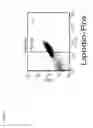
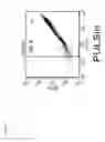


Sources:
- United States Patent and Trademark Office - verify current appl. status at the USPTO↗
Similar patent applications:
Recent applications in this class:
- » 20250171742 2025-05-29
BIOREACTOR FOR CELL EXPANSION AND CULTIVATION - » 20250163381 2025-05-22
PRODUCTION METHOD OF MESENCHYMAL STEM CELL - » 20250136944 2025-05-01
STEM CELL COMPOSITION - » 20250066735 2025-02-27
PREPARATION METHOD OF MESENCHYMAL STEM CELL (MSC) AND APPLICATION THEREOF IN KNEE PRODUCT - » 20240409898 2024-12-12
POTENCY ASSAY - » 20240392248 2024-11-28
HUMAN BONE MARROW-DERIVED MESENCHYMAL STEM CELL SHEETS AND METHODS FOR THEIR PRODUCTION - » 20240376434 2024-11-14
CELL EXPANSION METHODS AND COMPOSITIONS FOR USE THEREIN - » 20240327795 2024-10-03
METHODS OF GENERATING MESENCHYAL STEM CELLS WHICH SECRETE NEUROTROPHIC FACTORS IN A BIOREACTOR SYSTEM - » 20240287461 2024-08-29
PREPARATION AND STANDARDIZATION OF CELL SPHEROIDS FOR PROMOTING TISSUE REGENERATION - » 20240271098 2024-08-15
SYSTEM AND METHOD FOR EXTRACTION AND CRYOPRESERVATION OF BONE MARROW