INCREASING THE VIABILITY AND STRESS TOLERANCE OF VIABLE BIOLOGICAL MATERIAL
US20140329222A1
2014-11-06
14/332,910
2014-07-16
Abstract:
The present invention relates to a method for improving viability and/or stress tolerance of viable biological material and using the said material comprising applying hydrostatic pressure to said biological material; keeping the said viable biological material at the hydrostatic pressure for a predetermined time period; releasing the hydrostatic pressure; and using the said material for any desired purpose in accordance with any useful protocol. The usage of the said biological material incorporates any techniques, protocols that are applicable in the field of assisted reproductive techniques, biotechnical and/or biotechnological manipulations.
Inventors:
- Csaba Pribenszky 5 🇭🇺 Budapest, Hungary
- Miklós MOLNÁR 1 🇭🇺 Budapest, Hungary
- András HORVÁTH 1 🇭🇺 Ullo, Hungary
Assignee:
- APPLIED CELL TECHNOLOGY KFT. 1 🇭🇺 Szekesfehervar, Hungary
Interested in similar patents?
Get notified when new applications in this technology area are published.
Classification:
C12N5/061 » CPC main
Undifferentiated human, animal or plant cells, e.g. cell lines; Tissues; Cultivation or maintenance thereof; Culture media therefor; Animal cells or tissues; Human cells or tissues; Vertebrate cells; Germ cells Sperm cells, spermatogonia
C12N5/0604 » CPC further
Undifferentiated human, animal or plant cells, e.g. cell lines; Tissues; Cultivation or maintenance thereof; Culture media therefor; Animal cells or tissues; Human cells or tissues; Vertebrate cells; Embryonic cells ; Embryoid bodies Whole embryos; Culture medium therefor
C12N5/0609 » CPC further
Undifferentiated human, animal or plant cells, e.g. cell lines; Tissues; Cultivation or maintenance thereof; Culture media therefor; Animal cells or tissues; Human cells or tissues; Vertebrate cells; Germ cells Oocytes, oogonia
C12N5/0606 » CPC further
Undifferentiated human, animal or plant cells, e.g. cell lines; Tissues; Cultivation or maintenance thereof; Culture media therefor; Animal cells or tissues; Human cells or tissues; Vertebrate cells; Embryonic cells ; Embryoid bodies Pluripotent embryonic cells, e.g. embryonic stem cells [ES]
A61D19/04 » CPC further
Instruments or methods for reproduction or fertilisation for embryo transplantation
A61B17/43 » CPC further
Surgical instruments, devices or methods, e.g. tourniquets; Gynaecological or obstetrical instruments or methods for reproduction or fertilisation for artificial insemination
A61B17/435 » CPC further
Surgical instruments, devices or methods, e.g. tourniquets; Gynaecological or obstetrical instruments or methods for reproduction or fertilisation for embryo or ova transplantation
A61D19/02 » CPC further
Instruments or methods for reproduction or fertilisation for artificial insemination
C12N1/36 » CPC further
Microorganisms, e.g. protozoa; Compositions thereof ; Processes of propagating, maintaining or preserving microorganisms or compositions thereof; Processes of preparing or isolating a composition containing a microorganism; Culture media therefor Adaptation or attenuation of cells
Description
This is a continuation-in-part of application Ser. No. 13/632,677, filed Oct. 1, 2012, now abandoned, which is a continuation of Ser. No. 12/094,582, filed Aug. 25, 2008, now U.S. Pat. No. 8,278,031, which is the National Stage of International Application PCT/IB2006/054358, filed Nov. 21, 2006, the entireties of which are hereby incorporated by reference herein.
The present invention relates to a method for improving viability and/or stress tolerance of viable biological material and using the said material comprising applying hydrostatic pressure to said biological material; keeping the said viable biological material at the hydrostatic pressure for a predetermined time period; releasing the hydrostatic pressure; and using the said material for any desired purpose in accordance with any useful protocol. The usage of the said biological material incorporates any techniques, protocols that are applicable in the field of assisted reproductive techniques, biotechnical and/or biotechnological manipulations.
BACKGROUND ART
The effect of hydrostatic pressure as a stressor in connection with increased stress tolerance and shock proteins has been studied in chondrocytes, yeast and bacteria, but not yet in gametes, embryos or stem cells.
The physiological mechanisms by which microorganisms adapt to sublethal stresses are not yet understood well. Recent studies describe that instabilities caused by sublethal cold shock in the normal protein synthesis in bacteria are overcome by the synthesis of so-called cold-shock proteins (CSPs) (Phadtare et al., 1999). These CSPs are suspected to have many functions such as RNA chaperones (Graumann and Marahiel, 1999) or transcription activators (LaTena et al., 1991); it is assumed that they also play a role in the protection against freezing (Wouters et al., 1999). Further investigations found that the production of CSPs is induced not only by cold shock, but also by other environmental stresses. In Escherichia coli, for example, a type of CSP is produced by nutritional stress (Yamanaka et al., 1998).
Another trial showed that high hydrostatic pressure treatment provoked the production of certain cold-induced proteins and heat shock proteins (Welch et al., 1993). Since both cold shock and high pressure treatment increase CSP levels, trials were conducted about the possibility of cross-protection. Wemekamp-Kamphuis et al. (2002) found that the level of survival after pressurization of cold-shocked Listeria monocytogenes was 100-fold higher than that of the cells growing at 37° C.
Hydrostatic pressure in the range of 30-50 MPa usually inhibits the growth of various organisms: the initiation of DNA replication is one of the most pressuresensitive intracellular processes (Abe et al., 1999). The effects vary in severity depending upon the magnitude and duration of compression (Murakami and Zimmerman, 1973). The cell membrane is noted as a primary site of pressure damage (Palou et al., 1997). High hydrostatic pressure treatment can alter the membrane functionality such as active transport or passive permeability and therefore may perturb the physico-chemical balance of the cell (Yager and Chang, 1983; Aldridge and Bruner, 1985; Macdonald, 1987; Schuster and Sleytr, 2002; Routray et al., 2002). The application of pressure can lead to changes in protein structure, including partially or completely unfolded conformations. Pressure can cause the denaturation of proteins (Schmid et al., 1975; Weber and Drickamer, 1983; Jaenicke, 1991; Gross and Jaenicke, 1994; Silva et al., 2001). Recent reports state that hydrostatic pressure enhances the production of shock proteins (Welch et al., 1993; Wemekamp-Kamphuis et al., 2002).
The physical or biochemical processes at altered pressure conditions are governed by the principle of Le Chatelier: all reactions that are accompanied by a volume decrease speed up considerably (Murakami and Zimmerman, 1973; Welch et al., 1993; Palou et al., 1997). The accumulation of the pressure effects is lethal beyond a certain level: while irreversible changes of some biomolecules take place at higher pressures, at 300 MPa most bacteria and multicellular organisms die. Though tardigrades—in their active state they die between 100 and 200 MPa—can survive up to 600 MPa if they are in a dehydrated state (Seki and Toyoshima, 1998). An early publication showed that biological systems are able to tolerate high pressures as long as the pressure is reduced slowly (Johnson et al., 1954). Pribenszky et al. (2003, 2004) also explored the possibility of gradual retrieval of the pressurized embryos and found that gradual release of pressure significantly improves survival.
In response to various stress stimuli, heat shock genes are induced to express heat shock proteins (HSPs). Previous studies have revealed that expression of heat shock genes is regulated both at transcriptional and posttranscriptional level, and the rapid transcriptional induction of heat shock genes involves activation of the specific transcription factor, heat shock factor 1 (HSF1). Furthermore, the transcriptional induction can vary in intensity and kinetics in a signal- and cell-type-dependent manner. Kaarniranta et al. (1998) demonstrated that mechanical loading in the form of hydrostatic pressure increases heat shock gene expression in human chondrocyte-like cells. The response to continuous HHP was characterized by elevated mRNA and protein levels of HSP70, without activation of HSF1 and transcriptional induction of hsp70 gene. The increased expression of HSP70 was mediated through stabilization of hsp70 mRNA molecules. Interestingly, in contrast to static pressurization, cyclic hydrostatic loading did not result in the induction of heat shock genes. The findings of Kaarniranta et al. (1998) showed that hsp70 gene expression is regulated post transcriptionally without transcriptional induction in chondrocyte-like cells upon exposure to high continuous hydrostatic pressure. They suggested that the posttranscriptional regulation in the form of hsp70 mRNA stabilization provides an additional mode of heat shock gene regulation that is likely to be of significant importance in certain forms of stress.
Previously, the present inventors found that a sublethal shock, high hydrostatic pressure (HHP), significantly improves the post-thaw survival of frozen mouse blastocysts (Pribenszky et al., 2005a, WO2005022996). Similarly, at semen cryopreservation, the average post-thaw motility was significantly superior with pressure pre-treatment in each of the pressurized bovine semen samples compared to the samples frozen without previous pressurization. The result clearly describes the beneficial effect of a previous pressure treatment to the post thaw motility of cryopreserved bull semen (Pribenszky et al., 2005b). Further investigations for exploring the biological background and biochemical change during the HHP process will unveil the mechanism of its protective effects. These studies, however, involve the cryopreservation of the biological material after the HHP pre-treatment, which may be of low efficiency with a variety of biological material.
The process of semen storing at temperatures above 0° C. is well established to keep spermatozoa for a short period of time [Hackett, et al., 1982; Pinto, 1999; O'Shea et al., 1964]. With optimal semen treatment (dilution) and storage at optimal temperatures the semen can be inseminated with acceptable fertility results (but with obviously reduced conception rates compared to fresh semen insemination) within 1-2 days post collection [Gill et al., 1970; Goodman and, Cain, 1993; Han-op, 1954; ljaz and Ducharme, 1995; Katila et al., 1997] These methods follow very similar basic steps:
1. Semen collection.
2. Semen dilution at body temperature.
3. Optional centrifugation of the diluted semen. Re-extending the semen to adjust the optimal sperm-concentration.
4. Keeping the (re)extended semen at room temperature or 4-5° C. or any temperature that is above the freezing point of the sample.
5. Insemination of the semen.
Similarly to spermatozoa that suffer a loss of viability during storage, the survival capacity of embryos or oocytes also reduce once removed from their physiological maternal surrounding and experience further in vitro manipulations (for example for in vitro culture, activation, embryo transfer, splitting, sex determination, biopsy, in vitro maturation, ICSI, cloning or any type of biotechnological procedure). For this reason improving the viability/survival capacity of gametes and embryos before or after any procedure including from routine storage, insemination or transfer as far as the most complex biotechnological procedure is of great scientific and economic importance.
Similarly, for example during preservation of microorganisms, such as bacteria (e.g. freeze-drying), the viability of microorganisms is greatly compromised. Improving the efficacy of any process that comes together with improved viability bears immense scientific and economic significance.
As it is clear from the above, there is still a need in the art for the improvement of the viability of biological material that is widely used in biotechnology protocols. In particular, in vitro culture, storage, and manipulation of gametes and embryos require meticulously adjusted conditions to avoid or minimize the harmful effects of uncontrolled stress. However, the present inventors surprisingly found that a well-defined and properly applied stress may induce general adaptation and increase tolerance to various in vitro procedures. Treatment with sublethal doses of HHP resulted in increased morphological survival, fertilizing ability, or developmental potential after various in vitro or in vivo procedures. HHP treatment of spermatozoa, oocytes, embryos, and embryonic stem cells increased fertilizing ability, developmental competence, and differentiation and improved results after cryopreservation, parthenogenetic activation, intracytoplasmic sperm injection, and somatic cell nuclear transfer. Osmotic stress of oocytes resulted in higher developmental rates after cryopreservation, parthenogenetic activation, and somatic cell nuclear transfer. Although cellular and subcellular mechanisms supposedly contributing to these processes require further research, the new principle, i.e., to improve the stress tolerance by a defined HHP sublethal stress, outlines a completely new strategy in mammalian embryology, as well as cryopreservation of other cells and tissues, with remarkable theoretical and practical consequences.
The present inventors surprisingly found that by applying a hydrostatic pressure challenge the viability of biological materials can be improved significantly, and by the application of the method, many state of the art biotechnology protocols can be accomplished more efficiently. The present specification shows a wide range of examples on this finding: after applying the present method to embryo transfer or insemination, the conception rate and birth rate improved; by applying the present method to oocytes, their stress tolerance greatly increased, which resulted in improved cleavage rate and higher blastocyst formation rate; by applying the present method to semen, and then by following state of the art dilution and storage, the motility of the spermatozoa was preserved for a significantly longer period of time.
It was also surprising that the improvements were substantial even when avoiding to apply temperatures below the freezing point of the medium during any stage of the storage and/or manipulation of the biological material. This finding has significant practical implications for the usability of the present and similar HHP methods.
In this context we must emphasize that the present inventive concept equally applies to any different biotechnical/biotechnological protocol or procedure used in the assisted reproductive technologies (ART) and other procedures, and the choice of those is not limited with respect to the invention. The only necessary step to include in the improved protocols is the step of hydrostatic pressure challenge; the parameters of which can be easily optimized by a person skilled in the art when following the teachings of the present description.
Because semen freezing yields poor post-thaw survival of spermatozoa at boars (and horses as well), the most common tool of breeding at these species is the insemination of fresh, extended, extended and cooled or extended and chilled semen. By the use of HHP pre-treatment semen is significantly better preserved at the given temperature, and also, the time of storage with higher quality is considerably increased.
Similarly, in vitro and in vivo embryo production, in vitro culture of embryos, sexing, splitting, gene transfer, embryo transfer, oocyte maturation, activation, ICSI, cloning or any biotechnical/biotechnological procedure in the embryo, oocyte or sperm greatly reduce their viability/survival capacity. As an extrapolation of the above features, by the use of HHP pre-treatment gametes and embryos will enter any type of assisted reproductive technology (ART) or biotechnical/biotechnological procedure with an increased survival capacity.
DISCLOSURE OF THE INVENTION
Accordingly, the present invention relates to a method for improving viability and/or stress tolerance of viable biological material and using the said material comprising
(a) applying hydrostatic pressure to said viable biological material;
(b) keeping the said viable biological material at the hydrostatic pressure for a pre-determined time period;
(c) releasing the hydrostatic pressure;
(d) using the said material for any desired purpose in accordance with any useful protocol,
wherein said viable biological material is kept at temperatures above the freezing point of the medium during any stage of using thereof.
In an embodiment, pressure used in the method according to the invention is in the range of 1 to 200 MPa. In preferred embodiments, the pressure is preferably in the range of 10 to 100 MPa, more preferably 20 to 75 MPa, and most preferably 30 to 60 MPa.
In another embodiment, the hydrostatic pressure used in the method according to the invention is applied for a time period between ‘instantaneous’ and 300 minutes. In preferred embodiments, the pressure is applied preferably for a time period between 0.001 seconds and 600 minutes, preferably 1 seconds to 300 minutes, more preferably 10 seconds to 150 minutes, more preferably 20 seconds to 90 minutes, and most preferably 30 seconds to 60 minutes.
In other embodiments, the time period for releasing the pressure is between 10 sec and 2 hours, or between 1 min and 1 hour, or in other cases 10 min and 30 min. The release of pressure can be instantaneous.
In a preferred embodiment, the invention relates to a method where the pressure is applied, kept and released according to a predetermined pressure profile.
In another preferred embodiment, the invention relates to a method where the pressure is applied, kept and released according to a predetermined temperature profile.
In a preferred embodiment, the method according to the invention is used in connection with gametes, embryos and stem cells selected from the group consisting of oocytes, sperms, zygotes, morulas, blastocysts, embryos, stem cells of a vertebrate animal.
Preferred embodiments relate to a method wherein the said vertebrate animal is a fish, bird or a mammal, preferably bovine, equine, caprine, ovine, swine, other livestock, pets, primates, including human.
In another embodiment, the present invention relates to a method wherein the said biological material is a culture of micro-organisms.
In preferred embodiments of the present invention, the said culture of micro-organism is a bacterial culture.
The present invention further relates to any method as described above, wherein the usage of the said biological material incorporates any techniques, protocols that are applicable in the field of assisted reproductive techniques, biotechnical and/or biotechnological manipulations.
In a preferred embodiment, the protocol used in the method according to the present invention is freeze-drying.
In a further aspect, the method of the present invention applied to improve the stress tolerance of the viable biological material. In a preferred embodiment, tolerance against increased temperature is improved.
For the sublethal stressor, HHP treatment is chosen due to its unique and outstanding features: 1) acts instantly and uniformly at every point of the sample, 2) HHP features zero penetration problems or gradient effects, 3) HHP can be applied with the highest precision, consistency, reliability, and safety.
Experiments followed a common scheme. First, samples (spermatozoa, oocytes, embryos, or stem cells) are exposed to different levels (5-80 MPa) of hydrostatic pressure applied for various times in the range of 0.1-120 min to determine the sublethal zone.
In the context of the present invention, the term “sublethal” means the type of treatment that does not cause loss in cell's survival. In other words, the treatment that is not sufficient to cause death. The limit of the lethal treatment in this context is when treatment begins to cause significant losses in cell survival (p=0.05). If the treatment does not reach this level, but conveys at least 10% intensity of the treatment that caused significant losses (p=0.05), then the treatment was sublethal.
Generally, for HHP treatment, oocytes, embryos, spermatozoa, embryoid bodies, or other cells or tissues are loaded into suitable containers (e.g., 0.25- or 0.5-ml ministraw, 5-ml maxistraw, 100- to 500-ml transfusion bag) in conventional culture or extender media (e.g., TCM-199, M2, or G-MOPS for oocytes and embryos) and sealed hermetically, without air bubbles. Subsequently, containers are placed into the pressure chamber (filled with a liquid, for example distilled water) of the pressurizing device that was previously heated up to the required temperature (e.g., body or room temperature). The pressure chamber is closed, and the machine executes the pressure program according to the set parameters (magnitude and duration of the pressure, treatment temperature, pressure profile).
At the second phase of experiments, samples are exposed to this sublethal dose, then incubated for 5-120 min under normal culture conditions for recovery. Subsequently, the required intervention (insemination, parthenogenetic activation, in vitro fertilization, in vitro maturation or culture, enucleation followed by somatic cell nuclear transfer, in vitro differentiation) is performed. Results are assessed by investigating morphology as well as functional parameters, including motility, membrane integrity, fertilizing ability, and developmental competence. In each experiment, treatment groups are compared with a single control group where HHP treatment was omitted.
Surprisingly, the pressure tolerance limit of mammalian gametes, embryos and stem cells was found in the 20-80 MPa zone, where cells survived 30- to 120-min treatments without any loss of their viability, although the highest hydrostatic pressure that these cells normally encounter is less than 0.2 MPa. The optimal and sublethal pressure ranges for gametes, embryos and stem cells of different mammalian species are shown in the following table:
| Mouse | Porcine | Ovine | Bovine | Human | |
| Sperm | 30 MPa/90 min | 30 MPa/90 min | 20 MPa/90 min | ||
| Oocyte | 20 MPa/60 min | 20 MPa/60 min | 20 MPa/60 min | 20 MPa/60 min | |
| Embryo | 60 MPa/60 min | 20 MPa/60 min | 60 MPa/30 min | 60 MPa/60 min | |
| Embryonic | 60 MPa/30 min | ||||
| stem cells | |||||
| Mesenchymal | 60 MPa/60 min | 40 MPa/30 min | |||
| stem cells | |||||
The present invention is described in more detail by using mouse, ovine and bovine embryos, bovine and boar spermatozoa, pig oocytes, human stem cells and two bacteria species for the purpose of demonstrating the inventive concept. It should be apparent that the disclosed procedures equally apply to all mammalian, avian or fish gametes and embryos, which are candidates of any kind of ART, or more generally, to any type of viable biological material usable in biotechnical or biotechnological procedures, for example or regenerative procedures. It is obvious, however, that the results of the present invention are unexpected and/or advantageous, and such results, based on the diverse distribution of examples presented herein, are likely to be exhibited over a wide range of biological material.
In the context of the present invention, the expression ‘viable biological material’ refers in general to a part of or originating from a living organism that has a capacity for living, developing, or germinating under favorable conditions. Without limitation, the viable biological material can be a cell, cell culture, tissue sample, tissue culture, organ, and the like.
With respect to micro-organisms, the term refers to an organism that is microscopic, i.e. too small to be visible to the naked eye. Micro-organisms can be bacteria, fungi, archaea or eukaryotes. Micro-organisms are often described as single-celled, or unicellular organisms; however, some unicellular protists are visible to the naked eye, and some multicellular species are microscopic.
As highly developed eukaryotic organisms, mouse embryos are more susceptible to the effect of hydrostatic pressure than tardigrades and bacteria. The first objective therefore is to establish the basic features of mouse embryos under pressure concerning their morphology and survival.
Carefully designed experiments were conducted to investigate the pressure tolerance of mouse embryos. The choice of pressure and time scale used was defined to give the widest applicable range for later practical applications. Therefore, the pressure for the use in the method according to invention is selected in the range from 1 MPa to 150 MPa. More particularly, the hydrostatic pressure that can be applied to the expanded blastocyst stage embryos is 1, 5, 10, 15, 20, 25, 30, 40, 50, 60, 70, 80, 90, 100, 110, 120, 130, 140 or 150 MPa, or any value in between these intermediate ranges. Similarly, a wide period of time can be selected for the mouse embryos to be kept under high hydrostatic pressure. More particularly, the mouse embryos are kept under the selected pressure for a time period between 1 sec. and 6 hours, more specifically 1 s, 5 s, 10 s, 20 s, 30 s, 40 s, 50 s, 1 min, 2 min, 3 min, 4 min, 5 min, 6 min, 8 min, 10 min, 15 min, 20 min, 30 min, 40 min, 50 min, 60 min, 70 min, 80 min, 90 min, 120 min, 150 min, 180 min, 210 min, 240 min, 300 min or 360 min. The time the embryos survive under pressure reduces with increasing pressure.
Embryos can survive a substantial amount of pressure without any visible change in their morphology (e.g., 90 MPa for 1 s or 30 MPa for 2 h). The embryos compacted depending on the magnitude and the duration of the applied pressure treatment. Without limiting the scope of the invention by theory, we assume that pressure can not be directly responsible for squeezing the water out of the blastocysts. Based on the cited documents, the compaction of the embryos was due to the consequences of pressure induced production of different proteins (cold-shock proteins, CSPs), reversible alterations in protein structure and metabolic processes. Compacted embryos could regain their normal morphology after 4-5 hours of in vitro culture, and resume development similarly to controls (e.g., embryos challenged by 90 MPa for 30 min or 30 MPa for 3 h).
Embryos from the above mentioned ‘sublethal range’ (e.g. compacted embryos) can preferably be selected for later transfer or any ART or biotechnological procedure. After pressurization, expanded blastocysts become compacted and stay in this form for 3-4 hours, then they re-expand. Based on this phenomenon, embryos treated with pressure before can be selected. Since the morphological changes of the embryos and the beneficial effects of the pressure pre-treatment may come from the altered protein structure and/or features and/or the enhanced production of different pressure-induced proteins, the examination of these proteins can be indicative of the high hydrostatic pressure applied to the biological material before any further process.
The higher the magnitude of the pressure, the less time the embryos survive. Pressure impact exceeding a certain magnitude and duration caused irreversible changes: embryos became disintegrated after 2 hours of in vitro culture or were already disintegrated after decompression (e.g., embryos challenged by 90 MPa for 2 h or 30 MPa for 5 h). The person skilled in the art should be capable of determine these limitpressures and limit-times by routine experimentation with respect to the specific biological material used.
It will be appreciated that the survival rate of the pressurized embryos can be enhanced by gradual decompression thereof. Studies showed that the survival rate of the pressurized embryos increased strikingly if they were retrieved gradually. While 60 minutes at 90 MPa was lethal for all of the embryos, 80% survived when 120 min. gradual decompression was used. The decompression time is also a feature of the present invention which is up to the person skilled in the art to determine in view of the specific application. More particularly, the mouse embryos kept under the selected pressure are decompressed for a time period between 1 sec. and 4 hours, more specifically 1 s, 5 s, 10 s, 20 s, 30 s, 40 s, 50 s, 1 min, 2 min, 3 min, 4 min, 5 min, 6 min, 8 min, 10 min, 15 min, 20 min, 30 min, 40 min, 50 min, 60 min, 70 min, 80 min, 90 min, 120 min, 150 min, 180 min, 210 min or 240 min.
It will be appreciated by the person skilled in the art that the pressure treatment according to the present invention can be carried out according to any desired pressure profile. Therefore, the time dependence of the pressure as it applied, kept and released may vary according to different time—pressure curves, including all time point of the pressure treatment, i.e. during application of the pressure, during the course of the max pressure period, and during the pressure release phase. It is obvious that the pressure level at any given time point can be optimized and set according to easily performed preliminary experiments based on the present teaching without excess experimentation.
In another preferred embodiment, the method of the present invention can be carried out according to a predetermined temperature profile. The term ‘temperature profile’ refers to the time—temperature profile as it is measurable or set during the course of the pressure treatment, and it is independently controllable from the pressure applied. In preferred embodiments, the pressure treatment carried out at a single temperature, however, the biological material of interest may dictate the use of different temperatures at the different stages or parts of the stages of the pressure treatment. A set temperature may be any temperature, for example room temperature, ambient temperature, the native body temperature where the biological material is originating from, a temperature slightly elevated of the said native body temperature, and the like. The person skilled in the art can easily determine the applicability of any given temperature profile by analyzing the efficiency of the pressure treatment compared to a control experiment.
Without being limited by theory, a possible explanation of this feature could be that a considerable amount of CO2 is generated under pressure (Abe and Horikoshi 1995). The hydration and ionization of CO2 (HCO3− and H+) are facilitated by elevated pressure because the reaction is accompanied by a decrease in volume (−0.26 ml/mol) in a manner dependent on the magnitude of the pressure applied (Palou at al. 1997, Welch at al. 1993). The intracellularly produced carbon dioxide instantly dissolves, and then dissociates to give HCO3− and H+, thus also reducing the intracellular pH (Abe and Horikoshi 1995, 1997, 1998, Abe et al. 1999). It can be assumed that the equilibrium maintained by elevated pressure is lethal for the embryos at atmospheric pressure. It may be also hypothesized that the instant decrease of pressure causes elevated release of CO2 from its hydrated and ionized form from the cytoplasm, causing immediate death of the embryos. On condition of a certain decompression time, the plasma membrane proteins (H+-ATPase) (Schmid et al. 1975, Pequeux and Gilles 1978) reversibly inactivated by elevated hydrostatic pressure, start to function again, (together with passive diffusion) shifting the equilibrium gradually towards the physiological state.
After the treatment with carefully chosen pressurizing parameters, the embryos were cultured in vitro, then transferred into pseudopregnant mothers. The number of pups born after pressure-treatment of the embryos was higher than what was achievable without pressure treatment.
Similarly to the treatment of embryos, pig oocytes were also pressurized to examine their pressure-tolerance. Then, with carefully selected parameters, oocytes were pretreated, stressed with an electric impulse 10 times greater than the optimal value, and were examined for cleavage and further development until the blastocyst stage. The number of in vitro activated oocytes that cleaved and developed further was higher with pressure-treatment than without.
Similarly to the treatment of embryos, bull and boar semen was also pressurized to examine the pressure-tolerance of spermatozoa. Then, with carefully selected parameters, semen was pre-treated, and kept in vitro at different temperatures. The number of motile spermatozoa in the different time points after semen collection was higher with pressure-treatment than without.
The insemination of pressure treated boar semen yielded higher litter size than was achievable routinely in the facility.
Similarly to the above mentioned treatments, Escherichia coli and Lactobacillus plantarum samples were pressurized to examine if the survival of the bacteria can be increased after freeze-drying. The number of surviving bacteria in selected pressure/time parameters after freeze-drying was higher with pressure-treatment than without.
The present descriptions shows the improvement in the viability/survival capacity of biological material by hydrostatic pressure challenge using mouse blastocysts, pig oocytes, boar and bull spermatozoa and two bacterium species as model systems. This can be evaluated by transferring the pressurized embryos, following their treatment to culture medium and/or into pseudopregnant recipients; or in vitro activation; or inseminating the treated semen or counting the survival of bacteria after freeze-drying, respectively. In vitro development, implantation and further uterine development and cleavage and further development are obvious proofs of the embryos' and oocytes' viability. Similarly, the in vitro storage and insemination of the semen, yielding higher number of motile spermatozoa (in vitro) and higher pregnancy rates support the applicability of the said method.
The pressurization may be carried out by using any available pressurizing device that can be adapted to the protocols according to the present invention. Non-limiting examples of such instruments are for example the devices described in WO2005022996 or in the present document.
The present invention is further illustrated by the experimental examples described below, however, the scope of the invention will by no means be limited to the specific embodiments described in the examples.
BRIEF DESCRIPTION OF THE DRAWINGS
FIG. 1 shows the hatching rate of the control and treatment groups at different time points after the treatment.
FIG. 2 shows the morphological appearance of ovine blastocysts after HP treatment: 0 h post expansion at 60 MPa (a) and 40 MPa at (b); 4 h (c) and 18 h (d) after 40 MPa treatment an in vitro culture.
FIG. 3 shows the increased growth rate of MSC1 cells after HP treatment. Initial cell counts and cell counts after 48 hours culturing were evaluated on 400 or 600 bar treated and control samples.
EXAMPLES
Example 1
Survival of Mouse Embryos at Different Pressures on Room Temperature, Effect of Pressure Treatment on Implantation and Birth Rates Embryo Production and Culture
One-cell stage mouse embryos were collected from superovulated 6-8 week old CB6F1 donors and cultured at 37° C. with 5% CO2 and maximal humidity in air in G 1.2 and G 2.2 mediums (Vitrolife, Goteborg) to the expanded blastocyst stage.
Pressurization
Blastocysts were loaded into 0.08 ml plastic straws (7-9 embryos/straw) with M2 (Sigma St. Louis, Mo.). Straws, filled with M2 as pressure medium, were placed into the chamber of a special custom-made device that is capable of generating and precisely detecting hydrostatic pressure up to 150 MPa. Achieving the desired amount of pressure took 20 seconds to 5 minutes (10 MPa to 150 MPa, respectively); the duration of releasing the pressure was 2-4 seconds.
Transfer
For in vivo evaluation, embryos pressurized with 600 bar for 30 min were cultured in G 2.2 for 2 hours as above. Then, they were transferred (7-12 embryos per animal) into Day 3 pseudopregnant recipients. Untreated blastocysts were transferred as controls.
Evaluation and Statistical Analysis
Conclusions were drawn from the changes in the morphological appearance of embryos examined at 400× magnification during 24 hours of continued in vitro culture, and from birth rate of the transferred embryos. Microscopically unchanged morphology of the blastomeres, reexpansion of the blastocoel and hatching from the zona pellucida were signs of in vitro survival. The number of fetuses at the 18 day dissection of the pregnant females or birth of healthy pups was proof of in vivo survival of the embryos. The survival rates were compared to control by chi-square test.
In the present experiments embryos were exposed to different hydrostatic pressures from 10 to 150 MPa (by 10 MPa increments) for various times, between 1 sec to 300 min, at room temperature.
The treatment exceeding a certain amount of pressure and time caused reversible morphological changes. The expanded blastocysts compacted inside the zona pellucida: the blastocoel disappeared, the size of the blastomers reduced but their structural integrity showed no alteration. After 4-5 hours of in vitro culture these blastocysts re-expanded and hatched from the zona pellucida in 24 hours (a). Embryos receiving less impact showed no morphological change and hatched within 24 hours of in vitro culture (b), while embryos challenged with a greater impact did not re-expand from the compacted stage and disintegrated within 2 hours, or were already disintegrated after decompression (c).
For in vivo evaluation, challenged embryos were judged ‘survived’ (a&b) and ‘dead’ (c) after 2 hours of in vitro culture after decompression and were transferred into recipients separately. Twenty nine pressure treated embryo were transferred to pseudopregnant mothers, out of which 28 were born. This ratio was higher then what was achievable with non-treated embryos. This significant improvement over the state of the art data (around 85%) also shows the robustness of the pressure pre-treatment. When the optimal pressure and time parameters are applied, the improvement of the viability of biological material still can be significant even when the baseline values quite high and already satisfactory for the industry. However, in the field of ART, every percentage point may have added economical significance.
Example 2
Survival of Bull Spermatozoa at Different Pressures on Room Temperature, Effect of Pressure Treatment on the Prevention of the Decline of Sperm Motility
Although bull semen is usually stored frozen, the feasibility of the present method was further tested in an industrially important system.
Semen of 13 bulls was diluted to a sperm concentration of 8×10⁷/mL with AndroMed extender (MiniTub, Tiefenbach, Germany). Diluted sperm was loaded into 0.25-mL straws at 25° C. Straws were divided into treatment groups and non-treated control group. The treatment groups were pressurized with computer controlled pressure machine (Cryo-Innovation, Budapest, Hungary) with 9 different profiles. For total/progressive motility testing CASA apparatus, SpermVision Version 3.0 (Minitub, Tiefenbach, Germany) was used.
It was concluded that pressure-treatment below the 600 bar region does not affect negatively sperm survival. After 8 hours of semen storage at room temperature the motility of the treated/non treated samples were analysed again: the proportion of the motile cells were higher in the treated samples.
Example 3
Survival of Boar Spermatozoa at Different Pressures on Room Temperature, Effect of Pressure Treatment on the Prevention of the Decline of Sperm Motility
To further examine the applicability the method according to the invention, survival of boar spermatozoa was examined, where storage on room temperature is rather an industry standard.
Semen Collection
Semen was collected from boars twice a week. The filtered sperm-rich fraction was collected by gloved-hand technique into a 250-ml insulated vacuum bottle, then sperm was evaluated (Hancock and Hovell, 1959). The sperm-rich fractions of ejaculates with greater than 70% motile sperm were used.
Preparation of Semen
Semen preparation followed a method previously described (Almlid and Johnson, 1988; Maxwell and Johnson, 1997) with a minor modifications. Briefly, the semen was diluted 1:2 with 37° C. Beltsville Thawing Solution (BTS) extender in the insulated bottle then was cooled at room temperature (20-23° C.) for 1 h after collection. After cooling, semen was transferred into 10 ml tubes, centrifuged at room temperature for 3 min at 2400×g, and the supernatant solution was discarded. The pellets were resuspended in lactose and egg yolk diluent at room temperature. Then glycerol diluent (the second diluent) and Equex paste (Minitub, Tiefenbach, Germany) was added to the semen to give a final concentration of 6% glycerol and 0.5% Equex. Ministraws, 0.25 ml, (IMV, L'Aigle, France) were then filled with semen, straws were heat sealed. The concentration was set to provide 300×106 sperm/ml.
Pressurization
The straws were placed into the pressure chamber, filled with water as a pressure medium, of the pressurizing device, and the pre-determined pressure-protocol was applied. The custom-made pressurizing device was capable of providing precisely controlled pressure in the range of 10-1000 bars. It was made of stainless steel (KO 33) with the inner diameter of 20×220 mm, and was connected to a pressure gauge. A piston, moving in the pressure chamber generated the hydrostatic pressure. Speed of pressurization and depressurization was 200 bar/min.
Samples were pressurized at room temperature (RT) with either 200, 400 or 800 bars for either 40, 80 or 120 minutes. Non-pressurized samples were kept at room temperature for the corresponding time.
Evaluation
After 20 min incubation, two 5 μl drops were transferred onto glass slides and two 22 mm×22 mm cover-slips were applied. The samples were inserted in the microscope (Olympus BX 30), equipped with a 37° C. microscope stage and phase contrast optics (20×, Olympus, Japan) and 5-5 fields were evaluated from each drop by means of CASA apparatus, SpermVision Version 3.0 (Minitiib, Tiefenbach, Germany). Spermatozoa with VSL>10 μm/s and AOC>10 were considered progressive motile.
Results
After analyzing the motility parameters using mixed model (factors: time, pressure, date(random)), the pressure factor proved to be significant (p=0.001, total motility; p=0.0103, progressive motility). After multiple comparisons of the pressure treatments, the 800 bar treatment impact proved to be significantly (P<0.001, total motility; P<0.05, progressive motility) worse than the other levels (Table 1).
| TABLE 1 |
| Motility parameters (mean motility (std error)) following different pressure treatments |
| 40 min | 80 min | 120 min |
| tm | pm | tm | pm | tm | pm | |
| 200 bar | 78.5 (1.66) | 51.25 (7.5) | 85.67 (2.33) | 71.33 (5.46) | 80 (2) | 56.5 (4.5) |
| 400 bar | 77 (1.58) | 63.25 (5.76) | 78.67 (4.18) | 64 (7.77) | 80.5 (4.5) | 74 (2) |
| 800 bar | 67 (4.53) | 46.75 (4.5) | 64 (0.58) | 47.33 (2.19) | 76.5 (1.5) | 66 (1) |
| Atmospheric | 72.25 (2.56) | 47.75 (10.59) | 78.33 (4.1) | 60 (10.21) | 75 (4) | 45.5 (3.5) |
| pressure | ||||||
| tm: total motility; pm: progressive motility. There were 2 repetitions of each treatment combination. |
Next, it was examined whether the application of pressure treatment affects the sperm motility rates after the 5 hours cold acclimatization time. A mixed model was fitted again with pressure, time and examination (with two levels: before and after the 5 hours acclimatization time), interactions of the fix factors, and date as random factor. Only the pressure and examination factors proved to be significant (Table 2).
| TABLE 2 |
| Motility parameters (mean motility (std error)) |
| after 5 hours cold acclimatization time |
| 30 min | 60 min | 90 min | |
| P/t (24° C.) | ||||
| 200 bar | 71% | 65% | 68% | |
| 400 bar | 85% | 86% | 74% | |
| Control group I.: | 65% | |||
| P/t (38.5° C.) | ||||
| 200 bar | 91% | 94% | 76% | |
| 400 bar | 72% | 75% | 83% | |
| Control group II.: | 90% | |||
| tm: total motility; pm: progressive motility. There were 4, 3 and 2 repetitions of each pressure treatments during 40, 80 and 120 minutes time intervals, respectively. |
The total motility of the non-pressurized (atm) samples reduced significantly, while after the 200 and 400 bar treatments the motility did not reduce after 5 hours of cold acclimatization time, compared to the initial motility.
Example 4
Effect of Pressure Treatment on the Fertilizing Capacity of Boar Spermatozoa
Five sows, already excluded from production, have been inseminated with pressure treated boar semen, in order to observe the fertilizing capacity of pressure treated spermatozoa, as well as to investigate any malformations in the offspring.
Ejaculates of two boars were extended 1:3 with commercial extender, at body temperature, then samples were let to cool down to room temperature in 30 minutes, before filling extended semen into infusion bags. Infusion bags were placed into the pressure chamber of an automatic pressurizing device (Cryo-Innovation Ltd., Budapest, Hungary) and pressure program was run at room temperature.
The pressure treatment used was 300 bars for 90 minutes. After treatment, semen samples were further extended with the same commercial extender at room temperature, then 5 sows were inseminated within 1 hour after pressurization. Insemination was repeated 12 hours after the first insemination with the treated semen, left at 5° C. for the corresponding time.
At the ultrasound examination all the five saws were proved to be pregnant. Following normal delivery, 58 healthy piglets were born. The piglets were free from any defects and malformations.
It was concluded that the applied pressure treatment maintains the fertilizing capacity of boar spermatozoa, and does not cause any defect or malformation in the offspring. The average litter size in the farm was 9.8 piglets/sow, whereas with the applied pressure treatment resulted in 100% pregnancy rate and 11.6 average litter size. It was also concluded, therefore, that the applied pressure treatment increases the achievable average litter size, as well.
Example 5
Survival of Boar Semen after Pressure Treatment
For in vitro evaluation, semen was collected from Seghers boars (n=7), extended with commercial extender in 1:3 ratio, cooled to room temperature, and then split into 2 groups. Semen in the treatment group was filled into sterile transfusion bags and treated with 300-bar pressure for 90 min at 25° C. in a computer-controlled pressure device (Cryo-Innovation Ltd., Budapest, Hungary). After treatment, both groups were placed into a cooling thermostat set to 15° C. Total and progressive motility was assessed daily for 12 days by a computer-assisted semen analysis system. Results showed that the reduction of both total (TM) and progressive (PM) motility of the treated samples was significantly slower compared with control samples. On Day 5, the ratio of live (TM) to PM cells (control v. HHP treated) was as follows: TM, 55.4 v. 64.6%; PM, 36.6 v. 46.4%. On Day 11, the ratio of TM to PM cells was as follows: TM, 43 v. 53%; PM, 27 v. 31.4%.
In Vitro Results
| Procedures | ||||
| Species | after HHP | Category | HHP-treated | Control |
| Swine | Storage at 15° C. | Motility at day 5 | 64% | 55% |
| Motility at day 11 | 53% | 43% | ||
For in vivo evaluation, semen (boars, n=14) was prepared, split, and treated as described above. Hungarian Large White× Hungarian Landrace sows (n=103) were then inseminated with teated or non-treated semen. Inseminations were done in the routine production of 3 swine farms. For statistical analysis, a generalized linear mixed-effects model was used, with P<0.05 regarded as significant.
Mean pregnancy rates and litter weights were not different between the 2 groups (73 v. 74%; 16.34 v. 16.37 kg; HHP treated v. non-treated, respectively). However, the effect of treatment on the litter size was significant (12.4 v. 11.4; HHP treated v. non-treated). Moreover, there was a significant difference in treatment effects between the gilts and the sows: whereas treatment had no significant effect on the litter size of the sows (P=0.47), the litter size of the gilts increased (P<0.001), with a mean of 2.55±0.83 (SE).
In Vivo Results
| Procedures | ||||
| Species | after HHP | Category | HHP-treated | Control |
| Swine | Insemination | Average litter size | 12.4 | 11.4 |
Example 6
Survival (Cleavage Following In Vitro Activation) of Pig Oocytes after Different Pressure Treatments
In vitro matured pig oocytes were activated via in vitro electro-activation following different pressure treatment combinations, in order to determine the pressure tolerance of the oocytes.
Six hundred in vitro matured, denudated oocytes were divided into treatment groups (n=15-20 oocytes/group) and control groups. Groups were pressurized in 0.5 ml artificial straws in TCM-HEPES medium (straws were sealed with mineral oil and metal ball) with 200-400 bar for 30-90 min at 24° C. and at 38.5° C. Controls were kept in the same circumstances, and one control group was left in the IVM medium in the thermostat for the corresponding time. After treatments, oocytes were activated in vitro, and placed into culture medium into 38.5° C. thermostat for further development. Cleavage was checked 48 hours after activation. Table 3 shows the percentage of oocytes in the different groups, that cleaved after in vitro activation.
| TABLE 3 |
| Percentage of oocytes cleaved after pressure treatments at 24° |
| C. and at 38.5° C., 48 h after in vitro activation |
| 30 min | 60 min | 90 min | |
| P/t (24° C.) | ||||
| 200 bar | 71% | 65% | 68% | |
| 400 bar | 85% | 86% | 74% | |
| Control group I.: | 65% | |||
| P/t (38.5° C.) | ||||
| 200 bar | 91% | 94% | 76% | |
| 400 bar | 72% | 75% | 83% | |
| Control group II.; | 90% | |||
| Control group III. | 81% | |||
| (thermostat): | ||||
It was concluded that 200-400 bar treatments for 30-90 minutes were not harmful for the matured pig oocytes. Also, the 400 bar treatment for 30-60 minutes at 24° C., and the 200 bar treatment for 30-60 minutes at 38.5° C. yielded higher cleavage rates, than the corresponding control groups. Treatments were also made with 600-800 bar/30-90 minutes. In these groups no oocytes survived the treatments; these pressure parameters were detrimental.
Example 7
Survival of In Vitro Matured Pig Oocytes after Different Pressure-Treatment Combinations, Activated with an Electric Pulse 10 Times Stronger than the Optimal
In vitro matured pig oocytes were activated via 10× magnitude electro-activation in vitro, following different pressure treatment combinations, in order to examine, if treated oocytes better survive the detrimental electro-shock.
Three—six hundred in vitro matured oocytes (daily) were divided into treatment groups (n=15-20 oocytes/group) and control groups. Groups with cumulus cells were pressurized in 0.5 ml artificial straws in TCM-HEPES medium (straws were sealed with mineral oil and metal ball) with 200-800 bar for 30-120 min at 24° C. Controls were kept in the same circumstances, and one control group was left in the IVM medium in the thermostat for the corresponding time.
After treatments, oocytes were denuded with vortexing, then activated in vitro with an electric impulse 10 times stronger than the optimal one, and then placed into culture medium into 38.5° C. thermostat for further development. Cleavage was checked 48 hours after activation, blastocyst formation was examined on the 6th day. Experiments were repeated 3 times.
| TABLE 4 |
| Percentage of the oocytes in the different groups, that cleaved and |
| developed further after in vitro activation with 10x electric pulse. |
| P/t | 30 min | 60 min | 120 min | |
| 200 bar | 42% | 54% | 26% | |
| 400 bar | 35% | 35% | 29% | |
| 600 bar | 0% | 0% | 0% | |
| 800 bar | 0% | 0% | ||
| Control I. (24° C.): | 20% | |||
| Control II. | 10% | |||
| (thermostat): | ||||
| Blastocyst | ||||
| formation | ||||
| 200 bar | 47% | 44% | 32% | |
| 400 bar | 29% | 35% | 28% | |
| 600 bar | 0% | 0% | 0% | |
| 800 bar | 0% | 0% | ||
| Control I. (24° C.): | 28% | |||
| Control II. | 21% | |||
| (thermostat): | ||||
200-400 bar treatments for 30-60 minutes yielded significantly higher number of surviving oocytes than those without treatment or those treated with 600 or 800 bar. It was concluded that the 200 bar pressure treatments for 30 or 60 minutes proved to provide significantly superior cleavage rate and blastocyst rate compared to the control and other groups.
Example 8
Survival of Bovine Embryos after Pressure Treatment
This experiment was conducted to evaluate bovine embryo baro-tolerance, using 40, 60 and 80 MPa for 30 and 60 minutes as hydrostatic pressure treatment. After pressure, the embryos were evaluated by collapse of the blastocoele and then returned to culture in vitro, examined after 2, 24, 48 and 72 h. The embryo survival was evaluated upon morphological appearance and continued in vitro development: intactness of the blastocysts, re-expansion of the blastocoel, and hatching from the zona pellucida were the signs of survival.
Untreated blastocysts were used as controls. Three replicates were performed by using a total of 451 embryos.
Methods
Ovaries were collected from a local slaughterhouse and transported to the laboratory in saline solution (NaCl 0.9%) at 35° C. Cumulus oocyte complexes (COC) were aspirated from 2 to 8 mm diameter follicles. Only COC's with a homogenous cytoplasm and at least three layers of cumulus cells were used. The selected COCs were washed and transferred to a 150 μL drop of maturation medium under silicone oil and incubated for 22 h at 39° C. in 5% of CO2 in air.
Following maturation COCs were transferred to 150 μL drop of fertilization medium. Frozen semen from one ejaculated of a fertility bull was used for all replicates. Motile spermatozoa were obtained by the Percoll gradient method (Parrish et al., 1995) and were added into the fertilization drop in a final concentration of 1×10⁶ sptz/ml. Gametes were cultured in this medium for 18 h under the same conditions as those used for in vitro maturiation, being the day of in vitro fecundation considered as day 0.
After co-incubation, presumptive zygotes were culture in groups of 20 in 150 μl of synthetic oviductal fluid (SOFaaci; Holm et al., 1999), supplemented with 2.77 mM of myo-inositol and 5% FCS.
Cleavage rates were recorded at 48 h post—insemination (Day −2) and embryos rate in day 7, the embryos were graded according to the International Embryo Transfer Society (IETS) manual standard (Robertson an Nelson, 1998), just grade 1 blastocysts were considered for experiment.
For the application of hydrostatic pressure, the blastocysts were load into plastic straws with holding medium (TQC AB Tecnology, Brasil), without air bubbles. Straws were placed into the pressure chamber (Cryoinnovation Ltd, Budapest, Hungary) with water as pressure medium.
Pressure levels and time: 40, 60 and 80 MPa for 30 and 60 min at 32° C.
Results
Hydrostatic pressure treatment showed similar re-expansion rates to group control (Table 5).
With 24 hours of culture, the embryos subjected to 60 MPa for 30 minutes and 60 MPa for 60 minutes had a higher hatching rate when compared to the control group. With 48 hours, only 60 MPa for 1 hour maintaining the difference in the hatching rate. However, after 72 hours, the hatching rate did not differ between treatments, except in embryos subjected to pressure of 80 MPa for 1 hour, where the rate of onset was lower (Table 5).
| TABLE 5 |
| Re-expansion and hatched rates of embryos treated with different levels and time of |
| hydrostatic pressure. The re-expansion rates of HP groups were similar to that of control. |
| The hatching rates at 24 hours were significantly higher after 60 MPa treatment for |
| 30 or 60 minutes when compared to the control group. At 48 hours of culture, only 60 |
| MPa for 60 minutes resulted in a significant increase in hatching rate. |
| 2 h pos- | 24 h pos- | 48 h pos- | 72 h pos- | |
| pressure (%) | pressure (%) | pressure (%) | pressure (%) |
| N | Re- | Re- | Re- | Re- | ||||
| Treatment | Total | expansion | expansion | Hatched | expansion | Hatched | expansion | Hatched |
| Control | 134 | 94.78a | 97.76a | 42.5a | 97.76a | 79.1a | 98.51a | 93.3a |
| 40 MPa-30 min | 36 | 100a | 100a | 55.6a,b | 100a | 83.3a,b | 100a | 91.67a |
| 40 MPa-60 min | 48 | 100a | 95.83a | 35.42a | 100a | 85.42a,b | 97.92a | 91.67a |
| 60 MPa-30 min | 59 | 98.31a | 98.31a | 64.4b | 100a | 83.05a,b | 98.31a | 91.53a |
| 60 MPa-60 min | 57 | 100a | 100a | 57.9b | 98.25a | 89.47b | 98.25a | 96.49a |
| 80 MPa-30 min | 57 | 94.74a | 92.98a | 56.1a,b | 96.49a | 82.46a,b | 96.49a | 91.2a |
| 80 MPa-60 min | 60 | 96.67a | 98.33a | 40a | 95a | 75a | 95a | 80b |
| a,bDifferent superscripts in the same column indicate statistical difference(P < 0.05). |
In addition, it can be seen from FIG. 1 that 60 MPa HP treatment for 60 minutes resulted in a significant increase in hatching rate, when compared to the control group. However, after 80 MPa pressure for 60 minutes, the hatching rate was lower than in the control group.
Example 9
Survival of Ovine Embryos after HP Treatment Experimental Design
In the first experiment, two different HP treatments, 40 MPa and 60 MPa, were applied to ovine in vitro produced blastocysts for 70 min at 38° C. Unexposed groups were used as controls. In each group 20-22 embryos were used and each experiment was repeated three times. Parameters selected in Experiment 1 were applied in Experiment 2 to explore the effect of HP treatment on embryo quality by analysis of blastocyst cell number, nuclear picnosis and expression of a panel of genes associated with embryo developmental competence. The experiment was performed with three replicates.
Methods
Source of Oocytes and In Vitro Maturation (IVM)
Adult (4-6 years old) ovine ovaries (Sarda sheep) were collected at local slaughterhouses and transported to the laboratory within 1-2 h in Dulbecco Phosphate Buffered Saline (PBS) with antibiotics. Collection of cumulus-oocyte complexes (COCs) was performed in sterile Petri dishes containing 20 mM Hepes-buffered TCM 199 supplemented with 0.1% (w/v) polyvinyl alcohol and antibiotics. Only COCs showing several intact cumulus cell layers and uniform cytoplasm with homogenously distributed lipid droplets were selected and matured in vitro in TCM 199 supplemented with 10% heat-treated oestrus sheep serum (OSS), 0.1 IU mL−1 FSH, 0.1 IU mL−1 LH and 100 μM cysteamine. 30-35 COCs were cultured for 24 h in 5% CO; in air at 38.5° C. in four-well Petri dishes (Nunclon, Nalge, Nunc International, Denmark) with 600 μL of maturation medium, layered with 300 μL of mineral oil.
In Vitro Fertilization (IVF)
In vitro matured oocytes were fertilized in SOF medium+2% OSS for 22 h at 38.5° C. and 5% CO2, 5% O2; and 90% N2 under mineral oil in four-well Petri dishes with frozen-thawed spermatozoa selected by swim-up technique (1×10⁶ mL−1 spermatozoa).
In Vitro Embryo Culture
IVF zygotes were cultured for 7 days in groups in four-well Petri dishes in SOF+ essential and non-essential amino acids at oviductal concentration (Walker et al., 1996), +0.4% BSA under mineral oil, in maximum humidified atmosphere with 5% CO2, 5% O2, 90% N2 at 38.5° C. The first cleavage was registered between 24-26 h after the start of fertilization. Development to blastocyst stage was recorded on the 7th day of culture.
Embryo Treatment with HP
Day 7 in vitro-produced ovine blastocysts were pressure treated according to the following conditions:
−60 MPa, 70 min, 38° C.
−40 MPa, 70 min, 38° C.
Blastocysts were loaded into 0.5-mL straws with a 2 mL syringe, as described by Pribenszky et al. (Reprod. Fert. Dev. 2008). Straws were then placed into custom-made hydrostatic pressure chamber (Cryo-Innovation Ltd., Budapest, Hungary) containing water as pressure medium, and then the treatment program was run. After HP treatment, embryos were released from straws and cultured for 24 h in TCM 199+10% FCS in 5% CO2 in air at 38.5° C. Untreated blastocysts cultured in the same condition were used as controls.
Evaluation of Embryo Survival
Experiment 1
Embryo survival after different HP treatments was evaluated upon blastocyst morphological appearance, re-expansion of the blastocoel, and hatching from the zona pellucida.
Evaluation of Blastocyst Cell Number and Nuclear Picnosis
Experiment 2
After HP treatment (40 MPa, 70 min, 38° C.) followed by 24 h culture, evaluation of embryo quality was performed by total cell count, differential staining of the inner cell mass (ICM) and trophectoderm (TE) cell compartments and evaluation of nuclear picnosis. The total cell count of the blastocyst included picnotic cells.
Differential Staining
To differentially stain ICM and TE nuclei, hatched blastocysts of HP-treated and control groups were exposed to 1% Triton X-100 in 20 mM Hepes-buffered TCM 199 for 3-5 seconds, washed 3-5 times and incubated in the same medium containing 30 μg mL−1 propidium iodide (PI) for 30 seconds. The blastocysts were then transferred into ice-cold ethanol containing 10 μgmL−1 Hoechst 33342 for 15 min. The blastocysts were directly mounted into a small droplet of glycerol on a glass slide and examined under fluorescence confocal microscope (Leica TCS SP5). ICM nuclei appeared blue, caused by DNA labelling with Hoechst 33342, while TE cells appeared red due to staining of nuclear DNA with the membrane impermeable PI.
The number of picnotic nuclei was also determined by morphological appearance of blastocysts cell to confocal microscopy. Picnotic nuclei were characterized by very dense chromatin or by dispersed nuclear fragments. Picnotic cell index was calculated according to Hardy et al. (1989):
Picnotic cell index=(No of picnotic cells/total No of cells+No of picnotic cells)×100
Results
Experiment 1
Survival and In Vitro Development of Embryos after Two Pressure Treatments
In Experiment 1, ovine IVP blastocysts were exposed to two hydrostatic pressures. A collapse of the blastocoel inside the zona pellucida was observed at a proportion of the embryos after the HP treatment of the expanded blastocysts (FIG. 2). A significantly higher number of blastocysts collapsed inside the zona pellucida after treatment with 40 MPa compared to the group treated with 60 MPa (97% vs 12%). 253 The hatching rate of embryos treated at 40 MPa was significantly higher than that of 60 MPa-treated group (87.5 vs. 0%; P<0.01) and similar to that of control embryos (85.3%) after 24 h culture (Table 6). A proportion (17.7%) of the embryos treated with 60 MPa showed signs of degeneration in contrast to 40 MPa and control groups.
| TABLE 6 |
| Survival and hatching rates of ovine blastocysts cultured |
| 24 h following treatment with two different HP conditions |
| Developmental stage | |||
| No | after 24 h IVC | Degen- |
| Group | blastocysts | contracted | expanded | hatched | erated |
| HP: 60 MPa | 62 | 10 | 41 | 0a | 11 |
| (16.2%)a | (66.1%)a | (17.7%) | |||
| HP: 40 MPa | 64 | 4 | 4 | 56 | 0 |
| (6.25%)b | (6.25%)b | (87.5%)b | |||
| Control | 60 | 3 | 5 | 52 | 0 |
| (5%)b | (8.3%)b | (86.7%)b | |||
| Values in the same column with different superscripts differ significantly (P < 0.01). |
Experiment 2
Treatment Effects to Embryos
Cell Number and Nuclear Picnosis
Blastocysts exposed at 40 MPa showed higher total cell number compared with control embryos (161.3±8.7 v. 123.9±9.4, P<0.01). The number of ICM and TE cells were higher after HP treatment compared to control (ICM: 23.5±2.3 v. 18.5±2.3, P<0.05; TE: 142.2 d±6.5 v. 116±6.5 P<0.01). The number of picnotic nuclei in blastocysts HP treated was significantly lower than control. (Pl: 1.3±0.4% v. 3.8±0.6%, P<0.05). Results are summarized in Table 7.
| TABLE 7 |
| Effect of HP treatment on cell number and picnosis |
| of in vitro produced ovine embryos. |
| No | Embryos cell number | ||
| blas- | (mean ± SD) | Picnotic |
| Group | tocysts | Total | ICM | TE | index (%) |
| HP: | 47 | 161.3 ± 8.7a | 23.5 ± 2.3a | 142 ± 6.5a | 1.28 ± 0.4%a |
| 40 MPa | |||||
| Control | 51 | 123.9 ± 9.4b | 18.5 ± 2.3c | 116 ± 6.5b | 3.84 ± 0.6%c |
| Values in the same column with different superscripts differ significantly (a vs b P < 0.01; a vs c P < 0.05) |
Example 10
Survival of Bacteria after Different Pressure Treatment Combinations Followed by Freeze-Drying
For the experiment two microbes, Escherichia coli and Lactobacillus plantarum were used. Cell counts were determined on TGE (Tripton Glucose Extract) nutritive media (MERCK) and MRS agar (MERCK), respectively. Cell counts were determined before preparing the treatment groups, after pressure treatments, and after 96 hours of incubation at 37° C. following freeze-drying (c.f.u.). Pressure treatments were executed with 9 pressurizing machines at the same time according to the following table:
| Time |
| Pressure | 30 mm | 60 min | 90 min |
| 200 bar | Treatment group 1 | Treatment group 2 | Treatment group 3 |
| 400 bar | Treatment group 4 | Treatment group 5 | Treatment group 6 |
| 600 bar | Treatment group 7 | Treatment group 8 | Treatment group 9 |
Samples were filled into 0.5 ml sterile artificial straws, and were sealed with sterile iron ball. Freeze-drying was made in Edwards freeze-drying equipment. Experiments were replicated two times. Results are presented in the following tables.
| TABLE 8 |
| Number of living Lactobacillus plantarum cells (c.f.u./0.4 |
| m1) before and after freeze-drying (two repetitions). (Initial cell count |
| (c.f.u./ml) before treatment: 9.0 × 10⁶-1.1 × 10⁷) |
| Number of living cells\0.4 ml |
| Treatment | Before freeze- | After freeze- | After freeze- | |
| group No. | drying | drying I. | drying II. | |
| 1 | 1.30 × 10⁷ | 2.29 × 10⁶ | 2.56 × 10⁶ | |
| 2 | 7.80 × 10⁶ | 3.02 × 10⁶ | 2.54 × 10⁶ | |
| 3 | 8.80 × 10⁶ | 2.01 × 10⁶ | 2.05 × 10⁶ | |
| 4 | 5.70 × 10⁶ | 4.25 × 10⁶ | 4.19 × 10⁶ | |
| 5 | 6.38 × 10⁶ | 1.99 × 10⁶ | 2.10 × 10⁶ | |
| 6 | 1.26 × 10⁷ | 4.34 × 10⁶ | 4.13 × 10⁶ | |
| 7 | 7.50 × 10⁶ | 3.12 × 10⁶ | 3.59 × 10⁶ | |
| 8 | 8.70 × 10⁶ | 2.71 × 10⁶ | 3.88 × 10⁶ | |
| 9 | 8.30 × 10⁶ | 3.36 × 10⁶ | 4.83 × 10⁶ | |
| Control | 1.05 × 10⁷ | 2.56 × 10⁶ | 4.08 × 10⁶ | |
| TABLE 9 |
| Number of living Escherichia coli cells (c.f.u./0.4 m1) before |
| and after freeze-drying (two repetitions). (Initial cell count (c.f.u./ml) |
| before treatement 4.08 × 10⁸-4.10 × 10⁸) |
| Number of living cells\0.4 ml |
| Treatment | Before freeze- | After freeze- | After freeze- | |
| group No. | drying | drying I. | drying II. | |
| 1 | 6.10 × 10⁸ | 1.55 × 10⁷ | 1.65 × 10⁷ | |
| 2 | 3.20 × 10⁸ | 2.56 × 10⁸ | 2.90 × 10⁸ | |
| 3 | 4.80 × 10⁸ | 9.30 × 10⁷ | 9.0 × 10⁷ | |
| 4 | 3.90 × 10⁸ | 1.36 × 10⁸ | 1.00 × 10⁸ | |
| 5 | 5.90 × 10⁸ | 2.14 × 10⁸ | 1.80 × 10⁸ | |
| 6 | 5.95 × 10⁸ | 2.27 × 10⁸ | 1.70 × 10⁸ | |
| 7 | 6.10 × 10⁸ | 7.50 × 10⁷ | 9.50 × 10⁷ | |
| 8 | 6.60 × 10⁸ | 1.65 × 10⁸ | 7.80 × 10⁷ | |
| 9 | 6.20 × 10⁸ | 1.52 × 10⁷ | 1.21 × 10⁷ | |
| Control | 5.10 × 10⁸ | 2.50 × 10⁷ | 7.7 × 10⁷ | |
Treatment groups marked with bold numbers represent significantly higher cell survival rate compared to the control group. Amongst the treatment groups, groups No. 2 and No. 4 proved to be superior. It was concluded that a specific high hydrostatic pressure treatment before freeze-drying enhances significantly the cell survival rate after freze-drying.
Example 11
Survival of Bovine Embryos after Embryo-Biopsy or Sexing with/without Pressure Treatment
Oocyte Collection and In Vitro Maturation (IVM)
COCs (aspirated from ovaries from slaughter house) are matured in TCM-199 Earl's supplemented with FCS, LH (Sigma), FSH (Sigma), L-Glutamine, penicillin and streptomycin, covered with mineral oil, in 38° C. with 5.1% CO2 and maximal humidity in air for 22 hours.
Sperm Preparation, In Vitro Fertilization (IVF) and In Vitro Culture (IVC)
Fertilization medium is: TALP supplemented with BSA, penicilamin, hipotaurin, epinephrine and heparin covered with mineral oil.
Motile spermatozoa are obtained by centrifugation of frozen-thawed spermatozoa on a Percoll discontinuous density gradient (2 ml of 45% Percoll over 2 ml of 90% Percoll) for 20 min at 700 g at room temperature. After resuspension spermatozoa is added to the fertilization drops. Plates are incubated for 19 hr in 5% CO2 in humidified air at 39° C. Presumptive zygotes are then cultured in vitro in SOF droplets under mineral oil in a humidified atmosphere of 5% CO2 at 39° C.
Pressurization
Expanded blastocysts are loaded into 0.25 ml plastic straws without air-bubbles (7-9 embryos/straw), with embryo holding medium, then straws are sealed with PVC. Straws are placed into the pressurizing device (Cryo-Innovation Ltd., Budapest, Hungary). Embryos are exposed to different hydrostatic pressures from 60 to 90 MPa (by 10 MPa increments) for various times (15, 30, 45, 50, 60, 90, 100 minutes), at room temperature.
Results
The survival rate of the sexed embryos or embryos after biopsy is significantly higher with specially selected pressure treatment, than without.
Example 12
Survival of Bovine Embryos after Gene Transfer with/without Pressure Treatment
In the present experiment bovine embryos are exposed to hydrostatic pressure in order to find out if their behavior under altered pressure conditions is similar to that of the mouse embryos. After challenging with hydrostatic pressure, samples are subjected to gene transfer. After in vitro culture and transfer, the survival of the samples is enhanced compared to the samples that were not treated with pressure previously.
Example 13
Survival of Human Embryos after ICSI and Embryo Biopsy with/without Pressure Treatment
In the present experiment human embryos are exposed to hydrostatic pressure in order to find out if their behavior under altered pressure conditions is similar to that of the mouse embryos. After challenging with hydrostatic pressure, samples are subjected to ICSI or biopsy. After in vitro culture and transfer, the survival of the samples is enhanced compared to the samples that were not treated with pressure previously.
Example 14
Survival of Oocytes (Human, Bovine, Caprine, Swine) after Pressure Treatment, In Vitro Storage and Maturation
The aim of the present experiment is to prove that oocytes tolerate any process (including in vitro storage/maturation) with a lot higher efficacy if they are treated previously with hydrostatic pressure. Oocytes are treated with hydrostatic pressure and samples are kept in vitro after releasing the pressure. The in vitro and in vivo survival of the oocytes is enhanced compared to the samples that were not treated with pressure previously.
Example 15
Increasing the Growth Rate of Stem Cells
The aim of this experiment was to investigate the effects of high hydrostatic pressure treatment on stem cells. In this study, hESC-derived mesenchymal stem cell like (MSCl-2) cells were treated with high hydrostatic pressure applied by a custom-made pressurizing device. These cells were characterized in vitro based on the mesenchymal stem cells' functional and phenotypic criteria defined by the Mesenchymal and Tissue Stem Cell Committee of the International Society for Cellular Therapy (Dominici et al. 2006). These experiments proved that the established MSCl-2 cell lines exhibited the robust differentiation potential toward all three canonical mesodermal (osteoblastic, chondrogenic, adipogenic) directions. Investigating the phenotypic characteristics of the MSC1 cells, no expression of various embryonic (October4, Nanog, ABCG2, PODXL, or SSEA4), or hematopoietic (CD34, CD45, CD14, CD133, HLA-DR) stem cell markers was observed. Though, the cells were positive for the characteristic cell surface markers of MSCs (CD44, CD73, CD90, CD105). MSC1 cells exhibited significant inhibition of mitogen-activated lymphocyte proliferation, and thus presented immunosuppressive features (Varga et al, 2011).
Methods
The MSCl cells were derived from human embryonic stem cells (HUES9) as it has been described before (Varga et al. 2011). Briefly, HUES9 cells were cultured on mitotically inactivated mouse embryonic fibroblasts (MEF). After embryoid body (EB) formation the cells were trypsinized and further cultured in DMEM supplemented with 10% fetal bovine serum on a gelatinized 10-cm plate. When the cell culture grew confluent, the cells were trypsinized and split to 1:3 ratio. After two passages most of the cultured cells presented fibroblast-like morphology.
In order to investigate the effect of HHP on MSCl cells they were harvested and loaded into 0.25 ml straws and subjected to high hydrostatic pressure treatment. The samples were subjected to 400 and 600 bar pressure for 30 minutes at 24 C. degrees. After the pressure treatment, the cells were transferred into culturing dishes, and cultured in DMEM supplemented with 10% fetal bovine serum (FBS) on a gelatinized 10-cm plate. The growth rate was determined by microscopic counting of the cells.
Results
It can be clearly seen from FIG. 3 that the initial cell counts were similar in control and HHP treated samples. However, after 48 hours culturing, the cell count of HHP treated samples were remarkably higher than in the control samples. These results indicate that high hydrostatic pressure treatment has a positive effect on in vitro growth rate of stem cells.
The results presented in the present examples show that the pressure treatment applied prior to any type of assisted reproductive or biotechnological technique improves the survival (stress tolerance) of the gametes and embryos (and stem cells). Also, the presented data on mouse embryos, bull and boar spermatozoa, big oocytes and bacteria indicate the wide applicability of the inventive concept. The application of the method according to the present invention can be useful in improving success rates in all kind of assisted reproductive or biotechnological techniques, embryo-manipulation, including other mammalian species, humans not excluded. The present method also opens wide possibilities for other fields where manipulation of gametes and embryos can find its applications.
Because semen freezing yields poor post-thaw survival of spermatozoa at boars (and horses as well), the most common tool of breeding at these species is the insemination of fresh, extended, extended and cooled or extended and chilled semen. By the use of HHP pre-treatment semen is significantly better preserved at the given temperature, and also, the time of storage with higher quality is considerably increased.
Similarly, in vitro and in vivo embryo production, in vitro culture of embryos, sexing, splitting and using any biotechnical/biotechnological procedure, embryo transfer, oocyte maturation, ICSI or any biotechnical/biotechnological procedure in the oocyte or sperm greatly reduce their viability/survival capacity. As an extrapolation of the above features, by the use of HHP pre-treatment gametes and embryos will enter any type of assisted reproductive technology (ART) or biotechnical/biotechnological procedure with an increased survival capacity. [C1]
REFERENCES
- Abe, F., and Horikoshi, K. (1995). Hydrostatic pressure promotes the acidification of vacuoles in Saccharomyces cerevisiae. FEMS Microbiol Lett 130, 307-312.
- Abe, F., and Horikoshi, K. (1997). Vacuolar acidification in Saccharomyces cerevisiae induced by elevated hydrostatic pressure is transient and is mediated by vacuolar H+-ATPase. Extremophiles 1, 89-93.
- Abe, F., and Horikoshi, K. (1998). Analysis of intracellular pH in the yeast Saccharomyces cerevisiae under elevated hydrostatic pressure: a study in baro-(piezo-)physiology. Extremophiles 2, 223-228.
- Abe, F., Kato, C., and Horikoshi, K. (1999). Pressure-regulated metabolism in microorganisms. Trends Microbiol 7, 447-453.
- Aldridge, B. E., Bruner, L. J. (1985). Pressure effects on mechanisms of charge transport across bilayer membranes. Biochim Biophys Acta 817, 343-354.
- Almlid T., and Johnson L. A., 1988. Effects of glycerol concentration, equilibration time and temperature of glycerol addition on post-thaw viability of boar spermatozoa frozen in straws. J. Anim. Sci. 66:2899-2905.
- M. Dominici, K. Le Blanc, I. Mueller, et al., Minimal criteria for defining multipotent mesenchymal stromal cells. The International Society for Cellular Therapy position statement, Cytotherapy 8 (2006) 315-317.
- Gill H P, Kaufman C F, Foote R H, Kirk R W. (1970) Artificral insemination of beagle bitches with freshly collected, liquid stored, and frozen-stored semen. Am J Vet Res 1970; 31:1807-1813.
- Goodman M F, Cain J t. (1993) Retrospective evaluation of artificial insemination wrth chilled extended semen in the dog. J Reprod Feral 1993; 47:554. abstr.
- Graumann, P. L., Marahiel M. A. (1999). Cold shock proteins CspB and CspC are major stationary-phase-induced proteins in Bacillus subtilis. Arch Microbiol 171, 135-138.
- Gross M., and Jaenicke R., 1994. Proteins under pressure. The influence of high hydrostatic pressure on structure, function and assembly of proteins and protein complexes. Eur. J. Biochem. 221:617-630.
- Hackett, A J; Wolynetz, M S (1982): Reproductive performance of totally confined sheep bred with semen extended in a lactose-egg yolk-glycerol buffer and stored at 5 degrees C.
- Canadian Journal Of Comparative Medicine. Revue Canadienne De Medecine Comparee, Volume 46, Issue 3, July 1982, Pages 327-333.
- Hancock J. L., and Hovell G. L. R., 1959. The collection of boar semen. Vet. Res. 71:664-669.
- Hardy K, Handyside A H, Winston R M. The human blastocyst: cell number, death and allocation during late preimplantation development in vitro. Development. 1989 November; 107(3):597-604. Harrop A E. (1954) Artificial insemination of a bitch with preserved semen Br Vet J. 1954; 110:424-425.
- Holm P, Booth P J, Schmidt M H, Greve T, Callesen H. High bovine blastocyst development in a static in vitro production system using SOFaa medium supplemented with sodium citrate and myo-inositol with or without serum-proteins. Theriogenology. 1999 September; 52(4):683-700. Jaenicke, R. (1991). Protein stability and molecular adaptation to extreme conditions. Eur J Biochem 202, 715-728.
- Johnson F. H., Eyring H., Polissar M. J., 1954. The Cinetic Basis of Molecular Biology. Wiley, New York.
- Kaarniranta K., Elo M., Sironen R., Lammi M. J., Goldring M. B., Eriksson J. E., Sistonen L., and Helminen H. J., 1998. Hsp70 accumulation in chondrocytic cells exposed to high continuous hydrostatic pressure coincides with mRNA stabilization rather than transcriptional activation. Proc. Natl. Acad. Sci. USA 95: 2319-2324.
- Katila T, Combes G B, Varner D D, Blanchard T L (1997) Comparison of three containers used for the tranSPOrt of cooled stallion semen. Theriogenology 1997; 48: 1085-1092.
- LaTena A., Brandi A., Falcon M., Spurio R., Pon C. L., and Gualerzi C. O., 1991. Identification of a cold-shock transcriptional enhancer of the Escherichia coli major cold shock gene encoding nucleotide protein H—NS. Proc. Natl. Acad. Sci. USA 88, 10907-10911.
- Ijaz A, Ducharme R. (1995) Effect of various extenders and taunne on survival of stallion sperm cooled to 5′ C Theriogenology 1995, 44: 1039-1050.
- MacDonald, A. G. (1987). The role of membrane fluidity in complex processes under high pressure. In: Jonnasch, H. W., Marquis, R. E., Zimmerman, A. M., editors. Current Perspectives in High Pressure Biology. London: Academic Press pp. 207-223.
- Maxwell W. M. C., and Johnson L. A., 1997. Membrane status of boar spermatozoa after cooling or cryopreservation. Theriogenology 48:209-219.
- Murakami, T. H., Zimmerman, A. M. (1973). DNA syntheseis in Tetrahymena: a pressure study. Cytobios 7, 171-181.
- O'Shea, T., Wales, R G. Effects of potassium on ram spermatozoa during chilling to and storage at 5 degrees C. Journal Of Reproduction And Fertility, Volume 53, August 1964, Pages 121-132.
- Palou, E., Lopez-Malo, A., Barbosa-Canovas, G. V., Welti-Chanes, J., and Swanson, B. G. (1997). Kinetic analysis of Zygosaccharomyces bailii inactivation by high hydrostatic pressure. Lebensm.-Wiss. U. Technol. 30, 703-708.
- Parrish, J. J., J. L. A. Krogenaes and J. L. Susko-Parrish, 1995. Effect of bovine sperm separation by either swim-up or Percoll method on success of in vitro fertilization and early embryonic development. Theriogenology, 44: 859-869.
- Péqueux, A., and Gilles, R. (1978). Effects of high hydrostatic pressures on the activity of the membrane ATPases of some organs implicated in hydromineral regulation. Comp Biochem Physiol B Biochem Mol Biol 59, 207-212.
- Phadtare, S., Alasina, J., Inouye, M. (1999). Cold-shock response and cold-shock proteins. Cliff Opin Microbiol 2, 175-180.
- Pinto, C R; Paccamonti, D L; Eilts, B E (1999) Fertility in bitches artificially inseminated with extended, chilled semen. Theriogenology, Volume 52, Issue 4, September 1999, Pages 609-616.
- Pribenszky C., Molnar M., Cseh S., Solti L., 2005a. Improving post-thaw survival of cryopreserved mouse blastocysts by hydrostatic pressure challenge. Anim. Reprod. Sci. 87. 143-150.
- Pribenszky C., Molnar M., Solti L., Dengg J., Lederer J., 2005b. The effect of high hydrostatic pressure on the motility of fresh and frozen-thawed bull semen (pilot study). Reproduction in domestic animals 40. 338.
- Pribenszky C s., Molnar M., Cseh S., and Solti L., 2003. Viability of embryos after exposing to high hydrostatic pressure. Theriogenology 59, 329 (Abstract).
- Pribenszky C s., Molnar M., Cseh S., and Solti L., 2004. Survival of mouse blastocysts after low temperature preservation under high pressure. Acta Vet. Hung. 52, 479-487.
- Pribenszky C., Siqueira Filho E., Molnár M., Harnos A. and Rumpf R.: Improved post-warming developmental competence of open pulled straw-vitrified in vitro-produced bovine blastocysts by sublethal pressure pretreatement. Reproduction, Fertility and Development 20(1) 124-125 (2008).
- Robertson I, Nelson R E. 1998. Certification and identification of the embryo. In:
- Stringfellow D, Seidel S M (Ed.) Manual of the International Embryo Transfer Society. LETS, Savoy, I L, USA. p. 103-134.
- Routray R., Suzuki T., Strussmann C. A., Takai R., 2002. Factors effecting the uptake of DMSO by the eggs and embryos of medaka, Oryzias latipes. Theriogenology 58:1483-1496.
- Schmid, G., Ludemann, H. D., and Jaenicke, R. (1975) High pressure effects on the activity of glycolytic enzymes. Biophys Chem 3, 90-98.
- Schuster, B., Sleytr, U. B. (2002). The effect of hydrostatic pressure on S-layer-supported lipid membranes. Biochim Biophys Acta 1563, 29-34.
- Silva, J. L., Foguel, D., Royer, C. A. (2001). Pressure provides new insights into protein folding, dynamics and structure. Trends Biochem Sci 26, 612-618.
- Varga N, Vera Z, Rajnavölgyi E, Német K, Uher F, Sarkadi B, Apáti A. Mesenchymal stem cell like (MSC1) cells generated from human embryonic stem cells support pluripotent cell growth. Biochem Biophys Res Commun. 2011 Oct. 28; 414(3): 474-80.
- Weber, G., Drickamer, H. G. (1983). The effect of high pressure upon proteins and other biomolecules. Q Rev Biophys 16, 89-112.
- Walker S K, Hill J L, Kleemann D O, Nancarrow C D. Development of ovine embryos in synthetic oviductal fluid containing amino acids at oviductal fluid concentrations. Biol Reprod. 1996 September; 55(3):703-8.
- Welch, T. J., Farewell, A., Neidhardt, F. C., Bartlett, D. H. (1993). Stress response of Escherichia coli to elevated hydrostatic pressure. J Bacteriol 175, 7170-7177.
- Wemekamp-Kamphuis, H. H., Karatzas, A. K., Wouters, J. A., Abee, T. (2002). Enhanced levels of cold shock proteins in Listeria monocytogenes L028 upon exposure to low temperature and high hydrostatic pressure. Appl Environ Microbiol 68, 456-63.
- Wouters, J. A., Jeynov, B., Rombouts, F. M., de Vos, W. M., Kuipers, O. P., Abee, T. (1999). Analysis of the role of 7 kDa cold-shock proteins of Lactobacillus lactis MG1363 in cryoprotection. Microbiology 145, 3185-3194.
- Yager, P., Chang, E. L. (1983). Destabilization of a lipid non-bilayer phase by high pressure. Biochim Biophys Acta 731, 491-494.
- Yamanaka, K., Fang, L., Inouye, M. (1998). The CspA family in Escherichia coli: multiple gene duplication for stress adaptation. Mol Microbiol 27, 247-255.
Claims
1. Method for improving the viability and/or stress tolerance of viable biological material and using said material comprising
(a) applying hydrostatic pressure to said biological material;
(b) keeping said viable biological material at the hydrostatic pressure for a predetermined time period;
(c) releasing the hydrostatic pressure;
(d) using said material for any desired purpose in accordance with any useful protocol,
wherein said viable biological material is kept at temperatures above the freezing point of the medium during any stage of using thereof.
2. The method according to claim 1 wherein said hydrostatic pressure is in the range of 1 to 200 MPa, preferably 10 to 100 MPa, more preferably 20 to 75 MPa, and most preferably 30 to 60 MPa.
3. The method according to claim 1 wherein said hydrostatic pressure is applied for a time period between 0.001 seconds and 600 minutes, preferably 1 seconds to 300 minutes, more preferably 10 seconds to 150 minutes, more preferably 20 seconds to 90 minutes, and most preferably 30 seconds to 60 minutes.
4. The method according to claim 1 wherein said pressure is released gradually over a time period between instantaneous release and 6 hours.
5. The method according to claim 1 wherein said pressure is applied, kept and released according to a predetermined pressure profile.
6. The method according to claim 1 wherein said pressure is applied, kept and released according to a predetermined temperature profile.
7. The method according to claim 1 wherein the said biological material is a gamete or embryo, and is preferably selected from the group consisting of oocytes, sperms, zygotes, morulas, blastocysts, embryos, and stem cells of a vertebrate animal.
8. The method according to claim 7 wherein said vertebrate animal is a fish, bird or a mammal, preferably bovine, equine, caprine, ovine, swine, other livestocks, pets, primates, including human.
9. The method according to claim 1 wherein said biological material is a culture of micro-organisms.
10. The method according to claim 9, wherein said culture of micro-organism is a bacterial culture.
11. The method according to claim 1 wherein said usage of the said biological material incorporates any techniques or protocols that are applicable in the field of assisted reproductive techniques, biotechnical and/or biotechnological manipulations.
12. The method according to claim 1 wherein said usage is freeze-drying.
13. The method according to claim 1 wherein the said stress tolerance is tolerance against increased temperature.
14. A method for improving the viability of a viable biological material and using the material comprising:
(a) applying hydrostatic pressure to said biological material;
(b) keeping said viable biological material at the hydrostatic pressure for a predetermined time period;
(c) releasing the hydrostatic pressure;
(d) using the viable biological material in the field of assisted reproductive techniques,
wherein said viable biological material is kept at temperatures above the freezing point of the medium during any stage of using thereof, and wherein the viable biological material is selected from the group consisting of mammalian embryos, oocytes and sperm, and a bacterial culture.
Images & Drawings included:
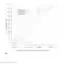

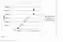
Sources:
- United States Patent and Trademark Office - verify current appl. status at the USPTO↗
Similar patent applications:
Recent applications in this class:
- » 20250115866 2025-04-10
COMPOSITION FOR SPERM TREATMENT, SPERM MOTILITY IMPROVING AGENT, SPERM FERTILIZING CAPABILITY RETAINING AGENT, SPERM MOTILITY IMPROVING METHOD, AND SPERM FERTILIZING CAPABILITY RETAINING METHOD - » 20250115865 2025-04-10
MAMMALIAN SPERM PREPARATION METHOD, ARTIFICIAL INSEMINATION METHOD, IN-VITRO FERTILIZATION METHOD, AND MEDIUM - » 20250019648 2025-01-16
A SPERM EXTENDER MEDIUM - » 20250002845 2025-01-02
METHOD OF SPERM SORTING USING LIPOSOMES AND/OR AN INHIBITOR OF SL03 POTASSIUM CHANNEL - » 20240344023 2024-10-17
HYDROXYUREA TO ENHANCE SPERM CELLS - » 20240228952 2024-07-11
APPARATUS AND METHOD FOR COLLECTION OF SPERM SAMPLES - » 20240132837 2024-04-25
APPARATUS AND METHOD FOR COLLECTION OF SPERM SAMPLES - » 20240084250 2024-03-14
METHODS AND DEVICES FOR SEPARATION OF MOTILE SPERM - » 20230399608 2023-12-14
PROCESS FOR ESTABLISHING A HUMAN TESTICULAR TISSUE CULTURE SYSTEM - » 20230303970 2023-09-28
METHOD FOR IMPROVED SPERM CELL POPULATIONS