Molecular detection assay using direct treatment with a bisulphite reagent
US20150086972A1
2015-03-26
14/342,093
2011-09-07
✅ Patent granted
US 9,732,375 B2
2017-08-15
WO; PCT/AU2011/001156; 20110907
WO; WO2013/033749; 20130314
Kenneth Horlick
Knobbe Martens Olson & Bear LLP
2033-06-16
Abstract:
A molecular detection assay including treating a biological sample directly with a bisulphite agent under conditions that allow cell disruption and nucleic acid treatment; removing the bisulphite agent from the treated sample; and detecting a target nucleic acid in the treated sample.
Assignee:
- HUMAN GENETIC SIGNATURES PTY. LTD. 1 🇦🇺 Randwick, NSW, Australia
- HUMAN GENETIC SIGNATURES PTY. LTD. 1 🇦🇺 Randwick, Australia
Applicant:
Interested in similar patents?
Get notified when new applications in this technology area are published.
Classification:
C12Q1/6806 » CPC main
Measuring or testing processes involving enzymes, nucleic acids or microorganisms ; Compositions therefor; Processes of preparing such compositions involving nucleic acids Preparing nucleic acids for analysis, e.g. for polymerase chain reaction [PCR] assay
C12Q1/689 » CPC further
Measuring or testing processes involving enzymes, nucleic acids or microorganisms ; Compositions therefor; Processes of preparing such compositions involving nucleic acids; Nucleic acid products used in the analysis of nucleic acids, e.g. primers or probes for detection or identification of organisms for bacteria
C12Q1/6886 » CPC further
Measuring or testing processes involving enzymes, nucleic acids or microorganisms ; Compositions therefor; Processes of preparing such compositions involving nucleic acids; Nucleic acid products used in the analysis of nucleic acids, e.g. primers or probes for diseases caused by alterations of genetic material for cancer
C12Q1/6893 » CPC further
Measuring or testing processes involving enzymes, nucleic acids or microorganisms ; Compositions therefor; Processes of preparing such compositions involving nucleic acids; Nucleic acid products used in the analysis of nucleic acids, e.g. primers or probes for detection or identification of organisms for protozoa
C12Q1/707 » CPC further
Measuring or testing processes involving enzymes, nucleic acids or microorganisms ; Compositions therefor; Processes of preparing such compositions involving virus or bacteriophage; Specific hybridization probes for hepatitis non-A, non-B Hepatitis, excluding hepatitis D
C12Q2600/158 » CPC further
Oligonucleotides characterized by their use Expression markers
C12Q1/68 IPC
Measuring or testing processes involving enzymes, nucleic acids or microorganisms ; Compositions therefor; Processes of preparing such compositions involving nucleic acids
C12Q1/70 IPC
Measuring or testing processes involving enzymes, nucleic acids or microorganisms ; Compositions therefor; Processes of preparing such compositions involving virus or bacteriophage
Description
TECHNICAL FIELD
The present invention relates to methods for processing of samples for molecular detection assays, particularly processing clinical samples for nucleic acid detection assays for disease detection.
BACKGROUND OF THE INVENTION
Molecular testing of humans has become increasingly important for medical diagnosis and patient management. Molecular testing of animals is gaining importance in veterinary applications. Nucleic acid testing is an essential tool for modern forensics, immigration, paternity assignment, and other human identity applications. Epigenetics is becoming increasingly important for cancer research, identification of biomarkers, predisposition factors, and potential drug targets. RNA Genotyping encompasses a range of applications used to analyse genetic differences between individuals or cells, in all areas of research, applied testing, and diagnostics. When dealing with biological samples, present molecular tests require specific pre-treatment of the samples prior to carrying out nucleic acid detection with techniques such as polymerase chain reaction (PCR). Sample pre-treatment is presently considered essential and many commercial kits and systems have been developed and are being used in the marketplace. Unfortunately, pre-treatment adds a costs to molecular testing and requires additional processing time and equipment.
Human Genetic Signatures Pty Ltd (Sydney, Australia) has developed a method for detection of microorganisms as disclosed in WO 2006/058393 and U.S. Pat. No. 7,833,942. The method involves treating microbial nucleic acid of a microorganism with an agent to form a simplified form of nucleic acid derived from the microbial nucleic acid having unique nucleic acid sequences that can be used to detect the microorganism.
The present inventor has developed an improved molecular detection assay that does not require sample pre-treatment as presently required in molecular detection assays.
SUMMARY OF THE INVENTION
The present invention relates to a molecular detection assay carried out on a biological sample without requiring a sample clean-up step or processing of the biological sample to lyse cells or purify nucleic acid.
In a first aspect, the present invention provides a molecular detection assay comprising:
(a) treating a biological sample directly with a bisulphite agent under conditions that allow cell disruption and nucleic acid treatment;
(b) removing the bisulphite agent from the treated sample; and
(c) detecting a target nucleic acid in the treated sample.
The biological sample does require cellular disruption or chemical/physical treatment prior to step (a).
The biological sample may be selected from faecal, nasal, blood, plasma, serum, buccal cells, pus, wound, concentrated filtrates, cerebrospinal fluid, semen, liquid based cytology (LBC), tissue, FFPE, laser captured cells, cultured cells, pelleted cells, bacterial cultures, bacterial colonies, viral suspension, aspirate, bronchial lavage, sputum sample, environmental sample, environmental concentrate, foodstuff, raw ingredient, water sample or water concentrate, and the like.
The nucleic acid detected may be DNA, RNA or combinations of both DNA and RNA.
The assay is suitable for detecting infectious disease, genetic disease or genetic trait in an animal. Preferably, the animal is a human.
The infectious disease may be caused by a microorganism including bacteria, viruses, viroid, yeast, fungi, prion, parasites, or amoeba.
The genetic disease may be cancer, mutation, copy number variation, inherited disorder, environmental induced disease, disease caused by exposure to carcinogens, disease characterised by expansion or reduction in a nucleotide repeat length in a genome.
The genetic trait may be susceptibility to cancer or any other disease where genetic and epigenetic changes contribute to the development of a disease state.
Preferably the bisulphite agent is sodium bisulphite or sodium metabisulphite. The bisulphite agent is preferably used at a concentration of about 2.5M to about 3.5M having a pH range of about 4.5 to about 5.5. More preferably, the bisulphite agent is used at a concentration of about 3M at a pH of about 5.0.
Preferably, step (a) involves heating between about 75° C. and 95° C. for about 1 minute to 30 minutes. More preferably, the sample is heated for about 10 to 20 minutes at a temperature of between about 80° C. and 95° C. It will be appreciated that the temperature and heating duration may vary depending on the sample being treated and the cellular source of the nucleic acid to be detected.
The bisulphite can be removed from the treated sample by any suitable means. Examples include column based purification or bead based purification are optimal but in some cases a simple precipitation step may be sufficient.
After removal of the bisulphite, the sample is preferably resuspended in elution buffer which has a pH of at least 10, more preferably between about pH 11.5 to about 12.5. A pH of at least about 12 has been found to be suitable for most samples.
The bisulphite agent modifies cytosine to uracil in treated nucleic acid. In double stranded DNA, the bisulphite agent modifies cytosine to uracil in each strand of complementary double stranded genomic DNA forming two derivative but non-complementary nucleic acid molecules.
In one preferred form, the derivative microbial nucleic acid substantially contains bases adenine (A), guanine (G), thymine (T) and uracil (U) and has substantially the same total number of bases as the corresponding untreated nucleic acid.
If the treated nucleic acid is amplified, then the amplified nucleic acid contains substantially the bases adenine (A), guanine (G) and thymine (T). The amplification may be carried out by any suitable means such as polymerase chain reaction (PCR), isothermal amplification, or signal amplification.
In a preferred form, the assay further includes providing nucleic acid primers or probes to the treated sample.
Preferably the treated sample undergoes an amplification reaction to form a target nucleic acid molecule specific for the microorganism or genetic indicator.
The target nucleic acid molecule is preferably detected by amplification. Examples of suitable amplification detection includes PCR, qPCR, Reverse transcriptase PCR, digital PCR, isothermal amplification or signal amplification.
The treated nucleic acid may also be detected by a sequencing method such Roche 454, ABI SOLiD or Ion torrent systems, Illumina Hi Seq SBS technology, Helicos Heliscope or SMRT technology employed by Pacific Biosciences or any other equivalent technology.
In a preferred form, the target nucleic acid molecule is detected by:
providing a detector ligand capable of binding to a target region of the microbial-specific nucleic acid molecule and allowing sufficient time for the detector ligand to bind to the target region; and
measuring binding of the detector ligand to the target region to detect the presence of the microbial-specific nucleic acid molecule.
In another preferred form, the target nucleic acid molecule is detected by separating an amplification product and visualising the separated product. Preferably, the amplification product is separated by electrophoresis and detected by visualising one or more bands on a gel.
Preferably, the target nucleic acid molecule does not occur naturally in a cell.
In a second aspect, the present invention provides a kit for a molecular detection assay for direct treatment of a sample, the kit comprising:
a bisulphite reagent;
nucleic acid elution and reaction reagents;
PCR primers for the target nucleic acid; and
instructions to carry out the assay.
The kit may further include nucleic acid separation or purification columns, PCR mastermix, reagents for PCR, control nucleic acid primers, reaction tubes, test tubes, swabs, and the like.
Throughout this specification, unless the context requires otherwise, the word “comprise”, or variations such as “comprises” or “comprising”, will be understood to imply the inclusion of a stated element, integer or step, or group of elements, integers or steps, but not the exclusion of any other element, integer or step, or group of elements, integers or steps.
Any discussion of documents, acts, materials, devices, articles or the like which has been included in the present specification is solely for the purpose of providing a context for the present invention. It is not to be taken as an admission that any or all of these matters form component of the prior art base or were common general knowledge in the field relevant to the present invention as it existed in Australia prior to development of the present invention.
In order that the present invention may be more clearly understood, preferred embodiments will be described with reference to the following drawings and examples.
BRIEF DESCRIPTION OF THE DRAWINGS
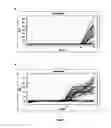
FIG. 1 shows results of C. difficile detection using the present invention. A. the Fam channel is specific for the tcdA/B genes of C. difficile; B. the Cy5 channel is specific for the extraction control.
FIG. 2 shows results of amplification of the three viruses from the same sample. A. Fam=Norovirus (single stranded RNA virus); B. Hex=Adenovirus (double stranded DNA virus; C. Cy5=Rotavirus (single stranded RNA virus
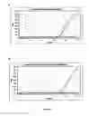
FIG. 3 shows the effect of spiking human cells into faecal material. A. shows human MRC5 cells heated for 15 minutes in 3M bisulphite reagent at 95° C. then resuspended in high pH buffer (12.3) and amplified for the human R-globin gene. B. shows the same cells spiked into faecal material heated for 15 minutes in 3M bisulphite reagent at 95° C. then resuspended in high pH buffer (12.3) and amplified for the human R-globin gene.
MODE(S) FOR CARRYING OUT THE INVENTION
Advantages
The present invention has many advantages over the prior art methods including the following.
-
- a. Lysis of the cells and nucleic acid conversion of the sample occurs simultaneously in the same tube and complete lysis and conversion can be achieved in less than 30 minutes, and even can be achieved by little as 10-20 minutes.
- b. Samples such as faecal material can be directly processed without loss of sensitivity.
- c. Tests based on the present invention have an advantage of being able performed on a primary patient sample without pre-treatment with enzymes such as proteinase K.
- d. The method does not require any expensive purification methods such as commercial purification kits presently on the market. These commercial kits are available for just about every sample type such as blood, faeces, cultured cells, FFPE, bacteria, viruses and parasites to name but a few. These commercial kits can cost thousands of dollars and can take 2-4 hours to purify a sample. Just about every lab in the world uses these kits for sample purification prior to any subsequent amplification step in molecular assays presently employed.
- e. After removal of the bisulphite the sample is then resuspended in a simple elution buffer that does not require any further treatment such as heat desulphonation, thus simplifying the process.
- f. The method can simultaneously detect both RNA and DNA viruses in the same tube from the same sample without any need to use a specialised viral purification kit. Both RNA and DNA viruses (even although one is single stranded and one is double stranded) are lysed and converted with equal efficiency by the present invention.
- g. Surprisingly the elution buffer, which is at high pH, does not degrade RNA. This finding goes against what has been taught in the prior art as prior art teaching specifically advises against the treatment of RNA at both high temperatures and pH.
- h. The method can lyse and convert all bacterial types using similar conditions thus a single patient sample can be used for multiple tests for C. difficile, Salmonella, Camplylobacter and parasites, etc thus removing the problem of a separate extraction method for each bacteria or target microorganism.
- i. The method is applicable to difficult to lyse samples such as parasites, which can form a tough outer shells that make them resistant to conventional lysis techniques. Thus target organisms such as Mycobacterium tuberculosis (TB) (very resistant to conventional lysis techniques) would be ideal to detect, as sensitive nucleic acid based diagnostic tests are required.
- j. The method is able to efficiently lyse and convert hepatitis C virus (HCV) directly from serum using as few as 10 copies of the virus demonstrating the utility to detect important RNA viruses at a very low level.
- k. The method also reduces sample processing time by many hours and removes the problem that the sample had to be purified then bisulphite converted and purified again as presently carried out.
- l. The method is suitable for the sensitive detection of colorectal cancer in patient faecal material. Such samples are non-invasive and would reduce the reliance on endoscopy as the primary means of detection. In addition, a ‘sample to result’ would be expected in less than three hours without complex sample processing procedure. The use of faecal material over serum samples for diagnosis has many advantages the most significant is that a high number of human cells are likely to be present in the sample, unlike serum samples where sensitivity is always a major problem.
Samples
The present invention is suitable for processing samples including faecal, nasal, blood, plasma, serum, buccal cells, pus, wound, concentrated filtrates, cerebrospinal fluid, semen, liquid based cytology (LBC), tissue, FFPE, laser captured cells, cultured cells, pelleted cells, bacterial cultures, bacterial colonies, viral suspension, aspirate, bronchial lavage, sputum sample, environmental sample, environmental concentrate, foodstuff, raw ingredient, water sample, water concentrate or the like.
Sample Treatment (HGS)
A preferred method of treating a sample according to the present invention is as follows. The sample is placed directly into 200 μl of 3M (range 2.5 to 3.5M) sodium bisulphite pH 5.0 (pH range 4.5 to 5.5) and heated at between 75° C. and 95° C. for between 10 and 20 minutes.
The bisulphite is removed from the treated sample by any suitable means. Examples include column based purification or bead based purification are optimal but in some cases a simple precipitation step may be sufficient.
After removal of the bisulphite, the sample is resuspended in elution buffer which has a pH of between 11.5 to 12.5 (>pH 12 preferred).
The sample is now ready for PCR amplification without further processing.
Removal of Bisulphite
Bisulphite removal can be used achieved by a commercially available method such as the MethylEasy™ technique (Human Genetic Signatures). Briefly, 240 μl of reagent #3 is added to the sample and the sample transferred to a centrifuge column. The column is then spun at 10,000×g to remove the bisulphite. The column is then washed ×2 with 300 μl of reagent #4 spinning at 10,000×g between washes. 20 to 50 μl of elution buffer is added and the sample spun into a clean collection tube. The sample is now ready for PCR amplification.
Sample Pre-Treatment
Current sample pre-treatment involves commercial kits sold by Qiagen Inc (Valencia, Calif. 91355 USA), Sigma Life Sciences (St Louis, Mo., USA), Invitrogen Corporation (Carlsbad, Calif. 92008, USA), and Promega Corporation (Madison, Wis. 53711 USA).
The present invention was compared with the following methods.
“In-House” Hospital Test
A sample of stool was heated at 95° C. for 10 minutes then diluted and the sample then amplified for the tcdB gene of C. difficile.
Qiagen Purification
QIAamp DNA Stool Mini Kit (50) Cat #51504.
Commercial Parasite Test
AusDiagnostics Gastrointestinal Parasites 5, Catalogue number: 6502.
Results
Faecal Samples
All faecal samples tested in Tables 1, 2, 3 and 4 were treated as follows:
A swab was placed in faecal material and then transferred to a tube containing 200 μl of 3M sodium bisulphite pH 5.0 mixed and then heated at 95° C. for 15 minutes.
Bisulphite was removed using the MethylEasy™ method and the samples were resuspended in 20-50 μl of elution buffer then amplified using standard conditions.
Table 1 shows the results of an “in-house” hospital test, the same samples tested using Qiagen purification and the same samples tested using the method according to the present invention. As can be seen from the results the “in-house” hospital test resulted in 7 false negative results and 1 false positive result (confirmed by EIA and culture as well as a commercially available molecular diagnostic kit) indicating that the method did not have the required sensitivity for the detection of C. difficile in primary patient samples. Using the Qiagen method, 4 samples did not have enough material for processing while 1 sample gave a false negative result (GDH positive by EIA). These results demonstrate the utility of the method according to the present invention (HGS method) for the rapid diagnosis of C. difficile. In addition, samples that have limited volumes are easily assayed using the HGS method. In addition, the Qiagen method requires at least 2 hours sample preparation time and having to weigh out predetermine amounts of faecal material.
| TABLE 1 |
| Results of the “in-house” hospital test vs Qiagen |
| purification vs HGS method for the detection of C. difficile. |
| Sample# | “In-house” | Qiagen | HGS | |
| 1 | NEG | NEG | NEG | |
| 2 | POS | POS | POS | |
| 3 | NEG | NEG | POS | |
| 4 | POS | POS | POS | |
| 5 | POS | POS | POS | |
| 6 | NEG | NEG | NEG | |
| 7 | POS | POS | POS | |
| 8 | NEG | POS | POS | |
| 9 | NEG | NEG | NEG | |
| 10 | NEG | NEG | NEG | |
| 11 | NEG | NEG | NEG | |
| 12 | NEG | POS | POS | |
| 13 | NEG | NEG | NEG | |
| 14 | NEG | NEG | NEG | |
| 15 | POS | NEG | NEG | |
| 16 | NEG | NEG | NEG | |
| 17 | NEG | NEG | NEG | |
| 18 | NEG | INSUFF | NEG | |
| 19 | NEG | NEG | NEG | |
| 20 | NEG | POS | POS | |
| 21 | POS | POS | POS | |
| 22 | POS | POS | POS | |
| 23 | NEG | NEG | NEG | |
| 24 | NEG | POS | POS | |
| 25 | POS | INSUFF | POS | |
| 26 | NEG | NEG | NEG | |
| 27 | NEG | POS | POS | |
| 28 | NEG | NEG | NEG | |
| 29 | NEG | NEG | NEG | |
| 30 | NEG | NEG | NEG | |
| 31 | POS | INSUFF | POS | |
| 32 | POS | POS | POS | |
| 33 | NEG | NEG | NEG | |
| 34 | NEG | NEG | NEG | |
| 35 | NEG | NEG | NEG | |
| 36 | NEG | NEG | NEG | |
| 37 | NEG | NEG | NEG | |
| 38 | NEG | POS | POS | |
| 39 | POS | POS | POS | |
| 40 | POS | POS | POS | |
| 41 | POS | POS | POS | |
| 42 | POS | POS | POS | |
| 43 | POS | INSUFF | POS | |
| 44 | NEG | NEG | NEG | |
| 45 | NEG | NEG | NEG | |
| TABLE 2 |
| Results of the Standard microscopy vs commercially available parasite |
| detection kit vs HGS method. |
| EasyPlex | ||||
| Gastrointestinal | Other Clinical | |||
| Sample# | Parasite Detected | Parasites 5 | HGS | Results |
| 1 | Giardia intestinalis | Negative | Giardia intestinalis | |
| 2 | Giardia intestinalis | Negative | Giardia intestinalis | |
| 3 | Giardia intestinalis | Negative | Giardia intestinalis | |
| 4 | Cryptosporidium | Negative | Cryptosporidium spp | |
| spp. | ||||
| 5 | Cryptosporidium | Negative | Cryptosporidium spp | |
| spp. | ||||
| 6 | Cryptosporidium | Negative | Cryptosporidium spp | |
| spp. | ||||
| 7 | Giardia intestinalis | Negative | Giardia intestinalis | |
| 8 | Giardia intestinalis | Negative | Giardia intestinalis | |
| 9 | Giardia intestinalis | Negative | Giardia intestinalis | |
| 10 | Cryptosporidium | Cryptosporidium spp. | Cryptosporidium spp and Entamoeba | |
| spp. | ||||
| 11 | Cryptosporidium | Cryptosporidium spp. | Cryptosporidium spp. | |
| spp. | ||||
| 12 | Cryptosporidium | Cryptosporidium spp. | Cryptosporidium spp. | |
| spp. | ||||
| 13 | Giardia intestinalis | Negative | Giardia intestinalis and Entamoeba | |
| 14 | Giardia intestinalis | Cryptosporidium spp. | Giardia intestinalis and Entamoeba | |
| 15 | Giardia intestinalis | Negative | Giardia intestinalis and Entamoeba | |
| 16 | Giardia intestinalis | Negative | Giardia intestinalis | |
| 17 | Giardia intestinalis | Negative | Giardia intestinalis | |
| 18 | D. fragilis, G. lamblia | Cryptosporidium spp. | Giardia intestinalis, Dientamoeba and | Salmonella Isolated |
| Salmonella spp. | ||||
| 19 | Giardia intestinalis | Giardia | Giardia intestinalis | |
| 20 | Giardia intestinalis | Negative | Giardia intestinalis | |
| 21 | Giardia intestinalis | Cryptosporidium spp. | Giardia intestinalis | |
| 22 | Cryptosporidium | Cryptosporidium spp. | Cryptosporidium spp. | |
| spp. | ||||
| 23 | Giardia intestinalis | Negative | Giardia intestinalis, Dientamoeba and | C. jejuni Isolated |
| Campy | ||||
| 24 | Giardia intestinalis | Negative | Giardia intestinalis | |
| 25 | Giardia intestinalis | Negative | Giardia intestinalis | |
| 26 | Giardia intestinalis | Negative | Giardia intestinalis | |
| 27 | NPI | Negative | Negative | |
| 28 | NPI | Negative | Negative | |
| 29 | NPI | Negative | Negative | |
| 30 | Cryptosporidium | Cryptosporidium spp. | Cryptosporidium spp. and Shigella | No Shigella Isolated |
| spp. | ||||
| 31 | Cryptosporidium | Cryptosporidium spp. | Cryptosporidium spp. and Shigella | No Shigella Isolated |
| spp. | ||||
| 32 | Cryptosporidium | Cryptosporidium spp. | Cryptosporidium spp. and Shigella | No Shigella isolated |
| spp. | ||||
| 33 | Cryptosporidium | Cryptosporidium spp. | Cryptosporidium spp. and Shigella | No Shigella Isolated |
| spp. | ||||
| 34 | Cryptosporidium | Cryptosporidium spp. | Cryptosporidium spp. and Shigella | No Shigella Isolated |
| spp. | ||||
| 35 | Cryptosporidium | Cryptosporidium spp. | Cryptosporidium spp. | |
| spp. | ||||
| 36 | Giardia intestinalis | Giardia | Giardia intestinalis, Entamoeba, | Shigella spp also |
| Shigella | isolated. | |||
| 37 | Giardia intestinalis | Negative | Giardia intestinalis | |
| 38 | Giardia intestinalis | Giardia | Giardia intestinalis, Entamoeba, | Shigella spp also |
| Shigella | isolated. | |||
| 39 | Giardia intestinalis | Giardia | Giardia intestinalis, Entamoeba, | Shigella spp also |
| Shigella | isolated. | |||
| 40 | NPI | Negative | Negative | |
| 41 | NPI | Negative | Giardia intestinalis | |
| 42 | NPI | Negative | Negative | |
| 43 | NPI | Negative | Giardia intestinalis | |
| 44 | Giardia intestinalis | Negative | Giardia intestinalis | |
| 45 | Giardia intestinalis | Negative | Giardia intestinalis | |
| 46 | NPI | Negative | Giardia intestinalis | |
| 47 | NPI | Negative | Giardia intestinalis | |
| 48 | NPI | Negative | Giardia intestinalis | |
| 49 | Giardia intestinalis | Negative | Giardia intestinalis | |
| 50 | Giardia intestinalis | Negative | Giardia intestinalis | |
| 51 | Giardia intestinalis | Negative | Giardia intestinalis | |
| 52 | NPI | Negative | Salmonella spp | Not Cultured |
| 53 | Giardia intestinalis | Negative | Giardia intestinalis | |
| 54 | Giardia intestinalis | Giardia | Giardia intestinalis | |
| All the samples highlighted belong to the same patient. |
Table 2 shows the results of a direct comparison of the standard microscopy, a commercial parasite test (Ausdiagnostic Easy-Plex Gastrointestinal parasite 5) and the HGS method. As can be seen from the results, the commercial method is unable to lyse many of the samples that contain the Giardia cysts. This is in contrast to the HGS method that easily detects all Giardia samples and also some samples that are missed by conventional microscopy. In addition, the HGS method seems to be universal for the lysis of bacteria and parasites alike as a number of samples contained both parasites and bacteria, results that were subsequently confirmed by the hospital.
Table 3 shows the results of the HGS method vs conventional culture methods for microbial detection. As can be seen the culture method failed to detect one sample that contained A Campylobacter spp that the molecular method detected. In addition no cross-reactivity was detected with samples that contained C. difficile. All positive culture results agreed with the HGS method indicating the improved sensitivity of molecular methods compared to conventional culture. Furthermore, the results of the HGS method are available in as little as 3 hours compared to overnight cultures required for conventional culture methods.
| TABLE 3 |
| Results of conventional culture methods for the |
| detection of enteric bacterial vs the HGS method |
| Sample | Culture | Campy | Salmon | |
| A01 | Salmon | N/A | 23.77 | |
| A02 | NPI | N/A | N/A | |
| A03 | Salmon | N/A | 29.7 | |
| A04 | Salmon | N/A | 26.06 | |
| A05 | Campy | 39.18 | N/A | |
| A06 | Campy | 44.37 | N/A | |
| A07 | Campy | 39.88 | N/A | |
| A08 | Salmon | N/A | 39.83 | |
| A09 | CD | N/A | N/A | |
| A10 | Campy | 35.39 | N/A | |
| A11 | NPI | 36.09 | N/A | |
| A12 | Campy | 35.72 | N/A | |
| A13 | Salmon | N/A | 30.21 | |
| A14 | Salmon | N/A | 30.24 | |
| A15 | Campy | 40.05 | N/A | |
| A16 | Salmon | N/A | 27.06 | |
| A17 | Campy | 36.26 | N/A | |
| A18 | Campy | 44.81 | N/A | |
| A19 | Campy | 26.57 | N/A | |
| A20 | Campy | 29.8 | N/A | |
| A21 | Salmon | N/A | 26.58 | |
| A22 | Campy | 34.78 | N/A | |
| A23 | Campy | 32.57 | N/A | |
| A24 | Salmon | N/A | 30.98 | |
| A25 | Campy | 31.18 | N/A | |
| A26 | Salmon | N/A | 45.32 | |
| A27 | Campy | 41.49 | N/A | |
| A28 | Campy | 38.69 | N/A | |
| A29 | Campy | 38.36 | N/A | |
| A30 | Salmon | N/A | 38.3 | |
| A31 | CD | N/A | N/A | |
| A32 | Salmon | N/A | 35.76 | |
| A33 | Campy | 35.61 | N/A | |
| A34 | CD | N/A | N/A | |
| A35 | Campy | 36.12 | N/A | |
| A36 | Campy | 39.96 | N/A | |
| A37 | Salmon | N/A | 37.53 | |
| A38 | Campy | 46.2 | N/A | |
| A39 | Campy | 35.96 | N/A | |
| A40 | Campy | 47.31 | N/A | |
| A41 | Salmon | N/A | 30.3 | |
| A42 | Salmon | N/A | 37.14 | |
| A43 | CD | N/A | N/A | |
Treatment Conditions
Four stool samples, 3 positive for C. difficile and 1 negative for C. difficile were assayed to determine the effect of temperature and time on the extraction efficiency. An extraction control (16s rDNA gene) was also included to determine the effect of conditions on the mixed flora that is present within stool samples. The Fam channel is specific for the tcdA/B genes of C. difficile while the Cy5 channel is specific for the extraction control. The results are shown in FIG. 1 and Table 4.
As can be seen from the results in Table 4 the preferred temperature and times for lysis of these samples lie between 85° C.-95° C. for 15 minutes. All positive samples and faecal flora were easily detected.
| TABLE 4 |
| The effect of time and temperature on the efficiency of the |
| HGS lysis/conversion method using stool samples. |
| tcdA/B | ExCon | tcdA/B | ExCon | tcdA/B | ExCon | |
| 10 min/85° C. | 15 min/85° C. | 20 min/85° C. |
| Sample 1 | 44.73 | 34.54 | 40.46 | 31.91 | 39.1 | 31.08 |
| Sample 2 | 41.57 | 35.09 | 41.59 | 30.31 | 40.23 | 29.09 |
| Sample 3 | N/A | 32.76 | N/A | 28.15 | N/A | 27.25 |
| Sample 4 | 42.6 | 30.19 | N/A | 30.26 | 46.22 | 28.91 |
| 10 min/90° C. | 15 min/90° C. | 20 min/90° C. |
| Sample 1 | 41.66 | 32.17 | 39.63 | 30.39 | 39.88 | 30.73 |
| Sample 2 | 41.19 | 29.05 | 41.25 | 27.53 | 41.33 | 25.17 |
| Sample 3 | N/A | N/A | N/A | 27.31 | N/A | 26.6 |
| Sample 4 | 42.63 | 28.69 | 43.88 | 27.27 | N/A | 27.86 |
| 10 min/95° C. | 15 min/95° C. | 20 min/95° C. |
| Sample 1 | 40.48 | 30.82 | 39.53 | 31.52 | 39.52 | 29.74 |
| Sample 2 | 40.38 | 26.41 | 40.3 | 26.31 | 42.21 | 30.52 |
| Sample 3 | N/A | 27.08 | N/A | 27.47 | N/A | 37.35 |
| Sample 4 | 41.93 | 27.64 | 40.31 | 27.76 | 42.2 | 26.66 |
| TABLE 5 |
| The effect of elution buffer pH on the ability of the method to efficiently detect HCV directly from serum samples |
| 70° C. | 75° C. | 80° C. | 90° C. |
| 5 min | 10 min. | 15 min | 5 min | 10 min | 15 min | 5 min | 10 min | 15 min | 5 min | 10 min | 15 min | |
| PCR-A-pH 11.5 | N/A | N/A | N/A | N/A | N/A | N/A | N/A | N/A | N/A | N/A | N/A | N/A |
| PCR-A-pH 12.5 | N/A | 36.83 | 36.01 | N/A | 33.27 | 34.8 | 36.88 | 33.02 | N/A | 34.45 | 34.22 | 33.14 |
| PCR-B-pH 11.5 | N/A | N/A | N/A | N/A | N/A | N/A | N/A | N/A | N/A | N/A | N/A | N/A |
| PCR-B-pH 12.5 | N/A | 39.34 | 37.17 | N/A | 35.76 | 36.2 | 40.98 | 35.05 | N/A | 36.21 | 36.48 | 35.51 |
10 μl of HCV (Acrometrix) were added to 200 μl of 3M sodium bisulphite and heated to 75° C., 75° C., 80° C. and 90° C. for 5, 10 or 15 minutes. Bisulphite was removed using a modified MethylEasy™ method and the samples were resuspended in 20 μl of elution buffer, 12 μl reverse transcribed then 2 μl amplified using standard conditions.
As can be seen from the results of Table 5 HCV RNA can easily withstand lysis conversion at 90° C., the highest temperature tested. Even more surprisingly the assay worked for RNA virus detection in the presence of the high pH buffer, which is contrary to the published literature indicating that RNA species need to be maintained around neutral pH.
These results combined with the results in Table 4 suggest that a temperature around 90° C. for 15 minutes could be a universal sample processing method for double stranded DNA containing organisms such as C. difficile and single stranded RNA viruses such as HCV. In addition, as C. difficile is a spore-containing organism the results indicate that the method is harsh enough to break open tough spores but gentle enough so that RNA containing viruses are not degraded. Again such data would not have been predicted by the prior art.
PCR A and B differ in the mastermix used (JumpStart (Sigma) and FastStart (Roche) respectively).
Serum
Dilutions of HCV (Zeptoometrix) were prepared then 10 μl of each dilution added to 200 μl of 3M sodium bisulphite and heated to 75° C. for 10 minutes.
Bisulphite was removed using the MethylEasy™ method and the samples were resuspended in 20 μl of elution buffer, 12 μl reverse transcribed then 2 μl amplified using standard conditions.
| TABLE 6 |
| Sensitive detection of HCV virus directly from serum. |
| HCV/PCR | IU/PCR | Positive | |
| 1\10 | 200 | 32.54 | 34.41 | |
| 1\100 | 20 | 37.07 | 38.32 | |
| 1\1000 | 2 | 37.54 | 38.63 | |
| 1\10000 | 0.2 | N/A | N/A | |
| 1/100000 | 0.02 | N/A | N/A | |
| Process | N/A | N/A | ||
As can be seen from Table 6 when using the HGS method to process the serum directly, as few as 2 IU of HCV can de efficiently lysed and converted simultaneously. These results demonstrate the excellent sensitivity that can be generated using RNA viruses as a target for the process.
Table 7 simultaneous lysis/conversion of both RNA viruses and DNA viruses under identical extraction conditions.
FIG. 2 shows amplification of the three viruses from the same sample.
A. Fam=Norovirus (single stranded RNA virus).
B. Hex=Adenovirus (double stranded DNA virus
C. Cy5=Rotavirus (single stranded RNA virus)
Norovirus, Adenovirus and Rotavirus qualitative samples were obtained from Zeptometrix. Samples of each virus (10 μl) were added to 3M sodium bisulphite and the samples heated at 90° C. for 10, 15 and 20 minutes. The bisulphite was then removed using a modified MethylEasy™ method and the samples were resuspended in 20 μl of elution buffer, 12 μl reverse transcribed then 2 μl amplified using standard conditions.
As can be seen from Table 7 after 15 minutes heating at 90° C. both the RNA and DNA viruses are efficiently lysed and converted. Both virus types were purified under identical conditions. These results demonstrate further the utility of the HGS method as a universal sample preparation method for the lysis and efficient conversion of both DNA and RNA viruses from the same sample. Again surprisingly the RNA viruses can easily withstand the 90° C. sample treatment and high pH buffer.
| TABLE 7 |
| Simultaneous lysis/conversion of both RNA viruses and DNA viruses under identical extraction conditions. |
| Fam Norovirus | Hex Adenovirus | Cy5 Rotavirus |
| 10 min | 15 min | 20 min | PC | 10 min | 15 min | 20 min | PC | 10 min | 15 min | 20 min | PC | |
| Neat | 31.75 | 29.67 | 30.09 | N/A | 36.32 | 32.36 | 36.37 | N/A | 34.5 | 32.13 | 37.35 | N/A |
| 1\10 | 35.61 | 33.25 | 31.96 | N/A | N/A | 36.45 | N/A | N/A | 40.04 | 34.5 | 38.6 | N/A |
| 1\100 | 40.51 | 38.87 | 35.84 | N/A | N/A | 44.67 | N/A | N/A | 39.32 | 38.57 | N/A | N/A |
Faecal samples were spiked with Norovirus, Adenovirus and Rotavirus qualitative samples obtained from Zeptometrix. Samples of virus (10 μl) were spiked into faecal material and the sample mixed. A swab was placed in faecal material and then transferred to a tube containing 200 μl of 3M sodium bisulphite pH 5.0 and then heated at 90° C. for 15 minutes.
Bisulphite was removed using a modified MethylEasy™ method and the samples were resuspended in 20 μl of elution buffer, 12 μl reverse transcribed then 2 μl amplified using standard conditions.
Table 8 the effect of spiking faecal samples with virus.
| Norovirus | Adenovirus | Rotavirus | Process | |
| Adenovirus spike | N/A | 41.1 | N/A | N/A |
| Norovirus spike | 39.45 | N/A | N/A | N/A |
| Rotavirus spike | N/A | N/A | 39.62 | N/A |
The results from Table 8 show that even when viral particles are spiked into faecal samples and the universal condition applied both DNA and RNA viruses are effectively lysed and converted. Thus the method should be applicable to any sample type for any micro-organism of interest.
Human Cells
To determine whether the present invention is also suitable for detection of human disease such as cancer, 5 μl, 10 μl and 20 μl of diluted human cells were incubated for 10 minutes or 15 minutes at 70° C., 80° C., 90° C. and 95° C. in 200 μl of 3M bisulphite. The samples were processed to remove the bisulphite solution and resuspended in 50 μl of buffer pH 12.3. Material (2 μl) was then amplified using primers and probes specific for the human β-globin gene. The results are shown in Table 9 and FIG. 3.
| TABLE 9 |
| Time/temperature optimisation using human MRC5 cells |
| 70° C. | 80° C. | 90° C. | 95° C. |
| 10 min | 15 min | 10 min | 15 min | 10 min | 15 min | 10 min | 15 min | |
| 5 ul cells | N/A | N/A | 42.15 | 43.63 | 38.62 | 33.19 | ND | ND |
| 10 ul cells | N/A | N/A | 40.72 | 36.45 | 35.04 | 33.31 | 37.11 | 33.8 |
| 20 ul cells | N/A | N/A | 45.08 | 37.03 | 36.38 | 32.49 | ND | ND |
| ND = not done. |
As can be seen from Table 9 the preferred time and temperature for simultaneous lysis and conversion of human cells without pre-treatment lies between 90-95° C. for 15 minutes.
FIG. 3 shows the effect of spiking human cells into faecal material. FIG. 3A shows human MRC5 cells heated for 15 minutes in 3M bisulphite reagent at 95° C. then resuspended in high pH buffer (12.3) and amplified for the human β-globin gene. FIG. 3B shows the same cells spiked into faecal material heated for 15 minutes in 3M bisulphite reagent at 95° C. then resuspended in high pH buffer (12.3) and amplified for the human β-globin gene. As can be seen from the results human cells can be lysed and efficiently converted in primary human faecal material without any need for sample pre-processing or DNA isolation.
The results indicate that methylation profiling of human cells can be carried out directly on faecal samples obtained from patient without the need for pre-treatment or sample purification prior to bisulphite. Thus the method would be ideal as a non-invasive and simple method for the diagnosis of colorectal cancer directly from primary patient material.
Diagnostic Test Kits
The present invention can be provided in the form of diagnostic kits to allow the ease of use in a diagnostic laboratory or the like. Table 10 and Table 11 show the reagents provided in a typical test kit for a microorganism, for example. A kit includes instructions for use. It will be appreciated that tubes, swabs and other laboratory equipment may not be provided in a kit as those materials are usually readily available in a diagnostic laboratory.
| TABLE 10 |
| Box 1 of 2 (Store at room temperature) |
| Component Name | Contents | |
| Reagent 1 (Alkali water) | 5 × 5 | mL | |
| Reagent 2 (Bisulphite) | 5 × 3.5 | g | |
| Reagent 3 (Binding reagent) | 5 × 5.8 | mL | |
| Reagent 4 (Wash buffer) | 5 × 3 | mL | |
| Reagent 5 (Elution buffer) | 5 × 3 | mL |
| Sheathed swabs | 100 | |
| Reaction Tubes (1.5 mL) | 100 | |
| Wash Tubes | 100 | |
| Collection Tubes (1.5 mL) | 100 | |
| Purification Columns | 100 | |
| Detailed user guide | 1 | |
| TABLE 11 |
| Box 2 of 2 (Store at −20° C. upon receipt) |
| PCR mastermix | 5 × vials | |
| PCR components | 5 × vials | |
Box 2 should be stored at −20° C. in a DNA “clean room” in a different location to where the samples will be processed.
Kits for specific microorganisms or genetic tests include PCR primers to allow amplification of the target nucleic acid molecule.
It will be appreciated by persons skilled in the art that numerous variations and/or modifications may be made to the invention as shown in the specific embodiments without departing from the spirit or scope of the invention as broadly described. The present embodiments are, therefore, to be considered in all respects as illustrative and not restrictive.
Claims
1. A molecular detection assay comprising:
treating a biological sample directly with a bisulphite agent under conditions that allow cell disruption and nucleic acid treatment;
removing the bisulphite agent from the treated sample; and
detecting a target nucleic acid in the treated sample.
2. The assay according to claim 1 wherein biological sample is selected from the group consisting of faecal, nasal, blood, plasma, serum, buccal cells, pus, wound, concentrated filtrates, cerebrospinal fluid, semen, liquid based cytology (LBC), tissue, FFPE, laser captured cells, cultured cells, pelleted cells, bacterial cultures, bacterial colonies, viral suspension, aspirate, bronchial lavage, sputum sample, environmental sample, environmental concentrate, foodstuff, raw ingredient, water sample and water concentrate.
3. The assay according to claim 1, wherein the assay is used to detect infectious disease, genetic disease or a genetic trait in an animal
4. The assay according to claim 3, wherein the infectious disease is caused by a microorganism.
5. The assay according to claim 4, wherein the microorganism is a bacterium, virus, viroid, yeast, fungi, prion, parasites, or amoeba.
6. The assay according to claim 2, wherein the genetic disease is cancer, a disease associated with copy number variation, an inherited disorder, an environmental induced disease, a disease caused by exposure to carcinogens, or a disease characterised by expansion or reduction in a nucleotide repeat length.
7. The assay according to claim 1, wherein the bisulphite agent is sodium bisulphite or sodium metabisulphite.
8. The assay according to claim 7, wherein the bisulphite agent is used at a concentration of 2.5M to 3.5M having a pH range of 4.5 to 5.5.
9. The assay according to claim 8, wherein the bisulphite agent is used at a concentration of 3M at a pH of 5.0.
10. The assay according to claim 1, wherein the treating step comprises heating between 75° C. and 95° C. for 1 to 30 minutes.
11. The assay according to claim 10, wherein the sample is heated for 10 to 20 minutes at a temperature of between 80° C. and 95° C.
12. The assay according to claim 1, wherein the bisulphite is removed by column based purification, bead based purification or precipitation.
13. The assay according to claim 1, wherein after removal of the bisulphite, the treated sample is resuspended in an elution buffer having a pH of at least 10.
14. The assay according to claim 13, wherein the elution buffer has a pH of 11.5 to 12.5.
15. The assay according to claim 14, wherein the elution buffer has pH of at least 12.
16. The assay according to claim 1, further including providing nucleic acid primers or probes to the treated sample.
17. The assay according to claim 16, wherein the target nucleic acid molecule is detected by amplification.
18. The assay according to claim 17, wherein the amplification is PCR, qPCR, reverse transcription PCR, digital PCR, isothermal amplification or signal amplification.
19. The assay according to claim 1, wherein the target nucleic acid is detected by sequencing the treated material.
20. (canceled)
Images & Drawings included:
Sources:
- United States Patent and Trademark Office - verify current appl. status at the USPTO↗
Recent applications in this class:
- » 20250290119 2025-09-18
METHODS AND COMPOSITIONS FOR MULTIPLEX CELL ANALYSIS - » 20250283149 2025-09-11
MULTIOMIC ANALYSIS OF CELL ANALYTES USING MICROFLUIDIC SYSTEMS - » 20250283148 2025-09-11
METHODS AND COMPOSITIONS FOR ENRICHING NUCLEIC ACIDS - » 20250283147 2025-09-11
COMPOSITIONS AND METHODS FOR ISOLATION OF CELL-FREE DNA - » 20250283146 2025-09-11
ANALYSIS OF RNA MODIFICATIONS - » 20250283145 2025-09-11
METHODS FOR BASE-LEVEL DETECTION OF METHYLATION IN NUCLEIC ACIDS - » 20250277253 2025-09-04
METHODS AND KITS FOR LABELING CELLULAR MOLECULES - » 20250270621 2025-08-28
METHODS, COMPOSITIONS, AND KITS FOR PREPARING NUCLEIC ACID LIBRARIES - » 20250270620 2025-08-28
Marked Items and Verification Methods - » 20250270619 2025-08-28
COMPOSITIONS, KITS AND METHODS FOR DIRECT AMPLIFICATION FROM CRUDE BIOLOGICAL SAMPLES