Bone graft delivery system and method for using same
US20150190148A1
2015-07-09
14/593,489
2015-01-09
✅ Patent granted
US 9,456,830 B2
2016-10-04
-
-
Christopher Beccia
Knobbe, Martens, Olson & Bear, LLP.
2035-01-09
Abstract:
A bone graft delivery system can include an elongate tube, a handle having a trigger, and a tip. The trigger is actuated to deliver bone graft material through the tube. The tip has one or more openings to deliver the bone graft material to a desired location and includes a surface suitable to act as a rasp for decorticating bone. A method for delivering bone graft material to a desired surgical location includes providing a bone graft delivery device, positioning the device adjacent the surgical location, decorticating bone, and delivering bone graft material to the surgical location.
Assignee:
- Surgical Device Exchange, LLC 3 🇺🇸 Boca Raton, FL, United States
Applicant:
Interested in similar patents?
Get notified when new applications in this technology area are published.
Classification:
A61B17/1659 » CPC main
Surgical instruments, devices or methods, e.g. tourniquets; Osteoclasts Bone cutting, breaking or removal means other than saws, e.g. ; Drills or chisels for bones; Trepans Surgical rasps, files, planes, or scrapers
A61F2/4601 » CPC further
Filters implantable into blood vessels; Prostheses, i.e. artificial substitutes or replacements for parts of the body; Appliances for connecting them with the body; Devices providing patency to, or preventing collapsing of, tubular structures of the body, e.g. stents; Prostheses implantable into the body; Joints; Special tools or methods for implanting or extracting artificial joints, accessories, bone grafts or substitutes, or particular adaptations therefor for introducing bone substitute, for implanting bone graft implants or for compacting them in the bone cavity
A61B17/1635 » CPC further
Surgical instruments, devices or methods, e.g. tourniquets; Osteoclasts Bone cutting, breaking or removal means other than saws, e.g. ; Drills or chisels for bones; Trepans for grafts, harvesting or transplants
A61B90/39 » CPC further
Instruments, implements or accessories specially adapted for surgery or diagnosis and not covered by any of the groups - , e.g. for luxation treatment or for protecting wound edges Markers, e.g. radio-opaque or breast lesions markers
A61B2090/3966 » CPC further
Instruments, implements or accessories specially adapted for surgery or diagnosis and not covered by any of the groups - , e.g. for luxation treatment or for protecting wound edges; Markers, e.g. radio-opaque or breast lesions markers Radiopaque markers visible in an X-ray image
A61F2002/2835 » CPC further
Filters implantable into blood vessels; Prostheses, i.e. artificial substitutes or replacements for parts of the body; Appliances for connecting them with the body; Devices providing patency to, or preventing collapsing of, tubular structures of the body, e.g. stents; Prostheses implantable into the body; Bones Bone graft implants for filling a bony defect or an endoprosthesis cavity, e.g. by synthetic material or biological material
A61B17/16 IPC
Surgical instruments, devices or methods, e.g. tourniquets Osteoclasts Bone cutting, breaking or removal means other than saws, e.g. ; Drills or chisels for bones; Trepans
A61F2/46 IPC
Filters implantable into blood vessels; Prostheses, i.e. artificial substitutes or replacements for parts of the body; Appliances for connecting them with the body; Devices providing patency to, or preventing collapsing of, tubular structures of the body, e.g. stents; Prostheses implantable into the body; Joints Special tools or methods for implanting or extracting artificial joints, accessories, bone grafts or substitutes, or particular adaptations therefor
A61F2/28 » CPC further
Filters implantable into blood vessels; Prostheses, i.e. artificial substitutes or replacements for parts of the body; Appliances for connecting them with the body; Devices providing patency to, or preventing collapsing of, tubular structures of the body, e.g. stents; Prostheses implantable into the body Bones
Description
CROSS-REFERENCE TO RELATED APPLICATIONS
This application is a continuation of U.S. application Ser. No. 13/485,641, entitled “BONE GRAFT DELIVERY SYSTEM AND METHOD FOR USING SAME,” filed May 31, 2012, which claims priority benefit of U.S. Provisional Application No. 61/492,316, entitled “BONE GRAFT DELIVERY SYSTEM AND METHOD FOR USING SAME,” filed Jun. 1, 2011, the entirety of each of which is hereby incorporated by reference herein.
BACKGROUND
1. Field
The present application relates to orthopedic surgery in general, and more particularly, to bone graft delivery systems and methods. 2. Description of the Related Art
In a bone grafting procedure, a surgeon places bone or a bone substitute into an area in a patient's body to provide a type of scaffold for bone growth and repair. Bone grafts can be used to help treat various orthopedic problems, for example, to fuse a joint or repair a fracture. Bone graft material can be, for example, autogenous (harvested from the patient's own body), allogeneic (harvested from another person, usually a cadaver), or synthetic. Many bone grafting procedures are performed via open surgery implantation. However, these procedures can be performed minimally invasively, for example, by using a needle to inject the bone graft material into the target location without requiring a surgical incision.
In some cases decortication of the bony area receiving the graft is performed prior to delivery of the bone graft material. Decortication removes superficial cortical bone and exposes the underlying cancellous bone, which can help accelerate the integration of the bone graft with the native bone.
SUMMARY
The devices, systems, and methods described herein allow for minimally invasive delivery of bone graft material to a desired location in a patient's body. In some embodiments, the devices, systems, and methods described herein also provide for bone decortication.
In some embodiments, a bone graft delivery system includes an elongate tube, a handle at a proximal end of the tube configured to be actuated to deliver bone graft material through the tube, and a tip at a distal end of the tube. The handle may include a trigger. The tip includes one or more openings configured to deliver the bone graft material to a desired location and a surface suitable to serve as a rasp for scraping bone.
In some embodiments, a method for delivering bone graft material to a surgical location includes providing a bone graft delivery device comprising an elongate tube and a distal tip having at least one opening for delivering the bone graft material to the surgical location and positioning the device adjacent the surgical location. The method further includes decorticating bone with the distal tip and delivering bone graft material through the tube and out the at least one opening of the tip.
BRIEF DESCRIPTION OF THE DRAWINGS
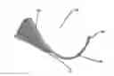
FIG. 1 illustrates a side view of an example embodiment of a bone graft delivery device;
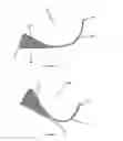
FIG. 2 illustrates a perspective view of the bone graft delivery device of FIG. 1; and
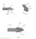
FIGS. 3-5 illustrates various views of a distal tip of the bone graft delivery device of FIGS. 1 and 2.
DETAILED DESCRIPTION
As shown in FIGS. 1 and 2, a bone graft delivery device 100 generally includes a handle 102 having a trigger 110 or other actuation mechanism, a tube 120 having a lumen therethrough, and a distal tip 130. In the illustrated embodiment, the bone graft delivery device 100 is similar to a caulking gun. The handle 102 can house a supply of the desired bone graft material. The bone graft material can be pre-loaded in the handle 102 or can be supplied to the handle via a cartridge that can be removably coupled to the handle 102.
In some embodiments, the device 100 can further include a plunger 112 that is retracted proximally to allow the handle to receive a cartridge or pre-loaded volume of bone graft material.
In use, the trigger 110 is actuated to deliver bone graft material through the tube 120 and distal tip 130 to a desired surgical location. In some embodiments, the plunger 112 is simultaneously pushed distally to help deliver bone graft material through the tube 120. In some embodiments, the trigger 110 or other actuation mechanism is configured to deliver a controlled release amount of bone graft material during actuation of the device, for example, ½ cc of bone graft material per complete squeeze of the trigger 110. The trigger 110 or other actuation mechanism may be operated manually or by mechanical, battery powered, electric, pneumatic, or any other means of force.
As shown in FIGS. 1 and 2, the tube 120 can include a permanent bend or curve that may be useful in positioning the device 100 at a desired location, for example, a space between two spinal discs. Alternatively, the tube 120 may be straight to deliver bone graft material directly into a desired location such as a disc space. In some embodiments, the tube 120 is somewhat flexible or repositionable and can be manipulated to bend or curve the tube 120 as needed to reach the desired location. In some embodiments, the tube 120 is made of a rigid material, for example, a plastic, composite, or metal, and is generally hollow to allow for the passage of bone graft material through the tube 120.
As shown in FIGS. 3-5, a distal end of the tube 120 includes a tip 130. In the illustrated embodiment, the tip 130 is somewhat bullet-shaped with a generally triangular cross-section; however, other shapes and configurations are also possible. In some embodiments, the tip 130 is pointed and/or sharp to dissect or split muscle and tissue as it is advanced through the patient's skin and body to the surgical location. Alternatively, the tip 130 can be blunt to allow for displacement of muscle without risk of cutting of nerves or other tissue. The tip has a single or multiple openings 132 in fluid communication with the tube 120 lumen and configured to deliver the bone graft material from the tube 120 to the desired location.
In some embodiments, at least one side or area of the tip 130 includes a series of jagged edges or other suitable surface 134 configured to serve as a rasp for scraping bone. The rasp may be operated manually or by mechanical, battery powered, electric, pneumatic, or any other means of force to allow for decortication of the area to receive the bone graft material.
The tip 130 may be made of a metallic, radiopaque material to facilitate visualization on, for example, fluoroscopy or x-ray. Alternatively, the tip 130 may be made of another material, for example a durable medical plastic or a composite material, and may include markers to facilitate visualization.
In one embodiment, the device 100 described herein may be used in minimally invasive spinal surgery. For example, in a conventional posterolateral spine procedure, screws and or fusion cages may be delivered to adjacent vertebrae using small incisions made in a patient's back. It may additionally be desirable to deliver bone graft material to the surgical location, e.g., to the transverse processes, disc spaces, or facet joints, through one of these small incisions. The device described herein is sized to be delivered through a minimally invasive opening made in the patient's skin (e.g., through a skin incision of 4 cm or less), and configured so that the tip can be positioned adjacent a pedicle screw or other desired location. The curvature of the tube 120 can facilitate positioning of the tip 130 at desired spinal locations and allows, for example, insertion of the device 100 through an incision over one vertebra, and positioning of the tip 130 at an adjacent vertebra. Alternatively, the device can be delivered through any desired opening made in the patient's skin (e.g., minimally invasive or open). The jagged edges or other surface 134 on the device can be used to decorticate desired bone locations, causing bleeding of the bone and creating a surface that promotes bone fusion. The trigger 110 or other actuation mechanism can then be actuated to deliver bone graft material through the tube 120 lumen and openings 132 in the tip 130 to promote fusion of the bone.
Although use of the device 100 has been described with respect to an example spinal procedure, the device 100 can also be used in other spinal procedures and other orthopedic applications to deliver bone graft material to other locations in the body (for example, the femur or tibia).
Various modifications to the implementations described in this disclosure may be readily apparent to those skilled in the art, and the generic principles defined herein may be applied to other implementations without departing from the spirit or scope of this disclosure. Thus, the disclosure is not intended to be limited to the implementations shown herein, but is to be accorded the widest scope consistent with the principles and features disclosed herein. Certain embodiments of the invention are encompassed in the claim set listed below.
Claims
1. (canceled)
2. (canceled)
3. (canceled)
4. (canceled)
5. (canceled)
6. (canceled)
7. (canceled)
8. (canceled)
9. (canceled)
10. (canceled)
11. (canceled)
12. (canceled)
13. (canceled)
14. (canceled)
15. (canceled)
16. (canceled)
17. (canceled)
18. (canceled)
19. A bone graft delivery system, comprising:
an elongate tube comprising a proximal end and a distal end; and
a tip at a distal end of the tube, the tip having one or more openings configured to deliver the bone graft material to a desired location, wherein the tip includes a surface configured to decorticate bone extending proximally of the one or more openings.
20. The system of claim 19, wherein the surface configured to decorticate bone comprises a rasping surface.
21. The system of claim 19, wherein the surface configured to decorticate bone comprises jagged edges.
22. The system of claim 19, wherein the tip is made of metal.
23. The system of claim 19, wherein the tip is made of a radiopaque material.
24. The system of claim 19, wherein the tip is made of a durable medical plastic.
25. The system of claim 19, wherein the tip is made of a composite material.
26. The system of claim 19, wherein the tip includes one or more radiopaque markers.
27. The system of claim 19, wherein the tip has a blunt end.
28. The system of claim 19, wherein the elongate tube is rigid.
29. The system of claim 19, wherein the elongate tube includes a permanent bend.
30. A method for delivering bone graft material to a surgical location, comprising:
positioning a bone graft delivery device adjacent the surgical location, the bone graft delivery device comprising an elongate tube and a distal tip, wherein the distal tip has a rasping surface and at least one opening for delivering the bone graft material to the surgical location;
decorticating bone with the rasping surface of the distal tip; and
with the rasping surface at the distal tip of the bone graft delivery device, delivering bone graft material through the tube and out the at least one opening of the distal tip.
31. The method of claim 30, wherein the bone graft material comprises autogenous, cadaveric and/or synthetic material.
32. The method of claim 30, wherein the bone graft delivery device is positioned at the surgical location through a minimally invasive opening in a patient's skin.
33. The method of claim 30, wherein the bone graft delivery device is positioned adjacent the spine and the distal tip decorticates a portion of the spine.
34. The method of claim 30, wherein decorticating bone with the distal tip comprises rasping bone with jagged edges of the distal tip.
35. The method of claim 30, wherein decorticating bone with the rasping surface of distal tip comprises actuating the distal tip by mechanical, battery powered, electric, pneumatic, or another means of force.
Images & Drawings included:
Sources:
- United States Patent and Trademark Office - verify current appl. status at the USPTO↗
Similar patent applications:
- » 14162102
Bone graft delivery system and method for using same - » 14214031
Bone graft delivery system and method for using same - » 16198754
Bone graft delivery system and method for using same - » 20150209156
Bone graft delivery system and method for using same - » 20160296344
Bone graft delivery system and method for using same - » 20170224397
Bone graft delivery system and method for using same - » 20170354514
Bone graft delivery system and method for using same - » 20170367846
Bone graft delivery system and method for using same - » 20180177608
Bone graft delivery system and method for using same - » 13485641
Bone graft delivery system and method for using same
Recent applications in this class:
- » 20250114103 2025-04-10
CARTILAGE REMOVAL TOOL AND METHOD - » 20250032131 2025-01-30
Bone Resection Method by Plunge Milling and Rasping During Total Ankle Arthroplasty - » 20240423646 2024-12-26
HAND-HELD MEDICAL INSTRUMENT - » 20240398426 2024-12-05
Reamer for Augmented Glenoid Implant - » 20240382217 2024-11-21
FEMORAL FINISHING RASP - » 20240130741 2024-04-25
Joint surface replacement system - » 20240074769 2024-03-07
Cartilage removal tool and method - » 20240041476 2024-02-08
Single-use joint decorticator apparatus - » 20240032945 2024-02-01
SHAVER WITH BLOOD VESSEL AND NERVE MONITORING FEATURES - » 20230389940 2023-12-07
SURGICAL INSTRUMENTS INCLUDING A SET OF CUTTING BLADES FOR PERFORMING AN OSTEOTOMY
Recent applications for this Assignee:
- » 14162102 2015-02-03
Bone graft delivery system and method for using same - » 13485641 2015-01-13
Bone graft delivery system and method for using same