Compositions and methods for altering human cutaneous microbiome to increase growth of and reduce proliferation
US20160089395A1
2016-03-31
14/869,802
2015-09-29
✅ Patent granted
US 9,370,476 B2
2016-06-21
-
-
Robert A Wax | Melissa Mercier
Fish & Richardson P.C.
2035-09-29
Abstract:
An antibacterial composition comprising arginine bicarbonate, zinc carbonate, preferably arginine bicarbonate and zinc carbonate (ABZC), in combination, plus one or more physiologically acceptable excipients, administered for the modification of cutaneous microfloras, generally to inhibit the growth of pathogenic Staphylococcus aureus bacteria by promoting the growth of non-pathogenic Staphylococcus epidermidis bacteria.
Inventors:
- Israel Kleinberg 12 🇺🇸 Smithtown, NY, United States
- Zegong Zhang 3 🇺🇸 Stony Brook, NY, United States
Assignee:
- The Research Foundation for the State University of New York 566 🇺🇸 Albany, NY, United States
Applicant:
Interested in similar patents?
Get notified when new applications in this technology area are published.
Classification:
A61K9/00 IPC
Medicinal preparations characterised by special physical form
A61K33/30 » CPC main
Medicinal preparations containing inorganic active ingredients; Heavy metals; Compounds thereof Zinc; Compounds thereof
A61Q15/00 » CPC further
Anti-perspirants or body deodorants
A61K2800/592 » CPC further
Properties of cosmetic compositions or active ingredients thereof or formulation aids used therein and process related aspects; Chemical, physico-chemical or functional or structural properties of particular ingredients; Mixtures Mixtures of compounds complementing their respective functions
A61K9/0014 » CPC further
Medicinal preparations characterised by special physical form; Galenical forms characterised by the site of application Skin, i.e. galenical aspects of topical compositions
A61K8/27 » CPC further
Cosmetics or similar toilet preparations characterised by the composition containing inorganic ingredients Zinc; Compounds thereof
A61K8/44 » CPC main
Cosmetics or similar toilet preparations characterised by the composition containing organic compounds containing nitrogen Aminocarboxylic acids or derivatives thereof, e.g. aminocarboxylic acids containing sulfur; Salts; Esters or N-acylated derivatives thereof
A61K31/198 » CPC further
Medicinal preparations containing organic active ingredients; Acids; Anhydrides, halides or salts thereof, e.g. sulfur acids, imidic, hydrazonic, hydroximic acids; Carboxylic acids, e.g. valproic acid having an amino group the amino and the carboxyl groups being attached to the same acyclic carbon chain, e.g. gamma-aminobutyric acid [GABA], beta-alanine, epsilon-aminocaproic acid, pantothenic acid Alpha-aminoacids, e.g. alanine, edetic acids [EDTA]
Description
FIELD OF THE INVENTION
The present invention relates to compositions and methods for selectively increasing the growth of Staphylococcus epidermidis and inhibiting the growth of Staphylococcus aureus bacteria in the cutaneous microbiome. More particularly, the present invention relates to compositions and methods for increasing the growth of Staphylococcus epidermidis and reducing the incidence of MRSA and MSSA by the selective inhibition of Staphylococcus aureus.
BACKGROUND OF THE INVENTION
The cutaneous microbiome in humans is comprised of a variety of microorganisms, of which staphylococci, corynebacteria and propionibacteria are among the most prominent (Starkemann et al., 2005, Troccaz et al., 2004, Jackman, 1982). These bacteria act upon odorless precursors contained in sweat per se, producing sugars, sugar amines, amino acids, and short chain carboxylic acids (SCCAs), of which some are degraded further to products that include odorants that are associated to a major extent with cutaneous odor (Zeng et al, 1991; Jackman, 1982).
One frequent undesirable member of the cutaneous microbiome, Staphylococcus aureus (Staph. aureus, including methicillin-resistant Staph. aureus (MRSA) and methicillin-susceptible Staph. aureus (MSSA)), has a well-known role in invasive infections in humans. It is one of the most problematic of human pathogens, because it is capable of wide infection and fatalities (see, e.g., David et al., 2010, Mainous III et al., 2006, Klevens et al., 2007). Antibiotics used against it have achieved limited success. Methicillin is effective but limited because of adaptation, which can result in the emergence of MRSA, which is representative of antibiotic failure occurring now more so with increasing frequency of use (see, e.g., David et al 2010, Chen et al 2006, Centers for Disease Control and Prevention 2003).
SUMMARY OF THE INVENTION
The present invention is directed to compositions of zinc salts and arginine and/or its salts for the selective inhibition of Staph. aureus growth and favoring growth of Staph. epidermidis.
The present invention is directed to a topical antibacterial composition including arginine or its salt, a zinc salt, and, optionally, a buffer for maintaining the pH of the composition at 6.0 or greater. The antibacterial compositions of the invention are useful in selectively inhibiting the growth of Staphylococcus aureus and increasing the growth of Staphylococcus epidermidis bacteria in the cutaneous microbiome.
BRIEF DESCRIPTION OF THE DRAWINGS
The invention will be more fully understood from the following detailed description taken in conjunction with the accompanying figures, in which:
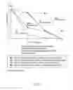
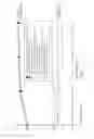
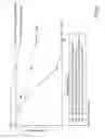
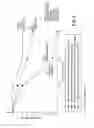
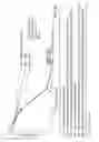
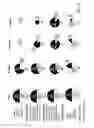
FIG. 1 is a graph showing the effect of arginine bicarbonate on growth of 8.3% (v/v) Staphylococcus epidermidis incubated with 12.0 mM zinc carbonate, 6.0 mM CIL and 24.0 mM arginine bicarbonate at 37° C. for 72 hours.
FIG. 2 is a graph showing the effect of arginine bicarbonate on growth of 8.3% (v/v) Staphylococcus aureus (MSSA), compared to 8.3% (v/v) Staphylococcus epidermidis, incubated with 12.0 mM zinc carbonate, 6.0 mM CIL and 24.0 mM arginine bicarbonate at 37° C. for 72 hours.
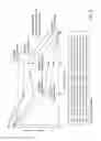
FIG. 3 is a graph showing the effect of arginine bicarbonate on growth of an 8.3% (v/v) 1:1 mixture of Staphylococcus epidermidis and Staphylococcus aureus (MSSA) incubated with 12.0 mM zinc carbonate and 6.0 mM CIL, with or without 24.0 mM arginine bicarbonate, at 37° C. for 72 hours.
FIG. 4 is a graph showing the effect of arginine bicarbonate on growth of 8.3% (v/v) Staphylococcus aureus (MSSA) incubatcd with 12.0 mM zinc carbonate and 6.0 mM CIL, with or without 24.0 mM arginine bicarbonate, at 37° C. for 72 hours.
FIG. 5 is a graph showing the effect of arginine bicarbonate on growth of 8.3% (v/v) Staphylococcus epidermidis incubated with 12.0 mM zinc carbonate and 6.0 mM CIL, with or without 24.0 mM arginine bicarbonate, at 37° C. for 72 hours.
FIG. 6 is a graph showing the effect of zinc carbonate on growth of 8.3% (v/v) Staphylococcus aureus (MSSA) incubated with 12.0 mM zinc carbonate and 6.0 mM CIL at 37° C. for 72 hours.
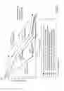
FIG. 7 is a graph showing the effect of arginine bicarbonate on the pH and growth of 8.3% (v/v) Staphylococcus aureus (MSSA), 8.3% (v/v) Staphylococcus epidermidis or an 8.3% (v/v) 1:1 mixture of Staphylococcus aureus (MSSA) and Staphylococcus epidermidis incubated with 12.0 mM zinc carbonate and 6.0 mM CIL, with or without 24.0 mM arginine bicarbonate, at 37° C. for 72 hours.
FIG. 8 is a graph showing the effect of arginine bicarbonate on the growth of 8.3% (v/v) Staphylococcus epidermidis or 8.3% (v/v) Staphylococcus aureus (MRSA) incubated with 12.0 mM zinc carbonate, 6.0 mM CIL and 24.0 mM arginine bicarbonate at 37° C. for 72 hours.
FIG. 9 is a graph showing the effect of arginine bicarbonate on the growth of 8.3% (v/v) Staphylococcus aureus (MRSA), compared to 8.3% (v/v) Staphylococcus epidermidis, incubated with 12.0 mM zinc carbonate and 6.0 mM CIL, with or without 24.0 mM arginine bicarbonate, at 37° C. for 72 hours.
FIG. 10 is a graph showing the effect of arginine bicarbonate on the growth of 8.3% (v/v) Staphylococcus epidermidis or 8.3% (v/v) Staphylococcus aureus (MRSA) incubated with 12.0 mM zinc carbonate, 6.0 mM CIL and 24.0 mM arginine bicarbonate, and modified versions of this medium, at 37° C. for 72 hours.
FIG. 11 is a graph showing the effect of arginine bicarbonate on the growth of an 8.3% (v/v) 1:1 mixture of Staphylococcus aureus (MRSA) and Staphylococcus epidermidis incubated with 12.0 mM zinc carbonate and 6.0 mM CIL, with or without 24.0 mM arginine bicarbonate, at 37° C. for 72 hours.
FIG. 12 is a graph showing the effcct of arginine bicarbonate on the growth of 8.3% (v/v) Staphylococcus aureus (MRSA) incubated at various dilutions with 12.0 mM zinc carbonate and 6.0 mM CIL, with or without 24.0 mM arginine bicarbonate, at 37° C. for 72 hours.
FIG. 13 is a graph showing the effect of arginine bicarbonate on live growth of 8.3% (v/v) Staphylococcus epidermidis incubated at various dilutions with 12.0 mM zinc carbonate and 6.0 mM CIL, with or without 24.0 mM arginine bicarbonate, at 37° C. for 72 hours.
FIG. 14 is a graph showing the effect of zinc carbonate on the growth of 8.3% (v/v) Staphylococcus aureus (MRSA) incubated at various dilutions with 12.0 mM zinc carbonate and 6.0 mM CIL at 37° C. for 72 hours.
FIG. 15 is a graph showing the pH responses of 8.3% (v/v) Staphlococcus epidermidis, 8.3% Staphylococcus aureus (MRSA) or an 8.3% (v/v) 1:1 mixture of Staphylococcus epidermidis and Staphylococcus aureus (MRSA) to 12.0 mM zinc carbonate and 6.0 mM CIL, with or without 24.0 mM arginine bicarbonate, at 37° C. for 72 hours.
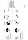
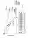
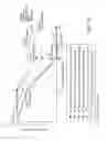
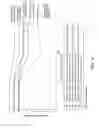
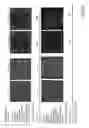
FIG. 16 is a photograph showing the effect of 24.0 mM arginine bicarbonate on growth of 8.3% (v/v) Staphylococcus epidermidis or 8.3% (v/v) Staphylococcus aureus (MSSA) incubated with 12.0 mM zinc carbonate, 6.0 mM CIL and 24.0 mM arginine bicarbonate, at 37° C. for 72 hours.
FIG. 17 is a photograph showing the effect of 24.0 mM arginine bicarbonate on growth of an 8.3% (v/v) 1:1 mixture of Staphylococcus epidermidis and Staphylococcus aureus (MSSA) incubated with 12.0 mM zinc carbonate and 6.0 mM CIL, with or without 24.0 mM arginine bicarbonate, at 37° C. for 72 hours.
FIG. 18 is a photograph showing the effect of 24.0 mM arginine bicarbonate on growth of an 8.3% (v/v) 1:1 mixture of Staphylococcus epidermidis and Staphylococcus aureus (MSSA) incubated with 12.0 mM zinc carbonate and 6.0 mM CIL, with or without 24.0 mM arginine bicarbonate, at 37° C. for 72 hours.
FIG. 19 is a photograph showing the effect of 24.0 mM arginine bicarbonate on growth of 8.3% (v/v) Staphylococcus epidermidis or 8.3% (v/v) Staphylococcus aureus (MRSA) incubated with 12.0 mM zinc carbonate, 6.0 mM CIL and 24.0 mM arginine bicarbonate, at 37° C. for 72 hours.
FIG. 20 is a photograph showing the effect of 24.0 mM arginine bicarbonate on growth of 8.3% (v/v) Staphylococcus epidermidis or 8.3% (v/v) Staphylococcus aureus (MRSA) incubated with 12.0 mM zinc carbonate and 6.0 mM CIL, with or without 24.0 mM arginine bicarbonate, at 37° C. for 0 hour.
FIG. 21 is a photograph showing the effect of 24.0 mM arginine bicarbonate on growth of 8.3% (v/v) Staphylococcus epidermidis or 8.3% (v/v) Staphylococcus aureus (MRSA) incubated with 12.0 mM zinc carbonate, 6.0 mM CIL and 24.0 mM arginine bicarbonate, and modified versions of this medium, at 37° C. for 24 hours.
FIG. 22 is a photograph showing the effect of 24.0 mM arginine bicarbonate on growth of an 8.3% (v/v) 1:1 mixture of Staphylococcus epidermidis and Staphylococcus aureus (MRSA) incubated with 12.0 mM zinc carbonate and 6.0 mM CIL, with or without 24.0 mM arginine bicarbonate, at 37° C. for 48 hours.
FIG. 23 is a photograph showing the effect of arginine bicarbonate on growth of an 8.3% (v/v) 1:1 mixture of Staphylococcus epidermidis and Staphylococcus aureus (MRSA) incubated with 12.0 mM zinc carbonate, 6.0 mM CIL and 24.0 mM arginine bicarbonate, with or without an additional 24 mM arginine bicarbonate added at indicated times, at 37° C. for 72 hours.
DETAILED DESCRIPTION OF THE INVENTION
Corynebacteria, staphylococci and proprionibacteria are among the main microorganisms present in the cutaneous microbiome, with Staph. epidermidis, C. striatum and P. avidum as prominent representative bacteria
Unexpectedly, it has been discovered that certain compositions including a zinc salt and arginine and/or its salt are useful as antibacterial compositions, inhibiting Staph. aureus growth while favoring Staph. epidermidis growth. This ability to select between Staph. aureus and Staph. epidermidis allows the treatment of significant physiological and health-related disease conditions caused by aberrant or excessive growth of Staph. aureus (see, e.g., Peacock et al., 2001, Uehara et al., 2000). Although Staph. aureus is capable of wide infection and fatalities (see, e.g., David et al., 2010, Mainous III et al., 2006, Klevens et al., 2007), current antibiotic treatments have achieved limited success due to the emergence of resistant Staph. aureus strains, e.g., MRSA (see, e.g., David et al 2010, Chen et al 2006, Centers for Disease Control and Prevention 2003). A recent discovery has shown that firmicidin (Gallo et al., 2013, Nakatsuji et al., 2012), a newly discovered antibiotic generated by Staph. epidermidis, can reduce Staph. aureus, but it is not known whether this will, like other antibiotics, succumb to adaptation and loss of effectiveness. From a commercial stand-point, this approach is likely to be costly.
Unlike traditional antibacterial treatments, the compositions of the present invention are aimed at modulating natural interactions between Staph. aureus and other prominent members of the cutaneous microflora, e.g., Staph. epidermidis (see, e.g., Frank et al., 2010, Vehara et al., 2000, Wertheim et al., 2005). These bacteria naturally compete, e.g., for local resources and attachment to mucosal sites (Frank et al., 2010). The compositions of the invention, rather than merely targeting Staph. aureus, render an ecological change that favors selection of desirable Staph. epidermidis over non-desirable Staph. aureus bacteria.
Because the compositions of the present invention derive their antibacterial effectiveness not only by targeting Staph. aureus directly, but also by enhancing the ability of other, non-pathogenic bacteria (e.g., Staph. epidermidis) to out-compete Staph. aureus. The compositions disclosed here are less likely to be susceptible to the emergence of resistant strains (e.g., MRSA) than traditional antibacterial treatments.
A further advantage of the present invention is that the compositions disclosed herein are effective in reducing cutaneous odor production. Thus, a single topical composition may be used as both deodorant and antibacterial treatment.
Antibacterial compositions as described herein are administered, preferably topically, for the treatment of any one or more symptoms desirable of change, e.g., Staph. aureus growth. Dosage forms are solid or free-flowing. Dosage forms include, but are not limited to, soaps, sprays, drops, aerosols, powders, roll-ons, lotions, creams, sticks, solutions, sachets, colloidal suspensions, films, patches and ointments.
Antibacterial compositions as described herein may have a pH of at least 6.0, or at least 7.0, or at least 8.0, or at least 9.0 upon topical administration.
Antibacterial compositions as described herein may optionally include one or more physiologically acceptable buffers sufficient to maintain the pH of said composition, e.g., at 6.0 or greater, at 7.0 or greater, at 8.0 or greater, or at 9.0 or greater upon topical application. Such buffers are generally known in the art, and may include, e.g., ACES, acetic acid, ADA, AMP, AMPD, bicine, bis-Tris, bis-Tris propane, BES, boric acid, cacodylate, CABS, CAPS, CAPSO, CHES, citric acid, diethanolamine, DIPSO, EPPS/HEPPS, ethanolamine, formic acid, glycine, glycylglycine, HEPES, HEPPSO, histidine, imidazole, lactic acid, maleic acid, malic acid, MES, MOPS, MOPSO, morpholine, phosphate, phosphoric acid, picolinic acid, PIPES, piperazine, piperidine, pivalic acid, POPSO, pyridine, succinic acid, TAPS, TAPSO, TEA, TES, tricine, and/or Tris.
Except where otherwise noted, the terms “axillary odor” and “foot odor” are used interchangeably herein, the terms “microbiome,” “microbiota,” and “microflora” are used interchangeably herein, the terms “foot,” “foot web,” “foot-web,” “toe,” “toe web” and “toe-web” are used interchangeably herein, and the terms “odor” and “malodor” are used interchangeably herein.
The terms “cutaneous” and “skin” refer, in the context of the present invention, regions of the human body including, e.g., the axilla, foot-webs and nasal atrium.
The terms “physiologically acceptable” and “physiologically-acceptable” denote, in the context of the present invention, “safe and effective when administered to humans and/or mammals in need thereof,” e.g., to reduce axillary odor, promote the growth of Staphylococcus epidermidis bacteria, inhibit the growth of Staphylococcus aureus bacteria, or any or all of the preceding.
Examples
The following examples are intended to illustrate, but not limit, the present disclosure.
Growth of Staph. aureus (MSSA or MRSA) and Staph. epidermidis when one or the other or a mixture of the two bacteria were incubated in the presence of (i) cysteine and (ii) isoleucine, leucine, phenylalanine. Zinc carbonate was also provided with and without arginine bicarbonate at 37° C. for 72 hours and with additional above ingredients adding into the cultural media in 37° C. water bath in 24 and 48 hours.
Materials and Methods for Growth Comparison Experiments between Staph. epidermidis and Staph. aureus
-
- (a) Preparation of Agar plates containing various bacterial growth media. Preparation included (i) BHI Blood agar (Fisher Scientific, Springfield, N.J. USA) and (ii) CHROMagar Staph. aureus agar (CHROMagar, Paris, France), especially prepared for the isolation and identification of Staph. aureus; if present, it results in colonies that show a characteristic mauve color that enables ease of identification (French, 2009, Han et al., 2007).
- (b) Stock solutions of CIL amino acids. These amino acids include cysteine, isoleucine and leucine with each present at a concentration of 72 mM. Aqueous solutions of each were sterilized by syringe filtering as described earlier (Zhang and Kleinberg, 2014).
- (c) Stock aqueous solutions of arginine bicarbonate at 144 mM and zinc carbonate at 72 mM. Stock solutions of 144 mM arginine bicarbonate were sterilized together with 72 mM zinc carbonate by syringe filtering. Zinc carbonate has a limited solubility and hence is sterilized by first autoclaving as a powder and then dissolving it until saturation in sterile distilled water is achieved. This means that at 72 mM and above, it may have to be used as a zinc carbonate suspension.
- (d) Rabbit coagulase plasma (PL 850) and Prolex Staph Xtra Latex kits (PL.1080). Both of these items are provided as a kit and are obtained from Pro-Lab Diagnostics, Austin, Tex. They are prepared for the identification of pathogenic staphylococci (e.g., Staph. aureus).
- (e) Experimental and control incubation mixtures containing Staph. epidermidis (ATCC 12228) and Staph. aureus (MSSA and/or MRSA). These incubation mixtures were prepared for comparison purposes and included MSSA (ATCC 25923) or MRSA (ATCC 33591) bacterial species mixed with the microorganism Staph. epidermidis. Pure cultures of Staph. epidermidis and Staph. aureus (MSSA or MRSA) were each prepared as 25% (v/v) bacterial suspensions in sterile distilled water. As above and as much as possible, bacterial pellets were broken up into fine particles, by stirring with a sterile TB syringe and a 25-27 gauge needle, if and when needed.
As a preparatory step, the resulting suspensions obtained were incubated in a shaking water bath at 37° C. for one hour, in order to deplete stored substrates acquired by some bacteria, during their preparatory growth period (Wijeyeweera and Kleinberg, 1989). The pH of each of the above bacterial suspensions was then measured by transferring 0.25 ml of such to a small sterile test-tube and measuring its pH. This made it easier to avoid any bacterial contamination during handling. Samples were then stored at 4° C. until time of inoculation of agar plates.
Preparation of Experimental and Control Samples
Preparation was performed according to information in Table 1 below.
| TABLE 1.1 |
| Experimental (A and B) and negative control (C) samples were |
| prepared according to the following ABC Composition Tables: |
| A. |
| Experimental samples (ml) |
| Composition | I | II | III | IV | V | VI | Final concentrations |
| Amino acids | Cys 72 mM | 0.225 | 0.225 | 0.225 | 0.225 | 0.225 | 0.225 | 6 mM |
| Ieu 72 mM | 0.225 | 0.225 | 0.225 | 0.225 | 0.225 | 0.225 | 6 mM | |
| Ileu 72 mM | 0.225 | 0.225 | 0.225 | 0.225 | 0.225 | 0.225 | 6 mM |
| Zinc Carbonate (72 mM) | 0.45 | 0.45 | 0.45 | 0.45 | 0.45 | 0.45 | 12 mM |
| Arg. Bicarbonate (144 mM) | 0.45 | 0.45 | 0.45 | — | — | — | 24 mM (IV, V, VI = 0 mM) |
| Staph. epidermidis (25%) | 0.45 | — | 0.90 | 0.45 | — | 0.90 | 8.3% | mixture | 4.15% |
| Staph. aureus 25% (MSSA or MRSA) | 0.45 | 0.90 | — | 0.45 | 0.90 | — | 8.3% | 4.15% |
| D-water | 0.225 | 0.225 | 0.225 | 0.675 | 0.675 | 0.675 | |
| Total volume (ml) | 2.70 | 2.70 | 2.70 | 2.70 | 2.70 | 2.70 | |
| B. |
| Experimental samples (ml) |
| Composition | IA | IIA | IIIA | IB | IIB | IIIB | Final concentrations |
| Amino acids | Cys 72 mM | 0.225 | 0.225 | 0.225 | 0.225 | 0.225 | 0.225 | 6 mM |
| Ieu 72 mM | 0.225 | 0.225 | 0.225 | 0.225 | 0.225 | 0.225 | 6 mM | |
| Ileu 72 mM | 0.225 | 0.225 | 0.225 | 0.225 | 0.225 | 0.225 | 6 mM |
| Zinc Carbonate (72 mM) | 0.45 | 0.45 | 0.45 | 0.45 | 0.45 | 0.45 | 12 mM |
| Arg. Bicarbonate (44 mM) | 0.45 | 0.45 | 0.45 | 0.45 | 0.45 | 0.45 | 24 mM |
| Staph. epidermidis (25%) | 0.45 | — | 0.90 | 0.45 | — | 0.90 | 8.3% | mixture | 4.15% |
| Staph. aureus 25% (MRSA) | 0.45 | 0.90 | — | 0.45 | 0.90 | — | 8.3% | 4.15% |
| D-water | 0.225 | 0.225 | 0.225 | 0.225 | 0.225 | 0.225 |
| Total volume (ml) | 2.70 | 2.70 | 2.70 | 2.70 | 2.70 | 2.70 |
| C. |
| Negative controls |
| Composition | 1 | 2 | 3 | Final concentrations | ||
| Amino acids | Cys 72 mM | — | — | — | — | |
| Ieu 72 mM | — | — | — | — | ||
| Ileu 72 mM | — | — | — | — |
| Zinc Carbonate (72 mM) | — | — | — | — | |
| Arg. Bicarbonate (144 mM) | — | — | — | — |
| Staph. epidermidis (25%) | 0.45 | — | 0.90 | 8.3% | mixture | 4.15% | |
| Staph. aureus 25% (MSSA or MRSA) | 0.45 | 0.90 | — | 8.3% | 4.15% | ||
| D-water | 1.80 | 1.80 | 1.80 | ||||
| Total volume (ml) | 2.70 | 2.70 | 2.70 | ||||
| Arginine bicarbonate is absent in IV, V and VI |
Dilutions of Experimental and Negative Control Samples and Inoculations of BHI Blood Agar and CHROMagar Staph. aureus Plates
Serial dilutions from 101 to 1010 of each of experimental samples I, II, III, IV, V, VI and control samples 1, 2, 3 (see Table 1) were prepared with sterile distilled water. Each dilution contained 0.1 ml of serial diluted sample and 0.9 ml of sterile distilled water. BHI Blood agar plates were then inoculated with a mixture of 100 μl of a 104 to 1010 concentration of Staph. epidermidis bacteria and 100 μl of a 104 to 1010 sample of Staph. aureus (MSSA or MRSA) mixture (Samples I, IV and Negative Control 1) onto CHROMagar Staph. aureus plates using sterile glass bars on a turning table, respectively.
Incubation Procedures
As a first precautionary step, all agar plates were incubated for 24 hours in a 37° C. incubator and examined thereafter for bacterial growth to ensure initial agar plate sterility. Plates were then inoculated with samples taken at times 0, 24, 48 and 72 hours in succession throughout the 4 days of incubation. Successive inoculations consisted of the transfer of bacterial samples from a prior incubation to a subsequent fresh sterile plate, followed by incubation at 37° C. for 24-48 hours and subsequently repeating the process.
Colony density was scored for each of the plates as follows: between 0 and 10 as 0—no colonies; 1—<10 colonies; 2—10 to 20 colonies; 3—20 to 30 colonies; 4—30 to 50 colonies: 5—50 to 100 colonies; 6—100 to 250 colonies; 7—250 to 500 colonies; 8—>500 colonics; 9—colonies almost fused to form a layer; 10—colonies forming a bacterial layer.
Differentiation of Colonies of Staph. aureus and Staph. epidermidis Derived from Growth on BHI Blood and CHROMagar SA Plates of Samples from Incubation Mixtures with Staph. aureus and Staph. epidermidis
Staph. aureus colonies are usually a golden yellow color and show large and complete blood hemolytic rings around the colonies that grow on BHI Blood agar plates. Use of the coagulase serum test (test procedure of Rabbit Coagulase Plasma provided by Pro-Lab Diagnostics, Austin, Tex. USA) and Prolex Staph Xtra Latex Test (Test Protocol of Prolix™ Staph Xtra Latex Kit provided by Pro Lab Diagnostics, Austin, Tex. USA) showed positive results. On CHROMagar Staph. aureus plates, where Staph. aureus colonies readily grow, they show, as pointed out above, a mauve color. In contrast, their counterpart, Staph. epidermidis colonies, are white and have no or small hemolytic rings around the colonies, when grown on BHI Blood agar plates. On CHROMagar Staph. aureus plates, Staph. epidermidis is unable to grow or able to form tiny white colonies. Coagulase serum and Prolex Staph Xtra Latex testing proved negative (i.e. no coagulation).
Inoculation of Samples Incubated in a Water Bath at 37° C. for 24 Hours and then Inoculated onto (i) BHI Blood Agar Plates and (ii) CHROMagar Staph. aureus Plates
Following the same serial dilution procedures, as done for the Day I incubation period, Samples I, II, III, IV, V, VI and 1, 2, 3 were diluted serially 104 to 1010 on BHI Blood agar plates. Similarly, samples of a mixture of Staph. epidermidis and Staph. aureus (I, IV and Negative Control 1) were prepared on CHROMagar Staph. aureus plates and incubated using the same procedures, as were used on Day 1, i.e. incubation at 37° C. for 24-48 hours.
Addition of Extra Ingredients to Samples, IA, IIA, IIIA and IB, IIB, IIIB Incubated as on Day 1, in a Water Bath at 37° C. for 24 Hours
-
- Under aseptic conditions, samples, IA, IIA, IIIA and IB, IIB, IIIB were each centrifuged and 1.35 ml of supernatant was removed from each of samples, IA, IIA, IIIA, and 1.125 ml of supernatant from samples, IB, IIB, IIIB, respectively.
- The table immediately below, lists additional ingredients introduced into samples:
| TABLE 1.2 | |
| Volumes (ml) added to experimental samples |
| Ingredients | IA | IIA | IIIA | IB | IIB | IIIB |
| Cys 72 mM | 0.225 | 0.225 | 0.225 | 0.225 | 0.225 | 0.225 |
| Ieu 72 mM | 0.225 | 0.225 | 0.225 | 0.225 | 0.225 | 0.225 |
| Ileu 72 mM | 0.225 | 0.225 | 0.225 | 0.225 | 0.225 | 0.225 |
| Zinc Carbonate | 0.225 | 0.225 | 0.225 | — | — | — |
| (72 mM) | ||||||
| Arg. Bicarbonate | 0.450 | 0.450 | 0.450 | 0.450 | 0.450 | 0.450 |
| (144 mM) | ||||||
Incubation of all experimental and control samples in a 37° C. water bath was continued for another 24 hours. Total incubation time to this point was 48 hours.
Day 3 in the Experimental Protocol (i.e., the 48-72 Hour Time Period).
This period consisted of bacterial growth on the medium agar plates inoculated on Day 2 and incubated at 37° C., (as above), on medium agar plates for another 24 hours and preparation of samples for incubation continuation for another 24 hours. Bacterial growth on BHI Blood agar and CHROMagar Staph. aureus plates was then determined as before.
The next step was inoculation of samples incubated in a 37° C. water bath for a total of 48 hours on the BHI Blood agar plates and CHROMagar Staph. aureus plates.
-
- The same procedures of serial dilutions, as was done on Day 1, was carried out here; i.e. all samples (I, II, III, IV, V, VI, 1, 2, 3 and IA, IIA, IIIA, IB, IIB, IIIB).
- Inoculated 104 to 1010 serial dilutions of samples on BHI Blood agar plates and the samples of the mixture of SE and SA (I, IA, IB, IV and Negative Control 1) on CHROMagar Staph. aureus plates were tested by following the same procedures as was done on Day 1.
- Plates were incubated as before at 37° C. between and for 24 and 48 hours.
Preparation of Samples for Incubation in a Water Bath at 37° C. for 48 Hours and Followed then for a Further 24 Hours - Additional ingredients were added to samples of IA, IIA, IIIA and IB, IIB, IIIB, which were each incubated in a 37° C. water bath for a total period of 48 hours.
- Samples IA, IIA, IIIA and samples IB, IIB, IIIB were centrifuged as before and 1.35 ml of supernatant was removed from samples, IA, IIA, IIIA; and 1.125 ml of supernatant was also removed from samples, IB, IIB, and IIIB, respectively.
- Table 1.3, below, was followed in order to serve as a guide for adding additional ingredients into the samples:
| TABLE 1.3 | |
| The (ml) volumes added to the experimental samples |
| Ingredients | IA | IIA | IIIA | IB | IIB | IIIB |
| Cys 72 mM | 0.225 | 0.225 | 0.225 | 0.225 | 0.225 | 0.225 |
| Ieu 72 mM | 0.225 | 0.225 | 0.225 | 0.225 | 0.225 | 0.225 |
| Ileu 72 mM | 0.225 | 0.225 | 0.225 | 0.225 | 0.225 | 0.225 |
| Zinc Carbonate | 0.225 | 0.225 | 0.225 | — | — | — |
| (72 mM) | ||||||
| Arg. Bicarbonate | 0.450 | 0.450 | 0.450 | 0.450 | 0.450 | 0.450 |
| (144 mM) | ||||||
-
- Incubation of all experimental and control samples in the water bath at 37° C. was extended for another 24 hours (i.e. 72 hours total).
Day 4 (72-96 Hours, i.e., the Last Part of the Instant Experimental Protocol)
Bacterial growth on medium agar plates inoculated on Day 3 was examined and then incubated in a water bath at 37° C. for a total of 72 hours.
Examination of Bacterial Growth on BHI Blood Agar and CHROMagar Staph. aureus plates inoculated on Day 3
The same methods were followed as was done on Day 4.
Inoculation of Samples Incubated at 37° C. for a Total of 72 Hours on BHI Blood Agar Plates and CHROMagar Staph. aureus Plates
-
- The same procedures of serial dilution were followed as was done on Day 1 for all samples (I, II, III, IV, V, VI, 1, 2, 3 and IA, IIA, IIIA, IB, IIB, IIIB).
- Inoculation of 104 to 1010 serial dilutions of samples on BHI Blood agar plates and the samples of the mixture of SE and SA (I, IA, IB, IV and 1) on CHROMagar Staph. aureus plates were the same as the procedures carried out on Day 1.
- Plates were then incubated at 37° C. for 24-48 hours.
Day 5 (End of Experiment, 96 Hours Total Duration)
Examination of bacterial growth on media agar plates inoculated on Day 4 and a review of the entire experiment was performed. Examination of bacterial growth on BHI Blood agar and CHROMagar Staph. aureus plates inoculated was performed on Day 4 by following the same methods as was done on Day 1.
Results
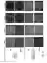
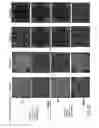
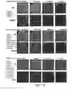
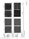
Overview of the bacterial growth of all samples on the BHI Blood agar plates and on the CHROMagar Staph. aureus plates in the 72 hour experiments reported herein are shown in Tables 1.4, 1.5 and 1.6. FIGS. 1-15 depict the effect of different media on bacterial growth. Photographs showing colony growth data from which the Figures were derived are set forth as FIGS. 16-23.
| TABLE 1.4 |
| Density (1-10*) of colonies of Staphylococcus epidermidis (SE) and Staphylococcus aureus |
| (MSSA) when incubated in media comprised of 6 mM cysteine, 6 mM isoleucine, 6 mM leucine |
| (i.e., 6 mM CIL) and 12 mM zinc carbonate, with or without 24 mM arginine bicarbonate |
| at 37° C. for 72 hours, compared with negative control (water only) |
| Medium-Cys, Ileu, | Medium-Cys, Ileu, | |
| Leu, zinc carbonate | Leu, zinc carbonate |
| Negative Control | with arginine | without arginine | |
| Medium (Water only) | bicarbonate | bicarbonate |
| Time of | Times of dilution of 8.3% bacteria incubated in media |
| Bacteria | Incubation | Plates | 104 | 105 | 106 | 107 | 104 | 105 | 106 | 107 | 104 | 105 | 106 | 107 |
| SE | 0 h | BHI | 9 | 9 | 8 | 8 | 9 | 9 | 8 | 8 | 9 | 9 | 8 | 8 |
| MSSA | Blood | 9 | 9 | 9 | 9 | 10 | 9 | 9 | 9 | 10 | 9 | 9 | 9 | |
| Mix | Agar | 9 | 9 | 9 | 9 | 10 | 9 | 9 | 9 | 10 | 9 | 9 | 9 | |
| CHRO | 9 | 9 | 9 | 9 | 10 | 9 | 9 | 9 | 10 | 9 | 9 | 9 | ||
| SE/SA | . . . | . . . | . . . | . . . | . . . | . . . | . . . | . . . | . . . | . . . | . . . | . . . | ||
| % (~) | ||||||||||||||
| SE | 24 h | BHI | 9 | 8 | 7 | 7 | 8 | 7 | 6 | 5 | 5 | 1 | 1 | 1 |
| MSSA | Blood | 9 | 8 | 7 | 5 | 7 | 3 | 2 | 1 | 8 | 7 | 5 | 4 | |
| Mix | Agar | 9 | 8 | 7 | 5 | 8 | 6 | 4 | 3 | 8 | 7 | 7 | 6 | |
| CHRO | 9 | 8 | 7 | 5 | 6 | 4 | 2 | 1 | 8 | 7 | 7 | 6 | ||
| SE/SA | . . . | . . . | 5/95 | 10/90 | . . . | 70/30 | 60/40 | 80/20 | . . . | 15/85 | 15/85 | 20/80 | ||
| % (~) | ||||||||||||||
| SE | 48 h | BHI | 9 | 8 | 7 | 6 | 5 | 2 | 1 | 1 | 0 | 0 | 0 | 0 |
| MSSA | Blood | 8 | 7 | 6 | 5 | 5 | 2 | 1 | 1 | 8 | 7 | 6 | 6 | |
| Mix | Agar | 9 | 8 | 7 | 6 | 6 | 2 | 1 | 1 | 8 | 7 | 6 | 5 | |
| CHRO | 9 | 7 | 6 | 5 | 5 | 2 | 1 | 1 | 8 | 7 | 6 | 5 | ||
| SE/SA | . . . | . . . | 10/90 | 20/80 | 10/90 | 15/85 | . . . | . . . | . . . | 5/95 | 5/95 | 5/95 | ||
| % (~) | ||||||||||||||
| SE | 72 h | BHI | 8 | 7 | 6 | 3 | 1 | 1 | 0 | 0 | 0 | 0 | 0 | 0 |
| MSSA | Blood | 8 | 6 | 4 | 3 | 0 | 0 | 0 | 0 | 7 | 2 | 1 | 1 | |
| Mix | Agar | 9 | 7 | 5 | 3 | 2 | 0 | 0 | 0 | 7 | 5 | 4 | 2 | |
| CHRO | 9 | 7 | 5 | 4 | 1 | 0 | 0 | 0 | 7 | 5 | 4 | X | ||
| SE/SA | . . . | 5/95 | 5/95 | 0 | 20/80 | . . . | . . . | . . . | . . . | 0 | 0 | . . . | ||
| % (~) | ||||||||||||||
| SE, Staph. epidermidis, | ||||||||||||||
| MSSA, Staph. aureus (MSSA), | ||||||||||||||
| Mix, mixture of Staph. epidermidis and Staph. aureus (MSSA), | ||||||||||||||
| CHRO, CHROMAgar medium plate selective for Staph. aureus, X, contamination | ||||||||||||||
| *Scale (0-10): 0, no colony; 1, <10; 2, 10-20; 3, 20-30; 4, 30-50; 5, 50-100; 6, 100-250; 7, 250-500; 8, >500; 9, colonies almost form a layer and are unable to count; 10, colonies form a layer |
| TABLE 1.5 |
| Density (1-10*) of colonies of Staphylococcus epidermidis (SE) and Staphyloccus aureus |
| (MRSA) when incubated in media comprised of 6 mM cysteine, 6 mM isoleucine, 6 mM leucine |
| (i.e., 6 mM CIL) and 12 mM zinc carbonate, with or without 24 mM arginine bicarbonate |
| at 37° C. for 72 hours, compared with negative control (water only) |
| Medium-Cys, Ileu, | Medium-Cys, Ileu, | |
| Leu, zinc carbonate | Leu, zinc carbonate |
| Negative Control | with arginine | without arginine | |
| Medium (Water only) | bicarbonate | bicarbonate |
| Time of | Times of dilution of 8.3% bacteria incubated in media |
| Bacteria | Incubation | Plates | 104 | 105 | 106 | 107 | 104 | 105 | 106 | 107 | 104 | 105 | 106 | 107 |
| SE | 0 h | BHI | 9 | 9 | 8 | 7 | 9 | 8 | 8 | 8 | 9 | 8 | 8 | 8 |
| MRSA | Blood | 10 | 9 | 8 | 8 | 10 | 9 | 8 | 8 | 9 | 8 | 8 | 8 | |
| Mix | Agar | 10 | 9 | 8 | 7 | 10 | 9 | 8 | 8 | 9 | 8 | 8 | 8 | |
| CHRO | 10 | 9 | 8 | 7 | 10 | 9 | 8 | 8 | 9 | 8 | 8 | 8 | ||
| SE/SA | . . . | . . . | . . . | . . . | . . . | . . . | . . . | . . . | . . . | . . . | . . . | . . . | ||
| % (~) | ||||||||||||||
| SE | 24 h | BHI | 9 | 9 | 8 | 8 | 8 | 8 | 8 | 8 | 7 | 6 | 6 | 6 |
| MRSA | Blood | 10 | 9 | 9 | 8 | 7 | 5 | 5 | 5 | 9 | 8 | 8 | 8 | |
| Mix | Agar | 10 | 9 | 8 | 8 | 8 | 7 | 5 | 5 | 9 | 8 | 8 | X | |
| CHRO | 10 | 9 | 8 | 8 | 6 | 5 | 3 | 2 | 8 | 8 | 8 | 6 | ||
| SE/SA | . . . | . . . | . . . | . . . | . . . | 60/40 | 80/20 | 80/20 | . . . | . . . | . . . | . . . | ||
| % (~) | ||||||||||||||
| SE | 48 h | BHI | 9 | 8 | 7 | 6 | 6 | 4 | 3 | 1-3 | 0 | 0 | 0 | 0 |
| MRSA | Blood | 9 | 8 | 7 | 7 | 1 | 1 | 0 | 0 | 7 | 6 | 3 | 1 | |
| Mix | Agar | 9 | 8 | 7 | 6 | 1 | 0 | 0 | 0 | 7 | 6 | 5 | 5 | |
| CHRO | 9 | 8 | 6 | 6 | 0 | 0 | 0 | 0 | 6 | 5 | 4 | 4 | ||
| SE/SA | . . . | . . . | . . . | 5/95 | . . . | . . . | . . . | . . . | 10/90 | 20/80 | 20/80 | 20/80 | ||
| % (~) | ||||||||||||||
| SE | 72 h | BHI | 8 | 8 | 7 | 6 | 5 | 5 | 4 | 3 | 1-3 | 0 | 1-3 | 0 |
| MRSA | Blood | 9 | 8 | 7 | 7 | 0 | 0 | 0 | 0 | 7 | 6 | 4 | 4 | |
| Mix | Agar | 8 | 8 | 7 | 6 | 1 | 1 | 1 | 1 | 7 | 7 | 6 | 6 | |
| CHRO | 8 | 8 | 6 | 5 | 0 | 1 | 1 | 0 | 7 | 6 | 6 | 6 | ||
| SE/SA | . . . | . . . | . . . | . . . | . . . | . . . | . . . | . . . | 5/95 | 10/90 | 5/95 | 5/95 | ||
| % (~) | ||||||||||||||
| SE, Staph. epidermidis, | ||||||||||||||
| MRSA, Staph. aureus (MRSA), | ||||||||||||||
| Mix, mixture of Staph. epidermidis and Staph. aureus (MRSA), | ||||||||||||||
| CHRO, CHROMAgar medium plate selective for Staph. aureus, X, contamination | ||||||||||||||
| *Scale (0-10): 0, no colony; 1, <10; 2, 10-20; 3, 20-30; 4, 30-50; 5, 50-100; 6, 100-250; 7, 250-500; 8, >500; 9, colonies almost form a layer and are unable to count; 10, colonies form a layer |
| TABLE 1.6 |
| Density (1-10*) of colonies of Staphylococcus epidermidis (SE) and Staphyloccus aureus (MRSA) when incubated |
| in media comprised of 6 mM cysteine, 6 mM isoleucine, 6 mM leucine (i.e., 6 mM CIL) and 12 mM zinc carbonate, with |
| or without 24 mM arginine bicarbonate at 37° C. for 72 hours, compared with negative control (water only) |
| Media containing 6 mM Cys, 6 mM Leu, 6 mM Ileu, 12 mM zinc | |
| carbonate, 24 mM arginine bicarbonate |
| Additional same | Additional 24 mM | ||
| No additional | above media added | arginine bicarbonate | |
| medium added | in 24 and 48 hours | added in 24 and 48 hours |
| Time of | Times of dilution of 8.3% bacteria incubated in media |
| Bacteria | Incubation | Plates | 104 | 105 | 106 | 107 | 104 | 105 | 106 | 107 | 104 | 105 | 106 | 107 |
| SE | 0 h | BHI | 9 | 8 | 8 | 8 | 9 | 8 | 8 | 8 | 9 | 8 | 8 | 8 |
| MRSA | Blood | 10 | 9 | 8 | 8 | 10 | 9 | 8 | 8 | 10 | 9 | 8 | 8 | |
| Mix | Agar | 10 | 9 | 8 | 8 | 10 | 9 | 8 | 8 | 10 | 9 | 8 | 8 | |
| CHRO | 10 | 9 | 8 | 8 | 10 | 9 | 8 | 8 | 10 | 9 | 8 | 8 | ||
| SE/SA | . . . | . . . | . . . | . . . | . . . | . . . | . . . | . . . | . . . | . . . | . . . | . . . | ||
| % (~) | ||||||||||||||
| SE | 24 hr | BHI | 8 | 8 | 8 | 8 | 8 | 8 | 8 | 8 | 8 | 8 | 8 | 8 |
| MRSA | Blood | 7 | 5 | 5 | 5 | 7 | 5 | 5 | 5 | 7 | 5 | 5 | 5 | |
| Mix | Agar | 8 | 7 | 5 | 5 | 8 | 7 | 5 | 5 | 8 | 7 | 5 | 5 | |
| CHRO | 6 | 5 | 3 | 2 | 6 | 5 | 3 | 2 | 6 | 5 | 3 | 2 | ||
| SE/SA | 70/30 | 60/40 | 80/20 | 80/20 | . . . | 60/40 | 80/20 | 80/20 | . . . | 60/40 | 80/20 | 80/20 | ||
| % (~) | ||||||||||||||
| SE | 48 h | BHI | 6 | 4 | 3 | 1 | 7 | 6 | 4 | 1 | 8 | 7 | 5 | 4 |
| MRSA | Blood | 1 | 1 | 0 | 0 | 1 | 0 | 0 | 0 | 1 | 1 | 0 | 0 | |
| Mix | Agar | 1 | 0 | 0 | 0 | 5 | 1 | 1 | 0 | 7 | 5 | 1 | 1 | |
| CHRO | 0 | 0 | 0 | 0 | 1 | 0 | 0 | 0 | 3 | 1 | 1 | 0 | ||
| SE/SA | . . . | . . . | . . . | . . . | 90/10 | . . . | . . . | . . . | 90/10 | 90/10 | . . . | . . . | ||
| % (~) | ||||||||||||||
| SE | 72 h | BHI | 5 | 5 | 4 | 3 | 6 | 5 | 4 | 2 | 7 | 5 | 4 | 4 |
| MRSA | Blood | 0 | 0 | 0 | 0 | 0 | 0 | 0 | 0 | 0 | 0 | 0 | 0 | |
| Mix | Agar | 1 | 1 | 1 | 1 | 3 | 0 | 0 | 1 | 7 | 7 | 6 | 5 | |
| CHRO | 0 | 1 | 1 | 0 | 0 | 0 | 0 | 0 | 5 | 5 | 4 | 1 | ||
| SE/SA | . . . | . . . | . . . | . . . | 100/0 | . . . | . . . | . . . | 90/10 | 80/20 | 80/20 | 80/20 | ||
| % (~) | ||||||||||||||
| SE, Staph. epidermidis, | ||||||||||||||
| MRSA, Staph. aureus (MRSA), | ||||||||||||||
| Mix, mixture of Staph. epidermidis and Staph. aureus (MRSA), | ||||||||||||||
| CHRO, CHROMAgar medium plate selective for Staph. aureus, X, contamination | ||||||||||||||
| *Scale (0-10): 0, no colony; 1, <10; 2, 10-20; 3, 20-30; 4, 30-50; 5, 50-100; 6, 100-250; 7, 250-500; 8, >500; 9, colonies almost form a layer and are unable to count; 10, colonies form a layer |
Tables 1.4-1.6, above, include the following elements:
-
- (a) Incubation of Staph. epidermidis and Staph. aureus and their combinations in the medium containing 12 mM zinc carbonate, 24 mM arginine bicarbonate, the CIL amino acids and their controls, showed:
- (i) Staph. aureus (MSSA or MRSA) quickly decreased, when incubated in the presence of arginine bicarbonate for 24 to 48 hours; all Staph. aureus organisms completely disappeared by 72 hours (see supporting FIGS. 1, 8, 16 and 19).
- (ii) Staph. epidermidis on the other hand decreased only slightly, when incubated with the medium containing arginine bicarbonate during the first 24 hours of incubation and decreased moderately or rapidly in the 48 to 72 hours thereafter (see relevant FIGS. 1, 2, 8, 9, 16 and 19).
- (iii) The mixture of Staph. aureus (MSSA or MRSA) and Staph. epidermidis also showed decreases, albeit only moderately, while being incubated in the medium containing arginine bicarbonate for 24 hours and where approximately 60-80% of survivors were Staph. epidermidis. The Staph. aureus/Staph. epidermidis mixture decreased quickly after 24 hours of incubation and almost all of the bacteria had disappeared by 72 hours (see FIGS. 3, 11, 18 and 22).
- (iv) In the negative control, both Staph. aureus (MSSA or MRSA) and Staph. epidermidis and the mixtures thereof incubated in sterile D-water, showed almost no reduction in 24 to 48 hours and very slight reduction in 48 to 72 hours (see FIGS. 1, 8, 16 and 19).
- (b) Incubating Staph. aureus (MSSA or MRSA), Staph. epidermidis and their combinations in a medium containing the CIL amino acids, and zinc carbonate without arginine bicarbonate exhibited:
- (i) Staph. aureus (MSSA or MRSA) that showed no or slight reduction, while incubating for 24 to 48 hours and then decreased slightly or moderately thereafter. Staph. aureus showed much slower reduction of its numbers in the medium without arginine bicarbonate than when incubated in medium containing arginine bicarbonate (see FIGS. 2, 9, 17 and 20).
- (ii) Staph. epidermidis showed moderate to rapid reduction in numbers during incubation for 24 hours and disappeared after 48 hours (see FIGS. 2, 9, 17 and 20).
- (iii) Within 72 hours, the mixture of Staph. aureus (MSSA or MRSA) and Staph. epidermidis decreased moderately, while incubating in medium without arginine bicarbonate. Also, within 72 hours, approximately 70-90% of survivors were Staph. aureus, whereas in the mixture incubated in the medium containing arginine, bacteria correspondingly decreased slowly in 24 hours. About 70-75% of survivors were Staph. epidermidis and the mixture rapidly decreased in 48 to 72 hours. Almost all bacteria disappeared by 72 hours (see FIGS. 3, 11, 18, 22 and 23).
- (c) The results of Staph. aureus (MRSA) and Staph. epidermidis being incubated in the medium including 12 mM zinc carbonate, 24 mM arginine bicarbonate, the CIL amino acids, and additional same medium or 24 mM arginine bicarbonate being added in 24 and 48 hours during 72 hours of incubation at 37° C. showed:
- (i) Slow Staph. epidermidis reduction during the first 24 hours and slower reduction after 48 to 72 hours, when additional same medium was added, at 24 and 48 hours. Staph. epidermidis even decreased, albeit more slowly, when additional 24 mM arginine bicarbonate was added after 24 and 48 hours, whereas Staph. aureus (MRSA) decreased, moderately to rapidly, after 48 hours with no microbial survivors after 72 hours. There were no differences among the incubation media and additional medium, whether arginine bicarbonate was or was not added (see FIG. 10 and Photo 21).
- (ii) The mixture of Staph. aureus (MRSA) and Staph. epidermidis decreased in a similar pattern, as did Staph. epidermidis with 60% of survivors being Staph. epidermidis after 24 hours of incubation and more than 90% Staph. epidermidis survivors after 48 to 72 hours of incubation (see FIG. 11 and Photo 23).
- (d) Staph. aureus (MSSA or MRSA) was incubated with 12 mM zinc carbonate, 24 mM arginine bicarbonate and the CIL amino acids and decreased more and faster than being incubated in medium without arginine bicarbonate. This occurred within 72 hours of incubation, especially after 24 hours of incubation, when compared to samples diluted 104 to 106 (see FIGS. 4 and 12). In contrast, Staph. epidermidis decreased much less and more slowly in media containing arginine bicarbonate than being incubated in media without arginine bicarbonate, especially during 72 hours of incubation (see FIGS. 5 and 13).
- (e) The pH values of Staph. epidermidis, Staph. aureus (MSSA or MRSA) and mixtures thereof, when incubated with zinc carbonate, CIL and with or without arginine bicarbonate, and additional same medium or 24 mM arginine bicarbonate being added at 24 and 48 hours during 72 hours of incubation at 37° C., in comparison to a negative control (see FIGS. 7 and 15).
- (i) pH values of SE, SA and their mixture incubated in media containing arginine bicarbonate were stable at pH 8.3 to 8.6.
- (ii) pH values of SE, SA and their mixture incubated in media without arginine bicarbonate stayed at lower pH levels i.e. 6.1 to 6.8.
- (iii) Bacteria incubated in sterile distilled water that served as negative controls, had similar pH values, as counterpart bacteria incubated in media without arginine bicarbonate at pH 6.0 to 6.4.
- (a) Incubation of Staph. epidermidis and Staph. aureus and their combinations in the medium containing 12 mM zinc carbonate, 24 mM arginine bicarbonate, the CIL amino acids and their controls, showed:
Discussion
The results obtained in the experiments above demonstrated that a medium of 12 mM zinc carbonate, 24 mM arginine bicarbonate and 6 mM CIL (i.e., 6 mM of each of cysteine, isoleucine and leucine), when incubated in a water bath at 37° C. for 72 hours, was able to bring about a decrease in both Staph. epidermidis (SE) and Staph. aureus (MSSA or MRSA) levels (FIGS. 1 and 8). However, such a medium favored much of a reduction of Staph. aureus (MSSA or MRSA) and did so significantly more rapidly than reduction of Staph. epidermidis (FIGS. 2 and 9). The number of both bacteria decreased sharply after 24 hours of incubation (FIGS. 2 and 9). This appeared to be due to substrate depletion, since addition of arginine bicarbonate to the medium during the Staph. epidermidis incubation only decreased its numbers slightly (FIG. 10). To be noted, Staph. aureus (MRSA) showed no positive selection at all. Almost all of the Staph. aureus (MRSA) bacteria involved had disappeared after 48 to 72 hours (FIG. 10).
In contrast (see FIGS. 4, 5, 12 and 13), when Staph. epidermidis was incubated without arginine bicarbonate present, its numbers decreased much sooner than when the medium contained arginine bicarbonate. Staph. aureus (MSSA or MRSA) showed opposite results.
This implies that the medium containing 12.0 mM zinc carbonate, 24.0 mM arginine bicarbonate and 6.0 mM CIL amino acids was able to inhibit the growth of Staph. aureus (MSSA or MRSA), while maintaining growth of Staph. epidermidis. In other words and needing emphasis is that arginine bicarbonate was able to support the growth of Staph. epidermidis, while not similarly benefiting Staph. aureus (MSSA or MRSA) at all.
As a Non-Limiting Explanation:
-
- (1) Media containing arginine bicarbonate was able to maintain the media pH at a constant 8.3-8.6 pH level during 72 hours of incubation (see FIGS. 7 and 15). This was beneficial for the growth of Staph. epidermidis, which has proven herein to be a major bacterium for maintenance of a normal skin microflora and for suppressing Staph. aureus (MSSA or MRSA), i.e. pathogens of considerable concern. The medium containing zinc carbonate and CIL, but with no arginine bicarbonate present, had a pH between 6.1 and 6.8 (see FIGS. 7 and 15), which evidently was able to inhibit the growth of Staph. aureus (MSSA or MRSA) slightly to moderately (see FIGS. 6 and 14). But, it was not able to strongly inhibit Staph. aureus (MSSA or MRSA), in a medium containing arginine bicarbonate (see FIGS. 6 and 14 vs. 4 and 12). In contrast, Staph. epidermidis was quickly reduced in this medium (FIGS. 5 and 13). This would most importantly imply that a reason for this is that the alkaline pH (8.3-8.6), which promoted the growth of Staph. epidermidis, and its anti-Staph. aureus effectiveness, resulting in reduction of the growth of Staph. aureus (MSSA or MRSA).
- (2) Evidently, as explanation, the pH may not have been the only factor to affect the survival of Staph. epidermidis and Staph. aureus.
Although the overall pH of the medium (zinc carbonate, arginine bicarbonate and CIL) and additional same medium or 24 mM arginine bicarbonate being added at 24 and 48 hours during 72 hours of incubation, was maintained at pH 8.3-8.6; it showed remarkably well that as more arginine bicarbonate was added to the medium, the density of Staph. epidermidis that was ultimately obtained was increased. Nonetheless and most importantly, this indicated that arginine bicarbonate can play a significant enhancement role in the growth of Staph. epidermidis and that this effect may be largely but not solely due to the elevated and sustained pH favored by the presence of arginine bicarbonate.
In contrast, Staph. aureus (MSSA or MRSA) incubated in the medium containing zinc carbonate, CIL and no arginine bicarbonate or in a sterile distilled water negative control (both of which show a pH in the range of 6.0-6.8) showed almost no reduction in growth after 72 hours of incubation in distilled water (see FIGS. 1, 7, 8 and 15). However, there was moderate reduction during incubation for 72 hours in a medium containing zinc carbonate, and CIL without arginine bicarbonate (see FIGS. 6 and 14). Accordingly, one can conclude that zinc carbonate is an important ingredient for suppression of Staph. aureus (MSSA and MRSA) growth, and plays thereof a significant inhibitory role as well.
The present invention is not limited in scope by the specific embodiments described herein. Indeed, various modifications of the invention in addition to those described herein will become apparent to those skilled in the art from the foregoing description and the accompanying figures. Such modifications are intended to fall within the scope of the appended claims.
It is further to be understood that all values are approximate, and are provided for description. Patents, patent applications, publications, product descriptions, and protocols are cited throughout this application, the disclosures of which are incorporated herein by reference in their entireties for all purposes.
REFERENCES
1. Centers for Disease Control and Prevention: Public health dispatch: outbreaks of community-associated methicillin-resistant Staphylococcus aureus skin infections—Los Angeles County, California, 2002-2003. MMWR Morb. Mortal. Wkly. Rep., 52:88, 2003.
2. Chen, A. E., Goldstein, M., Carroll, K., Song, X., Perl, T. M., Siberry, G. K.: Evolving epidemiology of pediatric Staphylococcus aureus cutaneous infections in a Baltimore hospital. Pediatr. Emerg. Care, 22:717-723, 2006.
3. David, M. Z., Daum, R. S.: Community-associated methicillin-resistant Staphylococcus aureus: epidemiology and clinical consequences of an emerging epidemic. Clin. Microbiol. Review, 23 (3):616-87, 2010.
4. Denepitiya, L., Kleinberg, I.: A comparison of the acid-base and aciduric properties of various serotypes of the bacterium Streptococcus mutants associated with dental plague. Arch. Oral Biol., 29:385-393, 1984.
5. Denepitiya, L., Kleinberg, I.: A comparison of the microbial compositions of pooled human dental Plaque and salivary sediment. Arch. Oral Biol., 27:739-845, 1982.
6. Emter, R., Natsch, A.: The sequential action of a dipeptidase and a β-lyase is required for the release of the human body odorant 3-methyl-3-sulfanylhexan-1-ol from a secreted cys-gly-(s) conjugate by Corynebacteria. J. Biol. Chem., 283 (30):20645-20652, 2008.
7. Frank, D. N., Feazel, L. M., Bessesen, M. T., Price, C. S., Janoff, E. N., Pace, N. R.: The human nasal microbiota and Staphylococcus aureus carriage. PLOS ONE 5 (5):e10598, 2010.
8. French, G. L.: Methods for screening for methicillin-resistant Staphylococcus aureus carriage. Clin. Microbiol. Infect. 15 (Suppl. 7):10-16, 2009.
9. Gallo, R. L., Nakatsuji, T.: Firmocidin, an antimicrobial molecule produced by Staphylococcus epidermidis. U.S. Patent Application Publication 2013/0331384 A1.
10. Han, Z., Lautenbach, E., Fishman, N., Nachamkin, I.: Evaluation of mannitol salt agar, CHROMagar Staph aureus and CHROMagar MRSA for detection of methicillin-resistant Staphylococcus aureus from nasal swab specimens. J. Med. Microbiol., 56 (1):43-46, 2007.
11. Jackman, P. J. H.: Body odor—the role of skin bacteria. Sem. Dermatol., 1 (2):J43-148, 1982.
12. Kleinberg, I., Codipilly, D.: Cysteine challenge testing: a powerful tool for examining oral malodour processes and treatments in vivo. Inter. Dental J., 52:221-228, 2002.
13. Kleinberg. I., Codipilly, D.: H2S generation and Eh reduction in cysteine challenge testing as a means of determining the potential of test products and treatments for inhibiting oral malodor. J. Breath Res., 2:1-9, 2008.
14. Kleinberg, I., Codipilly, D.: Modeling of the oral malodor system and methods of analysis. Quint. Int, 30:357-396, 1999.
15. Klevens, R. M., Morrison, M. A., Nadle, J., Petit, S., Gershman, K., Petit, S., Ray, S., Harrison, L. H., Lynfield, R., Dumyati, G., Townes, J. M., Craig, A. S., Zell, E. R., Fosheim, G. E., McDougal, L. K., Carey, R. B., Fridkin, S. K.: Invasive methicillin-resistant Staphylococcus aureus infections in the United States, J. Am. Med. Assoc., 298:1763-1771, 2007.
16. Leyden, J. J., McGinley, K.: Coryneform bacteria. The skin microflora and microbial skin disease. Cambridge Univ. Press, 102-141, 1992.
17. Leyden, J. J., McGonley, K. J., Hölzle. E., Labows, J. N., Kligman, A. M.: The microbiology of human axilla and its relationship to axillary odor. J. Inv. Derm., 77:413-416, 1981.
18. Mainous III, A. G., Hueston, W., Everett, C. J., Diaz, V. A.: Nasal Carriage of Staphylococcus aureus and Methicillin-Resistant S aureus in the United States 2001-2002. Ann. Fam. Med., 4 (2): 132-137, 2006.
19. Nakatsuji, T., Nam, S., Fenical, W., Gallo, R. L.: Skin commensal bacteria acts as antimicronial shield: Identification of firmocidin, a novel small-molecule antobiotoc produced by Staphylococcus epidermidis. J. Inv. Derm., 132:S114, 2012.
20. Nobel, W. C.: Staphylococci on the skin. The skin microflora and microbial skin disease. Cambridge Univ. Press, 135-152, 1992.
21. Pader, M.: Oral hygiene products and practice. Cosmetic science and technology series. New York, Basel: Marcel Dekker, 6:344-359, 1988.
22. Peacock, S. J., de Silva, I., Lowy, F. D.: What determines nasal carriage of Staphylococcus aureus? TRENDS Microbiol., 9 (12):605-610, 2001.
23. Sandham, H. J., Kleinberg, I.: Effect of glucose concentration on carbon dioxide production in a human salivary sediment system. Arch. Oral Biol., 15:1285, 1970.
24. Shehadeh, N., Kligman, A. M.,: The bacteria responsible for axillary odor II. J. Invest. Derm., 41:3, 1963.
25. Starkenmann, C., Niclass, Y., Troccaz, M., Clark, A. J.: Identification of the precursor of (S)-3 methyl-3-sulfanylhexan-1-ol, the sulfury malodour of human axilla sweat. Chem Biodivers., 2:705-716, 2005.
26. Taylor, D., Daulby, A., Grimshaw, S., James, G., Mercer, J., Vaziri, S.: Characterization of the microflora of the human axilla. Intern. J. Cosm. Scien., 25:137-145, 2003.
27. Troccaz, M., Starkenmann, C., Niclass, Y., Waal, Mvd., Clark, A. J.: 3 methyl-3-sulfanylhexan-1-ol, as a major descriptor for the human axilla-sweat odour profile. Chem Biodiversity, 1:1022-1035, 2004.
28. Uehara, Y., Nakama, H., Agematsu, K., Uchida, M., Kawakami, Y., Abdul Fattah, A. S. M., Maruchi, N.: Bacterial interference among nasal inhabitants: eradication of Staphylococcus aureus from nasal cavities by artificial implantation of Corynebacterium sp. J. Hosp. Infect., 44:127-133, 2000.
29. Wertheim, H. L. F., Melles, D. C., Vos, M. C., Leeumen, W. V., Belkum, A. V., Verbrugh, H. A., Nouwen, J. L.: The role of nasal carriage in Staphylococcus aureus infection. Lancet Infect. Dis. 5:751-62, 2005.
30. Wijeyeweera, R. L., Kleinberg, I.: Acid-base pH curves in vitro with mixtures of pure cultures of human oral microorganisms. Arch. Oral Biol., 34 (1):55-64, 1989.
31. Zeng, X. N., Leyden. J. J., Lawley, H. J., Sawano, K., Hohara, I., Preti, G.: Analysis of characteristic odors from human male axillae, J. Chem. Ecol., 17 (7): 1469-1492, 1991.
Claims
What is claimed is:1. An antibacterial composition, comprising:
a. arginine, or a salt thereof;
b. a zinc salt;
c. optionally, a buffer sufficient to maintain the pH of said composition at 6.0 or greater upon topical application; and
d. a physiologically-acceptable carrier suitable for topical application, said composition being capable of inhibiting the growth and/or metabolism of Staphylococcus aureus to a greater extent than the growth and/or metabolism of Staphylococcus epidermidis.
2. The composition of claim 1, further comprising phenylalanine.
3. The composition of claim 1, said composition being capable of inhibiting the growth and/or metabolism of malodor-generating microbiota present in the cutaneous regions of a subpart of the human body.
4. The composition of claim 3, wherein said cutaneous regions comprise the axilla, foot-webs and nasal atrium.
5. The composition of claim 1, wherein said zinc salt is selected from zinc carbonate and zinc bicarbonate.
6. The composition of claim 1, wherein said arginine, or a salt thereof is selected from arginine, arginine carbonate and arginine bicarbonate.
7. The composition of claim 1, wherein said zinc salt is zinc carbonate and said arginine salt is arginine bicarbonate.
8. The composition of claim 1, wherein said composition is provided as a topical formulation selected from the group selected from soap, spray, drop, aerosol, powder, roll-on, lotion, cream, stick, solution, sachet, colloidal suspension, film, patch and ointment.
9. A method for promoting the growth of Staphylococcus epidermidis and inhibiting the growth of Staphylococcus aureus in the cutaneous microbiome, comprising topically applying to said microbiome a composition including an arginine salt; a zinc salt; and a physiologically-acceptable carrier suitable for topical application.
Images & Drawings included:


Sources:
- United States Patent and Trademark Office - verify current appl. status at the USPTO↗
Similar patent applications:
Recent applications in this class:
- » 20250255900 2025-08-14
COMPOSITIONS FOR IMPROVING HEALTH - » 20250235476 2025-07-24
METHODS AND COMPOSITIONS THAT ARE ALTERNATIVES TO ANTIBIOTICS - » 20250213609 2025-07-03
ONCOLOGY TREATMENTS USING ZINC AGENTS - » 20250195565 2025-06-19
Pharmaceutical Composition for Postpartum Recovery and Treatment of Pelvic Floor Dysfunction - » 20250177439 2025-06-05
LOW DOSE PRE-MIX FEED FOR TRACE MINERAL SUPPLEMENTATION - » 20250170173 2025-05-29
METHODS OF INCREASING BLOOD OXYGEN SATURATION - » 20250134924 2025-05-01
Composition for Prevention and Treatment of Bacterial and Viral Infections and Inflammation - » 20250082676 2025-03-13
TRACE ELEMENT COMPOSITIONS, METHODS OF MAKING AND USE - » 20250082675 2025-03-13
TRACE ELEMENT COMPOSITIONS, METHODS OF MAKING AND USE - » 20250057877 2025-02-20
MULTIPLE USE WOUND AND HEALING CREAM
Recent applications for this Assignee:
- » 20250288713 2025-09-18
BIORESORBABLE ZINC-BASED WOUND CLOSURE DEVICES - » 20250279013 2025-09-04
TRAINING MODEL FOR MEDICAL APPLICATIONS - » 20250222143 2025-07-10
TARGETED RADIOTHERANOSTICS BASED ON POLYAZAMACROCYCLIC, MIXED-DONOR SCAFFOLDS LINKED TO A TARGETING VECTOR - » 20250205372 2025-06-26
RADIOPHARMACEUTICAL COMPOSITIONS, SYNTHETIC METHODS, AND METHODS OF TREATMENT - » 20250170277 2025-05-29
METHODS FOR INCREASING PLATELET COUNT BY INHIBITING BILIVERDIN IXBETA REDUCTASE - » 20250154212 2025-05-15
METHOD OF DISRUPTING MEMORY AND LIPOPEPTIDE FOR USE IN SUCH METHOD - » 20250099433 2025-03-27
COMPOSITION AND METHOD FOR TREATMENT OF GRAM NEGATIVE BACTERIAL INFECTION - » 20250097021 2025-03-20
QUANTUM NETWORK DEVICES, SYSTEMS, AND METHODS - » 20250064792 2025-02-27
MUSCLE REGENERATION - » 20250059795 2025-02-20
SYSTEMS AND METHODS FOR FORMING MULTIPLE 3D STRUCTURES FROM A CIRCULARLY-PACKED NETWORK OF STRUCTURAL ELEMENTS