Bi-specific antibodies and uses thereof
US20170056523A1
2017-03-02
15/123,243
2015-03-02
✅ Patent granted
US 10,188,742 B2
2019-01-29
WO; PCT/US2015/018365; 20150302
WO; WO2015/134411; 20150911
Lei Yao
Hannah M. Tien
2035-03-02
Abstract:
Disclosed herein is a bi-specific antibody that specifically directs a therapeutic agent to a cancer cell by targeting a tumor antigen of the cancer cell, and thereby suppresses the growth of the cancer or blocking the invasion or metastasis of the cancer. The bi-specific antibody of the present disclosure includes a first antigen binding site that binds to polyethylene glycol (PEG); and a second antigen binding site that binds to a target ligand, such as a tumor antigen.
Inventors:
- Tian-Lu Cheng 16 🇹🇼 Kaohsiung City, Taiwan
- KUO-HSIANG CHUANG 9 🇹🇼 TAIPEI, Taiwan
- Yu-Cheng Su 7 🇹🇼 Taipei, Taiwan
- Tian-Lu Cheng 12 🇹🇼 Kaohsiung, Taiwan
- STEVEN R ROFFLER 5 🇹🇼 Taipei City, Taiwan
- CHIEN-HAN KAO 4 🇹🇼 Kaohsiung City, Taiwan
- BING-MAE CHEN 4 🇹🇼 Taipei City, Taiwan
- YU-CHENG SU 7 🇹🇼 Taipei City, Taiwan
- HSIN-YI TUNG 4 🇹🇼 Taipei City, Taiwan
- Kuo-Hsiang Chuang 15 🇹🇼 Taipei City, Taiwan
- Chien-Han Kao 4 🇹🇼 Kaohsiung, Taiwan
- Bing-Mae Chen 4 🇹🇼 Taipei, Taiwan
- Hsin-Yi Tung 4 🇹🇼 Taipei, Taiwan
- Steven R Roffler 1 🇹🇼 Taipai, Taiwan
Assignee:
- KAOHSIUNG MEDICAL UNIVERSITY 129 🇹🇼 KAOHSIUNG CITY, Taiwan
- Academia Sinica 85 🇹🇼 Taipei City, Taiwan
- Kaohsiung Medical University 143 🇹🇼 Kaohsiung, Taiwan
Applicant:
Interested in similar patents?
Get notified when new applications in this technology area are published.
Classification:
C07K16/28 IPC
Immunoglobulins [IGs], e.g. monoclonal or polyclonal antibodies against material from animals or humans against receptors, cell surface antigens or cell surface determinants
A61K47/6879 » CPC further
Medicinal preparations characterised by the non-active ingredients used, e.g. carriers or inert additives; Targeting or modifying agents chemically bound to the active ingredient the non-active ingredient being chemically bound to the active ingredient, e.g. polymer-drug conjugates the non-active ingredient being a modifying agent the modifying agent being an antibody, an immunoglobulin or a fragment thereof, e.g. an Fc-fragment the modifying agent being an antibody or an immunoglobulin bearing at least one antigen-binding site the antibody being a hybrid immunoglobulin the immunoglobulin having two or more different antigen-binding sites, e.g. bispecific or multispecific immunoglobulin
A61K47/6897 » CPC further
Medicinal preparations characterised by the non-active ingredients used, e.g. carriers or inert additives; Targeting or modifying agents chemically bound to the active ingredient the non-active ingredient being chemically bound to the active ingredient, e.g. polymer-drug conjugates the non-active ingredient being a modifying agent the modifying agent being an antibody, an immunoglobulin or a fragment thereof, e.g. an Fc-fragment; Pre-targeting systems involving an antibody for targeting specific cells Pre-targeting systems with two or three steps using antibody conjugates; Ligand-antiligand therapies
A61K49/0002 » CPC further
Preparations for testing General or multifunctional contrast agents, e.g. chelated agents
C07K16/2803 » CPC further
Immunoglobulins [IGs], e.g. monoclonal or polyclonal antibodies against material from animals or humans against receptors, cell surface antigens or cell surface determinants against the immunoglobulin superfamily
C07K16/2863 » CPC further
Immunoglobulins [IGs], e.g. monoclonal or polyclonal antibodies against material from animals or humans against receptors, cell surface antigens or cell surface determinants against receptors for growth factors, growth regulators
C07K16/2887 » CPC further
Immunoglobulins [IGs], e.g. monoclonal or polyclonal antibodies against material from animals or humans against receptors, cell surface antigens or cell surface determinants against CD20
C07K16/30 » CPC further
Immunoglobulins [IGs], e.g. monoclonal or polyclonal antibodies against material from animals or humans against receptors, cell surface antigens or cell surface determinants from tumour cells
C07K16/3061 » CPC further
Immunoglobulins [IGs], e.g. monoclonal or polyclonal antibodies against material from animals or humans against receptors, cell surface antigens or cell surface determinants from tumour cells Blood cells
C07K16/32 » CPC further
Immunoglobulins [IGs], e.g. monoclonal or polyclonal antibodies against material from animals or humans against translation products of oncogenes
C07K16/44 » CPC further
Immunoglobulins [IGs], e.g. monoclonal or polyclonal antibodies against material not provided for elsewhere, e.g. haptens, metals, DNA, RNA, amino acids
C07K2317/24 » CPC further
Immunoglobulins specific features characterized by taxonomic origin containing regions, domains or residues from different species, e.g. chimeric, humanized or veneered
C07K2317/31 » CPC further
Immunoglobulins specific features characterized by aspects of specificity or valency multispecific
C07K2317/35 » CPC further
Immunoglobulins specific features characterized by aspects of specificity or valency Valency
C07K2317/622 » CPC further
Immunoglobulins specific features characterized by non-natural combinations of immunoglobulin fragments comprising only variable region components Single chain antibody (scFv)
C07K2317/624 » CPC further
Immunoglobulins specific features characterized by non-natural combinations of immunoglobulin fragments comprising only variable region components Disulfide-stabilized antibody (dsFv)
C07K2317/73 » CPC further
Immunoglobulins specific features characterized by effect upon binding to a cell or to an antigen Inducing cell death, e.g. apoptosis, necrosis or inhibition of cell proliferation
C07K16/00 IPC
Immunoglobulins [IGs], e.g. monoclonal or polyclonal antibodies
A61K39/00 IPC
Medicinal preparations containing antigens or antibodies
A61K49/00 IPC
Preparations for testing
A61K47/68 IPC
Medicinal preparations characterised by the non-active ingredients used, e.g. carriers or inert additives; Targeting or modifying agents chemically bound to the active ingredient the non-active ingredient being chemically bound to the active ingredient, e.g. polymer-drug conjugates the non-active ingredient being a modifying agent the modifying agent being an antibody, an immunoglobulin or a fragment thereof, e.g. an Fc-fragment
Description
This application contains a Sequence Listing in computer readable form. The computer readable form is incorporated herein by reference. This application is the National Stage of International Application No. PCT/US2015/018365 filed on Mar. 2, 2015, which claims priority to U.S. Provisional Application No. 61/946,997, filed Mar. 3, 2014, and U.S. Provisional Appl. No. 61/946,980, filed Mar. 3, 2014. The entire contents of these documents are incorporated herein by reference in their entireties. This application also contains a Sequence Listing in computer readable form. The computer readable form is incorporated herein by reference.
BACKGROUND OF THE INVENTION
1. Field of the Invention
The present disclosure relates to treatments of cancers. Specifically, the present disclosure relates to novel bi-specific antibodies and their uses for suppressing the growth or metastasis of cancers; and tracking the development of cancers.
2. Description of Related Art
Covalent attachment of poly(ethylene glycol) (PEGylation) to substances such as proteins, peptides, and nanoparticles (NPs) (e.g., liposomes, micelles, and the like) can increase drug bioavailability, enhance blood circulation half-life and hinder capture by the reticuloendothelial system (RES). These favorable attributes have led to the widespread use of PEGylation in the development of NPs including those available in clinical use, such as liposomal doxorubicin (Caelyx) for the treatment of ovarian and breast carcinomas and Kaposi's sarcoma and Genexol-PM® (Paclitaxel-loaded PEG-PLA micelles), approved for metastatic breast cancer, non-small cell lung cancer and ovarian cancer in South Korea. PEGylated nanoparticles (PEG-NPs) are highly regarded as the second generation of drug delivery systems and the mainstream of therapeutic or imaging agents.
PEGylated substances, particularly, PEG-NPs can accumulate in tumors due to the enhanced permeability and retention (EPR) effect caused by the abnormal structure of endothelial cells in tumors. PEG-NPs, however, often accumulate near tumors but do not penetrate into the tumor mass, and some drugs cannot easily diffuse from PEG-NPs to cancer cells. Therefore, several studies reported that chemical conjugation of antibodies to PEG-NPs increases specific targeting and intracellular uptake which improves therapeutic efficacy and the sensitivity of imaging. However, chemically linking antibodies to PEG-NPs is difficult to achieve. Most functional groups (e.g., amino, carboxyl, thiol groups) are abundant in ligands, which may cause loss of antibody function, or result in heterogeneous orientation of the antibody, thereby rendering it difficult to obtain a reproducible product after chemical conjugation. Further, chemical conjugation may also alter the structure of nano-carriers and encapsulated drugs. These problems limit the clinical applicability of targeted NPs.
In view of the foregoing, there exist in the related art, a need for an improved way of targeting PEGylated substances (e.g., PEG-NPs), which is reproducible and easy to use.
SUMMARY
The present disclosure provides humanized bi-specific antibodies and their uses for treating cancers or for tracking development of cancers.
Accordingly, it is the first object of the present disclosure to provide a bi-specific antibody (BsAb) that bind to two different epitopes, which are a PEG molecule (e.g., the terminal methoxy or hydroxyl group of the PEG, or the backbone of the PEG) and a target ligand (e.g., an epidermal growth factor receptor (EGFR), TAG72, CD19, or CD20). The BsAb of the present disclosure includes a first antigen binding site that binds to PEG and comprises a first light chain variable domain and a first heavy chain variable domain; a second antigen binding site that binds to a target ligand (e.g., a tumor antigen). Preferably, the BsAb of the present disclosure further includes a peptide linker between the first antigen binding site and the second antigen binding site. Optionally, the first antigen binding site may further include a first hinge domain.
The target ligand may be a protein selected from the group consisting of epidermal growth factor receptor (EGFR), human epidermal growth factor receptor (HER2), HER3, tumor-associated glycoprotein 72 (TAG-72), CD19 and, CD20.
In some embodiments, the first antigen binding site of the BsAb binds to the backbone of PEG and comprises a first VL-Ck domain at least 90% identical to SEQ ID NO: 1, a first VH-CH1 domain at least 90% identical to SEQ ID NO: 2, and a first hinge domain at least 90% identical to SEQ ID NO: 3; while the second antigen binding site of the BsAb binds to any of TAG-72, EGFR, or HER2 and comprises a single chain variable fragment (scFv) at least 90% identical to SEQ ID NO: 5, 7, or 8; and the peptide linker is at least 90% identical to SEQ ID NO: 4.
In some embodiments, the first antigen binding site binds to the backbone of PEG with the first VL-Ck domain at least 90% identical to SEQ ID NO: 9, and the first VH-CH1 domain at least 90% identical to SEQ ID NO: 10; while the second antigen binding site binds to EGFR or CD19 and comprises a scFv at least 90% identical to SEQ ID NO: 7 or 11; and the peptide linker is at least 90% identical to SEQ ID NO: 4.
In other embodiment, the first antigen binding site binds to the backbone of PEG and comprises a first VL-Ck domain at least 90% identical to SEQ ID NO: 9, a first VH-CH1 domain at least 90% identical to SEQ ID NO: 10, and a first HR-CH2-CH3 domain at least 90% identical to SEQ ID NO: 22; while the second antigen binding site binds to CD19 or HER2 and comprises a second VL-CH1 domain at least 90% identical to SEQ ID NO: 23 or 26, a second VH-Ck domain at least 90% identical to SEQ ID NO: 24 or 27, and a second HR-CH2-CH3 domain at least 90% identical to SEQ ID NO:25.
In another embodiment, the first antigen binding site binds to the terminal methoxy or hydroxyl group of PEG and comprises a first VL-Ck domain at least 90% identical to SEQ ID NO: 12, a first VH-CH1 domain at least 90% identical to SEQ ID NO: 13, and a first HR-CH2-CH3 domain at least 90% identical to SEQ ID NO: 22; while the second antigen binding site binds to CD19 or HER2 and comprises a second VL-CH1 domain at least 90% identical to SEQ ID NO: 23 or 26, a second VH-Ck domain at least 90% identical to SEQ ID NO: 24 or 27, and a second HR-CH2-CH3 domain at least 90% identical to SEQ ID NO: 25.
In still another embodiment, the first antigen binding site binds to the terminal methoxy or hydroxyl group of polyethylene glycol (PEG) and comprises a first VL-Ck domain at least 90% identical to SEQ ID NO: 12, a first VH-CH1 domain at least 90% identical to SEQ ID NO: 13, and a first HR-CH2-CH3 domain at least 90% identical to SEQ ID NO: 22; while the second antigen binding site binds to HER2 or EGFR, and comprises a humanized single chain variable fragment (scFv) at least 90% identical to SEQ ID NO: 15 or 16.
In further embodiments, the first antigen binding site binds to the terminal methoxy or hydroxyl group of polyethylene glycol (PEG) and comprises a humanized single chain variable fragment (scFv) at least 90% identical to SEQ ID NO: 17; while the second antigen binding site binds to CD19 or CD20 and comprises a first VL-Ck domain at least 90% identical to SEQ ID NO: 21, a first VH-CH1 domain at least 90% identical to SEQ ID NO: 20.
It is the second object of the present disclosure to provide a pharmaceutical kit for treating or tracking the development of cancers, including metastatic and/or drug-resistant cancers. The pharmaceutical kit includes at least, two components, which are respectively the bi-specific antibody described above; and a PEGylated substance that is either a therapeutic agent or an imaging agent. The therapeutic agent may be any of a protein, a peptide, or a nanoparticle containing therein a chemotherapeutic drug. The imaging agent may be a quantum dot (QD), a microbubble contrast agent, a fluorescence dye, an iron nanoparticle, a chelated radioisotope or a gold nanoparticle.
In practice, the bi-specific antibody and the PEGylated substance of the pharmaceutical kit are first mixed to form an assembly; and the assembly is then administered to the subject for treating cancers or for tracking cancers.
It is thus the third object of the present disclosure to provide a method of treating a subject suffering from the growth of a cancer. The method includes the steps of, administering the bi-specific antibody described above and a PEGylated substance containing a therapeutic agent, concurrently or sequentially to the subject in a dose sufficient to inhibit the growth or metastasis of the cancer of the subject. Preferably, the method comprises the steps of mixing the bi-specific antibody described above and the PEGylated substance containing a therapeutic agent to form an assembly, and administering the assembly to the subject in a dose sufficient to inhibit the growth or metastasis of the cancer of the subject. The dose administered to the subject is from about 0.1 to 50 mg/Kg body weight of the subject. In certain embodiments, the dose is administered to the subject from about 1 to 40 mg/Kg body weight of the subject, preferably from about 5 to 10 mg/Kg body weight of the subject. The dose can be administered in a single dose, or alternatively in more than one smaller doses.
Cancers, preferably those exhibit increased expression levels of EGFR, HER2, TAG72, CD19 or CD20 are treatable by the method of the present disclosure. In preferred embodiments, the method of the present disclosure is effective for treating a subject having breast cancer, head and neck cancer, colorectal cancer or ovarian cancer.
It is the fourth object of the present disclosure to provide a method of imaging tissues in a live subject. The method includes steps of, administering the bi-specific antibody described above and a PEGylated substance containing a therapeutic agent, concurrently or sequentially to the subject in an amount sufficient to imagine the tissues in the subject. Preferably, the method includes steps of: (a) mixing a first sufficient amount of any of the humanized bi-specific antibody of the present disclosure and a second sufficient amount of a PEGylated substance (e.g., a nanoparticle containing therein an imagine agent such as a quantum dot (PEG-QD) or a fluorescent dye) to form an assembly; (b) injecting the assembly of the step (a) to the subject; and (c) imaging the tissues of the subject by fluorescence imaging, electron spin resonance (ESR) imaging, gamma camera imaging, X-ray imaging, computed tomography (CT), or magnetic resonance imaging (MRI). According to some embodiments, the PEG-QD comprises a quantum dot nanocrystal selected from the group consisting of CdHgTe, CdSe, CdSe/ZnS, CdS, CdTe, CdTe/CdS, PbSe and PbS.
The details of one or more embodiments of the invention are set forth in the accompanying description below. Other features and advantages of the invention will be apparent from the detail descriptions, and from claims.
BRIEF DESCRIPTION OF THE DRAWINGS
The patent or application file contains at least one drawing executed in color. Copies of this patent or patent application publication with color drawing(s) will be provided by the Office upon request and payment of the necessary fee.
These and other features, aspects and advantages of the present invention will become better understood with reference to the following description, appended claims and the accompanying drawings, where:
FIG. 1 is a schematic diagram of IgG antibody with the domains indicated;
FIG. 2A is a schematic diagram of a dimeric BsAb structure in accordance to one embodiment of the present disclosure;
FIG. 2B is a schematic diagram of a monomeric BsAb structure in accordance to one embodiment of the present disclosure;
FIG. 2C is a schematic diagram of a monomeric BsAb structure in accordance to another embodiment of the present disclosure;
FIG. 2D is a schematic diagram of a “knob in hole” BsAb structure in accordance to one embodiment of the present disclosure;
FIG. 2E is a schematic diagram depicting the modified “knob in hole” BsAb structure having crossover heavy and light chains in accordance to one embodiment of the present disclosure;
FIG. 3 is a schematic diagram illustrating the one-step targeting and treating cancer by use of the humanized anti-mPEG BsAbs in accordance to one embodiment of the present disclosure;
FIG. 4 depicts the binding of anti-mPEG antibody secreted by hybridoma 15-2b to immobilized PEG molecules in accordance with one example of the present disclosure;
FIG. 5A is a schematic illustration of DNA constructs for humanized anti-PEG (hE11) BsAbs of example 1.2 in accordance with one embodiment of the present disclosure;
FIG. 5B is a schematic drawing of the structure of the humanized anti-PEG (hE11) BsAbs of example 1.2;
FIG. 5C illustrates the SDS-PAGE analysis of the humanized anti-PEG (hE11) BsAbs of example 1.2 in reducing or non-reducing conditions in accordance with one embodiment of the present disclosure;
FIG. 5D illustrates the western blot analysis of the humanized anti-PEG (hE11) BsAbs of example 1.2 in accordance with one embodiment of the present disclosure;
FIG. 6 illustrates the antigen-binding activity of the humanized anti-PEG (hE11) BsAbs of example 1.2 towards (A) mucin, (B) BSA-PEG5,000 or (C) BSA in accordance with one embodiment of the present disclosure;
FIG. 7 illustrates the cancer cell selectivity of the humanized anti-PEG (hE11) BsAbs of example 1.2 in accordance with one embodiment of the present disclosure;
FIG. 8A illustrates the cancer cell selectivity of the dimeric humanized anti-PEG (hE11) BsAbs of example 1.4 in Jurkat (TAG-72+), MDA-MB-468 (EGFR+) or BT-474 (HER2+) cells in accordance with one embodiment of the present disclosure;
FIG. 8B illustrates the binding activities of the dimeric humanized anti-PEG (hE11) BsAbs of example 1.4 with the PEGylated liposomal Texas Red in Jurkat (TAG-72+), MDA-MB-468 (EGFR+) or BT-474 (HER2+) cells in accordance with one embodiment of the present disclosure;
FIG. 8C illustrates the binding activities of the dimeric humanized anti-PEG (hE11) BsAbs of example 1.4 with the PEGylated Quantum Dot (Qdot655) in Jurkat (TAG-72+), MDA-MB-468 (EGFR+) or BT-474 (HER2+) cells in accordance with one embodiment of the present disclosure;
FIG. 9 is a schematic illustration of DNA constructs for humanized monovalent anti-PEG (hE11) BsAbs of example 2.1 and the structure of the monovalent BsAb in accordance with one embodiment of the present disclosure;
FIG. 10 illustrates the cancer cell selectivity of the humanized monovalent anti-PEG (hE11) BsAbs of example 2.1 in Jurkat (TAG-72+), MDA-MB-468 (EGFR+) or BT-474 (HER2+) cells in accordance with one embodiment of the present disclosure;
FIG. 11 illustrates the binding activities of the humanized monovalent anti-PEG (hE11) BsAbs of example 2.1 with the PEGylated liposomal Texas Red in Jurkat (TAG-72+), MDA-MB-468 (EGFR+) or BT-474 (HER2+) cells in accordance with one embodiment of the present disclosure;
FIG. 12 illustrates the binding activities of the humanized monovalent anti-PEG (hE11) BsAbs of example 2.1 with the PEGylated Quantum Dot (Qdot655) in Jurkat (TAG-72+), MDA-MB-468 (EGFR+) or BT-474 (HER2+) cells in accordance with one embodiment of the present disclosure;
FIG. 13A is a schematic illustration of DNA constructs for humanized monovalent anti-PEG (h6.3) BsAbs of example 2.2 and the structure of the BsAb in accordance with one embodiment of the present disclosure;
FIG. 13B illustrates the SDS-PAGE analysis of the humanized monovalent anti-PEG (h6.3) BsAbs of example 2.2 in reducing or non-reducing conditions in accordance with one embodiment of the present disclosure;
FIGS. 14 and 14B respectively illustrate the antigen-binding activity of the humanized monovalent anti-PEG (h6.3) BsAbs of example 2.2 towards (A) NH2-PEG10,000-NH2 and (B) BSA in accordance with one embodiment of the present disclosure;
FIG. 14C illustrates the binding activities of the humanized monovalent anti-PEG (h6.3) BsAbs of example 2.2 with the PEGylated Quantum Dot (Qdot655) or PEGylated liposomal Texas Red in Raji (CD19+) or A431 (EGFR+) cells in accordance with one embodiment of the present disclosure;
FIGS. 14D and 14E respectively illustrate the binding kinetics of the humanized monovalent anti-PEG (h6.3) and antiCD19 BsAbs of example 2.2 towards CH3-PEG5,000-Alexa647 in accordance with one embodiment of the present disclosure;
FIG. 15 are real-time images illustrating the endocytic activity of the humanized monovalent anti-PEG (h6.3) BsAbs of example 2.2 with the PEGylated Quantum Dot (Qdot655) in A431 (EGFR+) cells in accordance with one embodiment of the present disclosure;
FIGS. 16A to 16C are line graphs respectively illustrate the enhanced in-vitro cytotoxity of Lipo/DOX by the humanized monovalent anti-PEG (h6.3) BsAbs of example 2.2 in (A) A431 cells (EGFR+), (B) MDA-MB-468 cells (EGFR+) and (C) Raji cells (CD19+) in accordance with one embodiment of the present disclosure;
FIG. 17 illustrates the tumor imaging enhancement of the humanized monovalent anti-PEG (h6.3) BsAbs of example 2.2 targeted PEG-NIR797 against CD19+ and EGFR+ tumor in accordance with one embodiment of the present disclosure;
FIG. 18A is a schematic illustration of DNA constructs for humanized anti-mPEG BsAbs in accordance with one embodiment of the present disclosure;
FIG. 18B illustrates the SDS-PAGE analysis of the humanized BsAbs of example 2.3 in reducing or non-reducing condition in accordance with one embodiment of the present disclosure;
FIGS. 18C and 18D illustrate the respective binding activities of the humanized BsAbs of example 2.3 with the indicated PEG-NPs in SW480 cells (EGFR+) (FIG. 2C) and SK-BR-3 cells (HER2+) (FIG. 2D) in accordance with one embodiment of the present disclosure;
FIG. 19A illustrates the cancer cell selectivity of PEG-NPs treated with the humanized BsAbs of example 2.3 in SW480 cells (EGFR+) and SW620 cells (EGFR−) (FIG. 2C) in accordance with one embodiment of the present disclosure;
FIG. 19B illustrates the cancer cell selectivity of PEG-NPs treated with the PEG×EGFR or PEG×HER2 of example 2.3 in SK-BR-3 cells (HER2+) and MDA-MB-468 cells (HER2−) in accordance with one embodiment of the present disclosure;
FIG. 20 illustrates the enhanced in-vitro cytotoxity of Lipo/DOX by PEG×EGFR of example 2.3 in (A) SW480 cells (EGFR+), (B) SW620 cells (EGFR−), (C) SK-BR-3 cells (HER2+), and (D) MDA-MB-468 cells (HER2−) in accordance with one embodiment of the present disclosure;
FIG. 21 are in vivo imaging of PEG×EGFR of example 2.3 targeting Lipo/IR780 in accordance with one embodiment of the present disclosure; and
FIGS. 22A and 22B illustrate the respectively size of EGFR+ and EGFR− tumors treated with PEG×EGFR targeted Lipo/Dox in accordance with one embodiment of the present disclosure; and
FIG. 22C is a line graph illustrating the changes in body weight of the test animals in FIGS. 22A and 22B;
FIG. 23A is a schematic drawing of DNA constructs for humanized anti-mPEG (h15-2b) anti-CD19 BsAb and anti-mPEG (h15-2b) anti-CD20 BsAb in accordance with one embodiment of the present disclosure;
FIG. 23B illustrates the cancer cell selectivity of the BsAbs of example 2.4 in Raji cells in accordance with one embodiment of the present disclosure;
FIG. 23C illustrates the mPEG binding activity of the BsAbs of example 2.4 in accordance with one embodiment of the present disclosure;
FIG. 23D illustrates the dual binding activity of the BsAbs of example 2.4 in accordance with one embodiment of the present disclosure;
FIG. 24A is a schematic illustration of DNA constructs for humanized knob in hole anti-PEG (h15-2b) BsAbs of example 3.1 and the structures of the BsAbs in accordance with one embodiment of the present disclosure;
FIG. 24B is a schematic illustration of DNA constructs for humanized knob in hole anti-PEG (h6.3) BsAbs of example 3.1 and the structures of the BsAbs in accordance with one embodiment of the present disclosure;
FIG. 23C illustrates the SDS-PAGE analysis of the humanized knob in hole anti-PEG (h15-2b or h6.3) BsAbs of example 3.1 in non-reducing condition in accordance with one embodiment of the present disclosure;
FIG. 25 illustrates the cancer cell selectivity of the humanized knob in hole anti-PEG (h15-2b) BsAbs of example 3.1 in Ramous (CD19+), Raji (CD19+) and SKBR3 (HER2+) cells in accordance with one embodiment of the present disclosure;
FIG. 26 illustrates the dual binding activities of the humanized knob in hole anti-PEG (15-2b) BsAbs of example 3.1 with the PEGylated Quantum Dot (Qdot655) in Ramos (CD19+), Raji (CD19+) and SKBR3 (HER2+) cells in accordance with one embodiment of the present disclosure;
FIG. 27 illustrates the cancer cell selectivity of the humanized knob in hole anti-PEG (h6.3) BsAbs of example 3.1 in Ramos (CD19+), Raji (CD19+) and SKBR3 (HER2+) cells in accordance with one embodiment of the present disclosure;
FIG. 28 illustrates the dual binding activities of the humanized knob in hole anti-PEG (h15-2b or h6.3) BsAbs of example 3.1 with the PEGylated Quantum Dot (Qdot655) in Raji (CD19+) and SKBR3 (HER2+) cells in accordance with one embodiment of the present disclosure;
FIG. 29 is a schematic illustration of DNA constructs for BsAbs of example 4.1 in accordance with one embodiment of the present disclosure;
FIG. 30 illustrates the enhanced in-vitro cytotoxicity of Lipo/DOX by BsAbs of example 4.1 in (A) SKBR3 cells (HER2+) and (B) A431 cells (EGFR+) in accordance with one embodiment of the present disclosure; and
FIG. 31 illustrates the synergistic anti-cancer effects of Lipo/DOX by BsAbs of example 4.1 in (A) SKBR3 cells (HER2+) and (B) A431 cells (EGFR+) in accordance with one embodiment of the present disclosure; and
FIG. 32 illustrates the synergistic anti-cancer effects of Lipo/DOX by BsAbs of example 4.1 in SKBR3 cells (HER2+) in accordance with one embodiment of the present disclosure.
DESCRIPTION
The detailed description provided below in connection with the appended drawings is intended as a description of the present examples and is not intended to represent the only forms in which the present example may be constructed or utilized. The description sets forth the functions of the example and the sequence of steps for constructing and operating the example. However, the same or equivalent functions and sequences may be accomplished by different examples.
I. DEFINITION
The term “antibody” is used in the broadest sense and specifically covers monoclonal antibodies, polyclonal antibodies, multispecific antibodies (e.g., bi-specific antibodies), and antibody fragments so long as they exhibit the desired biological activity, that is, to specifically bind to an antigen when it preferentially recognizes its target antigen in a complex mixture of proteins and/or other molecules.
The term “monoclonal antibody” as used herein refers to an antibody obtained from a population of substantially homogeneous antibodies, and is not to be constructed as requiring production of the antibody by any particular method. In contrast to polyclonal antibodies which typically include different antibodies directed to different epitopes, each monoclonal antibody is directed against a single determinant (i.e., epitope) on the antigen. The monoclonal antibodies of the present disclosure may be made by the hybridoma method or by recombinant DNA methods. The monoclonal antibodies herein specifically include “chimeric” or “recombinant” antibodies, in which a portion of the heavy and/or light chain is identical with or homologous to corresponding sequences in antibodies derived from a particular species or belonging to a antibody class or subclass, while the remainder of the chain identical with or homogolous to corresponding sequences in antibodies derived from another species or belonging to another antibody class or subclass, as well as fragments of such antibodies, as long as they exhibit the desired biological activity.
“Humanized” forms of non-human (e.g., murine) antibodies are chimeric antibodies which contain minimal sequence derived from non-human immunoglobulin. Humanized antibodies are human immunoglobulins in which hypervarible region residues are replaced by hypervarible region residues from a non-human species such as mouse, rat, rabbit, or non-human primate having the desired specificity or affinity. In some instances, Fv framework region (FR) residues of the human immunoglobulin are replaced by corresponding non-human residues. In general, the humanized antibody will comprise substantially all of at least one, and typically two, variable domains, in which all or substantially all of the FR regions are those of a human immunoglobulin sequence. The humanized antibody may optionally comprise at least a portion of an immunoglobulin constant region (Fc), typically that of a human immunoglobulin.
An “isolated” antibody is one which has been identified and separated and/or recovered from a component of its nature environment. Containment components of its nature environment are materials which would interfere with therapeutic uses of the antibody of this invention, and may include enzymes, hormones, and other protenaceous or non-proteinaceous solutes. Isolated antibody includes the antibody in situ within recombinant cells. Ordinarily, isolated antibody will be prepared by at least one purification step.
The term “bi-specific antibody (BsAb)” refers to an antibody having specificities for at least two different antigens. For example, BsAb may have one arm having a specificity for one antigenic site, such as a tumor associated antigen, while the other arm recognizes a different target, for example, a haptan that is bound to a lethal agent (e.g., INF-α or a liposome containing an anti-cancer agent such as vinca alkaloid) or an imaging agent (e.g., a microbubble containing a contrast agent or a quantum dot or fluorescent dye). In preferred embodiments, the BsAb of the present disclosure has two antigen-binding sites, in which one is directed against a tumor antigen (e.g., TAG72, CD19, EGFR or HER2), while the other is directed against a hydrophilic polymer (e.g., polyethylene oxide (PEG)), that is bound to a nanoparticle containing a cancer therapeutic agent therein (e.g., Lipo/DOX).
The term “valent” as used herein refers to the presence of a specified number of binding sites in an antibody molecule. As such, the term “monovalent”, “divalent”, “trivalent” and tetravalent” refer to the presence of 1, 2, 3, and 4 binding sites, respectively in an antibody molecule. The BsAb of the present disclosure is at least “divalent”, and may be multivalent, such as tetravalent.
The term “linker” and “peptide linker” are interchangeably used in the present disclosure and refers to a peptide having natural or synthetic amino acid residues for connecting two polypeptides. For example, the peptide linker may be used to connect the VH and the VL to form the single chain variable fragment (e.g., scFv); or to connect the scFv to the full length antibody to form a BsAb of the present disclosure. Preferably, the linker is a peptide having at least 5 amino acid residues in length, such as 5 to 100 amino acid residues in length, more preferably 10 to 30 amino acid residues in length. The linker within scFv is a peptide of at least 5 amino acid residues in length, preferably 15 to 20 amino acid residues in length. In one example, the linker comprises a sequence of (GnS)m, with G=glycine, S=serine, n is a number between 1 to 4, and m is 1, 2 or 3. Preferably, the linker comprises a sequence of (G4S)3; or a sequence of (G3S) and (G3S2).
The term “PEGylated substance” as used herein refers to a substance coated with polyethylene glycol (PEG), which includes but is not limited to, a protein (e.g., a chemokine), a peptide (e.g., leuprolide) and a nanoparticle (NP) containing therein a therapeutic agent or an imagine agent. Materials known in the state of the art that may give rise to the nanoparticle includes mesoporpous silica, as well as the material that has a hydrophilic portion and a hydrophobic portion that forms a micelle structure capable of including a therapeutic agent (e.g., anti-cancer agent) or an imaging agent (e.g., a fluorescence dye, a quantum dot, a chelated radioisotope, a paramagnetic iron, gold nanoparticle or a contrast agent) within its structure. Suitable materials for forming nanoparticles in the present disclosure include, but are not limited to, mesoporpous silica; phospholipids such as phosphatidylcholine (PC), phosphatidylethanolamine (PE), phosphatidylserine (PS), phosphatidylglycerol (PG), phosphatidic acid (PA), phosphatidylinositol (PI), sphingomyelin (SPM), and the like, alone or in combination; biodegrable polymer such as polylactic acid (PLA), polyglycolic acid (PGA) poly(lactic-co-glycolic acid) (PLGA), polycaprolactone (PCL), polydioxanone (PDO), polyanhydrides, polyorthoesters, chitosan and the like, alone or in combination. Preferably, the PEGylated substance, such as a PEGylated NP, further contains a cancer therapeutic agent or an imagine agent within the micelle structure.
The terms “cancer” and “tumor” are used alternatively in the present disclosure and preferably refer to the physiological condition in mammals and especially in humans that is typically characterized by un-regulated cell growth. Cancers in this respect include metastases cancers, and/or drug-resistant cancers. Cancers, preferably those exhibit increased expression levels of TAG72, EGFR, HER2, CD19, and CD20. Accordingly, cancers or tumors treatable by the present disclosure are breast, lung, colon, colorectal, spleen, kidney, liver, bladder, head and neck, ovary, prostate, brain, pancreas, skin, bone, blood, thymus, uterus, testicles, cervix, and neuron. More specifically, the cancer is selected from the group consisting of breast cancer, colorectal cancer, head and neck cancer, colon cancer, hepatic cancer, non-Hodgkin's lymphoma, lymphoma, pancreatic cancer, lung cancer, gastric cancer, prostate cancer, brain tumor, retinoblastoma, ovarian cancer, cervical cancer, hematopoietic malignances, esophageal cancer, renal cell carcinoma, squamous cell carcinoma, glioma, and leukemia
The term “therapeutic agent(s)” as used herein refers to an agent utilized to treat, combat, ameliorate, prevent or improve a disease or a condition, such as a cancer, in a patient. Accordingly, therapeutic agent(s) for treating cancer preferably refers to cytotoxic agents that are known to improve the therapeutic effects of a cancer treatment; accordingly, cytotoxic agents as used in the present disclosure include, but are not limited to, radiation, chemotherapeutic agents, antibodies, and the like.
The term “drug-resistant cancer” as used herein refers to a cancer whose growth is not suppressed or retarded by the application of a well-known cytotoxic agent, which may be a chemotherapeutic agent, an antibody, a peptide or a combination thereof. In some embodiments, the drug is a chemotherapeutic agent. Examples of chemotherapeutic agent include alkylating agent such as nitrosoureas, cisplatin, or dacarbazine; antimetabolites such as folic acid, purine or pyrimidine antagonists; mitotic inhibitors such as vinca alkaloids; cytotoxic antibiotics and camptothecin derivatives. Preferred chemotherapeutic agent includes adriamycin, amifostine, bleomycin, busulfan, cisplatin, and/or other platinum compounds, preferably including carboplatin and/or oxaliplatin, camptothecin, CPT-11, cytosine arabinoside, chlorambucil, cyclophosphamide, cytarabine, daunorubicin, doxorubicin, docetaxel, dacarbazine, dactinomycin, etoposide, 5-fluorouracil (5-FU), fluoxuridine, gemcitabine, hydroxyurea, ifosfamide, idarubicin, interferon beta, irinotecan, L-asparaginase, L-aspartic acid, lomustine, mechlorethamine, mitomycin, methotrexate, mitoxantrone, megestrol, melphalan, mercaptopurine, mitotane, paclitaxel (taxol), plicamycin, pentostatin, streptozocin, topotecan, tamoxifen, teniposide, thioguanine, vinblastine, vincristine, and a combination thereof. In other embodiments, the drug is a chemokine (e.g., CC chemokine, CXC chemokine, C chemokine and CX3C chemokine) or a cytokine (e.g., interferone, interleukin, lymphokine, and tumor necrosis factor). In further embodiments, the drug is a peptide, preferably a peptide with cytotoxicity effects toward cancer cells. Preferably, the anti-cancer peptide is selected from the group consisting of leuprolide, goserelin, octreotide, histrelin, abarelix, cetrorelix, degarelix, cilengtide, ATN-161, and IM862.
The term “therapeutically effective amount” as used herein refers to an amount effective, at dosages, and for periods of time necessary, to achieve the desired therapeutically desired result with respect to the treatment of cancers, including metastatic and/or drug-resistant cancers.
The phrase “pharmaceutically acceptable” refers to molecular entities and compositions that are “generally regarded as safe”, e.g., that are physiologically tolerable and do not typically produce an allergic or similar untoward reaction, such as gastric upset, dizziness and the like, when administered to a human. Preferably, as used herein, the term “pharmaceutically acceptable” means approved by a regulatory agency of the Federal or a state government or listed in the U.S. Pharmacopeia or other generally recognized pharmacopeia for use in animals, and more particularly in humans.
The term “administered”, “administering” or “administration” are used interchangeably herein to refer means either directly administering a bi-specific antibody or a composition of the present disclosure.
The term “subject” or “patient” refers to an animal including the human species that is treatable with the compositions and/or methods of the present disclosure. The term “subject” or “patient” intended to refer to both the male and female gender unless one gender is specifically indicated. Accordingly, the term “subject” or “patient” comprises any mammal which may benefit from treatment of cancer. Examples of a “subject” or “patient” include, but are not limited to, a human, rat, mouse, guinea pig, monkey, pig, goat, cow, horse, dog, cat, bird and fowl. In an exemplary embodiment, the patient is a human.
The term “identical” or “percent identity” as used herein refers to two or more sequences or subsequences that are the same or have a specified percentage of amino acid residues that are the same, when compared and aligned for maximum correspondence. To determine the percent identity, the sequences are aligned for optimal comparison purposes (e.g., gaps can be introduced in the sequence of a first amino acid sequence for optimal alignment with a second amino acid sequence). The amino acid residues at corresponding amino acid positions are then compared. When a position in the first sequence is occupied by the same amino acid residue as the corresponding position in the second sequence, then the molecules are identical at that position. The percent identity between the two sequences is a function of the number of identical positions shared by the sequences (i.e., % identity=number of identical positions/total number of positions (e.g., overlapping positions)×100). In certain embodiments, the two sequences are the same length.
The singular forms “a”, “and”, and “the” are used herein to include plural referents unless the context clearly dictates otherwise.
II. DESCRIPTION OF THE INVENTION
Accordingly, it is the first aspect of the present disclosure to provide bi-specific antibodies (BsAbs) that convert a non-targeted PEGylated substance to tumor-targeted PEGylated substance and thereby suppress the growth of a cancer or blocking the invasion or metastasis of a cancer, including drug-resistant cancer.
1. The Structures of BsAbs of the Present Disclosure
Antibodies belong to the immunoglobulin class of proteins that includes IgG, IgA, IgE, IgM, and IgD. The most abundant immunoglobulin found in serum is IgG, whose schematic structure is illustrated in FIG. 1. The IgG structure has four chains, two light chains and two heavy chains; each light chain has two domains and each heavy chain has four domains. The antigen-binding site is located in the fragment antigen binding (Fab) region that contains a variable light (VL) and variable heavy (VH) chain domains as well as a constant light (CL) and constant heavy (CH1) domains. The CH2 and CH3 domain region of the heavy chain is called fragment crystallizable (Fc) region. A full length antibody heavy chain is therefore a polypeptide consisting of, from N-terminus to C-terminus, a VH, a CH1, a hinge region (HR), a CH2, and a CH3; abbreviated as VH-CH1-HR-CH2-CH3. A full length antibody light chain is a polypeptide consisting in N-terminus to C-terminus direction of a VL and a CL, abbreviated as VL-CL, in which the CL can be κ (kappa) or λ (lambda). The IgG is regarded as a heterotetramer having two heavy chains that are held together by disulfide bonds (—S—S—) between the CL domain and the CH1 domain and between the hinge regions of the two heavy chains.
As stated above in the “definition” section, the BsAbs refer to Abs having specificities for at least two different antigens; hence, BsAbs of the present disclosure is a recombinant Ab engineered to contain sequences capable of binding to different antigens. Accordingly, various recombinant bi-specific antibody formats have been developed in the present disclosure, and the schematic structures of these BsAbs are illustrated in FIGS. 2A to 2E.
In some embodiments, the BsAb of the present disclosure is a dimeric, tetravalent bi-specific antibody, in which the two heavy chains of a full length IgG directed to the first antigens are respectively fused to single chain variable fragments (e.g., scFv) directed to the second antigens via peptide linkers (FIG. 2A). The scFv, preferably a disulfide-stabilized scFv, consists of an antibody heavy chain variable domain (VH) and an antibody light chain variable domain (VL), and a linker; abbreviated as VH-linker-VL.
Alternatively, the BsAb of the present disclosure may be a monomeric, divalent bi-specific antibody, in which a VH-CH1 domain and a light chain VL-CL domain directed to a first antigen is fused via a peptide linker to a disulfide stabilized single chain domain directed to a second antigen (FIG. 2B).
In some embodiments, the BsAb of the present disclosure is a monomeric, divalent bi-specific antibody, in which a disulfide stabilized single chain domain directed to the first antigen is connected to a monomeric antibody directed to a second antigen via a peptide linker (FIG. 2C).
In other embodiments, the BsAb of the present disclosure has a “knob into hole” structure, in which a knob in the CH3 domain of the first heavy chain is created by replacing several amino acids with alternative amino acids, and a hole in the juxtaposed position at the CH3 domain of the second heavy chain is created by replacing appropriate amino acid with alternative ones. In addition, cysteine residues are introduced to form a disulfide bond linkage between the heavy chains. A schematically presentation of the “knob into hole” BsAb structure is as depicted in FIG. 2D.
In further embodiments, the “knob in hole” BsAb as depicted in FIG. 2D is further modified, in which a monomeric antibody heavy chain is crossovered with its light chain during transcription, and thereby creating a modified antibody heavy chain hetero-polypeptide consisting in N-terminus to C-terminus direction of a VH, a CL, a hinge region (HR), a CH2, and a knob-CH3; abbreviated as VH-CL-HR-CH2-knob-CH3; and a modified antibody light chain hetero-polypeptide consisting in N-terminus to C-terminus direction of a VL and a CH1; abbreviated as VL-CH1. FIG. 2E is a schematic drawing of this modified “knob into hole” BsAb structure, in which one monomeric antibody heavy chain is crossovered with its light chain, while the other monomeric antibody structure remains unchanged.
2. Antibody Preparation
Methods for preparing the BsAbs of the present disclosure are described in the Examples. In order to prepare a humanized BsAb, a non-human (e.g., murine) antibody is prepared and used as a starting material; relevant technology is briefly described in the following section.
2.1 Production of Murine Anti-mPEG Antibody
To produce the desired monoclonal antibodies, animals such as mice, rats or rabbits are first immunized with mPEG-derivatized proteins (i.e., the PEG molecule has a terminal methoxy group) molecule or PEG-derivatized proteins (i.e., the PEG molecule has a terminal hydroxyl group) at a suitable dose. Generally, adjuvant and the mPEG- or PEG-derivatized protein solution are mixed together when immunizing the animals with mPEG- or PEG-derivatized proteins. Examples of adjuvants useful for this invention include Freund's complete adjuvant (FCA), Freund's incomplete adjuvant (FIA), and aluminum hydroxide adjuvant. Immunization is generally carried out mainly by intravenous, subcutaneous, intraperitoneal or intramuscular injection of the antigen. The immunization interval is not particularly limited. Immunization may be carried out at intervals of several days to several weeks, preferably 2 to 3 weeks, for 1 to 10 times, preferably 2 to 5 times. Once antibody titers in serum samples diluted by 1000 fold reaches 2 or more in the absorbance level as the result of immunization, the animals are left for about 1 month
Then, re-immunization is carried out for at least once. Several days, preferably 3 to 5 days, after the final immunization, splenic cells and regional lymph nodes are removed. Blood samples are taken regularly after immunization and subject to centrifugation to separate sera. The resultant sera are then subject to measurement of antibody titers by any suitable method, which includes, and is not limited to, enzyme linked immunosorbent assay (ELISA), enzyme immunoassay (EIA), or radio immunoassay (RIA). In one preferred example, antibody titers are measured by ELISA. Then, final immunization is given to those animals showing high antibody titers to mPEG- or PEG-derived protein isoforms.
Antibody-producing cells are prepared from splenic cells and regional lymph nodes or the like of the immunized animals. In the preparation of antibody-producing cells, it is preferably to remove tissue debris and erythrocytes as much as possible. Commercial erythrocyte remover may be used to this purpose. Alternatively, a buffer ammonium chloride and Tris may be prepared and used.
The thus prepared antibody-producing cells should be immediately fused with immortal cells such as myeloma cells to produce hybridoma cells, which semi-eternally continue to proliferate while producing antibodies. Commonly available cell strains derived from an animal such as a mouse may be used. A preferable cell strain to be used in this invention should be those that fuse efficiently, support stable high level production of antibody and are sensitive to HAT selection medium, which contains hypoxanthine, thymidine and aminopterin, and should survive there only when fused with antibody-producing cells. Examples of myeloma cells include, but are not limited to, mouse myeloma cell line (such as myeloma FO cells) and human myeloma cell line (such as Karpas 707H).
Cell fusion is usually carried out by mixing splenic cells or lymph node cells with a commercial available myeloma cells in the presence of a cell-fusion promoter, such as PEG having an average molecular weight from about 200 to 20,000 daltons or the like. Alternatively, cell fusion may be carried out in a commercial cell fusion device utilizing electric stimulation such as electro-fusion. After the fusion, the resultant cells are then diluted and cultured in HAT medium.
Hybridomas of interest are then selected from the fused cells. The fused cells surviving cultured in HAT medium would form colonies. The supernatant of each culture well is then collected and examined for the presence or absence of antibody titers to mPEG- or PEG-derivatizeded proteins. As a method of confirmation, ELISA, EIA or RIA may be used, in which CH3-PEG750-NH2 or NH2-PEG3000-NH2 is coated onto the plates and used as a screening criteria. Once antibody-positive wells are identified, cells are then cultured in a HT medium, which does not contain aminopterin. After culturing for a while, antibody titers in the culture supernatant are confirmed again. Cells that are finally selected are then subject to cloning to obtain single cells. Clones that exhibit high specificity to mPEG- or PEG-derived proteins are selected, and are proliferated to some extent to establish hybridomas.
According to preferred embodiments of the present disclosure, 3 hybridomas, E11, 15-2b and 6-3, were selected. The 15-2b hybridoma produced an anti-mPEG monoclonal antibody that specifically bound to terminal methoxy or hydroxyl group, but not the backbone, of PEG. By contrast, the E11 and 6-3 hybridomas, produced anti-PEG backbone monoclonal antibodies that bound to the backbone, instead of the end methoxy or hydroxyl group of PEG.
In some embodiments, the anti-mPEG monoclonal antibodies were selected over the anti-PEG backbone monoclonal antibodies due to space homogeneity rendered by anti-mPEG Abs once they were bound with PEGylated nanoparticles. In other embodiments, the anti-PEG backbone monoclonal antibodies were selected over the anti-mPEG monoclonal antibodies.
The thus produced anti-mPEG or anti-PEG monoclonal antibodies may be isolated or prepared by any known method. For example, antibodies may be prepared from cultured supernatant obtained by culturing hybridomas in a medium with low serum concentration. Alternatively, hybridomas may be injected into abdominal cavities of animals and the resultant abdominal dropsies are collected to prepare antibodies. Antibodies may be purified or isolated by methods that employ affinity column, gel filtration chromatography, ion exchange chromatography or the like. Any of these known methods may be appropriately selected or used in combination.
Alternatively, anti-mPEG or anti-PEG monoclonal antibodies may be produced by DNA cloning. DNA encoding anti-mPEG or anti-PEG mAbs may be easily isolated and sequenced by use of conventional procedures, such as using oliognucleotide probes that are capable of binding specifically to genes encoding the heavy and light chains of the monoclonal antibodies. The hybridoma cells (e.g., E11, 6-3 or 15-2b hybridoma) serve as a preferred source of such DNA. Once isolated, the DNA may be placed into expression vectors, which are then transfected into host cells such as E. Coli cells, simian COS cells or Chinese hamster ovary (CHO) cells or myeloma cells that do not produce immunoglobulin proteins, to synthesize the desired monoclonal antibodies in the recombinant host cells.
The monoclonal antibodies thus produced and the DNA encoding such antibodies can then be used to produce chimeric antibodies (e.g., bi-specific antibodies), humanized antibodies and/or antibody fragments derived thereof.
2.2 Production of Humanized Anti-mPEG (15-2) or Anti-PEG (E11 or 6.3) Antibody
The major concern of a non-human origin monoclonal antibody is its immunogenicity to the recipient, in some cases, caused dangerous allergic reactions. Most monoclonal antibodies are of murine origin, and have been found to be immunogenic when injected to human. To reduce the immunogenicity of anti-mPEG or anti-PEG mAbs of this invention, humanized antibodies are produced by attaching variable domains in the heavy and light chains of murine anti-mPEG or anti-PEG Abs onto the constant regions of human antibodies.
To create humanized anti-mPEG or anti-PEG antibodies, the DNA encoding such antibodies was isolated and sequenced in accordance with methods described above in section 2.1, and then used to create humanized constructs. Detailed production method is set forth in the Examples.
According to preferred embodiments of the present disclosure, CDR (complementary determining region) grafting is employed, in which the CDR regions in the VH and VL genes of a human antibody are replaced with the appropriate CDR coding segments (such as those DNA segments in anti-mPEG or anti-PEG Abs that code amino acid segments responsible for binding PEG). The resulting antibodies therefore have variable regions in which only the CDRs are from the original mouse antibodies, while the framework regions in the VH and VL genes as well as the constant region genes (i.e., CK or CH1-H-CH2-CH3) are those of human IgG.
In preferred embodiments, the humanized anti-mPEG or anti-PEG Ab comprises a heavy chain variable domain and a light chain variable domain. Once produced, the humanized anti-mPEG or anti-PEG Abs may be purified according to standard procedures in the art, including cross-flow filtration, affinity column chromatography, gel filtration and the like. It should be understood that the humanized antibodies shall perform in a manner identical or substantially similar to that of murine anti-mPEG Abs. Preferably, the humanized anti-mPEG or anti-PEG Abs (either in the form of Fab or full length IgG) shall be more advantages to use in a human subject, as compared to the murine version. In some embodiments, the humanized anti-mPEG Abs are used in the production of bi-specific antibodies of the present disclosure. In other embodiments, the humanized anti-PEG Abs are used in the production of bi-specific antibodies of the present disclosure.
2.3 Production of Bi-Specific Monoclonal Antibodies (BsAbs)
To produce BsAbs, the humanized anti-mPEG or anti-PEG Abs (either in the form of Fab or a full length IgG) described above in Section 2.2 are further linked with antibodies or scFv that bind tumor antigens, so as to confer cancer targeting effect. Detailed production method is set forth in the Examples.
In general, DNA sequences of the above humanized anti-mPEG or anti-PEG Abs including the heavy and light chains of humanized anti-mPEG or anti-PEG sequences are ligated with DNA sequence of a desired antibody or scFv that binds a tumor antigen via use of a linker, then the chimeric sequence is cloned into an expression vector for transfecting a host cell, and subsequently purified in accordance with similar steps described above in section 2.2. The thus produced BsAbs may then be used to treat cancers or to track the developments of cancers with an aid of an imaging system.
Accordingly, humanized monomeric and dimeric antibodies are produced, with bi-specificities to both PEGylated molecules and tumor antigens, which include, but are not limited to, TAG72, EGFR, HER2, CD19, and CD20.
In some embodiments, monomeric BsAbs including PEG×EGFR (anti-PEG anti-EGFR), PEG×TAG72 (anti-PEG anti-TAG72), and PEG×HER2 (anti-PEG anti-HER2) are produced, with the anti-PEG portion derived from the hE11 Fab fragment. In another embodiment, monomeric h6.3Fab×EGFR (anti-PEG anti-EGFR) and h6.3Fab×CD19 (anti-PEG anti-CD19) are produced, in which h6.3 Fab, instead of hE11 Fab, is fused with scFv against EGFR or CD19. In a further embodiment, monomeric h15-2b Fab × EGFR scFv (anti-PEG anti-EGFR), h15-2b Fab × HER2 scFv (anti-PEG anti-HER2), are produced, in which h15-2b Fab is fused with scFv against EGFR or HER2. In still further embodiments, monomeric h15-2b scFv × CD19 Fab (anti-PEG anti-CD19) and h15-2b scFv × CD20 Fab (anti-PEG anti-CD20) are produced, in which h15-2b scFv is fused with Fab against CD19 or CD20.
In other embodiments, dimeric BsAbs, including PEG2×EGFR (anti-PEG anti-EGFR), PEG2×TAG72 (anti-PEG anti-TAG72), and PEG2×HER2 (anti-PEG anti-HER2) are produced. Unlike the monomeric BsAb, each dimeric BsAb includes a full length IgG, with each heavy chain being linked to the scFv that binds a tumor antigen (e.g., TAG 72, EGFR or HER2). Further, monomeric BsAbs of PEG×EGFR, PEG×HER2, and PEG×TAG72 of the present disclosure differ from their counterparts in the dimeric forms (i.e., PEG2×EGFR, PEG2×HER2, and PEG2×TAG72) in that they do not possess HR-CH2-CH3 domains in their respective structures.
In still some other embodiments, “knob in hole” BsAbs are created, in which DNA sequences encoding antibody heavy chains, particularly the CH3 domains of the two heavy chains, are designed to introduce specific and complementary interactions at the interface of the respective CH3 domains of the two heavy chains. For example, several amino acids are substituted with alternative amino acids in the first heavy chain CH3 domain to create a “knob” structure, and several amino acids in the second heavy chain CH3 domain are altered to create a “hole” such that antibody heavy chains expressed from these DNA sequences are unlikely to form a combination of just the first pairs or just the second pairs, but rather the “knob in hole” heavy chain pairs. The knob-in-hole technique is well known to those skilled in the art, and can be readily applied in forming the BsAbs of the present disclosure. Additionally, the “knob in hole” BsAbs may be further modified by crossing over the antibody heavy chain and the antibody light chain, and thereby creating an antibody heavy chain hetero-polypeptide consisting in N-terminus to C-terminus direction of a VH, a CL, a hinge region (HR), a CH2, and a knob-CH3; abbreviated as VH-CL-HR-CH2-knob-CH3; and an antibody light chain hetero-polypeptide consisting in N-terminus to C-terminus direction of a VL and a CH1; abbreviated as VL-CH1.
Accordingly, in one specific embodiment, a “knob in hole” anti-mPEG, anti-CD19 BsAb is produced. Specifically, two point mutations, S354C and T366W are introduced into the CH3 region of one h15-2b (anti-mPEG) heavy chain to create a knob structure; whereas additional four point mutations at S349C, T366S, L368A, and Y407V are introduced into the CH3 region of one BU12 (anti-CD19) heavy chain to generate a hole structure. In addition to creating the knob and hole structures on respective heavy chains, the Bu12-hole heavy chain may be further modified by crossing over with its light chain to generate a hetero heavy chain polypeptide and a hetero light chain polypeptide as described above. Therefore, each arms of the Y-shape h15-2b knob/Bu12-hole BsAb respectively recognize different antigens, that is, a PEGylated molecule and CD19. In one specific embodiment, h15-2b knob/HER2-hole BsAb is provided, in which the two arms of the Y-shape h15-2b knob/HER2-hole BsAb respectively recognize a PEGylated molecule and HER2.
The components and their respective amino acid sequences of BsAbs of the present disclosure are summarized in Tables 1 to 13.
| TABLE 1 |
| Amino Acid Sequence of PEG2xTAG72 |
| SEQ | ||
| ID | ||
| Name | Amino Acid Sequence | NO |
| Humanized | DVVMTQSPLSLPVTLGQPASISCRSSKSIVHSNGN | 1 |
| E11 VL-Cκ | TYLEWFQQRPGQSPRRLIYKVSKRMSGVPDRFSGS | |
| GSGTDFTLKISRVEAEDVGVYYCSQGSHVPPTFGG | ||
| GTKVEIKRTVAAPSVFIFPPSDEQLKSGTASVVCL | ||
| LNNFYPREAKVQWKVDNALQSGNSQESVTEQDSKD | ||
| STYSLSSTLTLSKADYEKHKLYACEVTHQGLSSPV | ||
| TKSFNRGEC | ||
| Humanized | QVQLVQSGAEVKKPGASVKVSCKASGYTFTTYTMN | 2 |
| E11 | WVRQAPGQGLEWMGYIIPSSGYVDYNQKFKGRVTM | |
| VH-CH1 | TRDTSTSTVYMELSSLRSEDTAVYYCVRSLDGYFW | |
| FAYWGQGTLVTVSSASTKGPSVFPLAPSSKSTSGG | ||
| TAALGCLVKDYFPEPVTVSWNSGALTSGVHTFPAV | ||
| LQSSGLYSLSSVVTVPSSSLGTQTYICNVNHKPSN | ||
| TKVDKRV | ||
| Hinge | EPKSCDKTHTCPPCPAPELLGGPSVFLFPPKPKDT | 3 |
| CH2-CH3 | LMISRTPEVTCVVVDVSHEDPEVKFNWYVDGVEVH | |
| NAKTKPREEQYNSTYRVVSVLTVLHQDWLNGKEYK | ||
| CKVSNKALPAPIEKTISKAKGQPREPQVYTLPPSR | ||
| DELTKNQVSLTCLVKGFYPSDIAVEWESNGQPENN | ||
| YKTTPPVLDSDGSFFLYSKLTVDKSRWQQGNVFSC | ||
| SVMHEALHNHYTQKSLSLSPGK | ||
| Peptide | VDLVTVSSASTGGGSGQLGGGGS | 4 |
| Linker | ||
| Hcc49 | QVQLVQSGAEVKKPGASVKVSCKASGYTFTDHAIH | 5 |
| dsFv | WVRQAPGQCLEWMGYFSPGNDDFKYSQKFQGRVTI | |
| TADKSASTAYMELSSLRSEDTAVYYCARSWIMQYW | ||
| GQGTLVTVSSGGGGSGGGGSGGGGSDIVMTQSPDS | ||
| LAVSLGERATINCKSSQSVLYSSNNKNYLAWYQQK | ||
| PGQPPKLLIYWASTRESGVPDRFSGSGSGTDFTLT | ||
| ISSLQAEDVAVYYCQQYYSYPLTFGCGTKVEIK | ||
| 6xHis Tag | TRHHHHHH | 6 |
| TABLE 2 |
| Amino Acid Sequence of PEG2xEGFR |
| SEQ | ||
| ID | ||
| Name | Amino Acid Sequence | NO |
| Humanized | DVVMTQSPLSLPVTLGQPASISCRSSKSIVHSN | 1 |
| E11 VL-Cκ | GNTYLEWFQQRPGQSPRRLIYKVSKRMSGVPDR | |
| FSGSGSGTDFTLKISRVEAEDVGVYYCSQGSHV | ||
| PPTFGGGTKVEIKRTVAAPSVFIFPPSDEQLKS | ||
| GTASVVCLLNNFYPREAKVQWKVDNALQSGNSQ | ||
| ESVTEQDSKDSTYSLSSTLTLSKADYEKHKLYA | ||
| CEVTHQGLSSPVTKSFNRGEC | ||
| Humanized | QVQLVQSGAEVKKPGASVKVSCKASGYTFTTYT | 2 |
| E11 | MNWVRQAPGQGLEWMGYIIPSSGYVDYNQKFKG | |
| VH-CH1 | RVTMTRDTSTSTVYMELSSLRSEDTAVYYCVRS | |
| LDGYFWFAYWGQGTLVTVSSASTKGPSVFPLAP | ||
| SSKSTSGGTAALGCLVKDYFPEPVTVSWNSGAL | ||
| TSGVHTFPAVLQSSGLYSLSSVVTVPSSSLGTQ | ||
| TYICNVNHKPSNTKVDKRV | ||
| Hinge | EPKSCDKTHTCPPCPAPELLGGPSVFLFPPKPK | 3 |
| CH2-CH3 | DTLMISRTPEVTCVVVDVSHEDPEVKFNWYVDG | |
| VEVHNAKTKPREEQYNSTYRVVSVLTVLHQDWL | ||
| NGKEYKCKVSNKALPAPIEKTISKAKGQPREPQ | ||
| VYTLPPSRDELTKNQVSLTCLVKGFYPSDIAVE | ||
| WESNGQPENNYKTTPPVLDSDGSFFLYSKLTVD | ||
| KSRWQQGNVFSCSVMHEALHNHYTQKSLSLSPG | ||
| K | ||
| Peptide | VDLVTVSSASTGGGSGQLGGGGS | 4 |
| Linker | ||
| 11F8 | QVQLQESGPGLVKPSQTLSLTCTVSGGSISSGD | 7 |
| anti-EGFR | YYWSWIRQPPGKCLEWIGYIYYSGSTDYNPSLK | |
| dsFv | SRVTMSVDTSKNQFSLKVNSVTAADTAVYYCAR | |
| VSIFGVGTFDYWGQGTLVTVSSGGGGSGGGGSG | ||
| GGGSEIVMTQSPATLSLSPGERATLSCRASQSV | ||
| SSYLAWYQQKPGQAPRLLIYDASNRATGIPARF | ||
| SGSGSGTDFTLTISSLEPEDFAVYYCHQYGSTP | ||
| LTFGCGTKAEIK | ||
| 6xH is Tag | TRHHHHHH | 6 |
| TABLE 3 |
| Amino Acid Sequence of PEG2xHER2 |
| SEQ | ||
| ID | ||
| Name | Amino Acid Sequence | NO |
| Humanized | DVVMTQSPLSLPVTLGQPASISCRSSKSIVHSN | 1 |
| E11 VL-Cκ | GNTYLEWFQQRPGQSPRRLIYKVSKRMSGVPDR | |
| FSGSGSGTDFTLKISRVEAEDVGVYYCSQGSHV | ||
| PPTFGGGTKVEIKRTVAAPSVFIFPPSDEQLKS | ||
| GTASVVCLLNNFYPREAKVQWKVDNALQSGNSQ | ||
| ESVTEQDSKDSTYSLSSTLTLSKADYEKHKLYA | ||
| CEVTHQGLSSPVTKSFNRGEC | ||
| Humanized | QVQLVQSGAEVKKPGASVKVSCKASGYTFTTYT | 2 |
| E11 | MNWVRQAPGQGLEWMGYIIPSSGYVDYNQKFKG | |
| VH-CH1 | RVTMTRDTSTSTVYMELSSLRSEDTAVYYCVRS | |
| LDGYFWFAYWGQGTLVTVSSASTKGPSVFPLAP | ||
| SSKSTSGGTAALGCLVKDYFPEPVTVSWNSGAL | ||
| TSGVHTFPAVLQSSGLYSLSSVVTVPSSSLGTQ | ||
| TYICNVNHKPSNTKVDKRV | ||
| Hinge | EPKSCDKTHTCPPCPAPELLGGPSVFLFPPKPK | 3 |
| CH2-CH3 | DTLMISRTPEVTCVVVDVSHEDPEVKFNWYVDG | |
| VEVHNAKTKPREEQYNSTYRVVSVLTVLHQDWL | ||
| NGKEYKCKVSNKALPAPIEKTISKAKGQPREPQ | ||
| VYTLPPSRDELTKNQVSLTCLVKGFYPSDIAVE | ||
| WESNGQPENNYKTTPPVLDSDGSFFLYSKLTVD | ||
| KSRWQQGNVFSCSVMHEALHNHYTQKSLSLSPG | ||
| K | ||
| Peptide | VDLVTVSSASTGGGSGQLGGGGS | 4 |
| Linker | ||
| C6ML3-9 | QVQLLQSGAEVKKPGESLKISCKGSGYSFTSYW | 8 |
| anti-HER2 | IAWVRQMPGKGLEYMGLIYPGDSDTKYSPSFQG | |
| dsFv | QVTISVDKSVSTAYLQWSSLKPSDSAVYFCARH | |
| DVGYCSSSNCAKWPEYFQHWGQGTLVTVSSGGG | ||
| GSGGGGSGGGGSQSVLTQPPSVSAAPGQKVTIS | ||
| CSGSSSNIGNNYVSWYQQLPGTAPKLLIYDHTN | ||
| RPAGVPDRFSGSKSGTSASLAISGFRSEDEADY | ||
| YCASWDYTLSGWVFGGGTKLTVLG | ||
| 6xH is Tag | TRHHHHHH | 6 |
| TABLE 4 |
| Amino Acid Sequence of h6.3FabxEGFR |
| SEQ | ||
| ID | ||
| Name | Amino Acid Sequence | NO |
| Humanized | DIVMTQSPDSLAVSLGERATINCKSSQSVLYSS | 9 |
| 6.3 VL-Cκ | NQMNYLAWYQQKPGQPPKLLIYWASTRESGVPD | |
| RFSGSGSGTDFTLTISSLQAEDVAVYYCLQYLS | ||
| SWTFGGGTKLEIKTYSLSSTLTLSKADYEKHKL | ||
| YACEVTHQGLSSPVTKSFNRGEC | ||
| Humanized | QVQLVQSGSELKKPGASVKVSCKASGYTFKNYG | 10 |
| 6.3 | MNWVRQAPGQGLEWMGWINTYTGQPIYANDFKG | |
| VH-CH1 | RFVFSLDTSVSTAYLQISSLKAEDTAVYYCARD | |
| WGPYWGQGTLVTVSSASTKGPSVFPLAPSSKST | ||
| SGGTAALGCLVKDYFPEPVTVSWNSGALTSGVH | ||
| TFPAVLQSSGLYSLSSVVTVPSSSLGTQTYICN | ||
| VNHKPSNTKVDKRVEPKSCDK | ||
| Peptide | VDLVTVSSASTGGGSGQLGGGGS | 4 |
| Linker | ||
| 11F8 | QVQLQESGPGLVKPSQTLSLTCTVSGGSISSGD | 7 |
| anti-EGFR | YYWSWIRQPPGKCLEWIGYIYYSGSTDYNPSLK | |
| dsFv | SRVTMSVDTSKNQFSLKVNSVTAADTAVYYCAR | |
| VSIFGVGTFDYWGQGTLVTVSSGGGGSGGGGSG | ||
| GGGSEIVMTQSPATLSLSPGERATLSCRASQSV | ||
| SSYLAWYQQKPGQAPRLLIYDASNRATGIPARF | ||
| SGSGSGTDFTLTISSLEPEDFAVYYCHQYGSTP | ||
| LTFGCGTKAEIK | ||
| 6xH is Tag | TRHHHHHH | 6 |
| TABLE 5 |
| Amino Acid Sequence of h6.3FabxCD19 |
| SEQ | ||
| ID | ||
| Name | Amino Acid Sequence | NO |
| Humanized | DIVMTQSPDSLAVSLGERATINCKSSQSVLYSS | 9 |
| 6.3 VL-Cκ | NQMNYLAWYQQKPGQPPKLLIYWASTRESGVPD | |
| RFSGSGSGTDFTLTISSLQAEDVAVYYCLQYLS | ||
| SWTFGGGTKLEIKTYSLSSTLTLSKADYEKHKL | ||
| YACEVTHQGLSSPVTKSFNRGEC | ||
| Humanized | QVQLVQSGSELKKPGASVKVSCKASGYTFKNYG | 10 |
| 6.3 | MNWVRQAPGQGLEWMGWINTYTGQPIYANDFKG | |
| VH-CH1 | RFVFSLDTSVSTAYLQISSLKAEDTAVYYCARD | |
| WGPYWGQGTLVTVSSASTKGPSVFPLAPSSKST | ||
| SGGTAALGCLVKDYFPEPVTVSWNSGALTSGVH | ||
| TFPAVLQSSGLYSLSSVVTVPSSSLGTQTYICN | ||
| VNHKPSNTKVDKRVEPKSCDK | ||
| Peptide | VDLVTVSSASTGGGSGQLGGGGS | 4 |
| Linker | ||
| hBU12 | QVQLQESGPGLVKPSQTLSLTCTVSGGSISTSG | 11 |
| dsFv | MGVGWIRQHPGKCLEWIGHIWWDDDKRYNPALK | |
| SRVTISVDTSKNQFSLKLSSVTAADTAVYYCAR | ||
| MELWSYYFDYWGQGTLVTVSSGGGGSGGGGSGG | ||
| GGSEIVLTQSPATLSLSPGERATLSCSASSSVS | ||
| YMHWYQQKPGQAPRLLIYDTSKLASGIPARFSG | ||
| SGSGTDFTLTISSLEPEDVAVYYCFQGSVYPFT | ||
| FGCGTKLEIKR | ||
| 6xH is Tag | TRHHHHHH | 6 |
| TABLE 6 |
| Amino Acid Sequence of h15-2b Fab x HER2 scFv |
| SEQ | ||
| ID | ||
| Name | Amino Acid Sequence | NO |
| Humanized | DIQMTQSPSSLSASVGDRVTITCKASQDVNTSV | 12 |
| 15-2b | AWYQQKPGKAPKLLIYWASTRHTGVPSRFSGSG | |
| VL-Cκ | SGTDFTFTISSLQPEDIATYYCLQYINYPYTFG | |
| QGTKLEIKRTVAAPSVFIFPPSDEQLKSGTASV | ||
| VCLLNNFYPREAKVQWKVDNALQSGNSQESVTE | ||
| QDSKDSTYSLSSTLTLSKADYEKHKLYACEVTH | ||
| QGLSSPVTKSFNRGEC | ||
| Humanized | EVQLVESGGGLVQPGGSLKLSCAASGFTFSNYW | 13 |
| 15-2b | MNWVRQASGKGLEWVGEIRSKSNNYATHYAESV | |
| VH-CH1 | KGRFTISRDDSKNTAYLQMNSLKTEDTAVYYCS | |
| NRYYWGQGTLVTVSSASTKGPSVFPLAPCSRST | ||
| SESTAALGCLVKDYFPEPVTVSWNSGALTSGVH | ||
| TFPAVLQSSGLYSLSSVVTVPSSNFGTQTYTCN | ||
| VDHKPSNTKVDKTVERK | ||
| G-MYC- | GEQKLISEEDLGGGGSGGGGSGGGGSQL | 14 |
| (G4S)3 | ||
| Linker | ||
| C6ML3-9 | QVQLLQSGAEVKKPGESLKISCKGSGYSFTSYW | 15 |
| (Anti-HER2) | IAWVRQMPGKGLEYMGLIYPGDSDTKYSPSFQG | |
| scFv | QVTISVDKSVSTAYLQWSSLKPSDSAVYFCARH | |
| DVGYCSSSNCAKWPEYFQHWGQGTLVTVSSGGG | ||
| GSGGGGSGGGGSQSVLTQPPSVSAAPGQKVTIS | ||
| CSGSSSNIGNNYVSWYQQLPGTAPKLLIYDHTN | ||
| RPAGVPDRFSGSKSGTSASLAISGFRSEDEADY | ||
| YCASWDYTLSGWVFGGGTKLTVLG | ||
| TABLE 7 |
| Amino Acid Sequence of h15-2b Fab x EGFR scFv |
| SEQ | ||
| ID | ||
| Name | Amino Acid Sequence | NO |
| Humanized | DIQMTQSPSSLSASVGDRVTITCKASQDVNT | 12 |
| 15-2b | SVAWYQQKPGKAPKLLIYWASTRHTGVPSRF | |
| VL-Cκ | SGSGSGTDFTFTISSLQPEDIATYYCLQYIN | |
| YPYTFGQGTKLEIKRTVAAPSVFIFPPSDEQ | ||
| LKSGTASVVCLLNNFYPREAKVQWKVDNALQ | ||
| SGNSQESVTEQDSKDSTYSLSSTLTLSKADY | ||
| EKHKLYACEVTHQGLSSPVTKSFNRGEC | ||
| Humanized | EVQLVESGGGLVQPGGSLKLSCAASGFTFSN | 13 |
| 15-2b | YWMNWVRQASGKGLEWVGEIRSKSNNYATHY | |
| VH-CH1 | AESVKGRFTISRDDSKNTAYLQMNSLKTEDT | |
| AVYYCSNRYYWGQGTLVTVSSASTKGPSVFP | ||
| LAPCSRSTSESTAALGCLVKDYFPEPVTVSW | ||
| NSGALTSGVHTFPAVLQSSGLYSLSSVVTVP | ||
| SSNFGTQTYTCNVDHKPSNTKVDKTVERK | ||
| G-MYC- | GEQKLISEEDLGGGGSGGGGSGGGGSQL | 14 |
| (G4S)3 | ||
| Linker | ||
| h528 | DIVMTQSPLSLPVTPGEPASISCRSSQNIVH | 16 |
| (Anti-EGFR) | NNGITYLEWYLQKPGQSPQLLIYKVSDRFSG | |
| scFv | VPDRFSGSGSGTDFTLKISRVEAEDVGVYYC | |
| FQGSHIPPTFGQGTKVEIKRAGGGGSGGGGS | ||
| GGGGSQVQLVQSGAEVKKPGASVKVSCKASG | ||
| YTFTSYWMHWVRQAPGQGLEWMGNIYPGSGG | ||
| TNYAEKFKNRVTMTRDTSISTAYMELSRLRS | ||
| DDTAVYYCARSGGPYFFDYWGQGTLVTVSS | ||
| TABLE 8 |
| Amino Acid Sequence of h15-2b scFv x CD19 Fab |
| SEQ | ||
| ID | ||
| Name | Amino Acid Sequence | NO |
| Humanized | DIQMTQSPSSLSASVGDRVTITCKASQDVNTS | 17 |
| 15-2b scFv | VAWYQQKPGKAPKLLIYWASTRHTGVPSRFSG | |
| SGSGTDFTFTISSLQPEDIATYYCLQYINYPY | ||
| TFGQGTKLEIKRGGGGSEVQLVESGGGLVQPG | ||
| GSLKLSCAASGFTFSNYWMNWVRQASGKGLEW | ||
| VGEIRSKSNNYATHYAESVKGRFTISRDDSKN | ||
| TAYLQMNSLKTEDTAVYYCTNRYYWGQGTLVT | ||
| VSS | ||
| G-MYC- | GEQKLISEEDLGGGGSGGGGSGGGGSQL | 14 |
| (G4S)3 | ||
| Linker | ||
| hHB1 2b | EVQLVESGGGLVQPGGSLRLSCAASGFTFS | 18 |
| (Anti-CD19) | SSWMNWVRQAPGKGLEWVGRIYPGDGDTNY | |
| VH-CH1 | NGKFKGRFTISRDDSKNSLYLQMNSLKTED | |
| TAVYYCARSGFITTVLDFDYWGQGTLVTVS | ||
| SASTKGPSVFPLAPSSKSTSGGTAALGCLV | ||
| KDYFPEPVTVSWNSGALTSGVHTFPAVLQS | ||
| SGLYSLSSVVTVPSSSLGTQTYICNVNHKP | ||
| SNTKVDKRV | ||
| hHB1 2b | EIVLTQSPDFQSVTPKEKVTITCRASESVD | 19 |
| (Anti-CD19) | TFGISFMNWFQQKPDQSPKLLIHAASNQGS | |
| VL-Cκ | GVPSRFSGSGSGTDFTLTINSLEAEDAATY | |
| YCQQSKEVPFTFGGGTKVEIKTVAAPSVFI | ||
| FPPSDEQLKSGTASVVCLLNNFYPREAKVQ | ||
| WKVDNALQSGNSQESVTEQDSKDSTYSLSS | ||
| TLTLSKADYEKHKLYACEVTHQGLSSPVTK | ||
| SFNRGEC | ||
| TABLE 9 |
| Amino Acid Sequence of h15-2b scFv x CD20 Fab |
| SEQ | ||
| ID | ||
| Name | Amino Acid Sequence | NO |
| Humanized | DIQMTQSPSSLSASVGDRVTITCKASQDVNTS | 17 |
| 15-2b scFv | VAWYQQKPGKAPKLLIYWASTRHTGVPSRFSG | |
| SGSGTDFTFTISSLQPEDIATYYCLQYINYPY | ||
| TFGQGTKLEIKRGGGGSEVQLVESGGGLVQPG | ||
| GSLKLSCAASGFTFSNYWMNWVRQASGKGLEW | ||
| VGEIRSKSNNYATHYAESVKGRFTISRDDSKN | ||
| TAYLQMNSLKTEDTAVYYCTNRYYWGQGTLVT | ||
| VSS | ||
| G-MYC- | GEQKLISEEDLGGGGSGGGGSGGGGSQL | 14 |
| (G4S)3 | ||
| Linker | ||
| 2F2 | MELGLSWIFLLAILKGVQCEVQLVESGGGLVQ | 20 |
| (Anti-CD20) | PGRSLRLSCAASGFTFNDYAMHWVRQAPGKGL | |
| VH-CH1 | EWVSTISWNSGSIGYADSVKGRFTISRDNAKK | |
| SLYLQMNSLRAEDTALYYCAKDIQYGNYYYGM | ||
| DVWGQGTTVTVSSASTKGPSVFPLAPSSKSTS | ||
| GGTAALGCLVKDYFPEPVTVSWNSGALTSGVH | ||
| TFPAVLQSSGLYSLSSVVTVPSSSLGTQTYIC | ||
| NVNHKPSNTKVDKRV | ||
| 2F2 | MEAPAQLLFLLLLWLPDTTGEIVLTQSPATLS | 21 |
| (Anti-CD20) | LSPGERATLSCRASQSVSSYLAWYQQKPGQAP | |
| VL-Cκ | RLLIYDASNRATGIPARFSGSGSGTDFTLTIS | |
| SLEPEDFAVYYCQQRSNWPITFGQGTRLEIKT | ||
| VAAPSVFIFPPSDEQLKSGTASVVCLLNNFYP | ||
| REAKVQWKVDNALQSGNSQESVTEQDSKDSTY | ||
| SLSSTLTLSKADYEKHKLYACEVTHQGLSSPV | ||
| TKSFNRGEC | ||
| TABLE 10 |
| Amino Acid Sequence of 15-2b knob/Bu12 hole |
| SEQ | ||
| ID | ||
| Name | Amino Acid Sequence | NO |
| 15-2b knob heavy chain |
| Humanized | DIQMTQSPSSLSASVGDRVTITCKASQDVNTSV | 12 |
| 15-2b | AWYQQKPGKAPKLLIYWASTRHTGVPSRFSGSG | |
| VL-Cκ | SGTDFTFTISSLQPEDIATYYCLQYINYPYTFG | |
| QGTKLEIKRTVAAPSVFIFPPSDEQLKSGTASV | ||
| VCLLNNFYPREAKVQWKVDNALQSGNSQESVTE | ||
| QDSKDSTYSLSSTLTLSKADYEKHKLYACEVTH | ||
| QGLSSPVTKSFNRGEC | ||
| Humanized | EVQLVESGGGLVQPGGSLKLSCAASGFTFSNYW | 13 |
| 15-2b | MNWVRQASGKGLEWVGEIRSKSNNYATHYAESV | |
| VH-CH1 | KGRFTISRDDSKNTAYLQMNSLKTEDTAVYYCT | |
| NRYYWGQGTLVTVSSASTKGPSVFPLAPCSRST | ||
| SESTAALGCLVKDYFPEPVTVSWNSGALTSGVH | ||
| TFPAVLQSSGLYSLSSVVTVPSSNFGTQTYTCN | ||
| VDHKPSNTK | ||
| Knob Hinge | EPKSCDKTHTCPPCPAPELLGGPSVFLFPPKPK | 22 |
| CH2-CH3 | DTLMISRTPEVTCVVVDVSHEDPEVKFNWYVDG | |
| VEVHNAKTKPREEQYNSTYRVVSVLTVLHQDWL | ||
| NGKEYKCKVSNKALPAPIEKTISKAKGQPREPQ | ||
| VYTLPPCRDELTKNQVSLWCLVKGFYPSDIAVE | ||
| WESNGQPENNYKTTPPVLDSDGSFFLYSKLTVD | ||
| KSRWQQGNVFSCSVMHEALHNHYTQKSLSLSPG | ||
| K |
| hBU12 hole |
| hBU12 | EIVLTQSPATLSLSPGERATLSCSASSSVSYMH | 23 |
| VL- | WYQQKPGQAPRLLIYDTSKLASGIPARFSGSGS | |
| crossover | GTDFTLTISSLEPEDVAVYYCFQGSVYPFTFGQ | |
| CH1 | GTKLEIKRSSASTKGPSVFPLAPSSKSTSGGTA | |
| ALGCLVKDYFPEPVTVSWNSGALTSGVHTFPAV | ||
| LQSSGLYSLSSVVTVPSSSLGTQTYICNVNHKP | ||
| SNTKVDKKV | ||
| hBU12 | QVQLQESGPGLVKPSQTLSLTCTVSGGSISTSG | 24 |
| VH- | MGVGWIRQHPGKGLEWIGHIWWDDDKRYNPALK | |
| crossover | SRVTISVDTSKNQFSLKLSSVTAADTAVYYCAR | |
| Cκ | MELWSYYFDYWGQGTLVTVSSASVAAPSVFIFP | |
| PSDEQLKSGTASVVCLLNNFYPREAKVQWKVDN | ||
| ALQSGNSQESVTEQDSKDSTYSLSSTLTLSKAD | ||
| YEKHKLYACEVTHQGLSSPVTKSFNRGEC | ||
| hole | DKTHTCPPCPAPELLGGPSVFLFPPKPKDTLMI | 25 |
| hinge-CH2- | SRTPEVTCVVVDVSHEDPEVKFNWYVDGVEVHN | |
| CH3 | AKTKPREEQYNSTYRVVSVLTVLHQDWLNGKEY | |
| KCKVSNKALPAPIEKTISKAKGQPREPQVCTLP | ||
| PSRDELTKNQVSLSCAVKGFYPSDIAVEWESNG | ||
| QPENNYKTTPPVLDSDGSFFLVSKLTVDKSRWQ | ||
| QGNVFSCSVMHEALHNHYTQKSLSLSPGK | ||
| TABLE 11 |
| Amino Acid Sequence of 15-2b knob/anti-HER2 hole |
| SEQ | ||
| ID | ||
| Name | Amino Acid Sequence | NO |
| 15-2b knob heavy chain |
| Humanized | DIQMTQSPSSLSASVGDRVTITCKASQDVNTS | 12 |
| 15-2b | VAWYQQKPGKAPKLLIYWASTRHTGVPSRFSG | |
| VL-Cκ | SGSGTDFTFTISSLQPEDIATYYCLQYINYPY | |
| TFGQGTKLEIKRTVAAPSVFIFPPSDEQLKSG | ||
| TASVVCLLNNFYPREAKVQWKVDNALQSGNSQ | ||
| ESVTEQDSKDSTYSLSSTLTLSKADYEKHKLY | ||
| ACEVTHQGLSSPVTKSFNRGEC | ||
| Humanized | EVQLVESGGGLVQPGGSLKLSCAASGFTFSNY | 13 |
| 15-2b | WMNWVRQASGKGLEWVGEIRSKSNNYATHYAE | |
| VH-CH1 | SVKGRFTISRDDSKNTAYLQMNSLKTEDTAVY | |
| YCSNRYYWGQGTLVTVSSASTKGPSVFPLAPC | ||
| SRSTSESTAALGCLVKDYFPEPVTVSWNSGAL | ||
| TSGVHTFPAVLQSSGLYSLSSVVTVPSSNFGT | ||
| QTYTCNVDHKPSNTKVDKTVERK | ||
| Knob Hinge | EPKSCDKTHTCPPCPAPELLGGPSVFLFPPKP | 22 |
| CH2-CH3 | KDTLMISRTPEVTCVVVDVSHEDPEVKFNWYV | |
| DGVEVHNAKTKPREEQYNSTYRVVSVLTVLHQ | ||
| DWLNGKEYKCKVSNKALPAPIEKTISKAKGQP | ||
| REPQVYTLPPCRDELTKNQVSLWCLVKGFYPS | ||
| DIAVEWESNGQPENNYKTTPPVLDSDGSFFLY | ||
| SKLTVDKSRWQQGNVFSCSVMHEALHNHYTQK | ||
| SLSLSPGK | ||
| Anti-HER2 hole |
| C6ML3-9 | QSVLTQPPSVSAAPGQKVTISCSGSSSNIGNN | 26 |
| VL- | YVSWYQQLPGTAPKLLIYDHTNRPAGVPDRFS | |
| crossover | GSKSGTSASLAISGFRSEDEADYYCASWDYTL | |
| CH1 | SGWVFGGGTKLTVLGSSASTKGPSVFPLAPSS | |
| KSTSGGTAALGCLVKDYFPEPVTVSWNSGALT | ||
| SGVHTFPAVLQSSGLYSLSSVVTVPSSSLGTQ | ||
| TYICNVNHKPSNTKVDKKV | ||
| C6ML3-9 | QVQLLQSGAEVKKPGESLKISCKGSGYSFTSY | 27 |
| VH- | WIAWVRQMPGKGLEYMGLIYPGDSDTKYSPSF | |
| crossover | QGQVTISVDKSVSTAYLQWSSLKPSDSAVYFC | |
| Cκ | ARHDVGYCSSSNCAKWPEYFQHWGQGTLVTVS | |
| SASVAAPSVFIFPPSDEQLKSGTASVVCLLNN | ||
| FYPREAKVQWKVDNALQSGNSQESVTEQDSKD | ||
| STYSLSSTLTLSKADYEKHKLYACEVTHQGLS | ||
| SPVTKSFNRGEC | ||
| hole | DKTHTCPPCPAPELLGGPSVFLFPPKPKDTLM | 25 |
| hinge-CH2- | ISRTPEVTCVVVDVSHEDPEVKFNWYVDGVEV | |
| CH3 | HNAKTKPREEQYNSTYRVVSVLTVLHQDWLNG | |
| KEYKCKVSNKALPAPIEKTISKAKGQPREPQV | ||
| CTLPPSRDELTKNQVSLSCAVKGFYPSDIAVE | ||
| WESNGQPENNYKTTPPVLDSDGSFFLVSKLTV | ||
| DKSRWQQGNVFSCSVMHEALHNHYTQKSLSLS | ||
| PGK | ||
| TABLE 12 |
| Amino Acid Sequence of h6.3 knob/BU12 hole |
| SEQ | ||
| ID | ||
| Name | Amino Acid Sequence | NO |
| h6.3 knob heavy chain |
| Humanized | DIVMTQSPDSLAVSLGERATINCKSSQSVLY | 9 |
| 6.3 VL-Cκ | SSNQMNYLAWYQQKPGQPPKLLIYWASTRES | |
| GVPDRFSGSGSGTDFTLTISSLQAEDVAVYY | ||
| CLQYLSSWTFGGGTKLEIKTYSLSSTLTLSK | ||
| ADYEKHKLYACEVTHQGLSSPVTKSFNRGEC | ||
| Humanized | QVQLVQSGSELKKPGASVKVSCKASGYTFKN | 10 |
| 6.3 | YGMNWVRQAPGQGLEWMGWINTYTGQPIYAN | |
| VH-CH1 | DFKGRFVFSLDTSVSTAYLQISSLKAEDTAV | |
| YYCARDWGPYWGQGTLVTVSSASTKGPSVFP | ||
| LAPSSKSTSGGTAALGCLVKDYFPEPVTVSW | ||
| NSGALTSGVHTFPAVLQSSGLYSLSSVVTVP | ||
| SSSLGTQTYICNVNHKPSNTKVDKRVEPKSC | ||
| DK | ||
| Knob Hinge | EPKSCDKTHTCPPCPAPELLGGPSVFLFPPK | 22 |
| CH2-CH3 | PKDTLMISRTPEVTCVVVDVSHEDPEVKFNW | |
| YVDGVEVHNAKTKPREEQYNSTYRVVSVLTV | ||
| LHQDWLNGKEYKCKVSNKALPAPIEKTISKA | ||
| KGQPREPQVYTLPPCRDELTKNQVSLWCLVK | ||
| GFYPSDIAVEWESNGQPENNYKTTPPVLDSD | ||
| GSFFLYSKLTVDKSRWQQGNVFSCSVMHEAL | ||
| HNHYTQKSLSLSPGK | ||
| BU12 hole |
| hBU12 | EIVLTQSPATLSLSPGERATLSCSASSSVSY | 23 |
| VL- | MHWYQQKPGQAPRLLIYDTSKLASGIPARFS | |
| crossover | GSGSGTDFTLTISSLEPEDVAVYYCFQGSVY | |
| CH1 | PFTFGQGTKLEIKRSSASTKGPSVFPLAPSS | |
| KSTSGGTAALGCLVKDYFPEPVTVSWNSGAL | ||
| TSGVHTFPAVLQSSGLYSLSSVVTVPSSSLG | ||
| TQTYICNVNHKPSNTKVDKKV | ||
| hBU12 | QVQLQESGPGLVKPSQTLSLTCTVSGGSIST | 24 |
| VH- | SGMGVGWIRQHPGKGLEWIGHIWWDDDKRYN | |
| crossover | PALKSRVTISVDTSKNQFSLKLSSVTAADTA | |
| Cκ | VYYCARMELWSYYFDYWGQGTLVTVSSASVA | |
| APSVFIFPPSDEQLKSGTASVVCLLNNFYPR | ||
| EAKVQWKVDNALQSGNSQESVTEQDSKDSTY | ||
| SLSSTLTLSKADYEKHKLYACEVTHQGLSSP | ||
| VTKSFNRGEC | ||
| hole | DKTHTCPPCPAPELLGGPSVFLFPPKPKDTL | 25 |
| hinge-CH2- | MISRTPEVTCVVVDVSHEDPEVKFNWYVDGV | |
| CH3 | EVHNAKTKPREEQYNSTYRVVSVLTVLHQDW | |
| LNGKEYKCKVSNKALPAPIEKTISKAKGQPR | ||
| EPQVCTLPPSRDELTKNQVSLSCAVKGFYPS | ||
| DIAVEWESNGQPENNYKTTPPVLDSDGSFFL | ||
| VSKLTVDKSRWQQGNVFSCSVMHEALHNHYT | ||
| QKSLSLSPGK | ||
| TABLE 13 |
| Amino Acid Sequence of |
| h6.3 knob/anti-HER2 hole |
| SEQ | |||
| Name | Amino Acid Sequence | ID NO | |
| h6.3 knob heavy chain |
| Humanized | DIVMTQSPDSLAVSLGERATINCKSSQS | 9 | |
| 6.3 VL-Cκ | VLYSSNQMNYLAWYQQKPGQPPKLLIYW | ||
| ASTRESGVPDRFSGSGSGTDFTLTISSL | |||
| QAEDVAVYYCLQYLSSWTFGGGTKLEIK | |||
| TYSLSSTLTLSKADYEKHKLYACEVTHQ | |||
| GLSSPVTKSFNRGEC | |||
| Humanized | QVQLVQSGSELKKPGASVKVSCKASGYT | 10 | |
| 6.3 | FKNYGMNWVRQAPGQGLEWMGWINTYTG | ||
| VH-CH1 | QPIYANDFKGRFVFSLDTSVSTAYLQIS | ||
| SLKAEDTAVYYCARDWGPYWGQGTLVTV | |||
| SSASTKGPSVFPLAPSSKSTSGGTAALG | |||
| CLVKDYFPEPVTVSWNSGALTSGVHTFP | |||
| AVLQSSGLYSLSSVVTVPSSSLGTQTYI | |||
| CNVNHKPSNTKVDKRVEPKSCDK | |||
| Knob Hinge | EPKSCDKTHTCPPCPAPELLGGPSVFLF | 22 | |
| CH2-CH3 | PPKPKDTLMISRTPEVTCVVVDVSHEDP | ||
| EVKFNWYVDGVEVHNAKTKPREEQYNST | |||
| YRVVSVLTVLHQDWLNGKEYKCKVSNKA | |||
| LPAPIEKTISKAKGQPREPQVYTLPPCR | |||
| DELTKNQVSLWCLVKGFYPSDIAVEWES | |||
| NGQPENNYKTTPPVLDSDGSFFLYSKLT | |||
| VDKSRWQQGNVFSCSVMHEALHNHYTQK | |||
| SLSLSPGK | |||
| Anti-HER2 hole |
| C6ML3-9 | QSVLTQPPSVSAAPGQKVTISCSGSSSN | 26 | |
| VL- | IGNNYVSWYQQLPGTAPKLLIYDHTNRP | ||
| crossover | AGVPDRFSGSKSGTSASLAISGFRSEDE | ||
| CH1 | ADYYCASWDYTLSGWVFGGGTKLTVLGS | ||
| SASTKGPSVFPLAPSSKSTSGGTAALGC | |||
| LVKDYFPEPVTVSWNSGALTSGVHTFPA | |||
| VLQSSGLYSLSSVVTVPSSSLGTQTYIC | |||
| NVNHKPSNTKVDKKV | |||
| C6ML3-9 | QVQLLQSGAEVKKPGESLKISCKGSGYS | 27 | |
| VH- | FTSYWIAWVRQMPGKGLEYMGLIYPGDS | ||
| crossover | DTKYSPSFQGQVTISVDKSVSTAYLQWS | ||
| Cκ | SLKPSDSAVYFCARHDVGYCSSSNCAKW | ||
| PEYFQHWGQGTLVTVSSASVAAPSVFIF | |||
| PPSDEQLKSGTASVVCLLNNFYPREAKV | |||
| QWKVDNALQSGNSQESVTEQDSKDSTYS | |||
| LSSTLTLSKADYEKHKLYACEVTHQGLS | |||
| SPVTKSFNRGEC | |||
| hole | DKTHTCPPCPAPELLGGPSVFLFPPKPK | 25 | |
| hinge- | DTLMISRTPEVTCVVVDVSHEDPEVKFN | ||
| CH2-CH3 | WYVDGVEVHNAKTKPREEQYNSTYRVVS | ||
| VLTVLHQDWLNGKEYKCKVSNKALPAPI | |||
| EKTISKAKGQPREPQVCTLPPSRDELTK | |||
| NQVSLSCAVKGFYPSDIAVEWESNGQPE | |||
| NNYKTTPPVLDSDGSFFLVSKLTVDKSR | |||
| WQQGNVFSCSVMHEALHNHYTQKSLSLS | |||
| PGK | |||
3. Pharmaceutical Kit
It is the second aspect of the present disclosure to provide a pharmaceutical kit for treating or imaging cancers, including metastatic and/or drug-resistant cancers. The pharmaceutical kit includes at least two components, the first component being the BsAbs of the present disclosure; and the second component being a PEGylated substance, which includes a cancer therapeutic agent (e.g., vinca alkaloid) or an imaging agent (e.g., a microbubble containing therein a contrast agent, or a quantum dot) inside the PEGylated substance. Typically, each component is contained in respective separate container. Preferably, the first and second components can be respectively present in the container in a dry solid form or as a suspension in a physiologically acceptable aqueous carrier. The kit may optionally include a physiologically acceptable aqueous carrier such as a saline for reconstitution of the dry components before injection. The two components will be reconstituted separately with the respective carriers, then mixed to form an assembly, which is administered to the subject (e.g., by injection).
4. One-Step Method of Targeting and Treating Cancer
Accordingly, it is the third aspect of the present disclosure to provide a one-step method of targeting and treating cancers, including metastatic and/or drug-resistant cancers. The method takes advantages of the pharmaceutical kit described in Section 3, in which the isolated humanized anti-PEG bi-specific antibody (BsAbs) as described in Section 2 is mixed with a PEGylated substance to form an assembly before being administered to the subject. Alternatively, the humanized BsAbs as described in Section 2 is injected to the test subject (e.g., human) first, then followed by the injection of a PEGylated substance (data not shown).
FIG. 3 is a schematic drawing illustrating the one-step targeting and treating cancer by use of the pharmaceutical kit 300 of the present disclosure. The pharmaceutical kit 300 includes a humanized anti-PEG BsAb 310 and a PEGylated substance 320. The humanized anti-PEG BsAb 310 is composed of a first antigen binding site 311 that selectively binds to PEGylated substance 320, which contains a cancer therapeutic agent within its structure; and a second antigen binding site 312 that selectively binds to a target protein, such as a tumor antigen 340. In this embodiment, the PEGylated substance 320 is depicted as a liposome or micelle, and is characterized in having a cancer therapeutic agent 321 within the liposome or micelle structure and a plurality of PEG molecules 322 extended from the surface of the liposome or micelle. Upon binding to the PEGylated substance 320, the first antigen binding site 311 of the BsAb 310 allows the BsAb 310 to orient the second antigen binding site 312 outward from the surface of the PEGylated substance 320, thereby converting the non-targeted PEGylated substance 320 to tumor cell-targeted PEGylated substance.
In practice, to achieve one-step targeting and treating purpose, the humanized anti-PEG BsAb 310 is mixed with the PEGylated substance 320 to form an assembly 330, the assembly 330 is then immediately administered (e.g., injection) to a subject (e.g., a human 360 as depicted in FIG. 3).
Accordingly, it is the third aspect of the present disclosure to provide a method of treating cancers. The method includes the step of, administering to the subject, the BsAb of the present disclosure and a PEGylated substance containing a cancer therapeutic agent therein, in a dose sufficient to inhibit the growth or metastasis of the cancer of the subject. The dose administered to the subject is from about 0.1 to 50 mg/Kg body weight of the subject. In certain embodiments, the dose is administered to the subject from about 1 to 40 mg/Kg body weight of the subject, such as 10 to 30 mg/Kg body weight of the subject. The dose can be administered in a single dose, or alternatively in more than one smaller doses.
The BsAb of the present disclosure and the PEGylated substance may be administered to a mammal, preferably human, by any route that may effectively transports the cancer therapeutic agent contained in the PEGylated substance to the appropriate or desired site of action, such as oral, nasal, pulmonary, transdermal, such as passive or iontophoretic delivery, or parenteral, e.g., rectal, depot, subcutaneous, intravenous, intramuscular, intranasal, intra-cerebella, ophthalmic solution or an ointment. It will be appreciated that the dosage of the present disclosure will vary from patient to patient not only for the cancer therapeutic agent selected, the route of administration, and the ability of the BsAb in combination with a PEGylated substance, to elicit a desired response in the patient, but also factors such as disease state or severity of the condition to be alleviated, age, sex, weight of the patient, the state of being of the patient, and the severity of the pathological condition being treated, concurrent medication or special diets then being followed by the patient, and other factors which those skilled in the art will recognize, with the appropriate dosage ultimately being at the discretion of the attendant physician. Dosage regimens may be adjusted to provide the improved therapeutic response. A therapeutically effective amount is also one in which any toxic or detrimental effects of the cancer therapeutic agent are outweighed by the therapeutically beneficial effects. Preferably, the BsAb and the PEGylated substance of the present disclosure are administered at a dosage and for a time such that the number and/or severity of the symptoms are decreased.
5. Method of Imaging a Targeted Tissues
The fourth aspect of the present disclosure is to provide a method of imaging tissues, particularly the cancerous tissues, of a live subject. The method also takes advantages of the pharmaceutical kit described in Section 3, in which the isolated humanized anti-PEG bi-specific antibody (BsAbs) as described in Section 2 is mixed with a PEGylated substance to form an assembly before being administered to the subject.
The method includes the steps of, (a) mixing a first sufficient amount of the humanized BsAb of the present disclosure and a second sufficient amount of a PEGylated quantum dot (PEG-QD) or a PEGylated liposome containing a fluorescent dye, to form an assembly; (b) injecting the assembly of the step (a) to a body portion of the subject; and (c) imaging the body portion of the subject by fluorescence imaging, electron spin resonance (ESR) imaging, X-ray imaging, computed tomography (CT), or magnetic resonance imaging (MRI). The PEG-QD includes a quantum dot nanocrystal selected from the group consisting of CdHgTe, CdSe, CdSe/ZnS, CdS, CdTe, CdTe/CdS, PbSe and PbS.
The present invention will now be described more specifically with reference to the following embodiments, which are provided for the purpose of demonstration rather than limitation.
Examples
Materials and Methods
Cells and Animals
Breast cancer cell line MCF-7, human T lymphocyte cell line Jurkat, ovarian cancer cell line OVCAR-3, epidermoid carcinoma cell line A431 (EGFR+) (ATCC CRL1555), B lymphocyte cell line Raji (CD19+) (ATCC CCL86), malignant melanoma cell line A-375, 293FT cells, B-lymphoblastoid cell line Ramos (CD19+) (ATCC CRL-1596), SW480 (EGFR+), SW620 human colon carcinoma cells, human breast adenocarcinoma cell line SKBR3 (HER2+), human breast adenocarcinoma cell line MDA-MB-468, BALB 3T3 cells, and GP2-293 retrovirus packaging cells were used in the present disclosure. In general, cells were cultured in Dulbecco's modified Eagle's medium (Sigma, St Louis, Mo., USA) supplemented with 10% fetal calf serum (HyClone, Logan, Utah), 100 U/mL penicillin and 100 μg/mL streptomycin at 37° C. in an atmosphere of 5% CO2 in air. A431, Raji and Ramos cells were grown in RPMI-1640 containing the same supplements but with 10% bovine serum source.
Female BALB/c nude mice (6-8 weeks old) were obtained from the National Laboratory Animal Center, Taipei, Taiwan. All animal experiments were performed in accordance with institutional guidelines and approved by the Laboratory Animal Facility and Pathology Core Committee of IBMS, Academia Sinica.
Generating Murine Anti-PEG Antibodies (Abs)
Hybridoma cells secreting anti-PEG Ab were generated by immunizing female BALB/c mice with mPEG-derived proteins or PEG-derived proteins as described previously (Su et al., Bioconjugate Chemistry (2010) 21(7), 1264-1270.). The hybridomas were then screened by ELISA. Specifically, 96-well plates were coated with 1 μg/well CH3-PEG750-NH2, NH2-PEG3000-NH2 (Sigma-Aldrich), or CH3-PEG5000-NH2 in 5 μL/well 0.1M NaHCO3/Na2CO3 for 3 hr at 37° C. and then blocked with 200 μL/well dilution buffer (2% skim mile in PBS) at 4° C. overnight. Graded concentrations of antibodies in 50 μL 2% skim milk were added to plates at room temperature for 1 hr. The plates were washed with PBS-T (PBS containing 0.05% Tween-20) 3 times and with PBS 2 times. HRP-conjugated goat anti-mouse IgMμchain (2 μg/mL) or HRP-conjugated donkey antimouse IgG Fc (2 μg/mL) in 50 μL dilution buffer were added for 1 hr. The plates were washed and peroxidase activity was measured by adding 100 μL/well TMB substrate solution (BioLegend, San Diego, Calif.) for 30 min at room temperature. After adding stop buffer (2N H2SO4, 50 μL/well), the absorbance (405 nm) were read. Selected hybridomas were cloned three times by limiting dilution in 96-well plates containing thymocyte feeder cells in HT medium (Sigma-Aldrich) supplemented with 15% fetal calf serum (Hyclone), and then three hybridoma cells, E11, 6-3, and 15-2b were produced, in which E11 and 6-3 secreted anti-PEG backbone Abs, whereas 15-2b secreted anti-mPEG Abs.
Construction of DNA Plasmids for PEG2×EGFR, PEG2×TAG72, PEG2×HER2, h6.3Fab×EGFR, and h6.3Fab×CD19
To generate the anti-PEG Fab or IgG based BsAbs, the mouse VL and VH domains of the anti-PEG antibodies were cloned from cDNA respectively prepared from the E11, 6-3, and 15-2b hybridoma cells. The humanized VL and VH domains of the anti-PEG (hE11, h6-3) and anti-mPEG (h15-2b) antibodies, generated by grafting the DNA sequences coding the complementarity-determining regions (CDRs) of the light and heavy chain variable region genes of E11, h6-3 and 15-25 into the framework regions of human IgG VL and VH genes, then were fused with the DNA sequence encoding the remaining human IgG1 constant region genes.
The Cκ, CH1, and partial CH1-hinge-CH2-CH3 (Fc), constant domains of human IgG1 were cloned from extracted human PBMC cDNA by using the primers in Table 14.
| TABLE 14 |
| Primers For Cloning Human |
| CH1, Cκ and Fc Fragments |
| SEQ | ||
| Name | Sequence (5′-3′) | ID NO |
| CH1 | ctggtcaccgtctcctcagcctccaccaagggaccatcg | 28 |
| (forward) | ||
| gtcgactttgtcacaagatttgggc | 29 | |
| (reverse) | ||
| Cκ | accaaggtggagatcaaacggactgtggctgcaccatct | 30 |
| (forward) | ||
| ctcgaggcactctcccctgttgaagc | 31 | |
| (reverse) | ||
| Fc | ggtggacaagagagttgagcccaaatcttgtgac | 32 |
| (forward) | ||
| caattgtccactgccacccccgcttga | 33 | |
| (reverse) | ||
The humanized variable domains (VL or VH) and human antibody constant domains (Cκ, CH1, or Hinge-CH2-CH3) were joined by overlap PCR. To this aim, all the humanized VL fragments were amplified by PCR to introduce a partial Cκ fragment at the C-terminus using primers in Table 15.
| TABLE 15 |
| Primers For Cloning the VL Domains of humanized |
| E11, 6-3, 15-2b anti-PEG fragments |
| SEQ | ||
| Name | Sequence (5′-3′) | ID NO |
| hE11VL | ggcccagccggccgatgttgt | 34 |
| (forward) | gatgactcagtc | |
| hE11VL- | gtgcagccacagtccgtttga | 35 |
| partial Cκ | tctccaccttggtc | |
| (reverse) | ||
| h6-3VL | ggcccagccggcc | 36 |
| (forward) | gacatcgtgatgacccag | |
| h6-3VL- | gtgcagccacagtccgtttga | 37 |
| partial Cκ | tttccaccttggtc | |
| (reverse) | ||
| h15-2bVL | ggcccagccggccgacatcca | 38 |
| (forward) | gatgacccag | |
| h15-2bVL- | gtgcagccacagtccgtttga | 39 |
| partial Cκ | tctccagcttggtc | |
| (reverse) | ||
The VL-partial Cκ and Cκ fragments were again joined by overlap PCR to generate VL-Cκ fragments using the forward primers of VL domains and the Cκ reverse primer as shown above in Tables 14 and 15.
Likewise, all the humanized VH fragments were amplified by PCR to introducing partial CH1 fragment at the C-terminus using primers shown in Table 16.
| TABLE 16 |
| Primers For Cloning the VH Domains of humanized |
| E11, 6-3, 15-2b anti-PEG Fragments |
| SEQ | ||
| Name | Sequence (5′-3′) | ID NO |
| hE11VH | agatctcaggtgcagctgg | 40 |
| (forward) | tgcag | |
| hE11VH- | tcccttggtggaggctgag | 41 |
| partial CH1 | gagacggtgaccaggg | |
| (reverse) | ||
| h6-3VLH | agatctcaggtgcagctgg | 42 |
| (forward) | tgcaatc | |
| h6-3VH- | gtcccttggtggaggctga | 43 |
| partial CH1 | ggagacggtgaccag | |
| (reverse) | ||
| h15-2bVH | agatctgaggtgcagctgg | 44 |
| (forward) | tggag | |
| h15-2bVH- | gcccttggtggaggctgag | 45 |
| partial CH1 | gagacggtgaccaggg | |
| (reverse) | ||
The VH-partial CH1, CH1 and Fc fragments were joined by overlap PCR to generate VH-CH1 or VH-CH1-hinge-CH2-CH3 fragments using the forward primers of VH domains, and the CH1 and Fc reverse primers as indicated above in Tables 14 and 16.
The hBU12 dsFv DNA fragment was synthesized by assembly PCR based on the VH and VL sequences of hBU12 described in U.S. Pat. No. 7,968,687 B2, the entirety of which is incorporated herein by reference. PCR was carried out as follows: 95° C. for 3 min; 10 cycles at 95° C. for 30 s, 63 to 53° C. touchdown for 1 min (decrease 1° C. every cycles), 72° C. for 1 min; 25 cycles at 95° C. for 30 s, 53° C. for 1 min, 72° C. for 1 min; 72° C. for 10 min. The VH and VL fragments were joined and amplified using P1 and P22 primers described in Table 17. The 11 F8 dsFv DNA fragment was synthesized by assembly PCR based on the VH and VL sequences of 11 F8 described in EP2332990 A1, the entirety of which is incorporated herein by reference. PCR was carried out as described above. The VH and VL fragments were assembled by assembly PCR using primers P23 to P34 and primers P35 to P44, respectively described in Table 18. The hCC49 scFv and DNS scFv DNA fragments were amplified by PCR using the primers as described in our previous studies (K C Chen et al., Bioconjugate Chemistry 22: 938-948, 2011). The production of C6ML3-9 dsFv plasmid was described in EP2258726A1. We next generated Sall-linker-MfeI-hBU12 scdsFv-MluI-6×His-ClaI by PCR using primers set forth in Table 19; whereas MfeI-linker-11F8 dsFv-MluI, MfeI-linker-DNS dsFv-MluI and MfeI-linker-hCC49 dsFv-MluI are generated by PCR using primers as described in Table 20.
| TABLE 17 |
| Primers For Cloning hBU12 dsFv |
| Primer | Primer Sequence | SEQ |
| name | (5′-xxxxxxx-3′) | ID NO |
| P1 | CAATTGCAGGTTCAGCTGCAAGAGTCT | 46 |
| GGCCCTGGGTTGGTTAAGCCC | ||
| P2 | CAGTACAAGTCAGACTGAGGGTCTGGG | 47 |
| AGGGCTTAACCAACCCAGGGCC | ||
| P3 | CAGTCTGACTTGTACTGTGTCTGGGGG | 48 |
| TTCAATCAGCACTTCTGGTATG | ||
| P4 | CTGGGTGCTGCCTAATCCAGCCTACAC | 49 |
| CCATACCAGAAGTGCTGATTG | ||
| P5 | GGATTAGGCAGCACCCAGGGAAGTGTC | 50 |
| TGGAGTGGATTGGACACATTTGG | ||
| P6 | AACAGTAATAGACAGCAACATCCTCTG | 51 |
| GCTCCAGGCTGCTGATTGTG | ||
| P7 | CAAGAGATATAACCCAGCCCTGAAGAG | 52 |
| CAGAGTGACAATCTCTGTGGATAC | ||
| P8 | GACAGCTTGAGGCTAAACTGGTTCTTG | 53 |
| GAGGTATCCACAGAGATTGTCAC | ||
| P9 | GTTTAGCCTCAAGCTGTCCAGTGTGAC | 54 |
| AGCTGCAGATACTGCTGTCTAC | ||
| P10 | AAACAGTAATAGACAGCAACATCCTCT | 55 |
| GGCTCCAGGCTGCTGATTGTG | ||
| P11 | GGAACTTTGGTCCTACTATTTTGACTA | 56 |
| CTGGGGCCAAGGCACCCTTG | ||
| P12 | GCCCCCTGACCCGCCACCTCCTGAGGA | 57 |
| GACTGTGACAAGGGTGCCTTGGCCCC | ||
| P13 | GGTGGATCGGGGGGTGGCGGATCTGAA | 58 |
| ATTGTTCTCACCCAGTCTCCAGCAAC | ||
| P14 | CAGGGTAGCCCTTTCCCCTGGAGAGAG | 59 |
| AGACAGGGTTGCTGGAGACTGGGTG | ||
| P15 | GGGGAAAGGGCTACCCTGAGCTGCAGT | 60 |
| GCCAGCTCAAGTGTAAGTTACATGC | ||
| P16 | CTGGGAGCCTGCCCTGGCTTCTGCTGG | 61 |
| TACCAGTGCATGTAACTTACACTTG | ||
| P17 | GCCAGGGCAGGCTCCCAGACTCCTGAT | 62 |
| TTATGACACATCCAAACTGGCTTC | ||
| P18 | CCAGACCCACTGCCACTGAACCTTGCT | 63 |
| GGAATACCAGAAGCCAGTTTGGATG | ||
| P19 | CAGTGGCAGTGGGTCTGGAACAGATTT | 64 |
| TACACTCACAATCAGCAGCCTGG | ||
| P20 | GAAAACAGTAATAGACAGCAACATCCT | 65 |
| CTGGCTCCAGGCTGCTGATTGTG | ||
| P21 | GCTGTCTATTACTGTTTTCAGGGGAGT | 66 |
| GTATACCCATTCACTTTTGGC | ||
| P22 | ACGCGTTCTTTTGATTTCCAACTTTGT | 67 |
| CCCGCAGCCAAAAGTGAATGGG | ||
| TABLE 18 |
| Primers For Cloning 11F8 dsFv |
| Primer | primer sequence | SEQ |
| name | (5′-xxxxxxx-3′) | ID NO |
| P23 | GTCGACCAATTGGGAGGTGGCGGATCCC | 68 |
| AGGTGCAGCTGCAGGAGTCGGG | ||
| P24 | ACAGGGTCTGTGAAGGCTTCACCAGTCC | 69 |
| TGGGCCCGACTCCTGCAGCTGC | ||
| P25 | AGCCTTCACAGACCCTGTCCCTCACCTG | 70 |
| CACTGTCTCTGGTGGCTCCATCAG | ||
| P26 | GGCGGATCCAACTCCAGTAGTAATCACC | 71 |
| ACTGCTGATGGAGCCACCAGAGAC | ||
| P27 | ACTGGAGTTGGATCCGCCAGCCCCCAGG | 72 |
| GAAGTGCCTGGAGTGGATTGGG | ||
| P28 | GGTTGTAGTCGGTGCTCCCACTGTAATA | 73 |
| GATGTACCCAATCCACTCCAGGCA | ||
| P29 | TGGGAGCACCGACTACAACCCGTCCCTC | 74 |
| AAGAGTCGAGTCACCATGTCCGTA | ||
| P30 | ACCTTCAGGGAAAACTGATTCTTGGACG | 75 |
| TGTCTACGGACATGGTGACTCG | ||
| P31 | TCAGTTTTCCCTGAAGGTCAACTCTGTG | 76 |
| ACCGCCGCAGACACGGCTGTGT | ||
| P32 | CCCCACTCCAAAAATCGACACTCTCGCA | 77 |
| CAGTAATACACAGCCGTGTCTGCGG | ||
| P33 | TCGATTTTTGGAGTGGGGACATTTGACT | 78 |
| ACTGGGGCCAGGGCACCCTGGT | ||
| P34 | ACCGCCCCCTGACCCGCCACCTCCGCTT | 79 |
| GAGACGGTGACCAGGGTGCCCTGGCC | ||
| P35 | GGATCGGGGGGTGGCGGATCTGAAATTG | 80 |
| TGATGACACAGTCTCCAGCCACCCTGTC | ||
| P36 | GCAGGAGAGGGTGGCTCTTTCCCCTGGA | 81 |
| GACAAAGACAGGGTGGCTGGAGAC | ||
| P37 | AGAGCCACCCTCTCCTGCAGGGCCAGTC | 82 |
| AGAGTGTTAGCAGCTACTTA | ||
| P38 | AGCCTGGCCAGGTTTCTGTTGGTACCAG | 83 |
| GCTAAGTAGCTGCTAACACT | ||
| P39 | CAGAAACCTGGCCAGGCTCCCAGGCTCC | 84 |
| TCATCTATGATGCATCCAACAG | ||
| P40 | ACTGCCACTGAACCTGGCTGGGATGCCA | 85 |
| GTGGCCCTGTTGGATGCATCATAG | ||
| P41 | GCCAGGTTCAGTGGCAGTGGGTCTGGGA | 86 |
| CAGACTTCACTCTCACCATCAG | ||
| P42 | AATACACTGCAAAATCTTCAGGCTCTAG | 87 |
| GCTGCTGATGGTGAGAGTGAAG | ||
| P43 | GAAGATTTTGCAGTGTATTACTGTCACC | 88 |
| AGTATGGTAGCACACCTCTCACTT | ||
| P44 | ACGCGTTTTGATCTCCGCCTTGGTCCCG | 89 |
| CAGCCGAAAGTGAGAGGTGTGCTA | ||
| TABLE 19 |
| Primers For Cloning SalI-linker-MfeI-hBU12 |
| scFv-MluI-6xHis-ClaI |
| SEQ ID | ||
| Name | Sequence (5′-3′) | NO |
| linker-MfeI-hBU12 | gtggtggttcaggacaatt | 90 |
| VH (forward) | gggaggtggcggatcccag | |
| gttcagctgcaagag | ||
| SalI-linker-MfeI- | gtcgacctggtcaccgtct | 91 |
| hBU12 VH (forward) | cctcagcctccaccggtgg | |
| tggttcaggacaat | ||
| hBU12VL-MluI-6xHis- | atcgatttaatgatgatga | 92 |
| ClaI (reverse) | tgatgatgacgcgttcttt | |
| tgatttccaactttg | ||
| TABLE 20 |
| Primers For Cloning MfeI-linker-11F8 |
| dsFv-MluI, MfeI-linker-DNS dsFv-MluI |
| and MfeI-linker-hCC49 dsFv-MluI |
| SEQ ID | |||
| Name | Sequence (5′-3′) | NO | |
| MfeI-h11F8 VH | caattgggaggtggcggatcc | 93 | |
| (forward) | caggtgcagctgcaggag | ||
| MluI-11F8VL | acgcgttttgatctccgcctt | 94 | |
| (reverse) | ggtc | ||
| MfeI-DNSVH | caattgggaggtggcggatcc | 95 | |
| (forward) | agtgaagtgaagcttgag | ||
| MluI-DNSVL | acgcgtccgttttatttccaa | 96 | |
| (reverse) | ctt | ||
| MfeI-hCC49VH | caattgggaggtggcggatcc | 97 | |
| (forward) | caggtgcagctggtgcag | ||
| MluI-hCC49VL | acgcgttttgatctccacctt | 98 | |
| (reverse) | ggtc | ||
The pLNCX-SfiI-mAGP3 VL-Cκ-XhoI-F2A-BgIII-mAGP3 VH-CH1-Sall-eB7-ClaI plasmid was used as template for further sub-cloning. These SfiI-VL-Cκ-XhoI or BgIII-VH-CH1-Sall or BgIII-VH-CH1-hinge-CH2-CH3-Sall fragments were sub-cloned into the template DNA plasmid described above by digesting with proper restriction enzyme to generate pLNCX-SfiI-anti-PEG VL-Cκ-XhoI-F2A-BgIII-anti-PEG VH-CH1-Sall-eB7-ClaI or pLNCX-SfiI-anti-PEG VL-Cκ-XhoI-F2A-BgIII-anti-PEG VH-CH1-hinge-CH2-CH3-Sall-eB7-ClaI plasmids. Furthermore, the single chain variable fragments (scFv or scdsFv) were sub-cloned into these plasmids by using Sall and ClaI or followed by MfeI and ClaI enzyme digestion.
Construction of DNA Plasmids for 15-2b Knob/Bu12 Hole, 15-2bknob/Anti-HER2 Hole, h6.3 Knob/Bu12 Hole, and h6.3 Knob/Anti-HER2 Hole
To construct the knob into hole BsAbs, VH—CH1 of h15-2b or h6-3 was fused with the modified human IgG1 CH2-CH3 domain to form the heavy chain of h15-2b-knob or h6-3-knob by assembly PCR. The heavy chain sequence, which is followed by a sequence derived from furin-2A (F2A) and the light chain sequence of h15-2b or h6-3, was cloned into lentival vector pLKOAS3W-hyg (RNAi core, Academia Sinica, Taipei, Taiwan) by use of NheI and PmeI restriction sites. We adopted the immunoglobulin domain crossover approach, alone with modifications of the locations of the CH1 and hinge regions, to generate BU12-hole and α-Her2-hole. In brief, VH-partial CH1-CK-partial hinge, with a human influenza virus hemagglutinin (HA) tag protein fused at the N terminal of the heavy chain was fused with human IgG1 CH2-CH3 domain to form the new heavy chain. VL-CH1-partial hinge sequences of α-Her2 Ab or BU12 were connected with the new heavy chain sequences by a F2A sequence and cloned into lentiviral vector pLKOAS3W-pur (RNAi core, Academia Sinica, Taipei, Taiwan) by use of SfiI and PmeI restriction sites. Primers used for cloning were as described in Table 21.
| TABLE 21 |
| Primers For Cloning knob into hole BsAbs |
| SEQ ID | |||
| Name | Sequence (5′-3′) | NO | |
| 15-2b-knob: |
| VL-Cκ | agatctgacatcca | 99 | |
| gatgacccag | |||
| (forward) | |||
| tatcgatgtttaaa | 100 | ||
| cctagcactctccc | |||
| ctgttgaa | |||
| (reverse) | |||
| VH-CH1 | ggcccagccggccg | 101 | |
| aggtgcagctggtg | |||
| gag | |||
| (forward) | |||
| aagttttttgtcga | 102 | ||
| ccgtgg | |||
| (reverse) | |||
| huIgG1- | gacaaaactcacac | 103 | |
| upper | atgcccaccgtgc | ||
| hinge | (forward) | ||
| CH1- | gcatgtgtgagttt | 104 | |
| partical | tgtcacaagatttg | ||
| hinge | ggctcaac | ||
| (reverse) | |||
| CH3 | ctcgagtttacccg | 105 | |
| gagacaggga | |||
| (reverse) | |||
| h6-3-knob: 104 |
| VL-Cκ | agatctgacatcgt | 106 | |
| gatgacccagtctc | |||
| (forward) | |||
| tatcgatgtttaaa | 107 | ||
| cctagcactctccc | |||
| ctgttgaa | |||
| (reverse) | |||
| VH-CH1 | caggtgcagctggt | 108 | |
| gcaat | |||
| (forward) | |||
| aactctcttgtcca | 109 | ||
| ccttgg | |||
| (reverse) | |||
| BU12-hole: |
| VH-Cκ- | agccggcccaggtt | 110 | |
| partial | cagctgcaagagtc | ||
| hinge | tggc | ||
| (forward) | |||
| gcatgtgtgagttt | 111 | ||
| tgtcacactctccc | |||
| ctgttgaagct | |||
| (reverse) | |||
| VL-partial | gaaattgttctcac | 112 | |
| VH-CH1- | ccagtctcc | ||
| upperHinge | (forward) | ||
| ttaacaagatttgg | 113 | ||
| gctcaac | |||
| (reverse) | |||
| partial | gttggaaatcaaaa | 114 | |
| VL-hCH1 | gatcctcagcctcc | ||
| accaagggcccatc | |||
| g | |||
| (forward) | |||
| α-Her2-hole: |
| VH | ggcccagccggccc | 115 | |
| aggtgcagctgttg | |||
| cagtctggg | |||
| (forward) | |||
| VH-Cκ- | aggtgcagctgttg | 116 | |
| partial | cagtctggg | ||
| hinge | (forward) | ||
| gcatgtgtgagttt | 117 | ||
| tgtcacactctccc | |||
| ctgttgaagct | |||
| (reverse) | |||
| VL- | ctgaccgtcctagg | 118 | |
| partial VH | ttcctcagcctcca | ||
| ccaagggcccatcg | |||
| (forward) | |||
| acctaggacggtca | 119 | ||
| gcttggtcccgccg | |||
| ccgaacacccagcc | |||
| cga | |||
| (reverse) | |||
| VL | ctgccagatctcag | 120 | |
| tctgtgttgacgca | |||
| g | |||
| (forward) | |||
| acctaggacggtca | 121 | ||
| gcttggtcccgccg | |||
| ccgaacacccagcc | |||
| cga | |||
| (reverse) | |||
| CH1-upper | ttaacaagatttgg | 122 | |
| hinge | gctcaac | ||
| (reverse) | |||
Constructing DNA Plasmids for 15-2b Fab×HER2 scFv, 15-2b Fab×EGFR scFv, 15-2b scFv×CD19 Fab, and 15-2b scFv×CD20 Fab
The VL-Cκ and VH-CH1 domains of the anti-mPEG antibody were cloned from the cDNA of the 15-2b hybridoma and humanized as described previously (Chuang K-H et al., J. Nucl. Med. 2010 (51): 933-941). The humanized anti-mPEG VL and VH segments were synthesized by assembly polymerase chain reaction (PCR) and were subcloned into retroviral vector pLNCX-anti-PEG-eB7) in the unique BgIII, Sall, SfiI, and XhoI restriction enzyme sites, respectively. Primers for cloning 15-2b Fab sequence are given in Table 22. The human anti-EGFR scFv was cloned based on the h528Fv DNA sequence (Makabe et al., (2008) J. Biol. Chem, 283, 1156-1166.). Therefore, h528Fv DNA sequence was generated by assembly PCR. Primers used in the cloning of anti-EGFR VH and VL are given in Table 23. Then, using Mfe-ahEGFR VL primer, ahEGFR VL-(G4S)2 primer, (G4S)2-ahEGFR VH primer and ahEGFR VH-stop-Cal primer to create human anti-EGFR scFv, which contained a myc tag and fifteen amino acid (GGGGS)3 flexible linker in front of the sequence.
A human anti-HER2 scFv and anti-DNS scFv were cloned from the pBub-YCMC plasmid (Shahied et al., (2004) J. Biol. Chem. 279, 53907-53914) and pLNCX-DNS-B7 (Chuang et al., (2006) Bioconjugate Chemistry 17, 707-714), respectively. Primers for cloning human anti-HER2 scFv and anti-DNS scFv are given in Tables 24 and 25, respectively. A myc tag and fifteen amino acid (GGGGS)3 flexible linker was placed between the anti-mPEG Fab and scFv genes to generate pLNCX-PEG×EGFR, pLNCX-PEG×HER2 and pLNCX-PEG×DNS plasmids by using Sall and Call restriction enzyme sites.
| TABLE 22 |
| Primers For Cloning h15-2b Fab Sequence |
| SEQ ID | |||
| Name of Primer | Sequence | NO | |
| h15-2b VH-CH1 |
| h15-2b Bgl-VH-1 | 5′-gaagatctgaggtgcagct | 123 | |
| (forward) | ggtggagtctgggggaggcttg | ||
| gtccag-3′ | |||
| h15-2b VH-2 | 5′-agaggctgcacaggagagt | 124 | |
| (reverse) | ttcagggaccccccaggctgga | ||
| ccaagcctcc-3′ | |||
| h15-2b VH-3 | 5′-tcctgtgcagcctctgggt | 125 | |
| (forward) | tcaccttcagtaactactggat | ||
| gaactgggtc-3′ | |||
| h15-2b VH-4 | 5′-gccaacccactccagccct | 126 | |
| (reverse) | ttcccggaagcctggcggaccc | ||
| agttcatcca-3′ | |||
| h15-2b VH-5 | 5′-ctggagtgggttggcgaaa | 127 | |
| (forward) | ttagatcgaaatctaataatta | ||
| tgcgacacat-3′ | |||
| h15-2b VH-6 | 5′-ggagatggtgaacctccct | 128 | |
| (reverse) | ttcacagactccgcataatgtg | ||
| tcgcataatt-3′ | |||
| h15-2b VH-7 | 5′-aggttcaccatctccagag | 129 | |
| (forward) | atgattcaaagaacacggcgta | ||
| tctgcaaatg-3′ | |||
| h15-2b VH-8 | 5′-gtaatacacggccgtgtcc | 130 | |
| (reverse) | tcggttttcaggctgttcattt | ||
| gcagatacgc-3′ | |||
| h15-2b VH-9 | 5′-acggccgtgtattactgtt | 131 | |
| (T935) | ccaacagatactactggggcca | ||
| (forward) | aggaaccctg-3′ | ||
| h15-2b VH-10 | 5′-acctttggtggaggctgag | 132 | |
| (reverse) | gagacggtgaccagggttcctt | ||
| ggcc-3′ | |||
| homo 3′ IgG2 | 5′-acgcgtcgactttgcgctc | 133 | |
| CH1-SalI | aactgtctt-3′ | ||
| (reverse) | |||
| h15-2b VL-Ck |
| h15-2b sfi-VL-1 | 5′-tgctggggcccagccggcc | 134 | |
| (forward) | gacatccagatgacccagtctc | ||
| ca-3′ | |||
| h15-2b VL-2 | 5′-ggtgactctgtctcctaca | 135 | |
| (reverse) | gatgcagacagggaggatggag | ||
| actgggtcat-3′ | |||
| h15-2b VL-3 | 5′-ggagacagagtcaccatca | 136 | |
| (forward) | cttgcaaggccagtcaggatgt | ||
| aaatacttct-3′ | |||
| h15-2b VL-4 | 5′-aggggctttccctggtttc | 137 | |
| (reverse) | tgctgataccaggctacagaag | ||
| tatttacatc-3′ | |||
| h15-2b VL-5 | 5′-ccagggaaagcccctaagc | 138 | |
| (forward) | tcctgatctactgggcatccac | ||
| ccggcacact-3′ | |||
| h15-2b VL-6 | 5′-cccagatccacttccactg | 139 | |
| (reverse) | aaccttgatgggaccccagtgt | ||
| gccgggtgga-3′ | |||
| h15-2b VL-7 | 5′-ggaagtggatctgggacag | 140 | |
| (forward) | attttactttcaccatcagcag | ||
| cctgcagcct-3′ | |||
| h15-2b VL-8 | 5′-gatatattgcagacagtaa | 141 | |
| (reverse) | tatgttgcaatatcttcaggct | ||
| gcaggctgct-3′ | |||
| h15-2b VL-9 | 5′-tgtctgcaatatatcaact | 142 | |
| (forward) | atccgtacacgtttggccaggg | ||
| gaccaagctg-3′ | |||
| h15-2b VL-10 | 5′-tggtgcagccacagtccgt | 143 | |
| (reverse) | ttgatctccagcttggtcccct | ||
| g-3′ | |||
| homo 3′ Ck | 5′-ccgctcgaggcactctccc | 144 | |
| cys-XhoI | ctgttgaagctctttgtgacgg | ||
| (reverse) | gcgagctcaggccctg-3′ | ||
| TABLE 23 |
| Primers for the cloning |
| of h528 (Anti-EGFR) scFv |
| Name of | SEQ ID | ||
| Primer | Sequence | NO | |
| h528VH01 | 5′-caggtgcaactggttcagag | 145 | |
| (forward) | cggcgcggaagtgaaaaagccgg | ||
| gcgcgtcggtt-3′ | |||
| h528VH02 | 5′-aaaggtatagcctgaggctt | 146 | |
| (reverse) | tgcagctcactttaaccgacgcg | ||
| cccgg-3′ | |||
| h528VH03 | 5′-tcaggctatacctttacgag | 147 | |
| (forward) | ctactggatgcattgggtgcgcc | ||
| aggcc-3′ | |||
| h528VH04 | 5′-aatgttacccatccattcca | 148 | |
| (reverse) | ggccctgacccggggcctggcgc | ||
| accca-3′ | |||
| h528VH05 | 5′-tggatgggtaacatttatcc | 149 | |
| (forward) | gggcagcggtggcaccaactatg | ||
| cggaa-3′ | |||
| h528VH06 | 5′-atcacgcgtcatggtcacgc | 150 | |
| (reverse) | ggttcttaaatttttccgcatag | ||
| ttggt-3′ | |||
| h528VH07 | 5′-accatgacgcgtgataccag | 151 | |
| (forward) | catttcgacggcctatatggaac | ||
| tgagc-3′ | |||
| h528VH08 | 5′-gtaatacacggcggtgtcat | 152 | |
| (reverse) | cgctacgcaggcggctcagttcc | ||
| atata-3′ | |||
| h528VH09 | 5′-accgccgtgtattactgcgc | 153 | |
| (forward) | gcgcagtggcggtccgtattttt | ||
| tcgat-3′ | |||
| h528VH10 | 5′-cgagctcacggtaaccagcg | 154 | |
| (reverse) | taccctggccccagtaatcgaaa | ||
| aaatacgg-3′ | |||
| (G4S)2-ahEGFR | 5′-ggcggtggtgggtcgggtgg | 155 | |
| VH (forward) | cggcggatctcaggtgcaactgg | ||
| tt-3′ | |||
| ahEGFR | 5′-ccatcgatttacgagctcac | 156 | |
| VH-stop-Cal | ggtaac-3′ | ||
| (reverse) | |||
| h528VL01 | 5′-gatattgtgatgacccagag | 157 | |
| (forward) | cccgctgagcctgccggtgaccc | ||
| caggc-3′ | |||
| h528VL02 | 5′-ctgcgagctgcggcagctaa | 158 | |
| (reverse) | tcgacgccggttcgcctggggtc | ||
| accgg-3′ | |||
| h528VL03 | 5′-tgccgcagctcgcagaacat | 159 | |
| (forward) | cgtgcataataacggcattacct | ||
| atctg-3′ | |||
| h528VL04 | 5′-cgggctttggcccggtttct | 160 | |
| (reverse) | gcagataccattccagataggta | ||
| atgcc-3′ | |||
| h528VL05 | 5′-ccgggccaaagcccgcagct | 161 | |
| (forward) | gttaatttataaagtgagcgatc | ||
| gcttt-3′ | |||
| h528VL06 | 5′-accgctgcccgaaaagcgat | 162 | |
| (reverse) | ccggcacgccgctaaagcgatcg | ||
| ctcac-3′ | |||
| h528VL07 | 5′-ttttcgggcagcggtagtgg | 163 | |
| (forward) | caccgattttacgctgaaaatta | ||
| gccgc-3′ | |||
| h528VL08 | 5′-gcagtaatacacgccaacat | 164 | |
| (reverse) | cctccgcttccacgcggctaatt | ||
| ttcag-3′ | |||
| h528VL09 | 5′-ggcgtgtattactgctttca | 165 | |
| (forward) | gggcagccatatcccgccaacct | ||
| ttggc-3′ | |||
| h528VL10 | 5′-cgcgcgtttaatttccactt | 166 | |
| (reverse) | tggtgccttggccaaaggttggc | ||
| gg-3′ | |||
| Mfe-ahEGFR | 5′-caattggatattgtgatgac | 167 | |
| VL (forward) | ccag-3′ | ||
| ahEGFR | 5′-cgacccaccaccgcccgagc | 168 | |
| VL-(G4S)2 | caccgccacccgcgcgtttaatt | ||
| (reverse) | tc-3′ | ||
| TABLE 24 |
| Primers for the cloning |
| of C6ML3-9 (Anti-HER2) scFv |
| Name of | SEQ ID | ||
| Primer | Sequence | NO | |
| Sal-G-myc- | 5′-acgcgtcgacggggaacaaaaa | 169 | |
| G4S | ctcatctcagaagaggatctgggag | ||
| (forward) | gcggtggcagt-3′ | ||
| G2S-G4SX2- | 5′-ggtggcagtggtggtggtggat | 170 | |
| MfeI | caggaggtggcggatcccaattgca | ||
| (forward) | ggtgcagctg-3′ | ||
| Her2 scFv- | 5′-atcgattcaacctaggacggtc | 171 | |
| stop-ClaI | agctt-3′ | ||
| (reverse) | |||
| TABLE 25 |
| Primers for the cloning of h15-2b scFv |
| Name of | SEQ ID | ||
| Primer | Sequence | NO | |
| mfe1-h15-2bVL | 5′-caattggacatccagatg | 172 | |
| (forward) | acccagtctcca-3′ | ||
| h15-2bscFv-ClaI | 5′-cccctgcaggcatcgatt | ||
| -Sbfl | tatgaggagacggtgac-3′ | 173 | |
| (reverse) | |||
Primers for cloning Human anti-CD19 VH and VL are given in Tables 26. The cloned human anti-CD19 VH and VL sequences (Table 13) were then fused with DNA sequence of h15-2b scFv to generate DNA construct for expressing BsAbs of PEG×CD19. Similarly, human anti-CD20 VH and VL were cloned by primers given in Table 27 and the cloned human anti-CD20 and anti-CD22 sequences were then fused with DNA sequence of h15-2b scFv to produce constructs for expressing BsAbs of PEG×CD20 and PEG×CD22, respectively.
| TABLE 26 |
| Primers for the cloning of |
| hHB12b (Anti-CD19) VL and VH |
| Name | SEQ ID | ||
| of Primer | Sequence | NO | |
| hHB12b VL |
| NaeI-X + | 5′-gccggccgagatcgtgc | 174 | |
| hHB12bVL-1 | tgacccagagccccgacttc | ||
| (forward) | cagagc-3′ | ||
| hHB12bVL-2 | 5′-ctctgcaggtgatggtc | 175 | |
| (reverse) | accttctccttgggggtcac | ||
| gctctggaagtcgggg-3′ | |||
| hHB12bVL-3 | 5′-gtgaccatcacctgcag | 176 | |
| (forward) | agccagcgagagcgtggaca | ||
| ccttcggcatcagcttc-3′ | |||
| hHB12bVL-4 | 5′-gctctggtcgggcttct | 177 | |
| (reverse) | gctggaaccagttcatgaag | ||
| ctgatgccgaaggtg-3′ | |||
| hHB12bVL-5 | 5′-gaagcccgaccagagcc | 178 | |
| (forward) | ccaagctgctgatccacgcc | ||
| gccagcaaccaggg-3′ | |||
| hHB12bVL-6 | 5′-cttccgctgccgctgaa | 179 | |
| (reverse) | tctgctgggcacgccgctgc | ||
| cctggttgctggcg-3′ | |||
| hHB12bVL-7 | 5′-ttcagcggcagcggaag | 180 | |
| (forward) | cggcaccgacttcaccctga | ||
| ccatcaacagcctgg-3′ | |||
| hHB12bVL-8 | 5′-ctctgctggcagtagta | 181 | |
| (reverse) | ggttgctgcgtcctcggcct | ||
| ccaggctgttgatggtc-3′ | |||
| hHB12bVL-9 | 5′-aacctactactgccagc | 182 | |
| (forward) | agagcaaggaggtgcccttc | ||
| accttcggcggcggc-3′ | |||
| DraIII + | 5′-gacactcggtgcagcca | 183 | |
| hHB12bVL-10 | cagtcttgatctccaccttg | ||
| (reverse) | gtgccgccgccgaag-3′ | ||
| hHB12b VH |
| HpaI + | 5′-gttaacgaggtgcagct | 184 | |
| hHB12bVH-1 | ggtggagagcggcggcggcc | ||
| (forward) | tggtgca-3′ | ||
| hHB12bVH-2 | 5′-cgctggcggcgcagctc | 185 | |
| (reverse) | agtctcaggctgccgccggg | ||
| ctgcaccaggccgccgc-3′ | |||
| hHB12bVH-3 | 5′-gctgcgccgccagcggc | 186 | |
| (forward) | ttcaccttcagcagcagctg | ||
| gatgaactgggtgagac-3′ | |||
| hHB12bVH-4 | 5′-gattctgcccacccact | 187 | |
| (reverse) | ccaggcccttgccgggggcc | ||
| tgtctcacccagttcatc | |||
| c-3′ | |||
| hHB12bVH-5 | 5′-gagtgggtgggcagaat | 188 | |
| (forward) | ctaccccggcgacggcgaca | ||
| ccaactacaacggcaagtt | |||
| c-3′ | |||
| hHB12bVH-6 | 5′-tcttgctgtcgtctctg | 189 | |
| (reverse) | ctgatggtgaatctgccctt | ||
| gaacttgccgttgtagtt | |||
| g-3′ | |||
| hHB12bVH-7 | 5′-ttcagcggcagcggaag | 190 | |
| (forward) | cggcaccgacttcaccctga | ||
| ccatcaacagcctgg-3′ | |||
| hHB12bVH-8 | 5′-atgaagccgcttctggc | 191 | |
| (reverse) | gcagtagtacacggcggtgt | ||
| cctcggtcttcaggctgt | |||
| t-3′ | |||
| hHB12bVH-9 | 5′-cgccagaagcggcttca | 192 | |
| (forward) | tcaccaccgtgctggacttc | ||
| gactactggggccaggg | |||
| c-3′ | |||
| ApaI + | 5′-gggccctttggtggagg | 193 | |
| hHB12bVH-10 | cgctgctcacggtcaccagg | ||
| (reverse) | gtgccctggccccagta | ||
| g-3′ | |||
| TABLE 27 |
| Primers for the cloning |
| of F2F (Anti-CD20) VL and VH |
| Name | SEQ ID | ||
| of Primer | Sequence | NO | |
| F2F VL |
| NaeI-X- | 5′-gccggccatggaagcc | 194 | |
| aCD20VL-1 | ccagctcagcttctcttcc | ||
| (forward) | tcctgctactctggc-3′ | ||
| aCD20VL-2 | 5′-ctggagactgtgtcaa | 195 | |
| (reverse) | cacaatttctccggtggta | ||
| tctgggagccagagtagca | |||
| ggaggaag-3′ | |||
| aCD20VL-3 | 5′-aattgtgttgacacag | 196 | |
| (forward) | tctccagccaccctgtctt | ||
| tgtctccaggggaaagagc | |||
| caccc-3′ | |||
| aCD20VL-4 | 5′-caggctaagtagctgc | 197 | |
| (reverse) | taacactctgactggccct | ||
| gcaggagagggtggctctt | |||
| tcccc-3′ | |||
| aCD20VL-5 | 5′-tgttagcagctactta | 198 | |
| (forward) | gcctggtaccaacagaaac | ||
| ctggccaggctcccaggct | |||
| cctc-3′ | |||
| aCD20VL-6 | 5′-ctggctgggatgccag | 199 | |
| (reverse) | tggccctgttggatgcatc | ||
| atagatgaggagcctggga | |||
| gcc-3′ | |||
| aCD20VL-7 | 5′-actggcatcccagcca | 200 | |
| (forward) | ggttcagtggcagtgggtc | ||
| tgggacagacttcactctc | |||
| accat-3′ | |||
| aCD20VL-8 | 5′-ctgacagtaataaact | 201 | |
| (reverse) | gcaaaatcttcaggctcta | ||
| ggctgctgatggtgagagt | |||
| gaagtctgtcc-3′ | |||
| aCD20VL-9 | 5′-gaagattttgcagttt | 202 | |
| (forward) | attactgtcagcagcgtag | ||
| caactggccgatcaccttc | |||
| ggccaagg-3′ | |||
| DraIII- | 5′-gacactcggtgcagcc | 203 | |
| aCD20VL-10 | acagttttaatctccagtc | ||
| (reverse) | gtgtcccttggccgaaggt | ||
| gatc-3′ | |||
| F2F VH |
| HpaI + | 5′-gttaacatggagttgg | 204 | |
| aCD20VH-1 | gactgagctggattttcct | ||
| (forward) | tttggctatttta-3′ | ||
| aCD20VH-2 | 5′-ctccaccagctgcact | 205 | |
| (reverse) | tcacactggacacctttta | ||
| aaatagccaaaaggaaaat | |||
| ccagc-3′ | |||
| aCD20VhH-3 | 5′-gaagtgcagctggtgg | 206 | |
| (forward) | agtctgggggaggcttggt | ||
| acagcctggcaggtccct | |||
| g-3′ | |||
| aCD20VH-4 | 5′-cataatcattaaaggt | 207 | |
| (reverse) | gaatccagaggctgcacag | ||
| gagagtctcagggacctgc | |||
| cagg-3′ | |||
| aCD20VH-5 | 5′-gcctctggattcacct | 208 | |
| (forward) | ttaatgattatgccatgca | ||
| ctgggtccggcaagctcca | |||
| gggaag-3′ | |||
| aCD20VH-6 | 5′-ggaaccactattccaa | 209 | |
| (reverse) | ctaatagttgagacccact | ||
| ccaggcccttccctggagc | |||
| ttgcc-3′ | |||
| aCD20VH-7 | 5′-tcaactattagttgga | 210 | |
| (forward) | atagtggttccataggcta | ||
| tgcggactctgtgaagggc | |||
| cgattc-3′ | |||
| aCD20VH-8 | 5′-gatacagggacttctt | 211 | |
| (reverse) | ggcgttgtctctggagatg | ||
| gtgaatcggcccttcacag | |||
| ag-3′ | |||
| aCD20VH-9 | 5′-cgccaagaagtccctg | 212 | |
| (forward) | tatctgcaaatgaacagtc | ||
| tgagagctgaggacacggc | |||
| c-3′ | |||
| aCD20VH-10 | 5′-gtagtagttgccgtac | 213 | |
| (reverse) | tgtatatcttttgcacagt | ||
| aatacaaggccgtgtcctc | |||
| agc-3′ | |||
| aCD20VH-11 | 5′-agatatacagtacggc | ||
| (forward) | aactactactacggtatgg | 214 | |
| acgtctggggccaagggac | |||
| cac-3′ | |||
| ApaI- | 5′-gggccctttggtggag | 215 | |
| aCD20VH-12 | gctgaggagacggtgaccg | ||
| (reverse) | tggtcccttggccc-3′ | ||
Production of Recombinant PEG2×TAG72, PEG2×EGFR, PEG2-HER2, PEG×TAG72, PEG×EGFR and PEG×HER BsAbs
CHO-K1/PEG2×TAG72 and CHO-K1/PEG2×DNS cells that stably secrete PEG2×TAG72 and PEG2×DNS BsAbs were generated by retroviral transduction of CHO-K1 Chinese hamster ovary cells. PEG2×TAG72, PEG2×EGFR, PEG2-HER2, PEG×TAG72, PEG×EGFR and PEG×HER BsAbs were produced by transient transfection of 293FT cells with corresponding plasmids. 293FT/h6.3Fab×CD19 and 293FT/h6.3Fab×EGFR cells that stably secreted h6.3Fab×CD19 and h6.3Fab×EGFR BsAbs were generated by lentiviral transduction. Recombinant lentiviral particles were packaged by co-transfection of pAS3w.Ppuro-pAS3w.Ppuro- h6.3Fab×CD19 and pAS3w.Ppuro- h6.3Fab×EGFR (7.5 μg) with pCMVΔR8.91 (6.75 μg) and pMD.G (0.75 μg) using TransIT-LT1 transfection reagent (Mirus Bio, Madison, Wis.) (45 μL) in 293FT cells grown in a 10 cm culture dish (90% confluency). After 48 hr, lentiviral particles were harvested and concentrated by ultracentrifugation (Beckman SW 41 Ti Ultracentrifuge Swing Bucket Rotor, 50,000 g, 1.5 hr, 4° C.). Lentiviral particles were suspended in culture medium containing 5 μg/mL polybrene and filtered through a 0.45 μm filter. 293FT cells were seeded in 6-well plates (1×10⁵ cells/well) one day before viral infection. Lentivirus containing medium was added to cells and then centrifuged for 1.5 hr (500 g, 32° C.). The cells were selected in puromycin (5 μg/mL) to generate stable cell lines. These anti-PEG BsAbs were purified by affinity chromatography on a TALON column. Briefly, the medium was harvested from CELLine adhere 1000 bioreactors (INTEGRA Biosciences AG, Switzerland) every 7-10 days. Poly-histidine-tagged BsAbs were purified on a Co2+-TALON column (GE Healthcare Life Sciences, Piscataway, N.J.). The columns were washed by 5-fold bed volumes of binding buffer (0.3 M NaCl/20 mM phosphate/HCl, pH 7.4) and followed by 10-fold bed volumes of washing buffer (0.3 M NaCl/20 mM phosphate/5 mM imidazole/HCl, pH 7.4). These poylhistidine-tagged BsAbs were eluted by elution buffer (0.3 M NaCl/20 mM phosphate/150 mM imidazole/HCl, pH 7.4). The eluted proteins were desalted on Sephadex G-25, equilibrated with PBS and concentrated by ultrafiltration. Protein concentrations were determined by the bicinchoninic acid protein assay (Pierce, Rockford, Ill., U.S.A.).
Production of PEG×EGFR, PEG×HER2 and PEG×DNS BsAbs
To produce desired BsAbs, the BALB 3T3 producer cells were transfected with plasmids of this invention as described above, and were subsequently sorted by FACS on a MoFlo™ XDP (Beckman coulter, Inc., Brea, Calif.) at 4° C. and then incubated into CELLine (INTEGRA Biosciences AG, Zizers, Switzerland) with 1% CCS DMEM. After collecting the culture medium, BsAbs were purified by mPEG affinity chromatography, which was made by coupling 36 mg of o-(2-aminoethyl)-o′-methylpolyethylene glycol 750 (Fluka-Sigma-Aldrich, St. Louis, Mo.) on 1 g of CNBr-activated Sepharose™ 4B (GE Healthcare, Little Chalfont, UK). This procedure was performed by following the instruction manual of CNBr-activated Sepharose™ 4 B (GE Healthcare).
Purification of Knob in Hole BsAbs
293FT cells stably expressing BsAbs were cultured in CellLine adhere 1000 (Integra Biosciences AG, Zizers, Switzerland) in DMEM with 10% low bovine IgG medium (serum was pre-absorbed with protein A resin) at a starting cell number of 5×10⁷. Culture supernatant was harvested every week. The pooled supernatant was centrifuged at 800 g for 10 min at 4° C. to remove cells and subsequently centrifuged at 15000 rpm for 25 min at 4° C. to remove cell debris. Later on, the supernatant was passed through a 0.45 μM filter and G25 column in phosphate saline buffer (PBS), and finally the bsAb was affinity purified using protein A sepharose. After protein A purification, the purified products were further purified by affinity chromatography by CNBr-activated Sepharose™ 4B (Sigma-Aldrich Chemical Co, St. Louis, Mo., USA) conjugated with 35 mg of methyl-PEG1000-NH2 per gram of CNBr activated Sepharose™ 4B. Subsequently, the purified bsAb were further purified by affinity chromatography using Pierce anti-HA agarose (Thermo Scientific, MA, USA). Purified bsAb fractions were dialyzed against 1000 volumes of PBS three times and concentrated using Amicon Ultra (30 kD cutoff) (Millipore).
Analysis of the Purified BsAbs
Five microgram of BsAbs (such as PEG2×TAG72, PEG2×DNS, mPEG×EGFR, mPEG×HER2, mPEG×DNS, 15-2b/BU12, h6.3/BU12, h6.3Fab×EGFR, and h6.3Fab×CD19 BsAbs) were electrophoresed in 10% SDS-PAGE gels under reducing or non-reducing conditions and then stained by Coomassie Blue.
ELISA
The anti-PEG binding specificity of BsAbs was measured by adding graded concentrations of PEG2×TAG72, PEG2×DNS, hCC49 scFv, h6.3Fab×EGFR or h6.3Fab×CD19 in 50 μL 2% skim milk to the mucin, BSA-PEG5000, NH2-PEG3k-NH2 or BSA coated plates at RT for 1 h. The plates were washed with PBS-T (PBS containing 0.05% Tween-20) three times. Rabbit anti-6×His (2 μg/mL) supplemented with HRP-conjugated goat anti-rabbit (2 μg/mL) or HRP-conjugated goat Ig anti human IgG Fab (2 μg/mL) (Jackson ImmunoResearch Laboratories, West Grove, Pa.) in 50 μL dilution buffer were added for 1 h at room temperature. The plates were washed with PBS-T (PBS containing 0.05% Tween-20) three times and with PBS two times. Bound peroxidase activity was measured by adding 150 μL/well ABTS substrate solution (BioLegend, San Diego, Calif.) for 30 min at room temperature. The absorbance (405 nm) of wells was measured in a microplate reader (Molecular Device, Menlo Park, Calif.).
Flow Cytometer Analysis
PEG2×TAG72 or control PEG2×DNS BsAbs (10 μg/mL) were incubated with A-375 (TAG72−), MCF-7 (TAG72+), Jurkat (TAG72+) or OVCAR-3 (TAG72+) cells at 4° C. for 30 min followed by FITC-labeled goat anti-human immunoglobulin second antibody (2 μg/mL) (Jackson ImmunoResearch Laboratories, West Grove, Pa.) or FITC-labeled 4arm-PEG (2 μg/mL). Tumor-specific targeting of PEGylated compounds was also examined by staining A431 (EGFRhigh), and Raji (CD19high) cell lines with 10 μg/mL of h6.3Fab×EGFR or h6.3Fab×CD19 BsAbs in PBS containing 0.05% BSA (staining buffer) at 4° C. for 30 min. The cells were washed with cold PBS for 3 times. PEG-Qdot655 (8 nM) (Invitrogen, Grand Island, N.Y.) or PEG-liposomal Texas-Red (100 nm size, 50 μM, lipid conc.) (FormuMax Scientific, Palo Alto, Calif.) in staining buffer was added to cells for 30 min at 4° C. Raji cells (CD19+) or SKBR3 cells (HER2+) were incubated with 10 μg/ml h15-2b-knob/BU12-hole, h15-2b-knob/anti-Her2-hole, h6-3-knob/BU12-hole or h6-3-knob/anti-Her2-hole BsAbs, washed and incubated with 0.25 μg/ml FITC-labeled goat anti-human IgG or 10 nM methoxy-PEG Qdot 655 at 4° C. for 30 min. After washing with cold PBS, the surface fluorescence of 104 viable cells was measured by FACScaliber flow cytometer (Becton Dickinson, Mountain View, Calif., USA) then analyzed with Flowjo (Tree Star Inc., San Carlos, Calif., USA).
Confocal Microscopy of BsAb-Targeted Nano-Particles
The coverslips (30 mm) in POC chambers were coated with 10 μg/mL poly-L-lysine in PBS for 30 min at room temperature. The coverslips were washed twice with PBS and then 5×10⁴ cells/chamber of A431 (EGFR+) tumor cells were seeded on the coverslips. A431 cells were incubated with 10 μg/mL of h6.3Fab×EGFR or h6.3Fab×CD19 BsAbs at 37° C. for 30 min containing 1 μg/mL of Hoechst 33342 and 100 nM of LysoTracker® Red DND-99 (Invitrogen Life Technologies Corporation, NY, USA). The cells were washed with culture medium for 2 times. Cell imaging was recorded with an Axiovert 200M Confocal Microscope (Carl Ziess Inc., Thornwood, N.Y.) after adding 16 nM of PEG-Qdot655 solution (Invitrogen Life Technologies Corporation, NY, USA).
Cytotoxicity Assay
A431 (EGFRhigh) and Raji (CD19high) cells (5000 cells/well) were seeded in 96-well plates overnight. Fifteen microgram per mL of h6.3Fab×EGFR or h6.3Fab×CD19 BsAbs were added to the cells for 30 min at 37° C. and followed by graded concentrations of free doxorubicin or PEGylated liposomal doxorubicin (Doxisome®, Taiwan Liposome Company Ltd., Taipei, Taiwan) was added to the cells in triplicate at 37° C. for 4 h. The cells were subsequently washed once and incubated for an additional 48 h in fresh culture medium and then pulsed for 16 h with 3H-thymidine (1 μCi/well). Results are expressed as percent inhibition of 3H-thymidine incorporation into cellular DNA in comparison to untreated cells.
In Vivo Optical Imaging of PEG-NIR797 Probes
BALB/c nude mice bearing Ramos (CD19+) and A431 (EGFR+) tumor (˜250 mm3) in their hind leg regions were intravenously injected with h6.3Fab×EGFR (50 μg) and PEG-NIR791 (50 μg). Pentobarbital anesthetized mice were sequentially imaged with an IVIS spectrum optical imaging system (excitation, 745 nm; emission, 840 nm; Perkin-Elmer, Inc., MA, USA) at 45 min, 24 and 48 hr after injection.
Detecting the Expressed Level of Tumor Markers on Colon and Breast Cancer Cells
EGFR expression was measured by staining SW480 or SW620 cells with 1 mg/ml Erbitux followed by 1 mg/ml FITC conjugated goat anti-human IgG Fc (Jackson Immuno-Research Laboratories, Westgrove, Pa.) at 4° C. The same procedure were used to measure HER2 expression of SK-BR-3 or MDA-MB-468 cells, which were stained by 1 mg/ml Herceptin followed by 1 mg/ml FITC conjugated goat anti-human IgG Fc. After extensive washing with ice cold PBS, the surface immunofluorescence of viable cells was measured with a FACScan flow cytometer (BD Biosciences, San Diego, Calif.) and fluorescence intensities were analyzed with Cellquest pro software (BD Biosciences).
Bi-Functional Assay of PEG×EGFR and PEG×HER2
Ninety-six well plates were coated with 2 μg/well of poly-L-lysine (40 μg/ml) in PBS for 5 min at room temperature, washed twice with PBS and then coated with 2×10⁵ cells/well of SW480 (EGFR+) or SK-BR-3 (HER2+) tumor cells. PEG×EGFR, PEG×HER2 and PEG×DNS (10 μg/ml) were added to the wells at room temperature for 1 h. The wells were then washed three times with DMEM and 200 ng/ml of Lipo/DOX, 66.7 ng/ml of Lipo/IR780, 100 ng/ml of SN38/PM, 600 ng/ml of FeOdots, 0.5 nM of AuNP and 0.5 nM of Qdot565nm were added to the wells for 20 mins. After extensive washing with DMEM, the concentrations of PEG-NPs were determined by adding 5 μg/ml of anti-PEG backbone Ab (6-3 Ab from 6-3 hybridoma) for 1 hr, and then DMEM washing three times. In order to amplifying the signals, 0.4 μg/ml of goat anti-mouse IgG Fc-HRP (Jackson ImmunoResearch Laboratories, Inc., PA, USA) was added to the wells. Washing wells fourth times with DMEM, followed by ABTS substrate before absorbance values at 405 nm were measured in a microplate reader (Molecular Device, Menlo Park, Calif., USA).
Non-Covalent Modification of PEG-NPs with PEG×EGFR and Peg×HER2
BsAbs were added to the PEG-NPs in BSA/PBS buffer (0.05% BSA in 1×PBS buffer) at 4° C. for 1 h at protein/PEG-NP ratios of 380-570 μg BsAb/μmol doxorubicin (for Lipo/DOX), 550 μg BsAbs/μmol FeOdot and 140 ng BsAbs/μmol Qdots. After PEG×EGFR or PEG×HER2 modification, PEG-NPs became αEGFR-NPs or αHER2-NPs.
Confirm the Conversion of Non-Targeted NPs to Targeted NPs
Ninety-six well plates were coated with 2 μg/well of poly-L-lysine (40 μg/ml) in PBS for 5 min at room temperature, washed twice with PBS and then coated with 2×10⁵ cells/well of SW480 (EGFR+), SW620 (EGFR−), SK-BR-3 (HER2+) or MDA-MB-468 (HER2−) tumor cells. SW480 (EGFR+) and SW620 (EGFR−) cells were incubated with 4 μg/ml of αEGFR-Lipo/DOX, 1 μg/ml of αEGFR-Lipo/IR780 and 4 μg/ml of FeOdots for 20 mins. After extensive washing with DMEM, the concentrations of PEGylated NPs were determined by adding 5 μg/ml of anti-PEG backbone Ab (6-3 Ab) for 1 hr, and then DMEM washing three times. In order to amplifying the signals, 0.4 μg/ml of goat anti-mouse IgG Fc-HRP (Jackson ImmunoResearch Laboratories, Inc., PA, USA) was added to the wells. Washing wells with DMEM, followed by adding ABTS substrate before absorbance values at 405 nm were measured in a microplate reader (Molecular Device, Menlo Park, Calif., USA). The same procedure was used to examine SK-BR-3 (HER2+) and MDA-MB-468 (HER2−) cells that were stained with 4 μg/ml of αHER2-Lipo/DOX, 4 μg/ml of FeOdots and 2 nM of αEGFR-Qdot565nm for 20 mins.
Confocal Microscopy of BsAb-Targeted NPs
Circular coverslips (18 mm) in 12 wells plate were coated with 20 μg/well of poly-L-lysine (40 μg/ml) in PBS for 5 min at room temperature. The coverslips were washed twice with PBS and then 4×10⁴ cells/well of SW480 (EGFR+), SW620 (EGFR−), SK-BR-3 (HER2+) or MDA-MB-468 (HER2−) tumor cells were coated on the coverslips. SW480 (EGFR+) and SW620 cells (EGFR−) were incubated with 300 ng/ml of αEGFR-Lipo/Rho and αDNS-Lipo/Rho at 37° C. for 1 h. The cells were fixed with 2% paraformaldehyde in PBS for 30 min at 4° C. and were stained with DAPI for 45 min at 4° C. Then, the coverslips were washed 4 times with PBS, and then mounted with fluorescent mounting medium (Dako, Glostrup, Denmark) on glass microscope slide. The fluorescent signals of αEGFR-Lipo/Rho and αDNS-Lipo/Rho were recorded with an Olympus FluoView™ FV1000 Confocal Microscope (Olympus Imaging America Inc., Center Valley, Pa.). The same procedure was used to image SK-BR-3 (HER2+) and MDA-MB-468 (HER2−) cells which were stained with 4 nM of αHER2-Qdot565nm and αDNS-Qdot565nm, respectively.
Targeting of BsAb-Targeted FeOdots by MR Imaging
MR imaging was performed with a clinical 3.0 T MR imager (Signa; GE Healthcare, Little Chalfont, UK). 1×10⁷ SW480 (EGFR+) or SW620 (EGFR−) cells were incubated with different concentrations of αEGFR-FeOdots or αDNS-FeOdots (7 μM, 14 μM and 28 μM) at 4° C. for 30 min. The cells were washed with PBS 3 times and then the accumulation of BsAbs-FeOdots were scanned by T2-weighted fast spin-echo sequence (TR/TE=2500 ms/60 ms). The same protocol was used to examine localization of αHER2-FeOdots and αDNS-FeOdots at SK-BR-3 (HER2+) and MDA-MB-468 (HER2−) cells.
In Vitro Cytotoxicity of BsAb-Targeted Lipo/DOX
SW480 (EGFR+) and SW620 (EGFR−) cells (3×10³/well) were seeded in 96-wells plates. 2 μg/ml or 4 μg/ml of αEGFR-Lipo/DOX, αDNS-Lipo/DOX, and Lipo/DOX were added to each well and incubated at 37° C. for 1 h. The medium was replenished and the cells were incubated for 72 h before cell viability was measured with the ATPlite™ Luminescence Assay System (Perkin-Elmer, Inc., Waltham, Mass.). Cell viability for SK-BR-3 (HER2+) or MDA-MB-468 (HER2−) cells incubated with αHER2-Lipo/DOX, αDNS-Lipo/DOX, or Lipo/DOX at 37° C. for 3 h were measured in accordance with the same procedures. Results were expressed as percent inhibition of luminescence as compared with untreated cells by the following formula: % inhibition=100×(treated luminescence/untreated luminescence). The standard deviation for each data point was averaged over three samples (n=3).
In Vivo Optical Imaging of BsAb-Lipo/IR780 and Lipo/IR780
BALB/c nude mice bearing SW480 (EGFR+) and SW620 (EGFR−) tumor (approximately 100 mm3) in their hind leg regions, were intravenously injected with αEGFR-Lipo/IR780, αDNS-Lipo/IR780, and Lipo/IR780 (100 μg per mouse), respectively. Pentobarbital anesthetized mice were sequentially imaged with an IVIS spectrum optical imaging system (excitation, 745 nm; emission, 840 nm; Perkin-Elmer, Inc., Waltham, Mass.) at 24, 48 and 72 h after injection.
Treatment of EGFR+ and EGFR− Tumors with BsAb-Lipo/DOX and Lipo/DOX
BALB/c nude mice (n=6) were inoculated s.c. with 4×10⁶ SW480 (EGFR+) cells and 1×10⁶ SW620 (EGFR−) cells in their hind leg regions. After tumor sizes reached to about 20 mm3, Lipo/DOX, αDNS-Lipo/DOX and αEGFR-Lipo/DOX were i.v. administered at 5 mg DOX/kg once weekly for 3 weeks, for a total dose of 15 mg DOX/kg. Other treatment groups included saline. Tumor measurements were performed 3 times a week using a caliper, and tumor sizes were calculated using the equation: (length×width×high)/2. Mice were weighted once a week to examine the toxicity.
Statistic Analysis.
Statistical significance of differences between mean values was estimated with JMP 9.0 software (SAS Institute, Inc., Cary, N.C.) using the nonparametric Mann-Whitney test. P-values in the cytotoxicity assay and in vivo toxicity <0.05 and the P-values in the in vivo treatment <0.01 were considered to be statistically significant.
Example 1
Production and Characterization of Dimeric Humanized Bi-Specific Antibodies (BsAbs)
1.1 Production of Murine Anti-mPEG or Anti-PEG Abs
In order to produce humanized BsAbs, three hybridoma cells, E11, 15-2b and 6-3, were identified, and their respective monoclonal Abs were collected by affinity chromatography. The binding specificity of the collected antibodies toward immobilized PEG was then determined. An exemplary binding specificity between monoclonal antibody produced by hybridoma 15-2b and PEG is illustrated in FIG. 4.
As evident from FIG. 4, monoclonal antibody produced by hybridoma 15-2b bound with CH3-PEG750-NH2, instead of NH2-PEG3000-NH2; which indicates that such monoclonal antibody specifically recognized the terminal methoxy group of the CH3-PEG molecules or the terminal hydroxyl group of PEG molecules (FIG. 4), and is thus termed anti-mPEG Ab; whereas the antibody produced by hybridoma 6-3 or E11 specifically recognized the backbone portion and not the terminal methoxy or hydroxyl group or the PEG molecules, and is thus termed anti-PEG Ab.
DNA encoding the anti-mPEG or anti-PEG Abs was then isolated and sequenced using conventional procedures (i.e., by using oligonucleotide probes that are capable of binding specifically to genes encoding the heavy and light chains of the monoclonal Abs).
1.2 Production of Dimeric Humanized Anti-PEG (hE11) Anti-TAG72 BsAb
To produce humanized Abs with bispecificity, the DNA sequence of murine anti-mPEG mAb of example 1.1 was humanized and fused with a humanized single-chain antibody fragment gene against tumor-associated glycoprotein 72 (TAG-72) antigen (hcc49 scFv) or a dansyl (DNS) hapten in accordance with procedures described in the Materials and Methods section.
FIG. 5A is a schematic illustration of the DNA constructs of the humanized bi-specific Abs prepared in this example. In general, each construct encoded in sequence, HA epitope tag (HA), the hE11 anti-PEG light chain, a F2A bicistronic element, the hE11 anti-PEG heavy chain, a hinge-CH2-CH3 domain, a linker peptide (L), an anti-tumor scFvsequence (e.g., hcc49 scFv for PEG2×TAG72 plasmid, and anti-dansyl scFv for the control PEG2×DNS plasmid), and a histidine tag. FIG. 5B is a schematic illustration of the dimeric humanized anti-PEG BsAb of this example. Accordingly, BsAbs including PEG2×TAG72 and PEG2×DNS were produced. SDS-PAGE analysis indicated that BsAbs were composed by a VH-CH1-H-CH2-CH3-scFv fragment (72 kDa) and light chain (35 kDa) under reducing condition (FIG. 5C, right panel); by contrast, a 230 kDa disulfide-linked BsAbs was observed under non-reducing condition (FIG. 5C, left panel). The result was further confirmed by a western blot analysis, in which the HA epitope tag on the N-terminus of the hE11 anti-PEG light chain and the His epitope tag present on the C-terminus of the scFv attached to the hE11 anti-PEG heavy chain were detected, demonstrating that the bispecific antibody was present in the expected conformation (FIG. 5D).
1.3 Characterizing the Function of BsAbs of Example 1.2
Bi-functional activity of the humanized hE11 BsAbs of example 1.2 was examined in this example.
Binding of the BsAbs was detected by ELISA. Microtiter plates were first coated with antigens and then PEG2×TAG72, PEG2×DNS BsAbs or hCC49 (anti-TAG72) single chain antibody was added. After the plates were extensively washed to remove unbound antibodies, the remaining bound antibodies in each well were detected with HRP-conjugated secondary antibody. The PEG2×TAG72 BsAb was able to bind to both mucin (TAG-72 tumor antigen) (FIG. 6A) as well as BSA-PEG (FIG. 6B) but not to BSA (FIG. 6C), demonstrating that PEG2×TAG72 displayed dual antigen specificity to both PEG and mucin. By contrast, the control PEG2×DNS BsAb bound to BSA-PEG but not to mucin. Likewise, the hcc49 scFv bound to mucin but not to BSA-PEG. In sum, PEG2×TAG72 anti-PEG (hE11) BsAb of example 1.2 can bind both PEG and tumor antigens.
To determine if the hE11 BsAbs of example 1.2 could bind target cells, MCF-7 breast cancer, Jurkat T cells and OVCAR-3 ovarian cancer cells, which express the TAG-72 antigen recognized by the hCC49 antibody, were incubated with PEG2×TAG72 and PEG2×DNS BsAbs. A-375 malignant melanoma cells, which do not express the TAG-72 antigen, were used as a negative control cell line. After washing unbound BsAbs from the cells, the BsAbs that remained bound to cells were detected with FITC-conjugated anti-human immunoglobulin antibody. Detection of surface immunofluorescence in a flow cytometer demonstrated that TAG-72 positive cells bound PEG2×TAG72 BsAb but not the control PEG2×DNS BsAb (FIG. 7, left panels). To test if PEG2×TAG72 could simultaneously bind to cancer cells and PEGylated molecules, the cells were first incubated with BsAbs, washed and then incubated with FITC-labeled PEG molecules. TAG-72 positive cells incubated with PEG2×TAG72 BsAb but not the control PEG2×DNS BsAb could bind PEG-FITC, demonstrating that PEG2×TAG72 acted as a true BsAb which could simultaneously bind tumor antigens and PEG molecules (FIG. 7, right panels).
1.4 Production and Characterization of Dimeric Humanized Anti-PEG (hE11) Anti-EGFR or Anti-HER BsAbs
To assess whether other cellular targets could be targeted by anti-PEG BsAbs, single-chain antibody fragment genes against epidermal growth factor receptor (EGFR) and the HER2 antigen was fused to the C-terminus of the heavy chain CH3 region gene of the humanized E11 antibody to generate PEG2×EGFR and PEG2×HER2, respectively in accordance with similar procedures of example 1.2.
Assessment of the ability of these BsAbs to bind cancer cells indicated that PEG2×TAG72 bound with Jurkat T cells (Tag-72 positive) but not MDA-MB-468 cells or BT-474 cells. By contrast, PEG2×EGFR BsAb bound to MDA-MB-468 cells (EGFR positive) but not the other two cells, whereas PEG2×HER2 BsAbs bound specifically with BT-474 cells (HER2 positive) (FIG. 8A). Thus, these BsAbs bound to respective target cells in an antigen-dependent manner.
The ability of the bispecific antibodies to simultaneously bind cancer cells and PEGylated compounds was further investigated by first incubating cells with BsAbs, washing unbound antibody from the cells and then adding PEG-liposomal Texas Red or PEG-Qdot655 (PEGylated quantum dots). Each BsAb selectively accumulated PEGylated liposomes (FIG. 8B) or PEGylated nanoparticles (FIG. 8C) at cells that expressed the corresponding target antigen on their surface.
In sum, the anti-PEG BsAbs of this example can simultaneously bind to target antigens and PEGylated substances to selectively accumulate PEGylated compounds and nanoparticles on their respective target cells.
Example 2
Production and Characterization of Monovalent Humanized BsAbs
2.1 Production and Characterization of Monovalent Anti-PEG (E11) BsAbs
Monovalent anti-PEG BsAbs were generated by fusing the Fab fragment of a humanized antibody derived from the anti-PEG antibody E11 to single chain antibodies with specificity for tumor-associated antigens. Specifically, the hE11 Fab fragment was fused a single-chain antibody fragment (scFv) derived from anti-TAG72, anti-EGFR (epidermal growth factor receptor) or anti-HER2/Neu antibodies (FIG. 9). CHO cells that stably expressed the monovalent BsAbs were generated and culture medium from each expression cell line was collected.
The binding specificity of monovalent BsAbs with their target proteins was measured by collecting the culture medium of BsAbs producing cells, adding the collected medium to cells that expressed the target protein, then determining the binding by ELSA. After washing the cells, the bound BsAbs were detected by FITC-labeled goat anti-human immunoglobulin second antibody. It was found that PEG×TAG72 bound to Jurkat T cells (Tag-72 positive), but not MDA-MB-468 cells or BT-474 cells. By contrast, PEG×EGFR BsAb bound to MDA-MB-468 cells (EGFR positive) but not the other two cells; whereas PEG×HER2 BsAb was found to bind with BT-474 cells (HER2 positive) specifically (FIG. 10). Thus, monovalent anti-PEG (hE11) BsAbs bound to target cells in an antigen-dependent and selective manner.
The ability of the monovalent BsAb to simultaneously bind with cancer cells and PEGylated compounds was examined by incubating target cells with BsAbs and PEGylated substances. After washing out the unbound antibody from the cells, PEG-liposomal Texas Red or PEG-Qdot655 (PEGylated quantum dots) were then added. Each BsAbs selectively accumulated PEGylated liposomes (FIG. 11) or PEGylated nanoparticles (FIG. 12) at cells that expressed the corresponding target antigen on their surface. Thus, monovalent anti-PEG (hE11) BsAbs can simultaneously bind to target antigens and PEGylated substances to selectively accumulate PEGylated compounds and nanoparticles on the surface of the target cells.
2.2 Production and Characterization of Monovalent Anti-PEG (h6.3) BsAbs
The humanized anti-PEG (h6.3) Fab was constructed as a single open reading frame by fusing VL-Cκ and VH-CH1 with a F2A (furin-2A) peptide linker, allowing the expression of light chain and heavy chain separately in accordance with procedures described in the “Materials and Methods” section. The single chain disulfide-stabilized variable fragments (dsFv) were linked to the C-termius of the CH1 domain in the 6.3Fab via a peptide linker to generate h6.3-11F8 (h6-3Fab×EGFR) and h6.3-hBU12 (h6.3Fab×CD19) BsAbs (FIG. 13A). These two BsAbs were then inserted into a lentiviral expression vector to generate stable 293FT producer cell lines. BsAbs (including h6-3Fab×EGFR and h6.3Fab×CD19) that were purified from the culture medium displayed the expected molecular sizes on a 10% SDS-PAGE (FIG. 13B).
Further, both h6-3Fab×EGFR and h6.3Fab×CD19 BsAbs bound to the NH2-PEG10,000-NH2, but not to the control BSA protein, indicating their binding specificity toward PEG molecule (FIGS. 14A and 14B). As to their binding specificity toward the target antigen, flow cytometer analysis revealed that h6.3Fab×EGFR, but not h6.3Fab×CD19, was capable of directing the PEGylated substance (including PEG-liposome Texas Red and PEG-Qdot655) to A431 cells (EGFR+); whereas h6.3Fab×CD19, but not h6.3Fab×EGFR, was capable of directing PEGylated substance to Raji cells (CD19+) (FIG. 14C). The PEG-binding kinetics of h6-3Fab×EGFR and h6.3Fab×CD19 BsAbs were verified by Microscale Thermophoresis (MST) (FIGS. 14D and 14E).
To verify whether BsAb-targeted substance could be internalized by antigen-positive cancer cells, live cell imaging was performed by staining cells with lysosome tracker and BsAbs, followed by the addition and incubation of PEG-Qdot655. It was found that h6.3Fab×EGFR treated A431 cells displayed red fluorescence within endocytic vesicles, whereas h6.3Fab×CD19 treated A431 cells failed to produce PEG-Qdot655 signals (FIG. 15). This observation indicates that h6.3Fab×EGFR mediated EGFR endocytosis allows the uptake of PEG-substance into A431 cells.
Next, the ability of whether h6.3Fab×CD19 and h6.3Fab×EGFR BsAbs could increase the cytotoxicity of the drug-loaded nanoparticles (NPs) to antigen-positive cancer cells was investigated. Raji (CD19+) and A431 (EGFR+) cells were incubated with h6.3Fab×CD19 and h6.3Fab×EGFR, followed by the addition of graded concentrations of Doxisome (i.e., PEGylated liposomal doxorubicin). When compared with Doxisome alone, both h6.3Fab×CD19 and h6.3Fab×EGFR enhanced cytotoxicity of Doxisome to Raji, MDA-MB-468 and A431 cells, respectively (FIG. 16). The results indicate that anti-PEG (h6.3) BsAbs may confer tumor selectivity and increase the cytotoxicity of a PEGylated NPs (e.g., doxisome) to antigen-positive cancer cells.
To determine tumor targeting of anti-PEG (h6.3) BsAbs-NPs in vivo, BALB/c nude mice bearing Ramos (CD19, left side) and SW480 (EGFR, right side) tumors in their hind leg regions were intravenously injected with h6.3Fab×EGFR and PEG-NIR797 probes. The mice were then imaged at 45 min, 24 and 48 hours after injection using an IVIS spectrum optical imaging system. Enhanced signals of PEG-NIR797 were observed in A431 tumors but not in Ramos tumors (FIG. 17), demonstrating that h6.3Fab×EGFR BsAbs preferentially deliver PEG probes to EGFR on antigen-positive tumors in vivo.
2.3 Production and Characterization of Monovalent Anti-mPEG (h15.2b) BsAbs
In this example, the DNA sequence of murine anti-mPEG mAb of example 1.1 was humanized and combined with another nucleic acid encoding a single chain variable fragment (scFv) of EGFR or HER2 in accordance with procedures described in the Materials and Methods section.
FIG. 18A is a schematic illustration of the DNA constructs of the humanized bi-specific Abs prepared in this example, and 3 BsAbs including PEG×EGFR, PEG×HER2 and PEG×DNS were produced. In general, each construct encoded in sequence, a signal peptide (SP), HA epitope tag (HA), the anti-mPEG light chain, a F2A bicistronic element, the anti-mPEG heavy chain Fd fragment, a myc epitope tag, a 15 amino acid flexible linker peptide (L) and scFv against an anti-tumor marker sequence, such as anti-EGFR scFv for PEG×EGFR plasmid, and anti-HER2 scFv for PEG×HER2 plasmid, and anti-dansyl scFv for the control PEG×DNS plasmid. Accordingly, 3 BsAbs including PEG×EGFR, PEG×HER2 and PEG×DNS were produced. SDS-PAGE analysis indicated that BsAbs were composed by a Fd-scFv fragment (56 kDa) and light chain (35 kDa) under reducing condition; by contrast, a 91 kDa disulfide-linked BsAbs was observed under non-reducing condition (FIG. 18B).
The thus produced humanized BsAbs were then subjected to bi-functional activity assay in antigen-positive or antigen-deficient cancer cells. Briefly, cells with over expressed EGFR (i.e., SW480, EGFR+) or HER2 (i.e., SK-BR-3, HER2+) were first incubated with humanized BsAbs of this example, and the unbound BsAbs were washed out with a buffer solution, various PEG-NPs (i.e., Lipo/DOX, Lipo/IR780, SN38/PM, FeOdot, AuNP, and Qdot565nm) were then added, and the respective binding activities of the BsAbs were detected by ELISA with an anti-mPEG antibody.
It is noted that PEG×EGFR, instead of the negative control PEG×DNS, mediated binding of all the tested PEG-NPs to EGFR+SW480 cancer cells (FIG. 18C). Likewise, PEG×HER2, but not PEG×DNS, mediated the binding of PEG-NPs to HER2+ SK-BR-3 cancer cells (FIG. 18D). In sum, both PEG×EGFR and PEG×HER2 display bi-functional binding activity, and are capable of mediating the cross-linking of PEG-NPs to cells that express the EGFR or HER2 tumor markers.
To evaluate whether the BsAb of this example may confer cancer cell specificity to the PEG-NP, the BsAbs of this example were added to various PEG-NPs (e.g., Lipo/DOX, Lipo/IR780 and FeOdot) to generate targeted PEG-NPs. Binding specificity was then measured by ELISA. Results are depicted in FIG. 19. It is found that PEG-NPs targeting depends on the anti-EGFR portion of the BsAb, for the control αDNS-NPs failed to bind to either SW480 (EGFR+) or SW620 (EGFR−) tumor cells (FIG. 19A) Likewise, incubating PEG-NPs with PEG×HER2, allowed the NPs to bind with SK-BR-3 (HER2+) tumor cells, but not MDA-MB-468 (HER2−) tumor cells (FIG. 19B). These results demonstrate that mixing PEG×EGFR or PEG×HER2 with PEG-NPs can endow the NPs with specificity to EGFR or HER2 on cancer cells.
The ability of the targeted PEG-NP in killing antigen-positive cancer cells were further investigated, and results are provided in FIG. 20. As depicted in FIG. 20, αEGFR-Lipo/DOX exhibited higher cytotoxicity to SW480 (EGFR+) cancer cells, as compared with that of Lipo/DOX or αDNS-Lipo/DOX (FIG. 20A). By contrast, αEGFR-Lipo/DOX displayed similar cytotoxicity as to that of Lipo/DOX or αDNS-Lipo/DOX to SW620 (EGFR−) tumor cells (FIG. 20B). Likewise, αHER2-Lipo/DOX was significantly more cytotoxic to SK-BR-3 (HER2+) cancer cells than that caused by Lipo/DOX or αDNS-Lipo/DOX (FIG. 20C). However, αHER2-Lipo/DOX displayed similar cytotoxicity to MDA-MB-468 (HER2−) cancer cells as compared with that of Lipo/DOX or αDNS-Lipo/DOX (FIG. 20D). Accordingly, it is reasonable to conclude that anti-mPEG BsAbs may confer tumor selectivity and increase the cytotoxicity of a PEG-NP (Lipo/DOX) to antigen-positive cancer cells.
To investigate tumor targeting of BsAbs-NPs in vivo, BALB/c nude mice bearing EGFR− SW620 (left side) and EGFR+ SW480 (right side) tumors in their hind leg regions were intravenously injected with αEGFR-Lipo/IR780, αDNS-Lipo/IR780 or Lipo/IR780. The mice were imaged at 24, 48 and 72 hrs after injection with an IVIS spectrum optical imaging system. The fluorescent signal of αEGFR-Lipo/IR780 was enhanced in SW480 (EGFR+) tumors as compared to SW620 (EGFR−) tumors from 24 to 72 hrs after probe injection (FIG. 21, bottom row). The fluorescent intensity of αEGFR-Lipo/IR780 in SW480 (EGFR+) tumor were 2.037, 2.318 and 2.328-fold greater at 24, 48 and 72 hrs than SW620 (EGFR−) tumor, respectively (Table 28). By contrast, Lipo/IR780 and αDNS-Lipo/IR780 localized more strongly in SW620 tumors, presumably by the EPR effect. These data indicate that αEGFR-Lipo/IR780 possessed selectivity for EGFR+ cancer cells, thereby facilitating enhanced accumulation in EGFR+ tumors.
| TABLE 28 |
| The region of interest (ROI) ratio of SW480 (EGFR+) to |
| SW620 (EGFR−) tumors was determined at the indicated times |
| Time |
| i.v. injection | ROI ratio | 24 h | 48 h | 72 h |
| Lipo/IR780 | EGFR + EGFR - | 0.93 | 0.93 | 0.92 |
| αDNS-Lipo/IR780 | EGFR + EGFR - | 1.09 | 0.97 | 1.01 |
| αEGFR-Lipo/IR780 | EGFR + EGFR - | 2.04 | 2.32 | 2.33 |
To examine whether PEG×EGFR of this example may increase the therapeutic efficacy of Lipo/DOX to EGFR+ tumors in vivo, BALB/c nude mice bearing SW480 (EGFR+) and SW620 (EGFR−) tumor in their hind leg regions were treated with Lipo/DOX αEGFR-Lipo/DOX, αDNS-Lipo/DOX or saline. It was found that αEGFR-Lipo/DOX suppressed the growth of SW480 (EGFR+) tumors significantly more than that treated by Lipo/DOX (P<0.01 on day 8 to 45) (FIG. 22A) without any apparent toxicity, as determined by mouse body weight (FIG. 22C). In the SW620 (EGFR−) tumor model, there were no significant differences between tumor sizes in mice treated with αEGFR-Lipo/DOX, αDNS-Lipo/DOX or Lipo/DOX (FIG. 22B). Accordingly, it is reasonable to conclude that PEG×EGFR of this example may indeed enhance the anti-tumor efficacy of Lipo/DOX to EGFR+ tumors in vivo.
2.4 Production and Characterization of Monovalent Anti-mPEG (h15.2b) Anti-CD19 or Anti-CD20 BsAbs
In this example, the humanized single chain variable fragment (scFv) of murine anti-mPEG mAb was combined with another nucleic acid encoding the monomeric IgG of CD19 or CD20 in accordance with procedures described in the Materials and Methods section.
FIG. 23A is a schematic illustration of the DNA constructs of the humanized bi-specific Abs prepared in this example. In general, each construct encoded in sequence, a signal peptide (SP), an anti-CD19 or anti-CD20 heavy chain sequence (VH-CH1), an anti-CD19 or anti-CD 20 light chain sequence (VL-Ck), an amino acid flexible linker peptide (L), and the anti-mPEG scFv. Accordingly, 2 anti-mPEG BsAbs respectively directed to CD19 and CD20 were produced. Binding results confirmed that the anti-mPEG BsAbs of the present example specifically bound to cells that positively expressed CD19 and CD20 (e.g., Raji cells) (FIG. 23B), as well as the terminal methoxy or hydroxyl group of PEG molecules (FIG. 23C). Further, once the anti-mPEG BsAbs of the present example was mixed with therapeutic nanoparticles (e.g., Lipo/DOX), they were able to target deliver the therapeutic nanoparticles to CD19 or CD-20 positive cancer cells (FIG. 23D).
Example 3
Construction and Characterization of Dimeric Humanized Knob in Hole BsAbs
3.1 Production of Knob in Hole Anti-PEG (h6.3 or h15.2b) Anti-HER2 or Anti-CD19 BsAbs
Recombinant DNA technology was utilized to create BsAbs derived from the cDNA coding regions of VH and VL of either an anti-HER2 antibody (C6) or an anti-CD19 antibodies (BU12), and the humanized anti-methoxy-PEG monoclonal h15-2b or the humanized anti-PEG antibody h6.3, with the employment of the “knobs-into-holes” strategy and immunoglobulin domain crossover approach, for heterodimer formation and correct antibody heavy chain and light assembly.
For generating correctly assembled antibody, the light chain Ck domain and heavy chain CH1 domain within the antigen binding fragment (Fab) of either BU12 or C6 antibody were exchanged, while the anti-PEG antibodies were kept unmodified. In particular, Cκ of the tumor antigen antibodies BU12 and C6 was replaced with the partial CH1 fragment and partial hinge of that antibody heavy chain, and the original CH1 fragment site within antibody heavy chain was replaced with the Cκ sequence of the antibody light chain. This allows the light chain to pair with its cognate heavy chain, instead of pairing with the heavy chain of the anti-PEG-knob antibody. Also, for heavy-chain heterodimer formation, a knob structure (T366W and S354C) was introduced into the h15-2b and h6.3 CH3 region and a hole structure (T366S, L368A, Y407V and Y349C) was introduced into CH3 of C6 and BU12, respectively. The construction maps of BsAbs constructed from the anti-mPEG (h15-2b)-knob antibody and BU12-hole or anti-HER2-hole antibodies are depicted in FIG. 24A; whereas the DNA maps of BsAbs constructed from the anti-PEG antibody (h6.3)-knob and BU12-hole or anti-HER2-hole antibodies are illustrated in FIG. 24B.
The DNA constructs of BsAbs were then inserted into a lentiviral expression vector to generate stable 293FT producer cell lines. BsAbs (including h15-2b knob+BU12-hole, h6.3+BU12-hole) that were purified from the culture medium displayed the expected molecular sizes on a 10% SDS-PAGE (FIG. 24C).
3.2 Characterization of Knob in Hole BsAbs of Example 3.1
In this example, the bi-specificity of the purified knob in hole BsAbs of example 3.1 was investigated. Briefly, Ramos cells (CD19+) and SKBR3 cells (HER2+) were used to verify whether purified BsAbs can bind to both PEG compounds and cancer cells that express the CD19 or Her2/neu tumor antigens. Briefly, Ramos cells (CD19+), Raji cells (CD19+) or SKBR3 cells (HER2+) were incubated with 10 μg/ml h15-2b-knob/BU12-hole or h15-2b-knob/anti-Her2-hole BsAbs, washed and incubated with 0.25 μg/ml FITC-labeled goat anti-human IgG or 10 nM methoxy-PEG Qdot 655 at 4° C. for 30 min. The surface fluorescence of viable cells was measured on a FACSCalibur. Results are illustrated in FIGS. 25 to 28.
It was found that h15-2b-knob/BU12-hole BsAbs bound to CD19-positive Ramos cells, but not to SKBR3 cells; whereas h15-2b-knob/anti-HER2-hole BsAbs bound to SKBR3 (Her2-positive) (FIG. 25). This indicates that h15-2b BsAbs retained the ability to specifically bind to cancer cells in an antigen-dependent fashion. More importantly, h15-2b-knob/BU12-hole could stably retain PEGylated Qdots at Ramos and Raji cells; and h15-2b-knob/anti-HER2-hole could retain the Qdots at SKBR3 cell (FIG. 26). Thus, these reagents acted as true bispecific molecules. Similar results were observed for h6.3-knob/BU12-hole BsAbs, which could bind to cells expressing CD19 (Ramos and Raji) as measured by FACs (FIG. 27). h6.3-knob/BU12-hole also effectively bound PEG-modified Qdots to Raji cells (FIG. 28), demonstrating that this BsAb could simultaneously bind to CD19 on cancer cells and the PEG molecule of a PEGylated nanoparticle.
Example 4
Construction and Characterization of Recombinant Intact Anti-Cancer BsAbs
4.1 Production of Herceptin/h-α-PEG and Erbitux/h-αPEG Abs
To create reagents that may synergistically attack cancer cells, a functional and humanized anti-PEG single-chain Ab (h-αPEG scFv) was fused to the C-terminal of commercial available targeted antibodies (including Herceptin and Erbitux) to form bi-functional Herceptin/h-αPEG, and Erbitux/h-αPEG Abs (FIG. 29). Accordingly, not only have the original anticancer effects of the herceptin and/or Erbitux antibodies been retained, but the newly produced BsAbs can also actively bind to PEGylated drugs at tumor sites, to produce synergistic anticancer effects (i.e., double-attack strategy).
4.2 Characterization of the Function of BsAbs of Example 4.1
In this example, the bi-functional activity of BsAbs of example 4.1 was investigated. Briefly, SKBR-3 human breast adenocarcinoma cells, which overexpress the HER2/c-erb-2 gene product, were coated in 96-well microtiter plates; then Herceptin/h-αPEG antibodies and control Herceptin antibodies were added to the microtiter plates. After the unbound bi-functional antibodies were washed out, PEGylated liposomes containing doxorubicin therein (herein Lipo-DOX) were added to the wells. Binding of the PEGylated compounds was determined by ELISA.
As expected, Herceptin/h-αPEG, but not control Herceptin antibodies, selectively bound Lipo-DOX to SKBR-3 cells (FIG. 30A). Similar results were also observed for A431 Human epithelial carcinoma cells, which exhibited an over expressed level of EGFR. Erbitux/h-αPEG, but not the control Erbitux antibodies, directed PEGylated compounds to be accumulated on the surface of A431 cells (EGFR+) (FIG. 30B).
The anticancer effects of using BsAbs of example 4.1 to target Lipo-Dox to cancer cells was further examined in vitro. Results are depicted in FIG. 31.
It was confirmed that Herceptin/h-αPEG (FIG. 31A) and Erbitux/h-αPEG (FIG. 31B), but not control Abs, when respectively combined with Lipo-Dox exhibited synergistic anti-cancer effects, indicating that the double-attack strategy help attain a higher level of tumor-killing effect.
In a similar experiment, HER-2 positive SKBR-3 cells were pre-incubated with 5 μg/mL Herceptin/h-αPEG or Herceptin at 37° C. for 1 h. After washing to remove unbound Abs, cells were then treated with graded concentrations of Lipo-Dox (9, 3, and 0.33 μg/mL) in triplicate for 6 h. Drug-containing medium was then replaced with fresh medium and allowed the cells to continue incubation for an additional 72 hr. Cellular ATP synthesis in the drug-treated cells was then compared with that of the untreated cells.
It was found that cytotoxicity level was much higher in cells pre-treated with bi-specific Herceptin antibody and Lipo-Dox, than that of the cells treated with either Herceptin alone or Lipo-Dox alone (FIG. 32). The finding is in line with that of FIG. 31, in which a synergistic killing effect caused by Lipo-Dox and the anti-PEG BsAb was observed.
It will be understood that the above description of embodiments is given by way of example only and that various modifications may be made by those with ordinary skill in the art. The above specification, examples and data provide a complete description of the structure and use of exemplary embodiments of the invention. Although various embodiments of the invention have been described above with a certain degree of particularity, or with reference to one or more individual embodiments, those with ordinary skill in the art could make numerous alterations to the disclosed embodiments without departing from the spirit or scope of this invention.
Claims
What is claimed is:1. A humanized bi-specific antibody against the backbone of polyethylene glycol (PEG) and a target ligand, comprising:
a first antigen binding site that binds to the PEG; and
a second antigen binding site that binds to the target ligand, which is any of EGFR, HER2, TAG-72, or CD19;
wherein,
the first antigen binding site comprises a first VL-Ck domain at least 90% identical to SEQ ID NOs: 1 or 9, and a first VH-CH1 domain at least 90% identical to SEQ ID NOs: 2 or 10; and
the second antigen binding site comprises a single chain variable fragment (scFv) at least 90% identical to SEQ ID NO: 5, 7, 8, 11, 15 or 16.
2. The humanized bi-specific antibody of claim 1, wherein the first antigen binding site further comprises a first HR-CH2-CH3 domain at least 90% identical to SEQ ID NO: 3 disposed between the first VH-CH1 domain and the scFv.
3. A humanized bi-specific antibody against the backbone of polyethylene glycol (PEG) and a target ligand, comprising:
a first antigen binding site that binds to the PEG; and
a second antigen binding site that binds to the target protein, which is CD19 or HER2
wherein,
the first antigen binding site comprises a first VL-Ck domain at least 90% identical to SEQ ID NO: 9, a first VH-CH1 domain at least 90% identical to SEQ ID NO: 10, and a first HR-CH2-CH3 domain at least 90% identical to SEQ ID NO: 22;
the second antigen binding site comprises a second VL-CH1 domain at least 90% identical to SEQ ID NO: 23 or 26, a second VL-Ck domain at least 90% identical to SEQ ID NO: 24 or 27, and a second HR-CH2-CH3 domain at least 90% identical to SEQ ID NO: 25.
4. A humanized bi-specific antibody against the terminal methoxy or hydroxyl group of polyethylene glycol (PEG) and a target ligand, comprising,
a first antigen binding site that binds to the PEG; and
a second antigen binding site that binds to the target ligand that is HER2 or CD19, wherein
the first antigen binding site comprises a first VL-Ck domain at least 90% identical to SEQ ID NO: 12, a first VH-CH1 domain at least 90% identical to SEQ ID NO: 13, and a first HR-CH2-CH3 domain at least 90% identical to SEQ ID NO: 22;
the second antigen binding site comprises a second VL-CH1 domain at least 90% identical to SEQ ID NO: 23 or 26, a second VH-Ck domain at least 90% identical to SEQ ID NO: 24 or 27, and a second HR-CH2-CH3 domain at least 90% identical to SEQ ID NO: 25.
5. A humanized bi-specific antibody against the terminal methoxy or hydroxyl group of polyethylene glycol (PEG) and a target ligand, comprising,
a first antigen binding site that binds to the PEG; and
a second antigen binding site that binds to the target ligand that is HER2 or EGFR;
wherein,
the first antigen binding site comprises the first VL-Ck domain at least 90% identical to SEQ ID NO: 12, the first VH-CH1 domain at least 90% identical to SEQ ID NO: 13, and the first HR-CH2-CH3 domain at least 90% identical to SEQ ID NO: 22; and
the second antigen binding site comprises a humanized single chain variable fragment (scFv) at least 90% identical to SEQ ID NO: 15 or 16.
6. A humanized bi-specific antibody against the terminal methoxy or hydroxyl group of polyethylene glycol (PEG) and a target ligand, comprising:
a first antigen binding site that binds to the PEG; and
a second antigen binding site that binds to the target protein, which is CD19 or CD20;
wherein,
the first antigen binding site comprises a humanized single chain variable fragment (scFv) at least 90% identical to SEQ ID NO: 17; and
the second antigen binding site comprises a first VL-Ck domain at least 90% identical to SEQ ID NO: 21, a first VH-CH1 domain at least 90% identical to SEQ ID NO: 20.
7. A pharmaceutical kit comprising the humanized bi-specific antibody of any of claims 1 to 6; and a PEGylated substance, wherein the substance is any of a protein, a peptide or a nanoparticle containing therein a chemotherapeutic drug or an imaging agent.
8. The pharmaceutical kit of claim 7, wherein the chemotherapeutic drug is any of adriamycin, amifostine, bleomycin, busulfan, cisplatin, carboplatin, oxaliplatin, camptothecin, CPT-11, cytosine arabinoside, chlorambucil, cyclophosphamide, cytarabine, daunorubicin, doxorubicin, docetaxel, dacarbazine, dactinomycin, etoposide, 5-fluorouracil (5-FU), fluoxuridine, gemcitabine, hydroxyurea, ifosfamide, idarubicin, interferon beta, irinotecan, L-asparaginase, L-aspartic acid, lomustine, mechlorethamine, mitomycin, methotrexate, mitoxantrone, megestrol, melphalan, mercaptopurine, mitotane, paclitaxel (taxol), plicamycin, pentostatin, streptozocin, topotecan, tamoxifen, teniposide, thioguanine, vinblastine, vincristine, SN38 or a combination thereof.
9. The pharmaceutical kit of claim 7, wherein the imaging agent is a quantum dot (QD), a microbubble contrast agent, a fluorescence dye, a chelated radioisotope a paramagnetic iron or a gold nanoparticle.
10. The pharmaceutical kit of claim 7, wherein the protein is a chemokine or a cytokine; and the peptide is any of leuprolide, goserelin, octreotide, histrelin, abarelix, cetrorelix, degarelix, cilengtide, ATN-161, or IM862.
11. A method for treating a subject suffering from a cancer comprising:
mixing a first amount of the humanized bi-specific antibody of any of claims 1 to 6 with a second amount of a PEGylated substance to form an assembly; and
administering a therapeutically effective amount of the assembly either sequentially or concurrently to the subject to inhibit the growth or metastasis of the cancer;
wherein the PEGylated substance is therapeutic and is any of a protein, a peptide, a nanoparticle containing therein a chemotherapeutic drug.
12. The method of claim 11, wherein the chemotherapeutic drug is any of adriamycin, amifostine, bleomycin, busulfan, cisplatin, carboplatin, oxaliplatin, camptothecin, CPT-11, cytosine arabinoside, chlorambucil, cyclophosphamide, cytarabine, daunorubicin, doxorubicin, docetaxel, dacarbazine, dactinomycin, etoposide, 5-fluorouracil (5-FU), fluoxuridine, gemcitabine, hydroxyurea, ifosfamide, idarubicin, interferon beta, irinotecan, L-asparaginase, L-aspartic acid, lomustine, mechlorethamine, mitomycin, methotrexate, mitoxantrone, megestrol, melphalan, mercaptopurine, mitotane, paclitaxel (taxol), plicamycin, pentostatin, streptozocin, topotecan, tamoxifen, teniposide, thioguanine, vinblastine, vincristine, SN38 or a combination thereof.
13. The method of claim 11, wherein the protein is a chemokine or a cytokine; and the peptide is any of leuprolide, goserelin, octreotide, histrelin, abarelix, cetrorelix, degarelix, cilengtide, ATN-161, or IM862.
14. The method of claim 11, wherein the cancer is any of breast cancer, colorectal cancer, colon cancer, hepatic cancer, non-Hodgkin's lymphoma, lymphoma, pancreatic cancer, lung cancer, gastric cancer, prostate cancer, brain tumor, retinoblastoma, ovary cancer, cervical cancer, hematopoietic malignances, esophageal cancer, renal cell carcinoma, squamous cell carcinoma, glioma, and leukemia.
15. A method of imaging tissues in a subject comprising:
(a) mixing a first sufficient amount of the humanized bi-specific antibody of any of claims 1 to 6 and a second sufficient amount of a PEGylated imaging agent to form an assembly;
(b) injecting the assembly of the step (a) to the subject; and
(c) imaging the tissues of the subject by fluorescence imaging, electron spin resonance (ESR) imaging, X-ray imaging, computed tomography (CT), or magnetic resonance imaging (MRI).
Images & Drawings included:


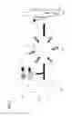
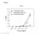
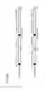
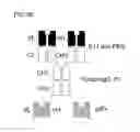
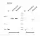
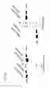
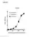
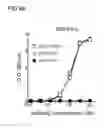
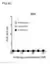
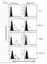
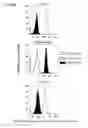
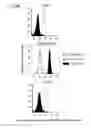
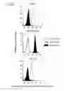
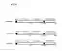
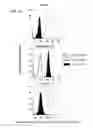
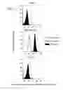
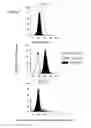
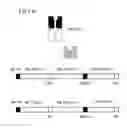
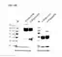
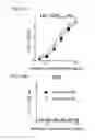
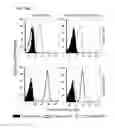
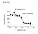
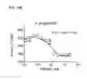
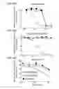
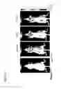
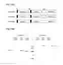
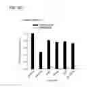
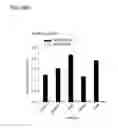
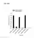
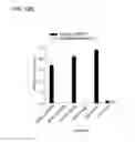

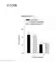
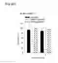
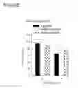
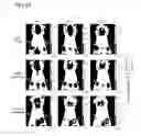
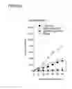
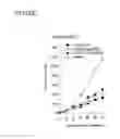
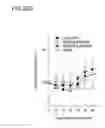
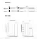

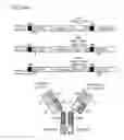
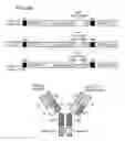
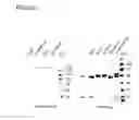
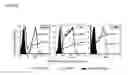
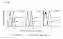
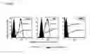
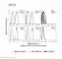
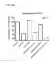
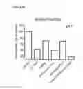
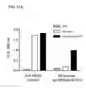
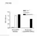
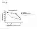
Sources:
- United States Patent and Trademark Office - verify current appl. status at the USPTO↗
Similar patent applications:
- » 20150071927
Anti-HER2 and anti-IGF-IR BI-specific antibodies and uses thereof - » 20190022245
Bi-specific antibodies and uses thereof - » 20200061205
Bi-specific antibodies and uses thereof - » 20210154319
BI-SPECIFIC ANTIBODIES AND USES THEREOF - » 20250074992
BI-SPECIFIC ANTIBODIES AND USE THEREOF - » 20210198370
VHS format bi-specific antibodies specific for HER2 and VEGF and use thereof - » 20200190192
PD-1/TIM-3 BI-SPECIFIC ANTIBODIES, COMPOSITIONS THEREOF, AND METHODS OF MAKING AND USING THE SAME - » 20200079850
PD-1/LAG3 BI-SPECIFIC ANTIBODIES, COMPOSITIONS THEREOF, AND METHODS OF MAKING AND USING THE SAME - » 20180326085
Bi-specific antibodies for enhanced tumor selectivity and inhibition and uses thereof - » 20220111064
BI-SPECIFIC ANTIBODIES FOR ENHANCED TUMOR SELECTIVITY AND INHIBITION AND USES THEREOF
Recent applications in this class:
- » 20170035908 2017-02-09
Detection of Early-Stage Pancreatic Adenocarcinoma - » 20140271687 2014-09-18
Methods of targeting T-cells to tumors
Recent applications for this Assignee:
- » 20250269018 2025-08-28
COMBINED CANCER THERAPY WITH AN EPITHELIAL CELL ADHESION MOLECULE (EPCAM) INHIBITOR AND A HEPATOCYTE GROWTH FACTOR RECEPTOR (HGFR) INHIBITOR - » 20250154561 2025-05-15
Method for detection of single nucleotide variant - » 20250129146 2025-04-24
COMBINED CANCER THERAPY WITH AN EPITHELIAL CELL ADHESION MOLECULE (EPCAM) INHIBITOR AND A WNT INHIBITOR - » 20240354599 2024-10-24
METHOD FOR DETERMINING PROBABILITY OF A KIDNEY STONE IN A SUBJECT BEING A URIC-ACID STONE - » 20240352477 2024-10-24
METHOD FOR PROMOTING GROWTH OF PLANTS - » 20240347203 2024-10-17
Methods and Systems for Predicting Risk of Cardiovascular Disease - » 20240316014 2024-09-26
METHOD FOR TREATING DEMYELINATING CONDITIONS - » 20240299567 2024-09-12
RECOMBINANT POLYPEPTIDES, CONJUGATES COMPRISING THE SAME, AND USES THEREOF - » 20240190866 2024-06-13
POLYHYDROXYLATED INDOLIZIDINE AND PYRROLIZIDINE DERIVATIVES AND USES THEREOF - » 20240174765 2024-05-30
ANTI- ENOLASE 1 (ENO1) ANTIBODY AND APPLICATIONS THEREOF