Injectable Osteogenic Formulations, and Methods of Use Thereof
US20170173046A1
2017-06-22
15/449,924
2017-03-04
Abstract:
Formulations and methods for growing bone in a site-specific location using an osteogenic molecule such as a prostaglandin, and a delivery vehicle which is preferably a polymer matrix.
Inventors:
- Albert G. Prescott 6 🇺🇸 Westford, MA, United States
- Sandy Marks 2 🇺🇸 Westborough, MA, United States
- Paul Ogden 1 🇺🇸 Princeton, MA, United States
Interested in similar patents?
Get notified when new applications in this technology area are published.
Classification:
A61K9/0019 » CPC further
Medicinal preparations characterised by special physical form; Galenical forms characterised by the site of application Injectable compositions; Intramuscular, intravenous, arterial, subcutaneous administration; Compositions to be administered through the skin in an invasive manner
A61K31/5575 » CPC main
Medicinal preparations containing organic active ingredients; Eicosanoids, e.g. leukotrienes or prostaglandins having a cyclopentane, e.g. prostaglandin E, prostaglandin F
A61K9/00 IPC
Medicinal preparations characterised by special physical form
A61K31/728 » CPC further
Medicinal preparations containing organic active ingredients; Carbohydrates; Sugars; Derivatives thereof; Polysaccharides, i.e. having more than five saccharide radicals attached to each other by glycosidic linkages; Derivatives thereof, e.g. ethers, esters; Glycosaminoglycans, i.e. mucopolysaccharides Hyaluronic acid
Description
CROSS REFERENCE TO RELATED APPLICATIONS
This application is a continuation in part of application Ser. No. 14/504,775, filed on Oct. 2, 2014, which is a divisional of application Ser. No. 11/426,097 filed on Jun. 23, 2006, which itself claims priority of Provisional application Ser. No. 60/693,391 filed on Jun. 23, 2005. The disclosures of each of these prior applications are incorporated herein by reference in their entireties.
FIELD
This invention relates to growing bone in a site-specific location.
BACKGROUND
The ability to grow bone at a site-specific location, in a minimally invasive manner, would have a profound impact on the health and quality of life for up to 50 million Americans who suffer bone defects and diseases. These diseases include post craniotomy resorption, periodontal disease, degenerative disk disease, osteoporosis, and aseptic osteolysis. Together, these diseases cost the United States Healthcare System in excess of $124 billion annually.
Cranial
Over 750,000 craniotomies are performed every year in the United States, to treat a variety of disorders including tumors, traumas, vascular lesions, seizures, decompression, cranioplasty, infections, intracranial cysts, and nerve decompression. Despite this, the skull has been one of the most difficult regions in which to use autograft techniques because of the cranium's propensity for resorption. Many materials and methods have been used including autologous bone grafts, metal plates (titanium, tantalum, stainless steel), hydroxyapatite cement, and methylmethacrylate, each with significant drawbacks.
Periodontal Disease.
The most common treatment for periodontal disease and tooth loosening is extraction and dental implantation or dentures. Over 20% of Americans, approximately 56 million people, have periodontal disease. Periodontal disease accounted for 10% of all dental costs in 1985.
Spinal Disorders
Spine disorder treatments are slowly trending toward minimally invasive techniques for penetrating past the muscle tissues surrounding the spine. Despite this, the solutions to degenerative disks (fusion, herniated disk repair and disk replacement) are still highly invasive procedures resulting in extensive surgical trauma and prolonged recovery times. Over 1 million spinal surgeries were performed last year and this number continues to rise.
Joint Replacements
Over ½ million knee and hip replacement surgeries are performed in the United States every year. The typical age of these patients is 65 years despite the fact that the average age of pain symptom onset is 40 years. The reason for this disparity is that the implants cause bone resorption at the interface, making their maximum useful life less than 15 years.
Overall, the ability to induce rapid, localized bone growth would have a substantial beneficial effect in all the above conditions by reducing morbidity, hospitalization time, recovery time, and costs. The ability to grow bone at specific sites would also have a substantial positive impact on the treatment of bone fractures resulting from osteoporosis. It is estimated that over 24 million people suffer from osteoporosis, resulting in 3 million fractures per year in the United States. In 1995 osteoporotic fractures were estimated at $13.8 billion in direct medical expenses.
| Disorder | Annual U.S. Cases | |
| Craniotomies | 750,000 | |
| Periodontal Disease | 28,000,000 | |
| Spinal Surgeries | 1,000,000 | |
| Aseptic Osteolysis | 500,000 | |
| Osteoporosis | 3,000,000 | |
| Total | 33,250,000 | |
Bone Morphogenic Proteins (BMPs) are osteogenic compounds. However, BMPs can cause ectopic ossification, making them somewhat difficult and risky to use.
SUMMARY
The invention comprises formulations and methodologies for treating degenerative bone conditions, bone fractures, and other bone-related conditions by combining an osteogenic compound with a delivery vehicle. The resulting combination may be injected or otherwise applied to (such as by an implant or other device, or by applying it to a site during surgery) a specific site, and cause bone growth at that site.
This invention features an osteogenic formulation comprising an osteogenic compound and a delivery vehicle. The osteogenic compound is preferably prostaglandin (PGE), and may comprise PGE1 and/or PGE2. One reason that prostaglandins are the preferred osteogenic compounds for the invention is that they do not induce bone synthesis at ectopic sites by non-bone cells.
The delivery vehicle may comprise a biodegradable matrix, which preferably comprises one or more polymers. Two preferred polymers are hyaluronic acid or a salt thereof, and/or a poly-glutamic acid.
The invention also features a method of employing an osteogenic compound, comprising providing prostaglandin (PGE) osteogenic compound, providing a biodegradable polymer matrix delivery vehicle for the PGE, mixing the PGE and the delivery vehicle, and delivering the mixture to a site. The PGE may comprise PGE1 and/or PGE2. The polymer preferably comprises hyaluronic acid or a salt thereof, and/or a poly-glutamic acid. The delivery is preferably accomplished with a syringe, but as described above can be accomplished by other know means for site-specific delivery of a treatment vehicle.
BRIEF DESCRIPTION OF THE DRAWINGS
The patent or application file contains at least one drawing executed in color. Copies of this patent or patent application publication with color drawings will be provided by the Office upon request and payment of the necessary fee.
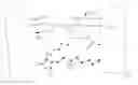
FIG. 1 is a color photograph of an in vivo test of an example of an osteogenic formulation.
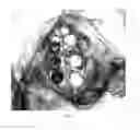
FIG. 2 is a series of color histological examination images of the test example of FIG. 1.
FIG. 3 illustrates a PGE1-Hyaluronic acid conjugate.
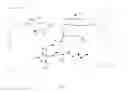
FIG. 4 are radiographs of two models in which four osteogenic formulations were tested.
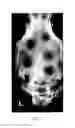
FIG. 5 is an enlargement of the left-most image from FIG. 7.
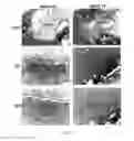
FIG. 6 is a series of three color histological examination images of the model depicted in FIGS. 4 and 5.
FIG. 7 is a series of three color histological examination images of the model depicted on the right side of FIG. 5.
FIG. 8 is a series of three color histological examination images of the left side of the model depicted on the right side of FIG. 4.
DETAILED DESCRIPTION
The following is a description of the preferred embodiments of the invention. The half-life of prostaglandin E1 (PGE1) is measured in minutes. PGE1 is rapidly metabolized in the lungs, therefore the delivery method is critical to widespread commercial use of PGE1 as an osteogenic compound. The invention is designed to stabilize and hold PGE1, which is by far the most potent osteogenic agent discovered to date, and deliver it via a gelatinous polymer matrix of hyaluronic acid, a material that is itself modestly osteogenic. PGE2 is less osteogenic than PGE1 but may still be useful in the invention. The matrix allows the osteogenic compound (preferably a prostaglandin) to make physical contact with the targeted bone surface, thus stimulating only the cells required for the healing process. The matrix releases the compound over a period of time so that the stimulation of the cells is maintained over a period of time until the process of osteogenesis is completed. Both the matrix and the compound are reabsorbed by the body. Because the material may be delivered by a syringe, more than one treatment can be readily accomplished. No surgical procedures are required.
The delivery matrix is a biodegradable polymer. The polymer should be gelatinous in nature and water soluble. This includes polymers such as carboxymethylcellulose, poly-glutamic acid (PGA), but preferably the polymer is hyaluronic acid. Hyaluronic acid is biocompatible and is itself mildly osteogenic.
The following are examples of producing the various preferred foiiiiulations.
Example of One Formulation
PGE1 is dissolved in pure water. In the event that the PGE1 won't completely dissolve, pure ethanol may be added until all the PGE1 is dissolved. The concentration of PGE1 may be varied as necessary.
Hyaluronic acid is hydrated in pure water. Hyaluronic acid concentrations may also be varied, but a concentration of greater than 30 mg/ml in pure water works well and may thus be diluted with the PGE1 solution.
The PGE solution is combined with the hyaluronic acid gel and allowed to mix. Mixing may be achieved in a mixer, a beaker with a stir bar and magnetic stirrer, or even by coupling two syringes together, each containing one of the two solutions. Solutions should be mixed until homogeneous. In addition, salts of hyaluronic acid may be used to vary the release of the PGE1. Sodium hyaluronate, calcium hyaluronate, and even ferric hyaluronate may be used. As the valence of the counter cation increases (i.e., Na+, Ca++, Fe+++ and so forth) the half life of hyaluronate increases, and the release rate of PGE1 decreases.
Following are examples of purified hyaluronic acid and poly-gamma-glutamic acid that may be produced for use as the matrix for the invention.
Example of Hyaluronic Acid Purification
-
- 1. Rooster combs are sliced and placed in ethanol. The ethanol is changed daily until it is no longer cloudy. Three days, 6 liters of ethanol used.
- 2. The ethanol is drained and the combs are placed in water with an antiseptic (Thymol) to prevent microbial growth.
- 3. The combs are mixed at less than 10° C. overnight or until the solution viscosity exceeds 500 cps. Steps 2 and 3 together take 3 days and use no ethanol.
- 4. The combs are strained from the extract. The extract is treated with NaCl to a final concentration of 0.2M.
- 5. The extract is centrifuged and added to 3 volumes of ethanol and the resulting stringy white precipitate is removed and stored under ethanol. Steps 4 and 5 together take 1 day and use 3 liters of ethanol.
- 6. Dissolve precipitate in DI water to approximately 1.5 mg/ml concentration. Though the actual concentration will change later, 0.75 to 5.0 mg/ml HA may be successfully precipitated with ethanol (and NaCl).
- 7. Add 100 ml of chloroform to every 1 liter of solution, mix overnight and centrifuge for 5 minutes at 4,000 RPMs. This step removes residual fats, lipids, certain proteins, and other materials that have been found to inhibit the Pronase® step. Steps 6 and 7 together take 2 days and use no ethanol.
- 8. Add the aqueous portion to a temperature-controlled reactor, add an antiseptic (Thymol), <0.5 mM CaCl2, heat to 37° C., adjust pH to 8.0 and add Pronase®. These are optimum Pronase® conditions per CalBiochem, Pronase® manufacturers.
- 9. Maintain pH at 8.0 via pH control and the addition of 0.2M Tris buffer. Run until no more Tris is required (typically overnight). This hydrolyzes proteins not removed by chloroform, as well as the link proteins responsible for binding HA to other GAGs. Steps 8 and 9 together take 1 day and use no ethanol.
- 10. Make up a solution of 100 mls of 2% CPC and 0.3M NaCl. Adjust the reactor contents to 0.3M, and add the CPC/NaCl solution to the reactor. It will change color from opaque to yellow. Allow it to mix for 15 minutes. Filter the reactor contents through a membrane filter (0.2 micron PES filter) and collect in a flask. This causes DNA, chondriotin sulfate, heparin, and other non-HA GAGs to complex and precipitate. They are subsequently removed by filtration. This step takes one-half day, and no ethanol.
- 11. Using a Pall-Filtron 30 kDa MWCO PES membrane, diafilter the solution against 5 volumes of 0.3M NaCl. This removes amino acids, peptides, Pronase®, CPC and other low MW contaminants.
- 12. Either precipitate with ethanol and dry under vacuum, or lyophilize the contents of the flask. This is the best way to store material until formulation. Formulation strength cannot be achieved through TFD at this time. Steps 11 and 12 together take one-half day and use 3 liters of ethanol.
- 13. Formulate to 10 mg/ml and verify properties against the traditional process. 10 mg/ml is a simple HA concentration that has been used often in the industry. One day, no ethanol.
The following describes the equipment used for diafiltration:
| DF Parameter | Value |
| 1. Three membrane setup (Used to | |
| determine MW cutoff for diafiltration) | |
| Membrane Type | Omega Polyethersulfon |
| Channel Depth | 40 mil |
| Membrane Area | 0.045 ft2/channel |
| Number of Channels | 3 in parallel |
| Trans-Membrane Pressure (TMP) | 18.5 psig |
| Pressure drop across membrane | 2.5 psig |
| Cross Flow Rate | 200 ml/min/channel |
| 2. Single membrane DF experimental setup | |
| (Used to purify HA from rooster comb) | |
| Membrane Type | Omega Polyethersulfon |
| Membrane Area | 1.0 ft2 |
| Trans-Membrane Pressure (TMP) | 8.0 psig |
| Cross Flow Rate | 1,000 ml/min |
| MWCO | 30k |
| Type | Centramate |
| Configuration | Open Channel |
The pump used to perform the diafiltration was a Cole-Paitiier Masterflex® L/S® Precision Standard Tubing Pump capable of over 1700 ml/min, SKU# EW-77911-00.
EXAMPLE 1
PGA Using Preferred Fermentation Method, and Purification to Medical Grade
Bacillus licheniformis ATCC 9945a was grown in Medium E. The fermentation was carried out at small scale, in shake flasks, at 37° C. Aeration was provided by diffusion. When the viscosity stopped rising (typically after about 3-5 days of fermentation), the fermentation broth was buffer exchanged via diafiltration using a filter with a molecular weight cut off (MWCO) of 30 kDa. The mixture of cells and PGA was then buffered in citric acid, and micro-filtered using a filter with an opening of 0.22 microns, to remove the host cells.
The filtrate was neutralized, and buffer exchanged with pure water and concentrated via diafiltration using a filter with a MWCO of 30 kDa. Material from this purification may be sterile filtered.
To describe the process in more detail, when the viscosity stopped rising, the fermentation broth was re-circulated through an Omega Polyethersulfon ultra-filtration cartridge by Pall Corporation with a 0.2 micron pore size. Once collected, the filtrate was re-circulated using an Omega Polyethersulfon ultra-filtration cartridge by Pall Corporation with a 0.16 micron pore size. The filtrate was collected and re-circulated through an Omega Polyethersulfon ultra-filtration cartridge by Pall Corporation with a 30 kda MWCO pore size. Five diafiltration volumes of solution were processed. At the end, the retentate was collected, sterilized by passing through a 0.22 micron filter, and precipitated in sterile ethanol and stored.
Material from this example has been used in rats in subsequent experiments with no inflammatory response. The molecular weight was determined to be 2 million Daltons using the following analytical MALLS method described in the Stock thesis that is incorporated by reference herein. PGA was dissolved at a concentration of 1 mg/ml in 0.1M citric acid, pH 2 to 3, with 0.05% sodium azide. The sample was degassed and 0.2 milliliters was injected at a flow rate of 0.5 mls/min. The SEC can utilize a TossoHaas TSK G5000PWXL, G6000PWXL, Waters Ultrahydrogel 1000 or 250. A Dawn DSP laser photometer from Wyatt technologies in conjunction with a Waters differential refractometer is used for detection.
This process is capable of making high molecular weight (when measured as described) poly-gamma-glutamic acid at purities up to and including pharmaceutical grade.
EXAMPLE 2
PGA from another Commercial Source Purified
A sample reported to be poly-gamma-glutamic acid in excess of 1 million Daltons was received from an offshore commercial supplier. The viscosity of a sample of known concentration seemed to be lower than would be the case if the PGA was indeed of the reported molecular weight. Analysis was impossible due to the large amount of contaminants, as evidenced by the off-white color noted when the sample was hydrated, and the fact that the hydrated sample had an odor similar to fermentation broth.
This material was re-circulated through an Omega Polyethersulfon ultra-filtration cartridge by Pall Corporation with a 0.2 micron pore size. Once collected, the filtrate was re-circulated using an Omega Polyethersulfon ultra-filtration cartridge by Pall Corporation with a 0.16 micron pore size. The filtrate was collected and re-circulated through an Omega Polyethersulfon ultra-filtration cartridge by Pall Corporation with a 30 kda MWCO pore size. Five diafiltration volumes of solution were processed. The resulting material was clear and odorless, supporting the production of low molecular weight, high purity PGA.
Example 3
PGA
Bacillus licheniformis ATCC 9945a was grown in Medium E. The fermentation was carried out at small scale, in shake flasks, at 37 C. Aeration was provided by diffusion. When the viscosity stopped rising, the fermentation broth was buffer exchanged via diafiltration using a filter with a molecular weight cut off (MWCO) of 30 kDa. The mixture of cells and PGA was then buffered in citric acid, and micro-filtered using a filter with an opening of 0.16 microns.
The filtrate was neutralized, and buffer exchanged with pure water and concentrated via diafiltration using a filter with a MWCO of 30 kDa. Material from this purification may be sterile filtered. Material from this example has been used in rats in subsequent experiments with no inflammatory response. The molecular weight was determined to be 2 million Daltons using the method described above in conjunction with example 1.
Example 4
PGA
Bacillus licheniformis ATCC 9945a was grown in Medium E. The feiiiientation was carried out at small scale, in shake flasks, at 37 C. Aeration was provided by diffusion. When the viscosity stopped rising, the pH of the fermentation broth was lowered to 2 by the addition of HCl. The cells were then removed by passing the broth through a 0.22 micron TFF filter and collecting the filtrate. The filtrate was then neutralized, and buffer exchanged with pure water and concentrated via diafiltration using a filter with a MWCO of 30 kDa. Material from this purification may be sterile filtered. Material from this example has been used in rats in subsequent experiments with no inflammatory response. The molecular weight was determined to be 2 million Daltons using the method described above in conjunction with example 1.
Example 5
PGA
Bacillus licheniformis ATCC 9945a was grown in Medium E. The fermentation was carried out at small scale, in shake flasks, at 37 C. Aeration was provided by diffusion. When the viscosity stopped rising, the pH of the fermentation broth was lowered to 2 by the addition of HCl. The cells were then removed by centrifugation at a speed over 10,000×g. The supernatant was then neutralized, and buffer exchanged with pure water and concentrated via diafiltration using a filter with a MWCO of 30 kDa. Material from this purification may be sterile filtered. Material from this example has been used in rats in subsequent experiments with no inflammatory response. The molecular weight was determined to be 2 million Daltons using the method described above in conjunction with example 1.
Several osteogenic formulations were tested in vivo.
In the first set of tests, we formulated three prototypes of the osteogenic formulations (identified as “LEAD”). The formulations were as follows:
| TABLE 1 | ||
| Prototype Designation | Mg PGE1/mL LEAD | mg HA/mL LEAD |
| LEAD20 | 160 | 17.65 |
| LEAD10 | 80 | 17.65 |
| LEAD05 | 40 | 17.65 |
These prototypes were submitted to an external contractor for bioreactivity testing, as well as bone growth and bone resorption. We also analyzed histological slides of some of the treated sites.
The model used was the rabbit cranial bur-hole model. Three Rabbits each had six, 5 mm bur-holes drilled in their skull. One side was left untreated while the other was filled with LEAD, as shown in the illustration. The animals were terminated at 6 weeks and examined.
Methodology
A critical size defect is the diameter of a bone wound such that beyond that amount, complete calcification of the wound will not occur during the lifetime of the animal. The critical size defect, in this case a 5 mm burr hole in a rabbit cranium, is easily reproducible and allows qualitative and quantitative evaluation of bone grafts and bone graft substitutes.
Work Performed
Critical size defects in rabbit skulls were created by using 5 mm drills, and filled with either vehicle control (C2), or treated with an osteogenic formula containing 20 mg/ml PGE (T2) and allowed to heal for 6 weeks.
Results
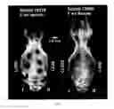
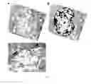
FIG. 1 includes micrographs of one model, with the three images in the left column of the un-treated control (60633 C2) at 1.25 power, 5 power, and 10 power, and the three images in the right column of the same model, but treated with LEAD (60633 T2).
The images in FIG. 1 are annotated with indications of the identification of the different tissues. The legend is as follows:
L=Lipid (Fat)
F=Fibrous
NB=New Bone
OB=Old Bone
V=Blood vessels
The low power micrographs (top row, FIG. 1) show that the bone on the control side (C2) grew such that the 5 mm was reduced only to 4.5 mm, while the bone on the treated side (T2) grew into the defect and reduced its diameter to almost 2.8 mm. In addition, whereas the new bone formed in the control site (C2) is dense and non-vascularized, the bone formed in the treated site (T2) is highly vascularized, and was growing and remodeling even when the study was terminated. See the 5× and 10× images. FIG. 1 as a whole clearly shows the 5 mm defect on the control side having healed to only 4.5 mm, with a dense layer of new bone that lacks any blood vessels needed for further growth. By contrast, the LEAD treated side is 2.8 mm, and the bone has numerous blood vessels, suggesting the bone would have continued to grow and the defect would have continued to heal had the test been carried out longer.
LEAD was found to have no inflammatory response. In addition, examination of bone formation versus bone resorption were examined. The results for this were as follows:
| TABLE 2 | ||||
| LEAD | LEAD | |||
| Control | Control | Formation | Resorption | |
| Prototype | Formation Score | Resorption Score | Score | Score |
| LEAD20 | 2 | 3 | 5 | 3 |
| LEAD10 | 3 | 4.5 | 4.5 | 3.5 |
| LEAD05 | 2.5 | 4 | 3 | 3 |
In all cases, the LEAD treated defects scored higher for bone formation and lower for bone resorption, versus controls.
We then embarked on two new development efforts. One was to chemically link the PGE1 to the polymer, thus creating a new molecule. The second, was to introduce an osteo-conductive bone substitute (Bi-Ostetic™ hydroxyapatite from Berkley Advanced Biomaterials).
In order to link the PGE1 to the HA via the amine groups (which is preferred since it would have minimal impact on material solubility), we used the mixed anhydride method of peptide synthesis (similar to Meienhofer et al., 1972).
- 1) 5 mg (1.4×10̂−5 mol) of PGE, is dissolved in 0.5 ml of dimethylformamide (DMF), then stirred in an ice bath at 0° C.
- 2) Chilled (0° C.) isobutyl chloroformate (2 ul, 1.6×10̂−5 mol) and triethylamine (TEA) (4 ul, 1.6×10̂−5 mol) is added to the above solution.
- 3) After 15 min, 8 uL (3.2×10̂−5 mol) of TEA is added, followed by the addition of 350 mg of hyaluronic acid dissolved in 2 ml of 1: 1 H20/DMF (pH=8.0) solution, and chilled.
- 4) The reaction is maintained at 0° C. for an hour, then allowed to react at room temperature (25 C) for 18 h.
- 5) The PGE-HA conjugate is lyophilized, and re-dissolved in a minimal amount of water.
- 6) The PGE-HA conjugate is then isolated by precipitation with a large excess of acetone.
- 7) The precipitate is washed over a fritted glass funnel with acetone and dried.
In theory, the resulting material has the chemical structure shown in FIG. 3.
A matrix experiment was designed using the same rabbit model. The data is described below, and is accompanied by additional illustrations, FIG. 4, which represents two animals for which data was obtained. The prototypes used, and their compositions, are in table 3, below.
| TABLE 3 | ||||
| Prototype | PGE1 | HA | HA-XL | Solids, Y/N |
| L20R | 160 | 17.64 | 0 | N |
| CL40 | 315.5 | 0 | 320 | N |
| 315.5 as HA, | ||||
| 4.5 as PGE1 | ||||
| CL20S | 157.75 | 0 | 160 | Y |
| 157.75 as HA, | ||||
| 2.25 as PGE1 | ||||
| L40S | 320 | 17.64 | 0 | Y |
As can be seen from the radio-graph in FIG. 5, L20R, which was the original formula tested, performed in a similar manner, healing the defects about halfway. CL40 however, almost healed the defect, as can be seen in the radio-graph.
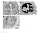
Histological examination of L20R and CL40, in FIGS. 6 and 7, respectively, showed that this was more than just calcification, and that new bone cells were numerous throughout the defects.
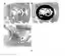
Examination of CL20S and L40S are more difficult due the radio-opacity of the filler material, as seen in FIG. 4. Histological examination in FIG. 8, shows bone cells growing on those solid pieces, but the benefit at the time of examination was inconclusive and more work is needed to understand whether this is a benefit or not.
The LEAD samples referred to above were formulated as follows:
LEAD 20
- 272 mg of PGE1 were dissolved in 0.7 ml of ethanol.
- 30 mg of HA were dissolved in 1 ml water.
- The two were combined and mixed thoroughly. The resulting concentrations of the formulation were:
- 160 mg of PGE1/mL of LEAD
- 17.65 mg HA/mL of LEAD
LEAD 10
- 136 mg of PGE1 were dissolved in 0.7 ml of ethanol.
- 30 mg of HA were dissolved in 1 ml water.
- The two were combined and mixed thoroughly. The resulting concentrations of the formulation were:
- 80 mg of PGE1/mL of LEAD
- 17.65 mg HA/mL of LEAD
LEAD 05
- 68 mg of PGE1 were dissolved in 0.7 ml of ethanol.
- 30 mg of HA were dissolved in 1 ml water.
- The two were combined and mixed thoroughly. The resulting concentrations of the formulation were:
- 40 mg of PGE1/mL of LEAD
- 17.65 mg HA/mL of LEAD
L20R Stands for LEAD 20, Regular and is the same as LEAD 20 above - 272 mg of PGE1 were dissolved in 0.7 ml of ethanol.
- 30 mg of HA were dissolved in 1 ml water.
- The two were combined and mixed thoroughly. The resulting concentrations of the formulation were:
- 160 mg of PGE1/mL of LEAD
- 17.65 mg HA/mL of LEAD
CL40 Stands for Cross-Linked 40, which Utilizes Cross Linked HA and PGE1 with a Final PGE1 Concentration of Double the Original LEAD 20 Formulation. - Cross Linked HA-PGE1 (aka, HA-XL) was created using the method described elsewhere.
- 282 mg of HA-XL (which we estimate to be 275 mg HA and 6.4 mg PGE1) was hydrated with 9.4 mL of water.
- 5100 mg of PGE1 was dissolved in 6.6 mL ethanol.
- Both solutions were combined and mixed thoroughly. The resulting concentrations were:
- 275 mg HA/16 mL=17.2 mg HA/mL LEAD
- (5100+6.4) mg PGE1/16 mL=319.15 mg PGE1/mL LEAD
CL20S Stands for Cross-Linked 20, which Utilizes Cross Linked HA and PGE1 with a Final PGE1 Concentration of the Original LEAD 20 Formulation, with Granular Solids q.s. - Cross Linked HA-PGE1 (aka, HA-XL) was created using the method described elsewhere herein.
- 282 mg of HA-XL (which we estimate to be 275 mg HA and 6.4 mg PGE1) was hydrated with 9.4 mL of water.
- 2550 mg of PGE1 was dissolved in 6.6 mL ethanol.
- Both solutions were combined and mixed thoroughly. The resulting concentrations were:
- 275 mg HA/16 mL=17.2 mg HA/mL LEAD
- (2550+6.4) mg PGE1/16 mL=159.8 mg PGE1/mL LEAD
- This material was then poured over granular hydroxyapatite to fill the void space in the surgical defect.
L20S Stands for LEAD 20, Regular and is the same as LEAD 20 above, with Granular Solids q.s. - 2550 mg of PGE1 were dissolved in 6.6 ml of ethanol.
- 282 mg of HA were dissolved in 9.4 ml water.
- The two were combined and mixed thoroughly. The resulting concentrations of the formulation were:
- 160 mg of PGE1/mL of LEAD
- 17.65 mg HA/mL of LEAD
- This material was then poured over granular hydroxyapatite to fill the void space in the surgical defect.
In order to be acceptable in the human clinic, PGE should be able to be delivered easily, non-invasively, and allow for proper dosing at the surface of the bone. We have achieved this by combining PGE with a hydrogel formulation of biological polymers such as hyaluronic acid (HA), poly-gamma-glutamic acid (PGA), and others. This allows PGE to be injected as needed, increases its half-life such that it becomes efficacious, and physically holds it resident in the location where bone growth is specifically desired.
When HA and/or PGA are used as the biodegradable polymer matrix delivery vehicle, the molecular weight can be selected to achieve desired results. Generally, higher molecular weights may lead to longer residence times, in situ. PGAs that have been used are generally in the range of from about 1.2 million Daltons to about 2.3 million Daltons. HAs that have been used, including in the formulations described above, are generally in the range of about 950,000 Daltons. Most generally, it is thought that the molecular weight of the polymer matrix should be at least about 500,000 Daltons, with molecular weights of at least about 750,000 Daltons preferred.
A number of implementations have been described. Nevertheless, it will be understood that additional modifications may be made without departing from the scope of the inventive concepts described herein, and, accordingly, other embodiments are within the scope of the following claims.
Claims
What is claimed is:1. A method of employing an osteogenic formulation, comprising:
providing prostaglandin El (PGE1);
providing a biodegradable polymer matrix delivery vehicle for the PGE1;
mixing the PGE1 and the delivery vehicle, to create an osteogenic formulation; and
delivering the formulation to a site in mammalian tissue.
2. The method of claim 1, wherein the polymer comprises hyaluronic acid or a salt thereof.
3. The method of claim 1, wherein the polymer comprises a poly-glutamic acid.
4. The method of claim 1, wherein delivery is accomplished with a syringe.
5. The method of claim 1, wherein the site is adjacent to bone.
6. The method of claim 5, wherein the delivery is accomplished with a syringe.
7. An osteogenic formulation, comprising:
prostaglandin El (PGE1); and
a biodegradable polymer matrix delivery vehicle for the PGE1 comprising hyaluronic acid or a salt thereof;
wherein the PGE1 and the delivery vehicle are mixed into an osteogenic formulation, for delivery to a site.
8. The formulation of claim 7, wherein the PGE1 is dissolved in water to create an osteogenic solution that is mixed with the delivery vehicle.
9. The formulation of claim 7, wherein the site is adjacent to bone, and the delivery is accomplished with a syringe.
10. The formulation of claim 7, wherein the mixing creates a homogeneous formulation.
11. The formulation of claim 7, wherein the osteogenic formulation is delivered to a site requiring stimulation of bone growth.
12. The formulation of claim 7, wherein the osteogenic formulation comes into physical contact with tissue in need of osteogenesis in vivo.
13. A composition of matter, comprising cross-linked prostaglandin El (PGE1) and hyaluronic acid.
14. The composition of matter of claim 13, used in an osteogenic formulation.
Images & Drawings included:
Sources:
- United States Patent and Trademark Office - verify current appl. status at the USPTO↗
Recent applications in this class:
- » 20250090549 2025-03-20
COMPOSITIONS AND METHODS FOR MUSCLE REGENERATION USING PROSTAGLANDIN E2 - » 20250090548 2025-03-20
J-SERIES PROSTAGLANDIN-ETHANOLAMIDES AS NOVEL THERAPEUTICS FOR SKIN AND/OR ORAL DISORDERS - » 20250064830 2025-02-27
TRANSDERMAL TREATMENT FOR ERECTILE DYSFUNCTION - » 20240415849 2024-12-19
COMPOSITIONS AND METHODS FOR MUSCLE REGENERATION USING PROSTAGLANDIN E2 - » 20240398830 2024-12-05
COMPOSITIONS AND USES THEREOF FOR TREATMENT OF NEUROLOGICAL DISORDERS - » 20240390390 2024-11-28
NITRIC OXIDE RELEASING PROSTAMIDE AS NEUROPROTECTIVE AGENT - » 20240238309 2024-07-18
PHARMACEUTICAL FORMULATIONS OF TREPROSTINIL PRODRUGS AND METHODS OF USE THEREOF - » 20240207287 2024-06-27
POLYMERIC IMPLANTS - » 20240180925 2024-06-06
STABLE OPHTHALMIC COMPOSITION COMPRISING LATANOPROST - » 20240156833 2024-05-16
Aqueous pharmaceutical compositions of prostaglandins