3-D TISSUE CULTURE BASED METHOD TO ASSESS MITOCHONDRIAL IMPAIRMENT
US20190079079A1
2019-03-14
16/085,334
2017-03-14
Abstract:
The present invention relates to a method and/or assay for the assessment of the metabolic effect of a candidate compound. The method and/or assay comprises exposing one or more 3-dimensional cell culture or tissue to one or more candidate compounds, and measuring, in at least one 3-dimensional cell culture or tissue, the effect of such exposure on the 3-dimensional cell culture or tissue-specific respiration rate (MTSRR) (FIG. 1B).
Interested in similar patents?
Get notified when new applications in this technology area are published.
Classification:
G01N33/5079 » CPC main
Investigating or analysing materials by specific methods not covered by groups -; Biological material, e.g. blood, urine ; Haemocytometers; Chemical analysis of biological material, e.g. blood, urine; Testing involving biospecific ligand binding methods; Immunological testing involving human or animal cells for testing or evaluating the effect of chemical or biological compounds, e.g. drugs, cosmetics involving cell organelles, e.g. Golgi complex, endoplasmic reticulum Mitochondria
C12N5/0667 » CPC further
Undifferentiated human, animal or plant cells, e.g. cell lines; Tissues; Cultivation or maintenance thereof; Culture media therefor; Animal cells or tissues; Human cells or tissues; Vertebrate cells; Cells of skeletal and connective tissues; Mesenchyme; Stem cells Adipose-derived stem cells [ADSC]; Adipose stromal stem cells
C12N5/0634 » CPC further
Undifferentiated human, animal or plant cells, e.g. cell lines; Tissues; Cultivation or maintenance thereof; Culture media therefor; Animal cells or tissues; Human cells or tissues; Vertebrate cells Cells from the blood or the immune system
G01N33/5047 » CPC further
Investigating or analysing materials by specific methods not covered by groups -; Biological material, e.g. blood, urine ; Haemocytometers; Chemical analysis of biological material, e.g. blood, urine; Testing involving biospecific ligand binding methods; Immunological testing involving human or animal cells for testing or evaluating the effect of chemical or biological compounds, e.g. drugs, cosmetics involving specific cell types Cells of the immune system
C12M23/12 » CPC further
Constructional details, e.g. recesses, hinges; Form or structure of the vessel Well or multiwell plates
C12N5/0695 » CPC further
Undifferentiated human, animal or plant cells, e.g. cell lines; Tissues; Cultivation or maintenance thereof; Culture media therefor; Animal cells or tissues; Human cells or tissues; Vertebrate cells; Tumour cells; Cancer cells Stem cells; Progenitor cells; Precursor cells
C12N5/0671 » CPC further
Undifferentiated human, animal or plant cells, e.g. cell lines; Tissues; Cultivation or maintenance thereof; Culture media therefor; Animal cells or tissues; Human cells or tissues; Vertebrate cells; Hepatocytes Three-dimensional culture, tissue culture or organ culture; Encapsulated cells
G01N33/5014 » CPC further
Investigating or analysing materials by specific methods not covered by groups -; Biological material, e.g. blood, urine ; Haemocytometers; Chemical analysis of biological material, e.g. blood, urine; Testing involving biospecific ligand binding methods; Immunological testing involving human or animal cells for testing or evaluating the effect of chemical or biological compounds, e.g. drugs, cosmetics for testing toxicity
C12N5/0656 » CPC further
Undifferentiated human, animal or plant cells, e.g. cell lines; Tissues; Cultivation or maintenance thereof; Culture media therefor; Animal cells or tissues; Human cells or tissues; Vertebrate cells; Cells of skeletal and connective tissues; Mesenchyme Adult fibroblasts
G01N33/50 IPC
Investigating or analysing materials by specific methods not covered by groups -; Biological material, e.g. blood, urine ; Haemocytometers Chemical analysis of biological material, e.g. blood, urine; Testing involving biospecific ligand binding methods; Immunological testing
Description
FIELD OF THE INVENTION
The present invention relates to a method and/or assay for the assessment of the metabolic effect of a candidate compound. The method and/or assay comprises exposing one or more 3-dimensional cell culture or tissue to a candidate compound.
Mitochondria play a critical role in generating most of the cell's energy as ATP. They are also involved in other metabolic processes such as urea generation, haem synthesis and fatty acid beta-oxidation. Disruption of mitochondrial function by drugs can result in cell death by necrosis or can signal cell death by apoptosis (e.g., following cytochrome c release).
Drugs that injure mitochondria usually do so by inhibiting respiratory complexes of the electron chain; inhibiting or uncoupling oxidative phosphorylation; inducing mitochondrial oxidative stress; or inhibiting DNA replication, transcription or translation.
It is important to test for mitochondrial toxicity early in drug development as impairment of mitochondrial function can induce various pathological conditions that are life threatening or can increase the progression of existing mitochondrial diseases.
Mitochondrial impairment has been reported to be involved, causative or at least related to different types of diseases, including Parkinson's disease, Alzheimer's disease, cardiac diseases, cancer, as well as cholesterol and lipid disorders.
Mitochondrial toxicity assays have been developed for this reason. Some of them, like the Mitochondrial ToxGlo™ Assay provided by Promega, comprise a cell-based assay method that uses a suspension of individual cells. The assay is based on the differential measurement of biomarkers associated with changes in cell membrane integrity and cellular ATP levels relative to vehicle-treated control cells during short exposure periods. Cell membrane integrity is first assessed by measuring the presence or absence of a distinct protease activity associated with necrosis. Next, ATP is measured after cell lysis. The two sets of data can be combined to produce profiles representative of mitochondrial dysfunction or non-mitochondrial related cytotoxic mechanisms.
Other Mitochondrial toxicity assays use 2d cell cultures, where cells are for example seeded in collagen I-coated microplates and adhered for 18-24 h before compound additions. For primary screening and secondary dose-response experiments, compounds at specific concentrations added to plated cells, and incubated for 24 h at 37° C., 5% CO2. Then, cell markers are added to carry out imaging cytometry analyses, which result in a measure of mitochondrial toxicity (Tsiper et al, 2012).
It is one object of the present invention to improve the methods from the prior art. It is another object of the present invention to increase the predictability of methods from the prior art. It is still another object of the present invention to further develop methods from the prior at in that they allow the investigation of the effects of long term exposure of cells to a candidate compound. It is still another object of the present invention to increase the sensitivity of the methods from the prior art in that they provide a more sensitive and precise prediction of mitochondrial toxicity of candidate compounds. It is still another object of the present invention to allow detection of mitochondrial impairment at toxin concentrations which do not yet affect cell viability, and cannot be detected in cell viability/cytotoxicity assays or mitochondrial toxicity assays according to the prior art.
Embodiments of the Invention
These and further objects are met with methods and means according to the independent claims of the present invention. The dependent claims are related to specific embodiments.
SUMMARY OF THE INVENTION
Before the invention is described in detail, it is to be understood that this invention is not limited to the particular component parts of the devices described or process steps of the methods described as such devices and methods may vary. It is also to be understood that the terminology used herein is for purposes of describing particular embodiments only, and is not intended to be limiting. It must be noted that, as used in the specification and the appended claims, the singular forms “a,” “an” and “the” include singular and/or plural referents unless the context clearly dictates otherwise. It is moreover to be understood that, in case parameter ranges are given which are delimited by numeric values, the ranges are deemed to include these limitation values.
According to one embodiment of the invention, a method and/or assay for the assessment of the metabolic effect of a candidate compound is provided, which comprises
-
- a) exposing one or more 3-dimensional cell culture or tissue to one or more candidate compounds, and
- b) measuring, in at least one 3-dimensional cell culture or tissue, the effect of such exposure on the 3-dimensional cell culture or tissue-specific respiration rate (MTSRR).
The terms “3-dimensional cell culture or tissue” and “3D microtissue” are used interchangeably herein. Surprisingly, the inventors have shown that with this assay, mitochondrial impairment can be detected at toxin concentrations which do not yet affect cell viability, and cannot be detected in cell viability/cytotoxicity assays or mitochondrial toxicity assays according to the prior art.
The inventors attribute this finding to the fact that a 3-dimensional cell culture or tissue is a more faithful reproduction of a natural tissue. In other words: it behaves more physiologically than, e.g., a 2D cell culture or a cell suspension. The 3-dimensional cell culture or tissue recapitulates the smallest functional unit of a tissue or organ, providing a morphological and functional analog to native tissue. They thus bridge the gap between in vitro and in vivo toxicity testing models. Without being bound to theory, the inventors explain this phenomenon with different effects. First, in a 3-dimensional cell culture or tissue, an interstitium and an extracellular can be built up. Second, in a 3-dimensional cell culture or tissue, cells can develop 3-dimensional cell-cell interactions. Third, in a 3-dimensional cell culture or tissue, drugs to be tested have to enter the tissue by diffusion from the outside, thus affecting cells in the outer cell mass more than cells in the inner cell mass. All these phenomena do not exist in 2D cell cultures and/or cell suspensions.
In one embodiment of the invention, the method and/or assay further comprises
-
- c) measuring, in the same or at least one other 3-dimensional cell culture or tissue, the effect of such exposure on cell viability and/or the cytotoxic effect (CV-CYT) in one or more cells of at least one 3-dimensional cell culture or tissue, and
- d) comparing the effects of steps b) and c) to provide an estimate on the mitochondrial toxicity of the one or more candidate compounds.
Steps a) and b), steps a) and c) and steps b) and c) can be carried out synchronously. In another embodiment, step a) can be carried out before steps b) and c), which can be carried out synchronously or subsequently.
As discussed already, mitochondrial toxicity assays from the prior art use either cell suspensions or 2D cell cultures. The inventors have unexpectedly shown that 3D microtissues exhibit a spare respiratory capacity which is more representative to the in vivo situation, i.e., to real tissues. Therefore, the use of 3D microtissues provides a more sensitive and precise prediction of mitochondrial toxicity of candidate compounds.
Generally, in drug discovery, preclinical testing, and clinical trials is estimated to cost between $500 million and $1.2 billion USD. Chances are that a drug will fail at any stage of development are enormous. Safety-related issues remain the primary reason for drug failure, largely because predicting which drugs will prove toxic in patients is extremely difficult. The standard assays discussed above suffer from limited throughput, loss of cell viability, and decrease in measurable tissue-specific functionality. In vivo animal tests are expensive, raise moral and ethical issues, and often fail to reveal important signs of toxicity in humans. In one further embodiment of the invention, said method and/or assay is thus used to determine the mitochondrial toxicity of a candidate compound. In this embodiment, the candidate compound is for example a candidate pharmaceutic, where potential toxic effects or other side effects are determined in a toxicity screening process. This embodiment also covers other compounds, like herbicides, fungicides or insecticides.
In one further embodiment of the invention, said method and/or assay is used to determine the chemotherapeutic efficacy of a candidate compound. In this embodiment, the candidate compound is a candidate chemotherapeutic, where the measured mitochondrial toxicity is a measure for potential chemotherapeutic efficacy.
In one further embodiment of the invention, the 3-dimensional cell culture or tissue is a spheroidal cell culture.
As discussed above, 3-dimensional cell culture or tissue have a significant advantage in the present context because they are a more faithful reproduction of a natural tissue, and behave more physiologically than, e.g., a 2D cell culture or a cell suspension. Further, they have a longer lifetime than 2D cell cultures or cell suspensions, which facilitates handling and allows for the testing of long term effects and off-target effects of candidate compounds. This is especially useful in screening of compounds that may later be used for long term treatments, or to which humans are exposed over long stretches of time.
In one further embodiment of the invention, said method and/or assay the 3-dimensional cell culture or tissue comprises primary cells. Compared to tumor cells or immortalized cells, primary cells have not accumulated mutations, and is thus more representative, or predictive, for the behavior of in vivo tissue. The transfer of primary cells into 3-dimensional cell cultures or tissues has two significant advantages: a) it further increases the physiologic similarity of the cells to in vivo tissues, and thus makes them even more representative, or predictive, and b) it increases the longevity of the primary cells compared to 2D cell cultures or cell suspensions.
Preferably, the 3-dimensional cell culture or tissue is produced by means of a hanging drop microtiter plate, as distributed by InspheroAG of Schlieren, Switzerland, under the brand “GravityPLUS™ Hanging Drop System”, and disclosed in international patent application WO2010031194A1. This approach allows the generation of 3-dimensional cell cultures or tissues in more complex 3D cell culture scenarios, such as when using primary cells, cell lines that are resistant to self-aggregation, or when generating co-culture microtissues.
In another embodiment, the 3-dimensional cell culture or tissue is produced in a ultra-low attachment (ULA) microtiter plate, as distributed by InspheroAG of Schlieren, Switzerland, under the brand “GravityTRAP™ Ultra-Low Attachment (ULA) Plate”.
In one further embodiment of the invention, said method and/or assay the 3-dimensional cell culture or tissue comprises hepatocytes. Hepatocytes in primary culture provide the closest in vitro model to human liver, which is the organ most effected by drug toxicity, and the only model that can produce a metabolic profile of a given drug that is similar to that found in vivo. Hepatocytes in primary culture express typical hepatic functions and express drug metabolizing enzymes. These advantages are particularly present in 3-dimensional cell cultures or tissues, which behave even more physiological, as the inventors have surprisingly shown, and do therefore more faithfully reflect the response of “true” liver tissue on drug exposure.
In other embodiments, the cells can be cardiac cells, neuronal cells, pancreatic islets, fibroblasts and keratinocytes. 3-dimensional cell cultures or tissues made from these cell types have specific applications in drug toxicity or efficacy screening.
In one further embodiment of the invention, the 3-dimensional cell culture or tissue comprises immortalized, malignant, cancerous and/or neoplastic cells.
In this embodiment, the effect of prior exposure on the microtissue-specific respiration rate (MTSRR) of the said cells can be used to investigate a potential chemotherapeutic efficacy of the tested molecule on cancer cells.
Further, cancer cells inherently alter their cellular machinery to change how they consume and utilize glucose. Inhibiting cancer specific key steps in the cellular respiration may disrupt cancer cell proliferation and survival without affecting normal cells.
In one further embodiment of the invention, the method and/or assay further comprises a step of determining, in the same or at least one other 3-dimensional cell culture or tissue, the effect of the exposure on the size thereof. This step can be carried out synchronously or subsequently with steps a) b) and/or c).
Preferably, the size determination refers to at least one parameter selected from the group consisting of:
-
- Diameter
- perimeter
- volume
- area of an optical cross section
The size can thus either be a parameter that has directly been measured, or a parameter which has been calculated on the basis of such measurements.
Preferably, the size determination of the 3-dimensional cell culture or tissue is carried out by means of an imaging device.
One example of such imaging device is the Cell3iMager distributed by, InSphero AG, Schlieren, CH, and manufactured by SCREEN, Japan. It allows analysis of spheroids by scanning multi-well plates in a bright-field. It computes estimated values based on spheroid size and density, together with spheroid number and area in each well. With easy and efficient operability, its vibration-free design protects cells from damage. An excellent application is also available to determine spheroid proliferation over time and to measure the granular distribution in 3D culture.
In another embodiment, the 3-dimensional cell culture or tissue further comprises at least one cell type selected from the group consisting of:
-
- Immune cells
- Stroma cells
- Fibroblasts
- Cancer stem cells
- Immortalized, malignant, cancerous and/or neoplastic cells that are known to be resistant or sensitive to a given anti-cancer agent,
Using immune cells in such 3-dimensional cell culture or tissue has a particular benefit because effects of the innate immune system can also be investigated. This is particularly useful in case an anti-cancer agent is investigated which co-acts with the immune system i.e., an immunotherapeutic agent, like an immune checkpoint inhibitor (see infra). These drugs activate the immune system to target cancer cells. Therefore, to test their efficacy in vitro, and to investigate potential relapses, these agents require the presence of immune cells.
Examples of suitable immune cells that can be used for such co-culture encompass, inter alia, tissue resident leukocytes, like macrophages (e.g., stellate macrophages/kupffer cells), CD8+ T cells, natural killer cells, natural killer T cells, and/or regulatory T cells.
Using connective tissue components, like stroma cells or fibroblasts, may help recreating the important tumor microenvironment in vitro, and thus increase the quality of the 3-dimensional cell culture or tissue, and this improve the predictive value of such culture or tissue for assays or screening purposes.
Cancer stem cells (CSCs) are cancer cells that possess characteristics associated with normal stem cells. Cancer stem cells sometimes share a feature with normal somatic stem cells in that they are naturally resistant to chemotherapeutic agents, because they because they express different transporter proteins, which actively export drugs. CSC are thus often able to survive first line cancer treatments and thus causing tumor relapse.
Co-culturing immortalized, malignant, cancerous and/or neoplastic cells with cancer stem cells can thus help to develop therapies which affect both the primary tumor and, at the same time, inhibit cancer stem cells.
The same rationale applies to the co-culture of a given immortalized, malignant, cancerous and/or neoplastic cell type with another immortalized, malignant, cancerous and/or neoplastic cell type that is known to be resistant or sensitive to a given anti-cancer agent.
In one embodiment, the microtissue-specific respiration rate is determined by simultaneous measurement of oxygen consumption rate (OCR) and extracellular acidification rate (ECAR), of one or more isolated 3D microtissues.
OCR is an indicator of mitochondrial respiration, and ECAR is largely the result of glycolysis. Such measurement can take place, e.g., in a Seahorse XF Analyzer, while other suitable devices exist on the market and can likewise be used. The Seahorse XF Analyzer is capable of performing real-time measurements of OCR and ECAR in extremely small volumes (about 2 μL) of medium above, usually, a monolayer of cells within a microplate well. Cellular oxygen consumption (respiration) and proton excretion (glycolysis) cause rapid, easily measurable changes to the concentrations of dissolved oxygen and free protons in this “transient microchamber” which are measured every few seconds by solid state sensor probes residing 200 microns above the cell monolayer. The instrument measures the concentrations for 2-5 minutes, and then calculates the OCR, and/or ECAR, respectively, e.g., in units of pmol/minute (OCR) and mpH/minute (ECAR). Total assay time is typically 60 to 90 minutes.
The inventors have for the first time shown that in this method, unexpectedly, 3D microtissues can be used instead of the cell monolayers that are disclosed in the secondary literature as well as in the product manual. Because of the microliter volumes that are provided by the wells, a spheroidal microtissue in such well provides a completely different microenvironment than a cell monolayer on the bottom thereof. Therefore, it was not foreseeable that a transfer of the respective method to 3D microtissues would be feasible and provide meaningful and reproducible results.
In one embodiment, in the determination of the microtissue-specific respiration rate (MTSRR), the spare respiratory capacity (“SPARE”) is used.
Spare Respiratory Capacity indicates the capability of the cell to respond to an energetic demand as well as how closely the cell is to respiring to its theoretical maximum. Spare Respiratory Capacity is defined as the difference between maximal respiration and basal respiration of a tissue and/or cell.
In order to determine spare respiratory capacity, one approach is to first measure the basal respiration rate, and then measure the maximal respiration rate. The arithmetic difference (“delta”) between the two is the spare respiratory capacity.
The basal respiration rate shows the energetic demand of the tissue or cell under baseline conditions, while the maximal oxygen consumption reflects the maximum rate of respiration a tissue or cell can achieve.
The basal respiration rate can be determined by measurement of oxygen consumption rate (OCR) in the first step of the measurement. The maximal respiration rate can be determined by measurement of oxygen consumption rate (OCR) in tissues or cells treated with an uncoupling agent, like carbonyl cyanide-4 (trifluoromethoxy) phenylhydrazone (FCCP). Such uncoupling agent mimic a physiological energy demand by stimulating the respiratory chain to operate at maximum capacity, by collapsing the proton gradient and disrupting the mitochondrial membrane potential. As a result, electron flow through the electron transport chain is uninhibited and oxygen is maximally consumed by complex IV.
According to further embodiments, in the determination of the microtissue-specific respiration rate (MTSRR), the basal respiration rate (“BASAL”) and/or the maximal respiration rate (“MAX”) are also used.
Primarily, the spare respiratory capacity (SPARE) is used in the determination of the microtissue-specific respiration rate (MTSRR), and basal respiration rate (BASAL) and maximal respiration rate (MAX) are used in case of inaccuracies or higher sensitivity compared to SPARE.
In one embodiment, the measurement of cell viability/cytotoxic effect determines the residual viability of one or more cells.
Cell viability/cytotoxic effect can for example be determined by quantitation of the ATP present in the cells. One such assay is the CellTiter-Glo Luminescent Cell Viability Assay (Promega, Madison, US), while other cell viability/cytotoxicity assays exist on the market and can likewise be used.
In another embodiment, the effect on microtissue-specific respiration rate and/or the effect on cell viability/cytotoxic effect is determined as inhibitory concentration, preferably as half maximal inhibitory concentration (IC50).
Said inhibitory concentration refers to the concentration of the candidate compound, to which the 3-dimensional cell culture or tissue is exposed in step a), as compared to an untreated 3-dimensional cell culture or tissue.
The variable used herein for IC50 with respect to microtissue-specific respiration rate is IC5O-MTSRR. As discussed above, MTSRR is preferably determined by determining spare respiratory capacity (SPARE). In such case, IC5O_MTSRR would be equal to IC5O_SPARE.
The variable used herein for IC50 with respect to cell viability/cytotoxicity is IC5O_CV/CYT.
In one further embodiment exposure step a) is carried out prior to subsequent measurement steps b) and/or c).
This embodiment allows exposure times of different lengths. In such way, short term effects and long term effects can be investigated. This embodiment has a surprising synergism with the claimed use of 3-dimensional cell cultures or tissues, which, as discussed above, have better longevity as compared to 2D cell cultures or cell suspensions, and are thus suitable to long-term exposure to candidate compounds, like drug candidates.
In contrast thereto, the use instructions of the Seahorse XF Analyzers provide that the test drugs are dispensed to the cells during the assay. For this purpose, the device provides an assay cartridge that accommodates up to four drugs for automated injection during the assay. The assay cartridge is first placed into the analyzer to allow automatic calibration of optical sensors. Next, the cell culture plate is inserted into the instrument. In such setting, however, no long term exposure tests can be carried out.
In yet another embodiment, exposure step a) is carried out in one or more first vessels, while the subsequent measurement steps b) and/or c) are carried out in one or more second and/or third vessels.
Exposure is carried out, preferably, in vessels of a first array (e.g., wells of a first microtiter plate), while the subsequent measurement is carried out in vessels of a second array (e.g., wells of a 2nd microtiter plate).
In still another embodiment, after exposure step a) the microtissues are transferred, by means of a suitable dispensing device, from the one or more first vessels to the one or more second and/or third vessels, to carry out measurement steps b) and/or c)
Said dispensing device is, e.g., a multichannel pipette with pipette tips that are either non-protein binding or pre-coated with a protein, to avoid that microtissues stick or adhere to the inner or outer surface of the pipette tip.
In another embodiment, the mitochondrial toxicity is determined on the basis of the quantitative relationship between
-
- a) the IC50 with respect to microtissue-specific respiration rate (IC5O_MTSRR)
- b) the IC50 with respect to cell viability/cytotoxicity (IC5O_CV/CYT).
Preferably,
-
- a) IC5O_MTSRR≥IC5O_CV/CYT means that the candidate compound has no specific mitochondrial toxicity
- b) IC5O_MTSRR<IC5O_CV/CYT and IC5O_MTSRR≥0.75×IC5O_CV/CYT means that the candidate compound has a low risk of mitochondrial toxicity
- c) IC5O_MTSRR<0.75×IC5O_CV/CYT means that the candidate compound has a high risk of mitochondrial toxicity
Experiments and Figures
While the invention has been illustrated and described in detail in the drawings and foregoing description, such illustration and description are to be considered illustrative or exemplary and not restrictive; the invention is not limited to the disclosed embodiments. Other variations to the disclosed embodiments can be understood and effected by those skilled in the art in practicing the claimed invention, from a study of the drawings, the disclosure, and the appended claims. In the claims, the word “comprising” does not exclude other elements or steps, and the indefinite article “a” or “an” does not exclude a plurality. The mere fact that certain measures are recited in mutually different dependent claims does not indicate that a combination of these measures cannot be used to advantage. Any reference signs in the claims should not be construed as limiting the scope.
EXAMPLE 1
Generation of 3D Microtissue
1.1. Thawing of Cryopreserved Hepatocytes
-
- Take chosen cell-vial from cryo-tank and transfer into water bath (37° C.); set timer on 2 minutes
- Pipette 40 ml of the Wash/Thawing medium into 50 ml tube
- Transfer the hepatocytes into the tube, wash with 1 ml medium
- Place 50 ml tube into centrifuge; start centrifugation for 5 min at 50 rcf (corresponds to 600 rpm) at RT.
- Remove supernatant
- Wash pellet with 20 ml wash buffer
- Place carefully 3 ml of Wash/Thawing medium into the 50 ml tube
- Re-suspend cell pellet with 2D cell culture medium
- Count hepatocytes with e.g. Trypan Blue
1.2. Hepatocyte Pre-Plating
-
- Use a collagen coated cell-culture dish for pre-seeding
- Seed Hepatocytes in a 6 cm dish: 100000-250000 hepatocytes/cm2; 0.05-0.5ml per cm2
- Place at 37° C. in a CO2 containing incubator
- Optionally: Exchange medium after attachment with Serum-free 2D-culture medium
- Harvest hepatocytes after 12-96 hours (daily medium exchange if longer than 12 hours)
- 3× wash with pre-warmed phosphate buffered saline the non-adherent hepatocytes away
- Add cell detachment solution such as collagenase/accutase mixtures to culture dish to dislodge the hepatocytes from the well 50-500 ul/cm2
- Incubate 5-30 min at 37° C. (visual observation until most cells are detached from the surface)
- Carefully transfer dispersed hepatocytes into a 50 ml tube prefilled with wash buffer
- Centrifuge for 5 min with 50 rcf (corresponds to 600 rpm) at RT
- Aspirate supernatant, add 1-50 ml wash medium to the pellet and re-suspend pellet by gentle pivoting of the tube
- Repeat centrifugation step for 5 min with 50 rcf (corresponds to 600 rpm) at RT
- Add 1 ml re-aggregation medium to the pellet and dissolve hepatocytes by gentle pivoting of the tube
- Count hepatocytes
1.3. Production of Microtissues
A. Preparation of GravityPLUS™ Plates (Insphero AG, CH)
-
- Fill Omnitray with 15 ml of 0.75× PBS/Amphothericin and add humidifier pad
- Place frame on Omnitray
- Put lid on the plate and label plate
B. Preparation of Cell Suspension
-
- Prepare cell suspension of defined cell number (i.e. 25′000 Heps/25′000 NPCs per ml) in falcon tubes
- Homogenize cell suspension by pivoting the tube
C. Seeding
-
- Use Viaflo electronic multichannel pipette (Integra Bioscience)
- Set Viaflo parameters: Repeat dispense: 40 ul, Speed 3, Repeat: 1× (for 96-well Viaflo)
- Empty cell suspension in reservoir (don't pour more than 30 ml in the reservoir, to ensure proper distribution of cells)
- Remove lid from GravityPLUS plates
- Prior Aspiration of cell suspension, gently pivot reservoir for homogeneous distribution of cells
- Start Viaflo program: Repeat dispense
- Place Viaflo horizontally on the inserts, avoid tapping of the drop on the top by gentle pressure of the Viaflo on the inserts
- Place lid back on plates
D. Transfer of Microtissues
-
- Pre-wet the GravityTRAP plate: Add 30-50 μl medium or PBS to the very bottom of each well and remove by aspiration or alternatively by flicking out (use a soak matt)
- Place the GravityPLUS plate right next to the receiver bottom plate. Take both, lid and frame together off the reservoir, and carefully latch the pins of the frame (still with the lid on) into all 3 notches of the receiver plate
- Apply 70 μl of medium which will release the hanging drop with the microtissue inside
- The “fallen” drop has an approx. volume of 60-70 μl
- Close the receiver plate with a lid and centrifuge the plate at 250× RCF for 30 sec-1 min. This will allow to get rid of trapped air bubbles and the microtissue to center at the bottom of the well
- Check transfer efficiency under the microscope
- Medium Replenishment: aspirate the medium by placing the tips right at the ledge of the GravityTRAP plate and remove the medium
- Refill wells of GravityTRAP plate with 50-80 μl medium
1.4. Media Used
| Wash | 2D cell | Re-aggregation | |
| Ingredient | buffer | culture medium | medium |
| Williams E Medium (GE | X | X | X |
| Healthcare) | |||
| Insulin-Transferrin- | X | X | X |
| Selenium (Gibco) | |||
| FBS (Fetal Calf Serum) | 20% | 10% | 20% |
| (Lonza) | |||
| HGF (Hepatocyte Growth | 20 μg/ml | ||
| Factor) (Peprotech) | |||
| 2 mM Glutamine, | X | X | X |
| Penicillin/Streptomycin, | |||
| Amphothericin, 0.1 μM | |||
| Dexamethasone | |||
EXAMPLE 2
Measuring Mitochondrial Toxicity with the Seahorse XF Analyzer
2.1 Treatment of 3D Microtissues
After generating 3D primary human hepatocytes microtissues (IPHH_02) of a diameter size of 370 um, the microtissues have been incubated with varying concentration of compounds for 48 h Thereby, the compounds were dissolved in 3D InSight™ Human Liver Maintenance Medium-TOX
2.2 Seahorse XF Analyzer Measurement
A. Seahorse Assay Medium Preparation
| Stock conc. | Assay conc. | Volume | ||
| Component | (mM) | (mM) | (mL) | |
| DMEM XF Base | — | — | 50 | |
| Glucose | 1000 | 10 | 0.5 | |
| Na-Pyruvate | 100 | 1 | 0.5 | |
| UltraGlutamine | 200 | 2 | 0.5 | |
| Adjust pH to 7.4 | ||||
B. Coating of Seahorse Spheroid plates
-
- 20 uL of Poly-D-Lysine (100 ug/mL diluted in cell culture grade water) were added to each well of the Seahorse Spheroid plate (Incubation for 20min @ RT)
- After coating, the Seahorse Spheroid plate was washed with 200 uL of sterile water and wash was removed (Washing step repeated once)
- Seahorse Spheroid plate has been air dried for a few minutes
C. Loading of Seahorse Spheroid Plate with Microtissues
-
- Warm up the prepared Seahorse assay medium (see above) to 37° C. and fill up the plate with 70 uL
- Use the Seahorse Capture Screen Insert Tool to accurately position the microtissues in the plate
- Flush the pipette tips several times with FCS before use
- After FCS-coating soak up the microtissues from the GravityTRAP plate in a volume of 20 uL
- Transfer the pipette tips into the holes of the Seahorse Capture Screen Insert tool
- Leave the tips in the tool for about 1 min to ensure complete spheroid transfer
- The spheroids are transferred to the wells by gravitation, so they fall out of the tips automatically and you do not have to use the trigger for that
- After completed microtissue transfer fill up the wells with pre-warmed Seahorse assay medium to a final volume of 180 uL
- Scan the plates in the Insphero Cell3iMager to localize microtissues on the plate before the Seahorse run
- Incubate microtissues for 1 h @ 37° C. w/o CO2
D. Seahorse XF Analyzer Run
-
- While cell incubation fill up the ports of the cartridge according to the Seahorse manufacturer's protocol
- Setup groups and parameters for the measurement (basal: 7 cycles, oligomycin 2 uM: 6 cycles, FCCP 0.5 uM: 3 cycles, FCCP 1 uM: 3 cycles, Rot/AA 0.5 uM: 9 cycles)
- Start the measurement
E. Cell Viability Assessment
-
- In parallel to the Seahorse XF Analyzer run, cell viability has been measured of different microtissues treated the same way as for the Seahorse experiment
E. Analysis and Prediction of Safety for Amiodarone as an Example
-
- By generating a Sigmoidal dose-response curve (variable slope) of all Respiratory
Spare Capacity values of each Amiodarone concentration, we were able to obtain an IC50_MTSRR of 25.3 μM
-
- By knowing that CMax of Amiodarone is 5.3 μM and dividing the IC50_MTSRR by CMax, we are able to generate a Margin of safety (MOS) of 4.8
- As this MOS of Amiodarone (4.8) is below 30, we have predicted Amiodarone to be a toxic compound, which correlates with the DILI classification (Gustaffson et al. 2013)
- Furthermore, we can identify the Mitochondrial toxicity risk range by considering the following scheme:
- IC5O_MTSRR≥IC5O_CV/CYT means that the candidate compound has no specific mitochondrial toxicity
- IC5O_MTSRR≤IC5O_CV/CYT and IC5O_MTSRR≥0.75×IC5O_CV/CYT means that the candidate compound has a low risk of mitochondrial toxicity
- IC5O_MTSRR<0.75×IC5O_CV/CYT means that the candidate compound has a high risk of mitochondrial toxicity
- Amiodarone's IC5O_MTSRR is smaller than 0.75×IC5O_CV/CYT which classifies Amiodarone as a compound with a high Mitotoxic risk
FIGURES
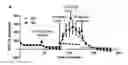
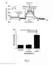
FIG. 1: Mitochondrial Bioenergetics 2D vs. 3D. A) OCR profile and B) Maximal Respiration. **** p<0.0001 unpaired t-test, two-tailed, Means+/−s.e.m.
FIG. 2: Outline of a general workflow. The chronologic order of the different steps is just a preferred embodiment, meaning that the treatment of the 3D microtissues with the molecules to be screened can also take place synchronously with the subsequent assays on viability (ATP content) and respiration rate. Further and not depicted in the general workflow, the steps can be accompanied by a step of size determination, e.g., by digital image analysis. It is also important to mention that in some embodiments, the assays (viability, respiration rate and size) can be made with the same 3D microtissue, while in other embodiments different 3D microtissues are being used which have however been pretreated in identical fashion.
FIG. 3: Analysis example for 3D primary human hepatocyte microtissues exposed to different candidate compounds. DILI classification: Drug-induced liver injury according to Gustafsson et al, 2014 (N=negative, P=positive). CMax=concentration of candidate compound used in Gustafsson et al, 2014; CTop=concentration of candidate compound used herein; IC50CV/Cyt determined by the CellTiter-Glo Luminescent Cell Viability Assay (Promega, Madison, US) as disclosed herein, IC50MTSRR determined as inhibition on spare respiratory capacity (SPARE), as disclosed herein; MOS: Margin of safety; High Mito-toxic risk: IC5O_MTSRR<0.75×IC5O_CV/CYT; Low Mito-toxic risk: IC5O_MTSRR<IC5O_CV/CYT and IC5O_MTSRR≥0.75×IC5O_CV/CYT; No Mito-toxic risk: IC5O_MTSRR≥IC5O_CV/CYT
Asteriks(*) show candidate drugs with respect to which the instant assay method delivers a different and more precise result with respect to mitochondrial toxicity than the general DILI classification according to Gustafsson et al., 2014. Fialuridine, which was categorized as liver toxic by Gustafsson et al. turned out to not have specific mitochondrial toxicity, while Metformin HCl, which was categorized as not liver toxic by Gustafsson et al., turned out to have a low mitochondrial toxicity.
REFERENCES
Tsiper M V et al. (2012); PloS one vol. 7 (10) p. e45226
Gustafsson et al, (2013); Tox Sciences 137(1), 189-211
Claims
1. Method and/or assay for the assessment of the metabolic effect of a candidate compound, which method and/or assay comprises
a) exposing one or more 3-dimensional cell culture or tissue to one or more candidate compounds, and
b) measuring, in at least one 3-dimensional cell culture or tissue, the effect of such exposure on the 3-dimensional cell culture or tissue-specific respiration rate (MTSRR),
c) measuring, in the same or at least one other 3-dimensional cell culture or tissue, the effect of such exposure on cell viability and/or the cytotoxic effect (CV-CYT) in one or more cells of at least one 3-dimensional cell culture or tissue, and
d) comparing the effects of steps b) and c) to provide an estimate on the mitochondrial toxicity of the one or more candidate compounds,
wherein the mitochondrial toxicity is determined on the basis of the quantitative relationship between the IC50 with respect to MTSRR (IC5O_MTSRR) and the IC50 with respect to CV-CYT (IC5O_CV/CYT).
2. (canceled)
3. The method and/or assay according to claim 1, wherein said method and/or assay is used to determine the mitochondrial toxicity of a candidate compound.
4. The method and/or assay according to claim 1, wherein said method and/or assay is used to determine the chemotherapeutic efficacy of a candidate compound.
5. The method and/or assay of claim 1, wherein the 3-dimensional cell culture or tissue is a spheroidal cell culture.
6. The method and/or assay of any of claim 1, wherein the 3-dimensional cell culture or tissue comprises primary cells.
7. The method and/or assay of claim 1, wherein the 3-dimensional cell culture or tissue comprises hepatocytes.
8. The method and/or assay of claim 1, wherein the 3-dimensional cell culture or tissue comprises immortalized, malignant, cancerous and/or neoplastic cells.
9. The method and/or assay according to claim 1, which method and/or assay further comprises a step of determining, in the same or at least one other 3-dimensional cell culture or tissue, the effect of the exposure on the size thereof.
10. The method and/or assay according to claim 9, in which method and/or assay the size determination refers to at least one parameter selected from the group consisting of:
Diameter
perimeter
volume
area of an optical cross section.
11. The method and/or assay according to claim 8, wherein the 3-dimensional cell culture or tissue further comprises at least one cell type selected from the group consisting of:
Immune cells
Stroma cells
Fibroblasts
Cancer stem cells
Immortalized, malignant, cancerous and/or neoplastic cells that are known to be resistant or sensitive to a given anti-cancer agent.
12. The method and/or assay according to claim 1, wherein the microtissue-specific respiration rate is determined by simultaneous measurement of oxygen consumption rate (OCR) and extracellular acidification rate (ECAR), of one or more isolated 3D microtissues
13. The method and/or assay according to claim 12, wherein in the determination of the microtissue-specific respiration rate (MTSRR), the spare respiratory capacity (“SPARE”) is used.
14. The method and/or assay according to claim 12, wherein in the determination of the microtissue-specific respiration rate (MTSRR), the basal respiration rate (“BASAL”) and/or the maximal respiration rate (“MAX”) are also used.
15. The method and/or assay according to claim 1, wherein the measurement of cell viability/cytotoxic effect determines the residual viability of one or more cells.
16. The method and/or assay according to claim 1, wherein the effect on microtissue-specific respiration rate and/or the effect on cell viability/cytotoxic effect is determined as inhibitory concentration, preferably as half maximal inhibitory concentration (IC50).
17. The method and/or assay according to claim 1, wherein exposure step a) is carried prior to subsequent measurement steps b) and/or c).
18. The method and/or assay according to claim 1, wherein exposure step a) is carried out in one or more first vessels, while the subsequent measurement steps b) and/or c) are carried out in one or more second and/or third vessels.
19. The method and/or assay according to claim 18, wherein after exposure step a) the microtissues are transferred, by means of a suitable dispensing device, from the one or more first vessels to the one or more second and/or third vessels, to carry out measurement steps b) and/or c)
20. (canceled)
21. The method and/or assay according to claim 1, wherein
IC5O-MTSRR≥IC5O-CV/CYT means that the candidate compound has no specific mitochondrial toxicity,
wherein IC5O-MTSRR<IC5O-CV/CYT and IC5O-MTSRR≥0.75×IC5O-CV/CYT means that the candidate compound has a low risk of mitochondrial toxicity, and
wherein IC5O-MTSRR<0.75×IC5O-CV/CYT means that the candidate compound has a high risk of mitochondrial toxicity
Images & Drawings included:


Sources:
- United States Patent and Trademark Office - verify current appl. status at the USPTO↗
Recent applications in this class:
- » 20240426811 2024-12-26
SCREENING FOR PHARMACEUTICAL COMPOUNDS FOR IMPROVING MITOCHONDRIAL FUNCTION - » 20240295545 2024-09-05
GLIOMA THERAPY - » 20220299500 2022-09-22
NDUFS2 SUBUNIT KNOCKOUT HUMAN CELL LINE - » 20210018493 2021-01-21
Methods of BH3 profiling - » 20200271640 2020-08-27
METHOD FOR SCREENING COMPOUNDS THAT MODULATE THE ACTIVITY OF THE ELECTRON TRANSPORT CHAIN - » 20200003762 2020-01-02
Diagnostics Platform for Mitochondrial Dysfunctions/Diseases - » 20180120300 2018-05-03
Mitochondrial apoptotic sensor - » 20180080926 2018-03-22
Peptide regulators of mitochondrial fusion and methods of use - » 20170292945 2017-10-12
Method for screening activator of mitochondrial activity - » 20160282337 2016-09-29
Methods, compositions and kits for assaying mitochondrial function