Marchand Salpingectomy—A laparoscopic surgical technique
US20190110787A1
2019-04-18
15/786,602
2017-10-18
✅ Patent granted
US 10,695,042 B2
2020-06-30
-
-
Anu Ramana
2038-03-22
Abstract:
The Marchand Salpingectomy is a fast, safe and minimally invasive procedure for removal of the fallopian tubes. The procedure involves minimal blood loss and gives the patient the benefit of permanent sterility as well as a decreased lifetime incidence of ovarian cancer. The procedure relies on two novel aspects of the technique which make the surgery significantly different than any surgery previously described as well as extremely minimally invasive.
Inventors:
- Greg J. Marchand 1 🇺🇸 Tempe, AZ, United States
- Greg J. Marchand 1 🇺🇸 Mesa, AZ, United States
Assignee:
- Marchand Institute for Minimally Invasive Surgery 3 🇺🇸 Mesa, AZ, United States
Applicant:
Interested in similar patents?
Get notified when new applications in this technology area are published.
Classification:
A61B18/1482 » CPC further
Surgical instruments, devices or methods for transferring non-mechanical forms of energy to or from the body by heating by passing a current through the tissue to be heated, e.g. high-frequency current; Probes or electrodes therefor having a long rigid shaft for accessing the inner body transcutaneously in minimal invasive surgery, e.g. laparoscopy
A61B17/3421 » CPC further
Surgical instruments, devices or methods, e.g. tourniquets; Trocars; Puncturing needles; Details of tips or shafts, e.g. grooves, expandable, bendable; Multiple coaxial sliding cannulas, e.g. for dilating Cannulas
A61B18/00 IPC
Surgical instruments, devices or methods for transferring non-mechanical forms of energy to or from the body
A61B17/3417 » CPC further
Surgical instruments, devices or methods, e.g. tourniquets; Trocars; Puncturing needles Details of tips or shafts, e.g. grooves, expandable, bendable; Multiple coaxial sliding cannulas, e.g. for dilating
A61B17/29 » CPC further
Surgical instruments, devices or methods, e.g. tourniquets; Surgical forceps Forceps for use in minimally invasive surgery
A61B2017/4233 » CPC further
Surgical instruments, devices or methods, e.g. tourniquets; Gynaecological or obstetrical instruments or methods Operations on Fallopian tubes, e.g. sterilization
A61B2018/00601 » CPC further
Surgical instruments, devices or methods for transferring non-mechanical forms of energy to or from the body for achieving a particular surgical effect Cutting
A61B17/02 IPC
Surgical instruments, devices or methods, e.g. tourniquets for holding wounds open; Tractors
A61B17/34 IPC
Surgical instruments, devices or methods, e.g. tourniquets Trocars; Puncturing needles
A61B17/0218 » CPC main
Surgical instruments, devices or methods, e.g. tourniquets for holding wounds open; Tractors for minimally invasive surgery
A61B17/42 » CPC further
Surgical instruments, devices or methods, e.g. tourniquets Gynaecological or obstetrical instruments or methods
A61B18/14 » CPC further
Surgical instruments, devices or methods for transferring non-mechanical forms of energy to or from the body by heating by passing a current through the tissue to be heated, e.g. high-frequency current Probes or electrodes therefor
Description
TECHNICAL FIELD
The present invented technique relates to Surgery, and in particular Surgery related to Gynecology and Laparoscopy. It is related to Surgery to remove the fallopian tubes.
BACKGROUND
Current surgical techniques exist to remove the fallopian tubes. This technique, secondary to unique, previously undescribed characteristics, is new.
SUMMARY OF THE TECHNIQUE
Known laparoscopic techniques include removal of the fallopian tubes using small holes. This technique uses an 11 mm and 5 mm laparoscopic trochar port in order to remove the fallopian tubes in a very fast and cosmetic manner with minimally blood loss.
The technique begins the patient prepped, draped and under general anesthesia as is common for laparoscopic techniques. Next, the procedure continues with placing a small incision and then an 11 mm trochar at the bottom of the umbilicus, into the abdominal cavity, and then placing a second incision of approximately 5 mm approximately 3 cm above the public symphesis in the mid-line, below the pubic hairline. The skin edge is pulled up approximately 3 more cm while placing the abdominal trochar. This gives the unique advantage of a trochar site higher on the abdomen without the disadvantage of a scar. Because the original incision was below the pubic hairline, the incision will ultimately return to this position following the surgery.
Next, a blunt bipolar laparoscopic device using bipolar energy is utilized in order to divide each fallopian tube from their origin at the uterus. The dissection is carried out the entire length of each fallopian through the broad ligament. The incision plane is kept as medial in the abdominal cavity as possible in order to avoid any possibility of damage to lateral structures. This is repeated on both sides until both fallopian tubes are free in the abdominal cavity.
The next important and unique aspect of the technique is also the removal of the fallopian tubes from by plunging the fallopian tubes through the 11 mm trochar port using a 5 mm grasper being utilized through the 5 mm port. Each fallopian tube is removed in this manner.
Following this, 30 cc of marcaine is injected into the abdominal cavity to help with postoperative pain, and the fascia for the 11 mm incision is closed with o vicryl. The skin for both incisions is closed with glue and covered with band-aids. The surgery is then considered complete.
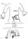
BRIEF DESCRIPTION OF DRAWINGS
FIG. 1: Drawing of action of pulling on skin edge to facilitate a higher entry into the abdominal cavity despite a lower incision below the public hairline.
FIG. 2: Drawing of the dissection of the fallopian tubes using a 5 mm bipolar device.
FIG. 3: Drawing of the removal of the fallopian tubes by using one port to plunge the tube through the other larger port.
Claims
1. This technique includes the unique aspect of the high placement of the 5 mm port which resides through an incision that is below the pubic hairline.
2. This technique includes the unique aspect of removing the fallopian tubes by plunging each tube individually through the 11 mm port using the 5 mm port and a 5 mm blunt grasper.
3. This technique represents a new surgical process that is unique and has the potential to decrease operative time while increasing patient safety.
Images & Drawings included:


Sources:
- United States Patent and Trademark Office - verify current appl. status at the USPTO↗
Recent applications in this class:
- » 20250261933 2025-08-21
Methods and Systems to Identify Actual Esophageal Tissue Changes During Cardiac Ablation - » 20250255596 2025-08-14
CATHETER TREATMENT SEGMENT - » 20250228543 2025-07-17
TRANSMISSION ASSEMBLY - » 20250228542 2025-07-17
MULTIPURPOSE MEDICAL DEVICE - » 20250221697 2025-07-10
DEVICES, SYSTEMS, AND METHODS FOR TISSUE TRACTION - » 20250221696 2025-07-10
Retraction System for Maxillofacial Surgery - » 20250213239 2025-07-03
DETACHABLE MULTI-RING TRACTION DEVICE - » 20250186032 2025-06-12
SURGICAL SMOKE FILTERING DEVICE - » 20250176955 2025-06-05
TISSUE OR ORGAN POSITION SYSTEM FOR INTRA-BODY USE IN A PATIENT AND METHODS OF POSITIONING TISSUES OR ORGANS WITHIN THE BODY OF A PATIENT - » 20250160885 2025-05-22
ACCESS TOOL FOR DELIVERING CARDIAC THERAPIES TO THE PERICARDIAL SPACE
Recent applications for this Assignee:
- » 20210186598 2021-06-24
Marchand advanced single port hysterectomy—a laparoscopic surgical technique - » 20190201032 2019-07-04
Non-morcellating minimally invasive surgical tissue removal system