Method for blood plasma protein activity preservation
US20200085747A1
2020-03-19
16/472,111
2016-12-21
✅ Patent granted
US 11,872,315 B2
2024-01-16
WO; PCT/CN2016/111228; 20161221
WO; WO2018/112780; 20180628
Nghi V Nguyen
Birch, Stewart, Kolasch & Birch, LLP
2038-08-17
Abstract:
A method for blood plasma protein activity preservation is provided. The method comprises the steps of mixing blood plasma with two or more protectants selected from the group consisting of triglyceride, glycerol, propylene glycol, alanine, serine, glycine, alginate, and sucrose to obtain a mixture; and lyophilizing the mixture.
Inventors:
- Cheng-Yao SU 3 🇹🇼 Taipei, Taiwan
- Cheng-Yao SU 5 🇹🇼 Taipei City, Taiwan
- Chung Chin SUN 3 🇹🇼 Taipei City, Taiwan
- Shan Shue WANG 2 🇹🇼 Taipei City, Taiwan
- Chung-Chin Sun 2 🇹🇼 Taipei, Taiwan
- Shan Shue Wang 1 🇹🇼 Taipei, Taiwan
Assignee:
- Chung Chin SUN 2 🇹🇼 Taipei City, Taiwan
- SUN, CHUNG CHIN 1 🇹🇼 Taipei, Taiwan
Applicant:
Interested in similar patents?
Get notified when new applications in this technology area are published.
Classification:
A61K35/16 » CPC further
Medicinal preparations containing materials or reaction products thereof with undetermined constitution; Materials from mammals; Compositions comprising non-specified tissues or cells; Compositions comprising non-embryonic stem cells; Genetically modified cells; Blood; Artificial blood Blood plasma; Blood serum
A61K9/19 » CPC main
Medicinal preparations characterised by special physical form; Particulate form, e.g. powders, Processes for size reducing of pure drugs or the resulting products, Pure drug nanoparticles lyophilised, i.e. freeze-dried, solutions or dispersions
Description
FIELD OF THE INVENTION
The present invention relates to a method for blood plasma protein activity preservation.
BACKGROUND OF THE INVENTION
Blood plasma is the pale yellow colored liquid component of blood that normally holds the blood cells in whole blood in suspension; this makes plasma the extracellular matrix of blood cells. It is mostly water and contains dissolved proteins (e.g., serum albumins, globulins, and fibrinogen), glucose, clotting factors, electrolytes, hormones, vitamins and carbon dioxide. Plasma plays a vital role in an intravascular osmotic effect that keeps electrolytes in balanced form and protects the body from infection and other blood disorders.
Blood plasma can be prepared by spinning a tube of fresh blood containing an anticoagulant in a centrifuge until the blood cells fall to the bottom of the tube. The use of blood plasma as a substitute for whole blood and for transfusion purposes was proposed in 1918. A dried plasma package for the armed forces was developed as it would reduce breakage and make the transportation, packaging, and storage much simpler. Serum albumin replaced dried plasma for combat use during the Korean War.
Plasma serves a variety of functions, from maintaining an appropriate blood pressure and volume to supplying critical proteins for blood clotting and immunity. It also serves as the medium for exchange of vital minerals such as sodium and potassium and helps to maintain a proper pH (acid-base) balance in the body, which is critical to cell function. Plasma as a blood product prepared from blood donations is used in blood transfusions, typically as Fresh Frozen Plasma (FFP) or Plasma Frozen within 24 hours after Phlebotomy (PF24). Plasma is frozen quickly after donation (up to 24 hours) to preserve clotting factors, stored up to one year, and thawed shortly before use. It is now usually used with thrombin in cases of excessive bleeding or to prevent bleeding in those patients with abnormal coagulation tests that are undergoing an invasive procedure. It is commonly transfused to trauma patients and patients with liver failure, severe infections, serious burns, or multiple clotting factor deficiencies. The use of FFP in hospital practice has risen by over 20% in the past few years and concern has been raised about the appropriateness of its clinical use.
The state of the art of plasma storage is to frozen plasma quickly within 24 hours after phlebotomy and store it typically as Fresh Frozen Plasma (FFP) up to one year. The FFP may be thawed shortly before use. However, such storage method has at least the following drawbacks: (1) plasma may not be stored for a long term unless it is frozen as FFP and stored in an ultra-low temperature refrigerator; and (2) the fibrinogen activity cannot be well maintained after thawing for use.
BRIEF SUMMARY OF THE INVENTION
The present invention provides a method for blood plasma protein activity preservation, comprising mixing blood plasma with two or more protectants selected from the group consisting of triglyceride, glycerol, propylene glycol, alanine, serine, glycine, alginate, and sucrose to obtain a mixture, and lyophilizing the mixture.
In certain embodiments of the present invention, the two or more protectants comprise a first protectant of glycerol, and a second protectant selected from the group consisting of triglyceride, alanine and serine.
In certain embodiments of the present invention, the two or more protectants comprise a first protectant of propylene glycol, and a second protectant selected from the group consisting of triglyceride, glycine, alginate and sucrose.
In certain embodiments of the present invention, the two or more protectants comprise glycerol, triglyceride and propylene glycol.
In certain embodiments of the present invention, the two or more protectants comprise glycerol, triglyceride and sucrose.
According to the present invention, the blood plasma may be further mixed, in the mixing step, with a protectant selected from the group consisting of dextran, albumin and gelatin.
It is to be understood that both the foregoing general description and the following detailed description are exemplary and explanatory only and are not restrictive of the invention.
BRIEF DESCRIPTION OF THE DRAWINGS
The foregoing summary, as well as the following detailed description of the invention, will be better understood when read in conjunction with the appended drawing. In the drawings:
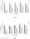
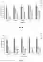
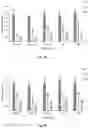
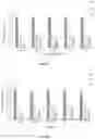
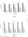
FIGS. 1A-1C show the growth factor levels in the plasma reconstituted from the plasma powder using albumin as a protectant. FIG. 1A shows the levels of PDGF-AB, FIG. 1B shows the levels of TGF-β1, and FIG. 1C shows the levels of VEGF. Amount of protectant used based on the volume of the plasma: % means % (w/v). Control: plasma only. The difference between data shown in the same style of bar (plasma reconstituted after the same period (1 hour, 1 month, or 6 months) of time after lyophilization) with different letters is statistically significant (P<0.05).
FIGS. 2A-2C show the growth factor levels in the plasma reconstituted from the plasma powder using alginate as a protectant. FIG. 2A shows the levels of PDGF-AB, FIG. 2B shows the levels of TGF-β1, and FIG. 2C shows the levels of VEGF. Amount of protectant used based on the volume of the plasma: % means % (w/v). Control: plasma only. The difference between data shown in the same style of bar (plasma reconstituted after the same period of time (1 hour, 1 month, or 6 months) after lyophilization) with different letters is statistically significant (P<0.05).
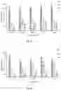
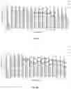
FIGS. 3A-3C show the growth factor levels in the plasma reconstituted from the plasma powder using dextran as a protectant. FIG. 3A shows the levels of PDGF-AB, FIG. 3B shows the levels of TGF-β1, and FIG. 3C shows the levels of VEGF. Amount of protectant used based on the volume of the plasma: % means % (w/v). Control: plasma only. The difference between data shown in the same style of bar (plasma reconstituted after the same period of time (1 hour, 1 month, or 6 months) after lyophilization) with different letters is statistically significant (P<0.05).
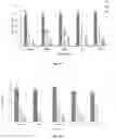
FIGS. 4A-4C show the growth factor levels in the plasma reconstituted from the plasma powder using gelatin as a protectant. FIG. 4A shows the levels of PDGF-AB, FIG. 4B shows the levels of TGF-β1, and FIG. 4C shows the levels of VEGF. Amount of protectant used based on the volume of the plasma: % means % (w/v). Control: plasma only. The difference between data shown in the same style of bar (plasma reconstituted after the same period of time (1 hour, 1 month, or 6 months) after lyophilization) with different letters is statistically significant (P<0.05).
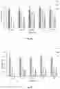
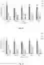
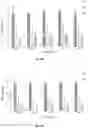
FIGS. 5A-5C show the growth factor levels in the plasma reconstituted from the plasma powder using glucose as a protectant. FIG. 5A shows the levels of PDGF-AB, FIG. 5B shows the levels of TGF-β1, and FIG. 5C shows the levels of VEGF. Amount of protectant used based on the volume of the plasma: % means % (w/v). Control: plasma only. The difference between data shown in the same style of bar (plasma reconstituted after the same period of time (1 hour, 1 month, or 6 months) after lyophilization) with different letters is statistically significant (P<0.05).
FIGS. 6A-6C show the growth factor levels in the plasma reconstituted from the plasma powder using glutamic acid as a protectant. FIG. 6A shows the levels of PDGF-AB, FIG. 6B shows the levels of TGF-β1, and FIG. 6C shows the levels of VEGF. Amount of protectant used based on the volume of the plasma: % means % (w/v). Control: plasma only. The difference between data shown in the same style of bar (plasma reconstituted after the same period of time (1 hour, 1 month, or 6 months) after lyophilization) with different letters is statistically significant (P<0.05).
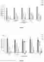
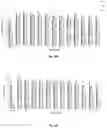
FIGS. 7A-7C show the growth factor levels in the plasma reconstituted from the plasma powder using glycerol as a protectant. FIG. 7A shows the levels of PDGF-AB, FIG. 7B shows the levels of TGF-β1, and FIG. 7C shows the levels of VEGF. Amount of protectant used based on the volume of the plasma: % means % (v/v). Control: plasma only. The difference between data shown in the same style of bar (plasma reconstituted after the same period of time (1 hour, 1 month, or 6 months) after lyophilization) with different letters is statistically significant (P<0.05).
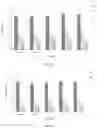
FIGS. 8A-8C show the growth factor levels in the plasma reconstituted from the plasma powder using glycine as a protectant. FIG. 8A shows the levels of PDGF-AB, FIG. 8B shows the levels of TGF-β1, and FIG. 8C shows the levels of VEGF. Amount of protectant used based on the volume of the plasma: % means % (w/v). Control: plasma only. The difference between data shown in the same style of bar (plasma reconstituted after the same period of time (1 hour, 1 month, or 6 months) after lyophilization) with different letters is statistically significant (P<0.05).
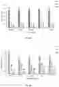
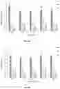
FIGS. 9A-9C show the growth factor levels in the plasma reconstituted from the plasma powder using propylene glycol as a protectant. FIG. 9A shows the levels of PDGF-AB, FIG. 9B shows the levels of TGF-β1, and FIG. 9C shows the levels of VEGF. Amount of protectant used based on the volume of the plasma: % means % (v/v). Control: plasma only. The difference between data shown in the same style of bar (plasma reconstituted after the same period of time (1 hour, 1 month, or 6 months) after lyophilization) with different letters is statistically significant (P<0.05).
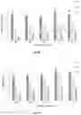
FIGS. 10A-10C show the growth factor levels in the plasma reconstituted from the plasma powder using sucrose as a protectant. FIG. 10A shows the levels of PDGF-AB, FIG. 10B shows the levels of TGF-β1, and FIG. 10C shows the levels of VEGF. Amount of protectant used based on the volume of the plasma: % means % (w/v). Control: plasma only. The difference between data shown in the same style of bar (plasma reconstituted after the same period of time (1 hour, 1 month, or 6 months) after lyophilization) with different letters is statistically significant (P<0.05).
FIGS. 11A-11C show the growth factor levels in the plasma reconstituted from the plasma powder using trehalose as a protectant. FIG. 11A shows the levels of PDGF-AB, FIG. 11B shows the levels of TGF-β1, and FIG. 11C shows the levels of VEGF. Amount of protectant used based on the volume of the plasma: % means % (w/v). Control: plasma only. The difference between data shown in the same style of bar (plasma reconstituted after the same period of time (1 hour, 1 month, or 6 months) after lyophilization) with different letters is statistically significant (P<0.05).
FIGS. 12A-12C show the growth factor levels in the plasma reconstituted from the plasma powder using triglyceride as a protectant. FIG. 12A shows the levels of PDGF-AB, FIG. 12B shows the levels of TGF-β1, and FIG. 12C shows the levels of VEGF. Amount of protectant used based on the volume of the plasma: % means % (v/v). Control: plasma only. The difference between data shown in the same style of bar (plasma reconstituted after the same period of time (1 hour, 1 month, or 6 months) after lyophilization) with different letters is statistically significant (P<0.05).
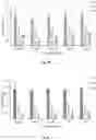
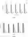
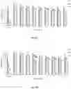
FIGS. 13A-13C show the growth factor levels in the plasma reconstituted from the plasma powder using two (2) protectants. FIG. 13A shows the levels of PDGF-AB, FIG. 13B shows the levels of TGF-β1, and FIG. 13C shows the levels of VEGF. Amount of protectant used based on the volume of the plasma: % means % (v/v) for triglyceride, glycerol and propylene glycol, and means % (w/v) for other protectants. Control: plasma only. 1: 2% glycerol+2% Dextran; 2: 0.1% Triglyceride+2% Dextran; 3: 0.16% glycerol+3% Albumin; 4: 0.1% Triglyceride+2% Propylene glycol; 5: 0.1% Triglyceride+2% glycerol; 6: 0.8% Glycine+1.6% Dextran; 7: 1.6% Glycine+1.6% Propylene glycol; 8: 0.8% Propylene glycol+4% Alginate; 9: 0.4% Propylene glycol+2.4% Albumin; 10: 1.6% Dextran+1.0% Propylene glycol; 11: 1% glycerol+2% Alanine; 12: 0.6% glycerol+1.2% Serine; 13: 0.08% Propylene glycol+2% Sucrose; 14: 0.08% Dextran+3.2% Albumin; and 15: 0.8% Glycine+1% Trehalose. The difference between data shown in the same style of bar (plasma reconstituted after the same period of time (1 hour, 1 month, or 6 months) after lyophilization) with different letters is statistically significant (P<0.05).
FIGS. 14A-14C show the growth factor levels in the plasma reconstituted from the plasma powder using three (3) protectants. FIG. 14A shows the levels of PDGF-AB, FIG. 14B shows the levels of TGF-β1, and FIG. 14C shows the levels of VEGF. Amount of protectant used based on the volume of the plasma: % means % (v/v) for triglyceride, glycerol and propylene glycol, and means % (w/v) for other protectants. Control: plasma only. 1: 4% Glutamic acid+0.4% Propylene glycol+0.04% Dextran; 2: 0.4% Triglyceride+4% glycerol+0.8% Dextran; 3: 0.1% Triglyceride+4% glycerol+0.3% Sucrose; 4: 1% Triglyceride+1.6% glycerol+0.8% Propylene glycol; 5: 0.4% Glutamic acid+0.4% Albumin+4% Gelatin; 6: 1% Glutamic acid+4% Albumin+0.8% Dextran; 7: 0.01% Triglyceride+4% Dextran+0.8% Albumin; 8: 0.04% Triglyceride+0.08% glycerol+4% Albumin; 9: 4% Triglyceride+0.4% glycerol+0.8% Glycine; 10: 3% Trehalose+4% Alginate+0.8% Dextran; 11: 0.1% Glutamic acid+1% Trehalose+4% Dextran; 12: 3% Glutamic acid+3% Trehalose+3% Alginate. The difference between data shown in the same style of bar (plasma reconstituted after the same period of time (1 hour, 1 month, or 6 months) after lyophilization) with different letters is statistically significant (P<0.05).
FIGS. 15A-15C show the growth factor levels in the plasma reconstituted from the plasma powder using four (4) protectants. FIG. 15A shows the levels of PDGF-AB, FIG. 15B shows the levels of TGF-β1, and FIG. 15C shows the levels of VEGF. Amount of protectant used based on the volume of the plasma: % means % (v/v) for triglyceride, glycerol and propylene glycol, and means % (w/v) for other protectants. Control: plasma only. 1: 0.4% Albumin+0.8% Polyethylene glycol+4% glycerol+0.4% Glycine; 2: 1% Polyethylene glycol+0.4% Gelatin+0.4% Glutamic acid+0.4% Glucose; 3: 4% Triglyceride+0.4% Albumin+0.4% Dextran+0.4% glycerol; 4: 0.04% Triglyceride+0.4% Albumin+0.4% Polyethylene glycol+0.4% Glucose; 5: 0.01% Albumin+2% Dextran+0.4% serine+4% sucrose; 6: 0.04% Triglyceride+4% Albumin+0.4% Glycine+0.4% Trehalose; 7: 4% Albumin+0.4% Polyethylene glycol+0.4% Alanine+4% Trehalose; 8: 0.4% Triglyceride+4% Gelatin+0.4% Alginate+0.4% Glycine; 9: 2% Gelatin+2% Alginate+0.4% Glycine+0.4% Trehalose. The difference between data shown in the same style of bar (plasma reconstituted after the same period of time (1 hour, 1 month, or 6 months) after lyophilization) with different letters is statistically significant (P<0.05).
DETAILED DESCRIPTION OF THE INVENTION
The present invention provides a method for blood plasma protein activity preservation, comprising mixing blood plasma with two or more protectants selected from the group consisting of triglyceride, glycerol, propylene glycol, alanine, serine, glycine, alginate, and sucrose to obtain a mixture, and lyophilizing the mixture.
According to the present invention, the protectants may be added in the following amounts: (1) 0.01-10% (v/v) triglyceride based on the volume of the plasma, preferably 0.05-2% (v/v); (2) 0.01%-10% (v/v) glycerol based on the volume of the plasma, preferably 0.5-5% (v/v); (3) 0.01%-10% (v/v) propylene glycol based on the volume of the plasma, preferably 0.5-5% (v/v); (4) 0.01%-10% (w/v) alanine based on the volume of the plasma, preferably 0.5-5% (w/v); (5) 0.01%-10% (w/v) serine based on the volume of the plasma, preferably 0.05-5% (w/v); (6) 0.01%-10% (w/v) glycine based on the volume of the plasma, preferably 0.5-5% (w/v); (7) 0.01%-10% (w/v) alginate based on the volume of the plasma, preferably 0.5-10% (w/v); (8) 0.01%-10% (w/v) sucrose based on the volume of the plasma, preferably 0.5%-5% (w/v).
In certain embodiments of the present invention, the two or more protectants comprise a first protectant of glycerol, and a second protectant selected from the group consisting of triglyceride, alanine and serine. In some embodiments, the two or more protectants are glycerol and triglyceride. In some embodiments, the two or more protectants are glycerol and alanine. In some embodiments, the two or more protectants are glycerol and serine.
In certain embodiments of the present invention, the two or more protectants comprise a first protectant of propylene glycol, and a second protectant selected from the group consisting of triglyceride, glycine, alginate and sucrose. In some embodiments, the two or more protectants are propylene glycol and triglyceride. In some embodiments, the two or more protectants are propylene glycol and glycine. In some embodiments, the two or more protectants are propylene glycol and alginate. In some embodiments, the two or more protectants are propylene glycol and sucrose.
In certain embodiments of the present invention, the two or more protectants comprise glycerol, triglyceride and propylene glycol. In one embodiment, the two or more protectants are glycerol, triglyceride and propylene glycol. For example, the following amounts of protectants may be added (based on the volume of plasma): about 1% (v/v) triglyceride, about 1.6% (v/v) glycerol, and about 0.8% (v/v) propylene glycol.
In certain embodiments of the present invention, the two or more protectants comprise glycerol, triglyceride and sucrose. In one embodiment, the two or more protectants are glycerol, triglyceride and sucrose. For example, the following amounts of protectants may be added (based on the volume of plasma): about 0.1% (v/v) triglyceride, about 4% (v/v) glycerol, and about 2% (w/v) sucrose.
According to the present invention, the blood plasma may be further mixed, in the mixing step, with a protectant selected from the group consisting of dextran, albumin and gelatin.
The present invention is further illustrated by the following examples, which are provided for the purpose of demonstration rather than limitation.
Example 1: Blood Plasma Isolation
Whole blood were collected from volunteer donors must be performed by personal trained in phlebotomy/venipuncture using a double blood bag system (about 50 ml) (TerumoBCT, Japan) with anticoagulant (1 ml of Anticoagulant Citrate Dextrose (ACD) Solution Formula/per 10 ml of blood). After blood collection, gently mix the blood by inverting the tube several times to ensure thorough mixing with anticoagulant. For thorough mixing of blood collected into citrate tubes, it is recommended to invert the tube 3-4 times, while ACD tubes should be inverted eight times. Blood samples should be maintained at temperate conditions (20-24° C.) and centrifuged within 4 hours of blood collection. To separate the plasma, centrifuge the blood samples at 1200×g for 10 minutes at 22° C. If needed, RCF for a centrifuge can be calculated. After centrifugation, the plasma layer will be the upper layer of the separated blood and appear a clear, straw-yellow colored fluid.
Example 2: Plasma Lyophilized Powder Preparation
An appropriate amount of protectants was added to freshly collected plasma and mixed thoroughly to obtain a mixture. The mixture was then lyophilized to powder.
| TABLE 1 |
| Amount of protectants used |
| Protectants | Amount | |
| Triglyceride | 0.01%-10% (v/v) | |
| Glycerol | 0.01%-10% (v/v) | |
| propylene glycol | 0.01%-10% (v/v) | |
| Alanine | 0.1%-5% (w/v) | |
| Serine | 0.01%-10% (w/v) | |
| Glycine | 0.01%-10% (w/v) | |
| Alginate | 0.01%-10% (w/v) | |
Example 3: Fibrinogen Activity Examination
20 mg plasma powder one hour, one month and six months after lyophilization, respectively, was dissolved in 1 mL saline and mixed thoroughly. The reconstituted plasma was mixed with thrombin solution (35-45 IU/mL) at a ratio of 1:1 by volume. (1) Clotting Activity: Fibrinogen activity was examined by observing clotting formation and evaluated as excellent (+++), good (++), fair (+), or no activity (−). (2) Viscosity: The viscosity of thrombin-added plasma is considered as positively correlated to fibrinogen activity and as evaluated as excellent (+++), good (++), fair (+), or no viscosity (−). The results are shown in Tables 2-5 below.
| TABLE 2 |
| Fibrinogen activity (one protectant) |
| Amount (based on the | ||
| volume of the plasma; | ||
| % means % (v/v) for | ||
| triglyceride, glycerol |
| and propylene glycol, | One Hour | One month | Six months |
| and means % (w/v) for | Clotting | Clotting | Clotting | ||||
| Protectants | other protectants) | activity | Viscosity | activity | Viscosity | activity | Viscosity |
| — | (plasma only) | − | − | − | − | − | − | |
| Ester - triglyceride | 0.01% | (v/v) | + | + | + | − | − | − |
| 0.1% | (v/v) | + | + | − | − | − | − | |
| 1% | (v/v) | + | + | − | − | − | − | |
| 10% | (v/v) | + | + | − | − | − | − | |
| Glycerol | 0.01% | (v/v) | + | + | − | − | − | − |
| 0.1% | (v/v) | + | + | − | − | − | − | |
| 1% | (v/v) | + | + | + | − | − | − | |
| 10% | (v/v) | + | + | + | − | − | − | |
| Amino Acid - | 0.01% | (w/v) | + | − | − | − | − | − |
| glycine | 0.1% | (w/v) | + | − | − | − | − | − |
| 1% | (w/v) | + | − | − | − | − | − | |
| 10% | (w/v) | + | − | − | − | − | − | |
| Amino Acid - | 0.01% | (w/v) | − | − | − | − | − | − |
| alanine | 0.1% | (w/v) | + | − | − | − | − | − |
| 1% | (w/v) | − | − | − | − | − | − | |
| 10% | (w/v) | − | − | − | − | − | − | |
| Amino Acid - | 0.01% | (w/v) | + | − | − | − | − | − |
| glutamic acid | 0.1% | (w/v) | + | − | − | − | − | − |
| 1% | (w/v) | + | + | − | − | − | − | |
| 10% | (w/v) | + | − | − | − | − | − | |
| Amino Acid - | 0.01% | (w/v) | + | − | − | − | − | − |
| serine | 0.1% | (w/v) | + | + | − | − | − | − |
| 1% | (w/v) | + | + | − | − | − | − | |
| 10% | (w/v) | + | − | − | − | − | − | |
| Albumin | 0.01% | (w/v) | + | + | + | − | − | − |
| 0.1% | (w/v) | + | + | + | + | − | − | |
| 1% | (w/v) | + | + | + | + | + | − | |
| 10% | (w/v) | + | + | + | − | − | − | |
| Gelatin | 0.01% | (w/v) | + | + | + | − | − | − |
| 0.1% | (w/v) | + | + | + | + | − | − | |
| 1% | (w/v) | + | + | + | + | + | − | |
| 10% | (w/v) | + | + | + | − | − | − | |
| Synthetic Polymer - | 0.01% | (v/v) | + | + | + | − | − | − |
| propylene glycol | 0.1% | (v/v) | + | + | + | − | − | − |
| 1% | (v/v) | + | + | + | + | − | − | |
| 10% | (v/v) | + | + | + | + | − | − | |
| Polysaccharide - | 0.01% | (w/v) | + | + | + | + | + | + |
| dextran | 0.1% | (w/v) | + | + | + | + | + | + |
| 1% | (w/v) | + | + | + | + | + | + | |
| 10% | (w/v) | + | + | + | + | + | + | |
| Polysaccharide - | 0.01% | (w/v) | + | − | − | − | − | − |
| alginate | 0.1% | (w/v) | + | − | − | − | − | − |
| 1% | (w/v) | + | + | − | − | − | − | |
| 10% | (w/v) | + | + | − | − | − | − | |
| Monosaccharide - | 0.01% | (w/v) | − | − | − | − | − | − |
| glucose | 0.1% | (w/v) | + | − | − | − | − | − |
| 1% | (w/v) | + | − | − | − | − | − | |
| 10% | (w/v) | − | − | − | − | − | − | |
| Disaccharide - | 0.01% | (w/v) | − | − | − | − | − | − |
| trehalose | 0.1% | (w/v) | + | − | − | − | − | − |
| 1% | (w/v) | + | − | − | − | − | − | |
| 10% | (w/v) | + | − | − | − | − | − | |
| Disaccharide - | 0.01% | (w/v) | + | + | − | − | − | − |
| sucrose | 0.1% | (w/v) | + | + | − | − | − | − |
| 1% | (w/v) | + | + | − | − | − | − | |
| 10% | (w/v) | + | + | − | − | − | − | |
| TABLE 3 |
| Fibrinogen activity (two protectants) |
| Protectants/Amounts (based | |
| on the volume of the plasma; | |
| % means % (v/v) for |
| triglyceride, glycerol and | One Hour | One month | Six months |
| propylene glycol, and means | Clotting | Clotting | Clotting | |||
| % (w/v) for other protectants) | activity | Viscosity | activity | Viscosity | activity | Viscosity |
| — (plasma only) | − | − | − | − | − | − |
| 0.1% Triglyceride + 2% glycerol | ++ | ++ | + | + | + | − |
| 0.1% Triglyceride + 2% Glycine | + | + | − | − | − | − |
| 0.1% Triglyceride + 2% Glutamic acid | + | + | − | − | − | − |
| 0.1% Triglyceride + 2% Albumin | ++ | + | − | − | − | − |
| 0.1% Triglyceride + 2% Gelatin | ++ | ++ | − | − | − | − |
| 0.1% Triglyceride + 2% Propylene glycol | ++ | ++ | + | + | + | − |
| 0.1% Triglyceride + 2% Dextran | ++ | ++ | ++ | + | + | − |
| 0.1% Triglyceride + 2% Alginate | ++ | ++ | − | − | − | − |
| 0.1% Triglyceride + 1% glucose | + | + | − | − | − | − |
| 0.1% Triglyceride + 1% Trehalose | + | + | + | − | − | − |
| 1% glycerol + 2% Alanine | + | + | + | + | + | − |
| 0.6% glycerol + 1.2% Serine | + | + | + | + | + | − |
| 0.16% glycerol + 3% Albumin | ++ | ++ | ++ | + | + | − |
| 2% glycerol + 2% Dextran | ++ | ++ | ++ | ++ | + | + |
| 0.08% glycerol + 2% Gelatin | ++ | ++ | + | − | − | − |
| 1% glycerol + 2% Alginate | + | + | + | − | − | − |
| 0.6% glycerol + 2% sucrose | + | + | + | − | − | − |
| 0.8% Glycine + 1.6% Albumin | + | − | − | − | − | − |
| 1.6% Glycine + 2% Propylene glycol | + | + | + | + | + | + |
| 0.8% Glycine +1.6% Dextran | + | + | + | + | + | + |
| 0.8% Glycine + 1% Trehalose | + | + | + | − | − | − |
| 2% Propylene glycol + 4% Alginate | + | + | + | + | + | + |
| 3.2% Propylene glycol + 1% Gelatin | + | + | + | − | + | − |
| 0.4% Propylene glycol + 2.4% Albumin | + | + | + | + | + | + |
| 0.08% Propylene glycol + 2% Sucrose | + | + | + | + | + | − |
| 0.08% Dextran + 3.2% Albumin | + | + | + | + | + | − |
| 0.06% Dextran + 1.6% Gelatin | + | + | + | + | − | − |
| 1.6% Dextran + 1.0% Propylene glycol | + | + | + | + | + | + |
| 2% Dextran + 1% Trehalose | + | + | + | − | − | − |
| TABLE 4 |
| Fibrinogen activity (three protectants) |
| Protectants/Amounts (based | |
| on the volume of the plasma; | |
| % means % (v/v) for |
| triglyceride, glycerol and | One Hour | One month | Six months |
| propylene glycol, and means | Clotting | Clotting | Clotting | |||
| % (w/v) for other protectants) | activity | Viscosity | activity | Viscosity | activity | Viscosity |
| — (plasma only) | − | − | − | − | − | − |
| 4% Triglyceride + 0.4% | ++ | ++ | ++ | + | + | − |
| glycerol + 0.8% Glycine | ||||||
| 1% Triglyceride + 1.6% | ++ | ++ | ++ | ++ | + | + |
| glycerol + 0.8% Propylene | ||||||
| glycol | ||||||
| 0.4% Triglyceride + 4% | ++ | ++ | ++ | ++ | + | + |
| glycerol + 0.8% Dextran | ||||||
| 0.1% Triglyceride + 4% | ++ | ++ | ++ | ++ | + | + |
| glycerol + 2% Sucrose | ||||||
| 0.04% Triglyceride + 0.08% | ++ | ++ | + | + | + | + |
| glycerol + 4% Albumin | ||||||
| 0.01% Triglyceride + 4% | ++ | ++ | + | + | + | + |
| Dextran + 0.8% Albumin | ||||||
| 4% Glutamic acid + 2% | ++ | ++ | ++ | ++ | ++ | ++ |
| Propylene glycol + 0.04% | ||||||
| Dextran | ||||||
| 1% Glutamic acid + 4% | ++ | ++ | + | + | + | + |
| Albumin + 0.8% Dextran | ||||||
| 0.4% Glutamic acid + 0.4% | ++ | ++ | + | + | + | + |
| Albumin + 4% Gelatin | ||||||
| 0.3% Trehalose + 4% | ++ | ++ | + | + | + | − |
| Alginate + 0.8% Dextran | ||||||
| 0.1% Glutamic acid + 1% | + | + | + | + | + | − |
| Trehalose + 4% Dextran | ||||||
| 3% Glutamic acid + 1% | ++ | ++ | − | − | − | − |
| Trehalose + 3% Alginate | ||||||
| TABLE 5 |
| Fibrinogen activity (four protectants) |
| Protectants/Amounts (based | |
| on the volume of the plasma; | |
| % means % (v/v) for |
| triglyceride, glycerol and | One Hour | One month | Six months |
| propylene glycol, and means | Clotting | Clotting | Clotting | |||
| % (w/v) for other protectants) | activity | Viscosity | activity | Viscosity | activity | Viscosity |
| — (plasma only) | − | − | − | − | − | − |
| 4% Triglyceride + 0.4% | ++ | ++ | ++ | ++ | + | + |
| Albumin + 0.4% Dextran + | ||||||
| 4% glycerol | ||||||
| 0.4% Triglyceride + 4% | ++ | ++ | ++ | + | − | − |
| Gelatin + 0.4% Alginate + | ||||||
| 0.4% Glycine | ||||||
| 0.04% Triglyceride + 0.4% | ++ | ++ | ++ | + | + | + |
| Albumin + 0.4% Polyethylene | ||||||
| glycol + 1% Glucose | ||||||
| 0.04% Triglyceride + 4% | ++ | ++ | + | + | + | − |
| Albumin + 0.4% Glycine + | ||||||
| 1% Trehalose | ||||||
| 1% Polyethylene glycol + | +++ | ++ | ++ | + | + | + |
| 0.4% Gelatin + 0.4% Glutamic | ||||||
| acid + 1% Glucose | ||||||
| 0.01% Albumin + 2% | ++ | ++ | + | + | + | − |
| Dextran + 0.4% serine + | ||||||
| 2% sucrose | ||||||
| 2% Gelatin + 2% Alginate + | ++ | ++ | + | − | − | − |
| 0.4% Glycine + 1% Trehalose | ||||||
| 4% Albumin + 0.4% | ++ | ++ | + | + | + | − |
| Polyethylene glycol + 0.4% | ||||||
| Alanine + 1% Trehalose | ||||||
| 0.4% Albumin + 0.8% | +++ | +++ | +++ | ++ | + | + |
| Polyethylene glycol + 4% | ||||||
| glycerol + 0.4% Glycine | ||||||
The protectants triglyceride, glycerol, propylene glycol, alanine, serine, glycine, alginate, and sucrose when used alone in the preparation of plasma lyophilized powder, are not able to preserve the fibrinogen activity for up to one month or six months. However, the fibrinogen activity may be preserved for up to one month or six months when these protectants are used in combination.
Example 4: Growth Factor Level Examination
20 mg plasma powder one hour, one month and six months after lyophilization, respectively, was dissolved in 1 mL saline and mixed thoroughly. The samples were analyzed within 1 hour after reconstitution by commercially available immunoassays. Standards and samples were assayed in triplicate, and mean values were calculated. The results were multiplied by the dilution factor applied to the samples.
PDGF-AB, TGF-β1, and VEGF levels were measured by ELISA assay.
1. PDGF-AB: PDGF-AB level was assayed using DueSet® ELISA kits (# DY222, R&D Systems, Minneapolis, Minn.). Samples were diluted 20 times in the Reagent Diluent. The plates were incubated for 2 hours, washed, and incubated with enzyme conjugated antibodies to PDGF-AB for an additional 2 hours at room temperature. The wells were washed using the Wash Buffer, then the Substrate Solution was added for 20 minutes at room temperature. Wells were protected from light. Stop Solution was added to each well, and the absorptions at 450 nm were determined using a microplate reader (Gen5, Biotek, VT, USA). The range detectable dose was 15.6-1000 pg/ml.
2. TGF-β1: TGF-β1 level was determined by DueSet® ELISA kits (# DY240, R&D Systems). Samples were diluted 20-fold in the Reagent Diluent. A dilution series of TGF-β1 standards was prepared in 100-μl volumes in 96-well microliter plates coated with TGF-β-receptor II. Before analysis of TGF-β1, acid activation and neutralization was performed to activate latent TGF-β1 to the immunoreactive form. For this purpose, 0.5 ml samples were mixed with 0.1 ml of 1N HCl, incubated at room temperature for 10 minutes, neutralized by an addition of 0.1 ml of 1.2N NaOH/0.5M HEPES (N-[2-hydroxyethyl] piperazine-N( )-[2-ethanesulfonic acid]) from Sigma (H-7523), and centrifuged. The supernatant fraction was then assayed for total TGF-β1 content. Aliquots (50 μl) were applied in duplicate to the microliter plate, which was then covered and incubated for 2 h at room temperature. The wells were then washed, enzyme-conjugated polyclonal antibody to TGF-b1 was added, and incubation continued for 2 h at room temperature. Measurements were completed as described above. The range detection limit of TGF-β1 was 31.20-2000 pg/ml.
3. VEGF: VEGF level was assayed using DueSet® ELISA kits (# DY293B, R&D Systems, Minneapolis, Minn.). Samples were diluted 2-fold in Reagent Diluent. The range detectable dose is typically less than 31.2-2000 pg/ml. 100 μl of assay reagent diluent were added to each well, followed by 100 μl of standard (VEGF standard). The plates were covered with adhesives strips and incubated for 2 h at room temperature. The wells were washed 4 times and then incubated with enzyme-conjugated VEGF for 2 h at room temperature. Measurements were completed as described above.
All tests were repeated three times, and the results were analyzed by one-way ANOVA, F-test and Duncan test by SPSS22 software, and expressed as Means±SD. Means in the same bar stripe of storage time with different letters are significantly different (P<0.05). The results are shown in FIGS. 1A-15C.
It will be appreciated by those skilled in the art that changes could be made to the embodiments described above without departing from the broad inventive concept thereof. It is understood, therefore, that this invention is not limited to the particular embodiments disclosed, but it is intended to cover modifications within the spirit and scope of the present invention as defined by the appended claims.
Claims
What is claimed is:1. A method for blood plasma protein activity preservation, comprising mixing blood plasma with two or more protectants selected from the group consisting of triglyceride, glycerol, propylene glycol, alanine, serine, glycine, alginate, and sucrose to obtain a mixture; and lyophilizing the mixture.
2. The method of claim 1, wherein the two or more protectants comprise a first protectant of glycerol and a second protectant selected from the group consisting of triglyceride, alanine, and serine.
3. The method of claim 1, wherein the two or more protectants comprise a first protectant of propylene glycol and a second protectant selected from the group consisting of triglyceride, glycine, alginate, and sucrose.
4. The method of claim 1, wherein the two or more protectants comprise glycerol, triglyceride, and propylene glycol.
5. The method of claim 2, wherein the two or more protectants comprise glycerol, triglyceride, and sucrose.
6. The method of claim 1, wherein in the mixing step the blood plasma is further mixed with a protectant selected from the group consisting of dextran, albumin, and gelatin.
7. The method of claim 2, wherein in the mixing step the blood plasma is further mixed with a protectant selected from the group consisting of dextran, albumin, and gelatin.
8. The method of claim 3, wherein in the mixing step the blood plasma is further mixed with a protectant selected from the group consisting of dextran, albumin, and gelatin.
9. The method of claim 4, wherein in the mixing step the blood plasma is further mixed with a protectant selected from the group consisting of dextran, albumin, and gelatin.
10. The method of claim 5, wherein in the mixing step the blood plasma is further mixed with a protectant selected from the group consisting of dextran, albumin, and gelatin.
Images & Drawings included:


Sources:
- United States Patent and Trademark Office - verify current appl. status at the USPTO↗
Recent applications in this class:
- » 20250255818 2025-08-14
LYOPHILIZATION METHOD - » 20250241859 2025-07-31
ANTIBODY FORMULATIONS - » 20250213484 2025-07-03
A FREEZE-DRYING PROCESS FOR AN ADC - » 20250195439 2025-06-19
METHOD FOR TREATING LOCAL LESION DISEASE - » 20250195438 2025-06-19
LYOPHILIZED MESENCHYMAL STEM CELL DERIVED SECRETOME AND USES THEREOF - » 20250195437 2025-06-19
CONTINUOUS SPIN FREEZE-DRYING OF NUCLEIC ACID CONTAINING COMPOSITIONS - » 20250186350 2025-06-12
COMPOSITIONS AND METHODS FOR LYOPHILIZATION OF BACTERIA OR LISTERIA STRAINS - » 20250186349 2025-06-12
LYOPHILIZED FORMULATION, AND PREPARATION METHOD THEREFOR AND USE THEREOF - » 20250170067 2025-05-29
Carrier-Binding Agent Compositions and Methods of Making and Using the Same - » 20250161221 2025-05-22
Lyophilized Pharmaceutical Compositions Comprising A Lipid Nanoparticle
Recent applications for this Assignee:
- » 20210059240 2021-03-04
NOVEL METHOD FOR BLOOD SERUM PROTEIN ACTIVITY PRESERVATION