Processes, methods and rationale for manufacturing pre-probiotic dietary supplements aimed at protecting the human body from the harmful effects of electromagnetic radiations through quantum entanglement of DNA repair genes elicited by natural superluminal particles inducing photosynthesis via Cherenkov effect
US20220401502A1
2022-12-22
17/343,501
2021-06-09
✅ Patent granted
US 11,969,452 B2
2024-04-30
-
-
Satyendra K Singh
2041-09-06
Abstract:
This invention relates to a product that is formulated with the goal of protecting the human body from the harmful effects of ionizing and non-ionizing electromagnetic radiations. Examples of ionizing radiations addressed by this invention include, but are not limited to, gamma- and X-rays. Non-ionizing electromagnetic radiations include, but are not limited to, radio waves such as those used for mobile telecommunication comprising the fifth generation technology standard for cellular networks, also known as 5G.
This invention is based on biological quantum entanglement occurring at the level of DNA repair genes of commensal microorganisms (cyanobacteria) that are extremely resistant to the killing effects of electromagnetic radiations. The DNA repair genes that are described in this invention are those involved with the repair of DNA lesions observed as consequence of exposure to ionizing and non-ionizing radiations. Expression of these genes in cyanobacteria is induced by their culture in a medium of carbonated mineral water naturally containing the radioactive isotopes 228U and its product of decay, 226Ra. Superluminal particles emitted by these isotopes are the source of Cherenkov radiation that, in turn, stimulates photosynthesis in the cyanobacteria. Photosynthesis is a phenomenon based on quantum entanglement; simultaneous induction of DNA repair genes and photosynthesis, enables entanglement between the DNA repair genes of cyanobacteria and those of commensal microorganisms that are part of the human microbiota and are cultured, under the form of a proprietary probiotic blend described in detail in this document, together with cyanobacteria in the culture medium. The culture medium, in addition to naturally radioactive water, is formulated to enhance horizontal gene transfer thanks to the presence of silica from Equisetum arvense. Radio-resistant edible cyanobacteria (that are classified as prebiotics), and entangled probiotic microorganisms are encapsulated for human consumption after a conventional process of freeze-drying. Transfer of resistance to the harmful effects of electromagnetic radiations to human cells is achieved through a Lactococcus phage-encoded protein, ORF252, that is the evolutionary precursor of human proteins involved in DNA repair and cancer protection. An application of this invention consists in its use for protecting normal (healthy) human cells subjected to radiation therapy, at the same time maximizing the therapeutic effects on radiation therapy. This document also comprises the original manufacturing methods of the pre-probiotic formula briefly described above.
Assignee:
- Marco Ruggiero 1 🇺🇸 Gilbert, AZ, United States
Applicant:
Interested in similar patents?
Get notified when new applications in this technology area are published.
Classification:
A61K9/0014 » CPC further
Medicinal preparations characterised by special physical form; Galenical forms characterised by the site of application Skin, i.e. galenical aspects of topical compositions
A61K9/00 IPC
Medicinal preparations characterised by special physical form
A61P39/00 » CPC further
General protective or antinoxious agents
A61K9/06 » CPC further
Medicinal preparations characterised by special physical form Ointments; Bases therefor; Other semi-solid forms, e.g. creams, sticks, gels
A61K9/19 IPC
Medicinal preparations characterised by special physical form; Particulate form, e.g. powders, Processes for size reducing of pure drugs or the resulting products, Pure drug nanoparticles lyophilised, i.e. freeze-dried, solutions or dispersions
A61K36/11 » CPC main
Medicinal preparations of undetermined constitution containing material from algae, lichens, fungi or plants, or derivatives thereof, e.g. traditional herbal medicines Pteridophyta or Filicophyta (ferns)
A61K35/748 » CPC further
Medicinal preparations containing materials or reaction products thereof with undetermined constitution; Microorganisms or materials therefrom; Bacteria Cyanobacteria, i.e. blue-green bacteria or blue-green algae, e.g. spirulina
Description
CROSS-REFERENCE TO RELATED U.S. APPLICATIONS DATA
This application claims the benefit of U.S. Provisional Patent Application No. 63/037,549. EFS ID. 39686529. Confirmation No. 6309. Filed on Jun. 10, 2020.
GOVERNMENT CONTRACT
Not applicable
STATEMENT RE. FEDERALLY SPONSORED RESEARCH/DEVELOPMENT
Not applicable
REFERENCES CITED
Patent Documents
- WO2008133547A1 11/2008 (https://patents.google.com/patent/WO2008133547A1/en)
- CA2165811C 11/2005 (https://patents.google.com/patent/CA2165811C/en)
OTHER PUBLICATIONS
- Casamassima F, Ruggiero C, Caramella D, Tinacci E, Villari N, Ruggiero M. Hematopoietic bone marrow recovery after radiation therapy: MRI evaluation. Blood. 1989 May 1; 73(6):1677-81
- Orlandini G E, Ruggiero L, Gulisano M, Ruggiero M, Villari N, Casamassima F. Magnetic resonance (MR) evaluation of bone marrow in vertebral bodies. Arch Ital Anat Embriol. 1991 Apr-Jun;96(2):93-100
- Ruggiero M, Casamassima F, Magnelli L, Pacini S, Pierce J H, Greenberger J S, Chiarugi V P. Mitogenic signal transduction: a common target for oncogenes that induce resistance to ionizing radiations. Biochem Biophys Res Commun. 1992 183(2):652-8. doi: 10.1016/0006-291x(92)90532-p
- Vincenzini M T, Marraccini P, Iantomasi T, Favilli F, Pacini S, Ruggiero M. Altered metabolism of glutathione in cells transformed by oncogenes which cause resistance to ionizing radiations. FEBS Lett. 1993 Apr 12; 320(3):219-23. doi: 10.1016/0014-5793(93)80590-q
- Pacini, S., Vincenzini, M. T., Marraccini, P, Iantomasi, T., Santucci, M. A., Casamassima, F. and Ruggiero, M.Molecular mechanisms responsible for radioresistance in cells transformed by oncogenes involved in lung cancer. In: Motta, G. (Ed): “Lung Cancer. Frontiers in Science and Treatment”, pp. 195-200, L. P. Graf. Publisher, 1994.
- Pacini, S., Santucci, M. A., Milano, F., Casamassima, F., Greenberger, J. S., Pierce, J. H. and Ruggiero, M. Increased formation of diacylglycerol in tumor cells that are resistant to ionizing radiations. Radiat. Oncol. Invest. 2: 20-24, 1994. doi: 10.1002/roi.2970020104
- Pacini S, Ruggiero M, Casamassima F, Santucci M A, Milano F, Ranaldi F, Vanni S, Giachetti E. Study of second messenger levels and of sugar catabolism enzyme activities in transformed cells resistant to ionizing radiations. Biochem Mol Biol Int. 1995 September;37(1):81-8
- Chiarugi V, Magnelli L, Cinelli M, Turchetti A, Ruggiero M. Dominant oncogenes, tumor suppressors, and radiosensitivity. Cell Mol Biol Res. 1995; 41(3):161-6
- Mazzanti R, Fantappie O, Fabrizio P, Pacini S, Relli P, Casamassima F, Milano F, Ruggiero M. Conferring drug resistance by MDR1 gene transfection increases susceptibility to irradiation and lipid peroxidation in 3T3 cell line. Free Radic Biol Med. 1996; 20(4):601-6. doi: 10.1016/0891-5849(95)02063-2
- Santucci M A, Greenberger J S, Pierce J H, Babini L, Barbieri E, Pacini S, Rigacci S, Pazzagli C, Berti A, Raugei G, and Ruggiero M. Overexpression of a synthetic phosphotyrosine protein phosphatase gene increases radiosensitivity in vitro of normal and transformed murine NIH/3T3 fibroblasts. Radiat. Oncol. Invest. 4: 1-8, 1996. doi: 10.1002/(SICI)1520-6823(1996)4:1<1::AID-ROI1>3.0.CO;2-1
- Pacini S, Milano F, Pinzani P, Pazzagli M, Gulisano M, Ruggiero M, Casamassima F. Effects of gemcitabine in normal and transformed human lung cell cultures: cytotoxicity and increase in radiation sensitivity. Tumori. 1999 Nov-Dec;85(6):503-7
- Pacini S, Vannelli G B, Barni T, Ruggiero M, Sardi I, Pacini P, Gulisano M. Effect of 0.2 T static magnetic field on human neurons: remodeling and inhibition of signal transduction without genome instability. Neurosci Lett. 1999 Jun 4; 267(3):185-8. doi: 10.1016/s0304-3940(99)00362-6
- Pacini S, Aterini S, Pacini P, Ruggiero C, Gulisano M, Ruggiero M. Influence of static magnetic field on the antiproliferative effects of vitamin D on human breast cancer cells. Oncol Res. 1999; 11(6):265-71
- Casamassima F, Pacini S, Panichi M, Milano F, Pinzani P, Pazzagli M, and Ruggiero M. Nucleoside analogues as radiosensitizers in the treatment of lung cancer. In “Bioluminescence and chemiluminescence: perspectives for the 21st century” Roda A., Pazzagli, M., Kricka L. J. and Stanley, P. E. Eds. John Wiley and Sons, Chichester, U. K. pp. 551-554, 1999.
- Pacini S, Ruggiero M, Sardi I, Aterini S, Gulisano F, Gulisano M. Exposure to global system for mobile communication (GSM) cellular phone radiofrequency alters gene expression, proliferation, and morphology of human skin fibroblasts. Oncol Res. 2002; 13(1):19-24. doi: 10.3727/096504002108747926
- Pacini S, Gulisano M, Peruzzi B, Sgambati E, Gheri G, Gheri Bryk S, Vannucchi S, Polli G, Ruggiero M. Effects of 0.2 T static magnetic field on human skin fibroblasts. Cancer Detect Prey. 2003; 27(5):327-32. doi: 10.1016/50361-090x(03)00124-7
- Ruggiero M, Bottaro D P, Liguri G, Gulisano M, Peruzzi B, Pacini S. 0.2 T magnetic field inhibits angiogenesis in chick embryo chorioallantoic membrane. Bioelectromagnetics. 2004 Jul;25(5):390-6. doi: 10.1002/bem.20008
- Ruggiero M. Static magnetic fields, blood and genes: an intriguing relationship. Cancer Biol Ther. 2008 June;7(6):820-1. Doi: 10.4161/cbt.7.6.6299
- Ruggiero M., Aterini S. Electromagnetic Fields. (2015). In: Schwab M. (eds) Encyclopedia of Cancer. Springer, Berlin, Heidelberg. doi: https://doi.org/10.1007/978-3-642-27841-9_1843-4
- Ruggiero M. Alzheimer DNA vaccine and relativistic time dilation. J Neurol Stroke. 2017 7(7): 1-2. doi: 10.15406/jnsk.2017.07.00263
- Ruggiero M. A novel approach to Klotho aimed at delaying and reversing aging. Bio Accent Open Journal HIV. 2017 3(2): 0-29. https://bioaccent.org/hiv/hiv29.pdf
- Ruggiero M. and Pacini S. On the impact of quantum biology and relativistic time dilation in autism. AIMS Molecular Science. 2018 5(1): 90-95. doi: 10.3934/molsci.2018.1.90
- Ruggiero M. How Quantum Biology Can Eradicate Heart Diseases. Open Journal Cardiol Heart Dis. 2018 1(4): 1-3. doi: 10.31031/OJCHD.2018.01.000520
- Pacini S. and Ruggiero M. Color Doppler Evaluation of Isovolumetric Relaxation Time and of Signals Arising from Axons of the Median Nerve as a Means to Evaluate Mitochondrial Functionality in the Context of Immunotherapy of Cancer and Chronic Conditions Associated with Mitochondrial Dysfunction. Am J of Immunol. 2019 (15): 22-32. doi: 10.3844/ajisp.2019.22.32
- Badri H, Monsieurs P, Coninx I, Wattiez R, Leys N. Molecular investigation of the radiation resistance of edible cyanobacterium Arthrospira sp. PCC 8005. Microbiologyopen. 2015; 4(2):187-207. doi:10.1002/mbo3.229
- Levin S G, Young R W, Stohler R L. Estimation of median human lethal radiation dose computed from data on occupants of reinforced concrete structures in Nagasaki, Japan. Health Phys. 1992; 63(5):522-531. doi:10.1097/00004032-199211000-00003
- Puértolas-Balint F, Schroeder B O. Does an Apple a Day Also Keep the Microbes Away? The Interplay Between Diet, Microbiota, and Host Defense Peptides at the Intestinal Mucosal Barrier. Front Immunol. 2020; 11:1164. Published 2020 Jun 9. doi:10.3389/fimmu.2020.01164
- Fleckenstein J, Schnug E. Issues of uranium and radioactivity in natural mineral waters. Landbauforschung Völkenrode 51(2001)4, pp. 149-157
- Badri H, Monsieurs P, Coninx I, Wattiez R, Leys N. Molecular investigation of the radiation resistance of edible cyanobacterium Arthrospira sp. PCC 8005. Microbiologyopen. 2015; 4(2):187-207. doi:10.1002/mbo3.229
- Pacini S, Ruggiero M. Natural Plasmids in a Swiss Fermented Milk and Colostrum Product assessed by Microbiome Array. Madridge J Immunol. 2019; 3(2): 100-108. DOI: 10.18689/mjim-1000123
- Pacini S, Ruggiero M. Phage composition of a fermented milk and colostrum product assessed by microbiome array; putative role of open reading frames in reference to cell signaling and neurological development. J Neurol Stroke. 2020; 10(2):80-90. DOI: 10.15406/jnsk.2020.10.00416
- Blythe J, Ruggiero M, Pacini S. Intermittent fasting and probiotic yogurt consumption are associated with reduction of serum alpha-N-acetylgalactosaminidase and increased urinary excretion of lipophilic toxicants. Madridge J Immunol. 2017; 1(1): 23-27. doi: 10.18689/mjim-1000107
- Carter M, Pacini S, Ruggiero M. Consumption of an Extremely Biodiverse Probiotic and a Supplement based on Microbial Chondroitin Sulfate is Associated with Very Low Serum Alpha-N-acetylgalactosaminidase (Nagalase) Activity and Decrease of C-reactive Protein Values. Am J Immunol 2020; 16: 8-18. DOI: 10.3844/ajisp.2020.8.18
- Antonucci N, Pacini S, Ruggiero M. Use of an Extremely Biodiverse Probiotic and a Supplement Based on Microbial Chondroitin Sulfate is Associated with a Significant Decrease of Serum Free Kappa Light Chains as well as a Trend Toward Normalization of Kappa/Lambda Ratio and of Plasma Cell Bone Marrow Infiltration in a Case of Multiple Myeloma. Am J Immunol 2019; 15: 5-9. DOI: 10.3844/ajisp.2019.5.9
- Zunaid I R, Pacini S, Ruggiero M. Significance of hydrophobic and charged sequence similarities in sodium-bile acid cotransporter and vitamin D-binding protein macrophage activating factor. bioRxiv 2020. DOI: https://doi.org/10.1101/2020.03.03.975524
- The UniProt Consortium, UniProt: a worldwide hub of protein knowledge, Nucleic Acids Research, Volume 47, Issue D1, 8 Jan. 2019, Pages D506-D515, https://doi.org/10.1093/nar/gkyl049
- Marletto C et al 2018 J. Phys. Commun. 2 101001. https://doi.org/10.1088/2399-6528/aae224
- Kovalenko V., Levchenko P G, Shutov S V and Bordiuk A Yu. Investigation of the nature of light scattering by water. Ukr. J. Phys. Opt. 2009, V10, Nol pp 38-53. PACS: 42.62.-b, 61.20.-p, 61.25.-f, 78.35+c, 78.40.Dw. UDC: 591.044; 577.3
- Gupta S, Gupta C, Prakash D (2017) Prebiotic Efficiency of Blue Green Algae on Probiotics Microorganisms. J Microbiol Exp 4(4): 00120 DOI: 10.15406/jmen.2017.04.00120
- Karkos P D, Leong S C, Karkos C D, Sivaji N, Assimakopoulos D A. Spirulina in clinical practice: evidence-based human applications. Evid Based Complement Alternat Med. 2011; 2011:531053. doi:10.1093/ecam/nen058
- Bhat V B, Madyastha K M. Scavenging of peroxynitrite by phycocyanin and phycocyanobilin from Spirulina platensis: protection against oxidative damage to DNA [published correction appears in Biochem Biophys Res Commun 2001 Aug 10;286(1):228]. Biochem Biophys Res Commun. 2001; 285(2):262-266. doi:10.1006/bbrc.2001.5195
- Mihai, Cosmin Teodor & Rotinberg, Pincu & Brinza, Florin & Vochita, Gabriela. (2014). Extremely low-frequency electromagnetic fields cause DNA strand breaks in normal cells. Journal of environmental health science & engineering. 12. 15. 10.1186/2052-336X-12-15
- IARC Working Group on the Evaluation of Carcinogenic Risks to Humans. Non-Ionizing Radiation, Part 1: Static and Extremely Low-Frequency (ELF) Electric and Magnetic Fields. Lyon (F R): International Agency for Research on Cancer; 2002. (IARC Monographs on the Evaluation of Carcinogenic Risks to Humans, No. 80.) Available from: https://www.ncbi.nlm.nih.gov/books/NBK390731/
- Simkó M, Mattsson M O. 5G Wireless Communication and Health Effects-A Pragmatic Review Based on Available Studies Regarding 6 to 100 GHz. Int J Environ Res Public Health. 2019;16(18):3406. Published 2019 Sep 13. doi:10.3390/ijerph16183406
- Kostoff R N, Heroux P, Aschner M, Tsatsakis A. Adverse health effects of 5G mobile networking technology under real-life conditions. Toxicol Lett. 2020; 323:35-40. doi: 10.1016/j.toxlet.2020.01.020
- Mazo V K, Gmoshinskii I V, Sokolova A G, Zorin S N, Danilina L L, Litvinova A V, Radchenko S N. Effect of biologically active food additives containing autolysate of baker's yeast and Spirulina on intestinal permeability in an experiment. Vopr Pitan. 1999; 68:17-19
- Qishen P, Guo B J, Kolman A. Radioprotective effect of extract from Spirulina platensis in mouse bone marrow cells studied by using the micronucleus test. Toxicol Lett. 1989; 48(2):165-169. doi:10.1016/0378-4274(89)90171-9
- Ran, Chongzhao et al. “In Vivo Photoactivation Without ‘Light’: Use of Cherenkov Radiation to Overcome the Penetration Limit of Light.” Molecular Imaging and Biology 14.2 (2012): 156-162. http://dx.doi.org/10.1007/s11307-011-0489-z
- Brookes J C. Quantum effects in biology: golden rule in enzymes, olfaction, photosynthesis and magnetodetection. Proc Math Phys Eng Sci. 2017;473(2201):20160822. doi:10.1098/rspa.2016.0822
- Mothersill C, Smith R, Wang J, et al. Biological Entanglement-Like Effect After Communication of Fish Prior to X-Ray Exposure. Dose Response. 2018;16(1):1559325817750067. Published 2018 Feb 15. doi:10.1177/1559325817750067
- Crabtree D P E, Herrera B J, Kang S. The response of human bacteria to static magnetic field and radiofrequency electromagnetic field. J Microbiol. 2017; 55(10):809-815. doi:10.1007/s12275-017-7208-7
- Taylor C Wallace, Francisco Guarner, Karen Madsen, Michael D Cabana, Glenn Gibson, Eric Hentges, Mary Ellen Sanders, Human gut microbiota and its relationship to health and disease, Nutrition Reviews, Volume 69, Issue 7, 1 Jul. 2011, Pages 392-403, https://doi.org/10.1111/j 0.1753-4887.2011.00402.x
- Van Belleghem J D, Da̧browska K, Vaneechoutte M, Barr J J, Bollyky P L. Interactions between Bacteriophage, Bacteria, and the Mammalian Immune System. Viruses. 2018;11(1):10. Published 2018 Dec 25. doi:10.3390/v11010010
- Górski A, Jończyk-Matysiak E, Lusiak-Szelachowska M, Miȩdzybrodzki R, Weber-Da̧browska B, Borysowski J. The Potential of Phage Therapy in Sepsis. Front Immunol. 2017; 8:1783. Published 2017 Dec 11. doi:10.3389/fimmu.2017.01783
- Kesari K K, Kumar S, Behari J. 900-MHz microwave radiation promotes oxidation in rat brain. Electromagn Biol Med. 2011; 30(4):219-234. doi:10.3109/15368378.2011.587930
- Lu Y S, Huang B T, Huang Y X. Reactive oxygen species formation and apoptosis in human peripheral blood mononuclear cell induced by 900 MHz mobile phone radiation. Oxid Med Cell Longev. 2012; 2012:740280. doi:10.1155/2012/740280
- López-Furelos A, Salas-Sánchez A A, Ares-Pena F J, Leiro-Vidal J M, López-Martín E. Exposure to radiation from single or combined radio frequencies provokes macrophage dysfunction in the RAW 264.7 cell line. Int J Radiat Biol. 2018; 94(6):607-618. doi: 10.1080/09553002.2018.1465610
- Johansson O. Disturbance of the immune system by electromagnetic fields-A potentially underlying cause for cellular damage and tissue repair reduction which could lead to disease and impairment. Pathophysiology. 2009; 16(2-3): 157-177. doi: 10.1016/j.pathophys.2009.03 0.004
- Pacini S, Morucci G, Punzi T, et al. Effect of paricalcitol and GcMAF on angiogenesis and human peripheral blood mononuclear cell proliferation and signaling. J Nephrol. 2012; 25(4):577-581. doi:10.5301/jn.5000035
- Koca O, Gokce A M, Akyuz M, Ercan F, Yurdakul N, Karaman M I. A new problem in inflammatory bladder diseases: use of mobile phones!. Int Braz J Urol. 2014; 40(4):520-525. doi:10.1590/51677-5538.1BJU.2014.04.11
- Labrie S, Moineau S. Complete genomic sequence of bacteriophage ul36: demonstration of phage heterogeneity within the P335 quasi-species of lactococcal phages. Virology. 2002; 296(2):308-320. doi:10.1006/viro.2002.1401
- Ploquin M, Bransi A, Paquet E R, et al. Functional and structural basis for a bacteriophage homolog of human RAD52. Curr Biol. 2008; 18(15):1142-1146. doi:10.1016/j.cub.2008.06.071
- Nguyen D, Liao W, Zeng S X, Lu H. Reviving the guardian of the genome: Small molecule activators of p53. Pharmacol Ther. 2017; 178:92-108. doi:10.1016/j.pharmthera.2017.03.013
- Kitayner M, Rozenberg H, Kessler N, et al. Structural basis of DNA recognition by p53 tetramers. Mol Cell. 2006; 22(6):741-753. doi:10.1016/j.molce1.2006.05.015
- Rieper E, Anders J, Vedral V. Quantum entanglement between the electron clouds of nucleic acids in DNA. https://arxiv.org/abs/1006.4053
- Hameroff S R, Craddock T J, Tuszynski J A. Quantum effects in the understanding of consciousness. J Integr Neurosci. 2014; 13(2):229-252. doi:10.1142/S0219635214400093
- Geesink, Hans & Meijer, Dirk. (2018). Is the Fabric of Reality Guided by a Semi-Harmonic, Toroidal Background Field? Int J Struct Page 15 of 15 Comput Biol 2(1): 1-15
- Grandy J K. The DNA molecule is autopoietic, dynamic, evolving, and a form of consciousness. International Journal of Arts & Sciences. ISSN: 1944-6934:: 4(20):7-30 (2011). https://publicationslist.org/data/john.grandy/ref-2/IJAS %202011.pdf
- Pastuzyn E D, Day C E, Kearns R B, et al. The Neuronal Gene Arc Encodes a Repurposed Retrotransposon Gag Protein that Mediates Intercellular RNA Transfer [published correction appears in Cell. 2018 Mar 22;173(1):275]. Cell. 2018;172(1-2):275-288.e18. doi:10.1016/j.ce11.2017.12.024
- Ghosh S, Hönscheid A, Dückers G, et al. Human RAD52-a novel player in DNA repair in cancer and immunodeficiency. Haematologica. 2017;102(2):e69-e72. doi:10.3324/haemato1.2016.155838
- Lau A, Kanaar R, Jackson S P, O'Connor M J. Suppression of retroviral infection by the RAD52 DNA repair protein. EMBO J. 2004; 23(16):3421-3429. doi:10.1038/sj.emboj 0.7600348
- Pallag A, Filip G A, Olteanu D, et al. Equisetum arvense L. Extract Induces Antibacterial Activity and Modulates Oxidative Stress, Inflammation, and Apoptosis in Endothelial Vascular Cells Exposed to Hyperosmotic Stress. Oxid Med Cell Longev. 2018; 2018:3060525. Published 2018 Feb 14. doi:10.1155/2018/3060525
- Bhat A A, Ahamad B, Rehman M U, Ahmad P. Impact of ethanolic extract of Equisetum arvense (EA1) on pancreatic carcinoma AsPC-1 cells. Saudi J Biol Sci. 2020; 27(5):1260-1264. doi:10.1016/j.sjbs.2020.01.029
- Steinborn C, Potterat O, Meyer U, et al. In Vitro Anti-inflammatory Effects of Equisetum arvense Are Not Solely Mediated by Silica. Planta Medica. 2018 May; 84(8):519-526. DOI: 10.1055/s-0043-123075
- Sand K K, Jelavić S. Mineral Facilitated Horizontal Gene Transfer: A New Principle for Evolution of Life?. Front Microbiol. 2018; 9:2217. Published 2018 Sep 25. doi:10.3389/fmicb.2018.02217
- Zhanwen, Xiao, Mingxiang, Xua, Taizo, Ohgi, Keisuke Sagisaka, Daisuke Fujita. Controlled assembly of DNA nanostructures on silanized silicon and mica surfaces for future molecular devices. Superlattices and Microstructures. Vol 32, Issues 4-6, 2002, Pages 215-220. https://doi.org/10.1016/S0749-6036(03)00021-1
- Kurian P, Dunston G, Lindesay J. How quantum entanglement in DNA synchronizes double-strand breakage by type II restriction endonucleases. J Theor Biol. 2016; 391:102-112. doi:10.1016/jitbi.2015.11.018
- Cademartiri R, Anany H, Gross I, Bhayani R, Griffiths M, Brook M A. Immobilization of bacteriophages on modified silica particles. Biomaterials. 2010; 31(7):1904-1910. doi: 10.1016/j.biomaterials.2009.11.029
- Ruggiero M. (2020) Experimental evidence for the role of natural radioactivity in influencing viability of commensal microorganisms. bioRxiv. doi: https://doi.org/10.1101/2020.07.21.214676
- Ruggiero, M. (2021). Experimental Evidence for the Role of Natural Radioactivity in Influencing Viability of Commensal Microorganisms. American Journal of Immunology, 17(1), 14-24. https://doi.org/10.3844/ajisp.2021.14.24
COPYRIGHT & TRADEMARK NOTICES
A portion of the disclosure of this patent document may contain material which is subject to copyright protection. This patent document may show and/or describe matter which is or mat become trade dress of the owner. The copyright and trade dress owner has no objection to the facsimile reproduction by any one of the patent document or the patent disclosure, as it appears in the Patent and Trademark Office patent files or records, but otherwise reserves all copyrights and trade dress rights whatsoever.
TECHNICAL FIELD
This invention relates to the formula of a pre-probiotic product that has the goal of protecting the human body from the harmful effects of electromagnetic radiations. These include ionizing radiations such as gamma- or X-rays as well as non-ionizing radiations such as radio waves such as those used for mobile telecommunication comprising the fifth generation technology standard for cellular networks, also known as 5G.
PRIOR ART
To the knowledge of the inventor, there are no products whose rationale, formula and manufacturing processes lead to protection against the harmful effects of electromagnetic radiations by transferring radio-resistance through phenomena of biological quantum entanglement using live microorganisms primed by superluminal particles. As far as protection against radio waves is concerned, there exist patents and patent applications for devices or materials aimed at protecting the human body against electromagnetic radiation such as those emitted by electronic devices, but none uses living micro-organisms able to transfer radio-resistance, and all describe shields such as, for example, gold sands that are supposed to prevent interaction between electromagnetic radiations emitted by mobile phones and the human body (WO2008133547A1). Other inventions describe devices emitting electromagnetic fields that are supposed to interfere with harmful electromagnetic radiations, thus neutralizing their effects on the human body (CA2165811C).
Inventor's Expertise
Since 1989, the inventor Dr. Marco Ruggiero, MD, PhD, has accumulated a vast research experience in the area of magnetic and electromagnetic fields as they relate to biological effects and health risks. The results of his research experience are published in the peer-reviewed articles listed below as wells as in book chapters that include a dedicated chapter in the prestigious Encyclopedia of Cancer (doi: https://doi.org/10.1007/978-3-642-27841-9_1843-4).
Hematopoietic bone marrow recovery after radiation therapy: Mill evaluation.
Casamassima F, Ruggiero C, Caramella D, Tinacci E, Villari N, Ruggiero M.
Blood. 1989 May 1;73(6):1677-81
Magnetic resonance (MR) evaluation of bone marrow in vertebral bodies.
Orlandini GE, Ruggiero L, Gulisano M, Ruggiero M, Villari N, Casamassima F.
Arch Ital Anat Embriol. 1991 Apr-Jun;96(2):93-100
Mitogenic signal transduction: a common target for oncogenes that induce resistance to ionizing radiations. Ruggiero M, Casamassima F, Magnelli L, Pacini S, Pierce J H, Greenberger J S, Chiarugi V P.
Biochem Biophys Res Commun. 1992 183(2):652-8. doi: 10.1016/0006-291x(92)90532-p
Altered metabolism of glutathione in cells transformed by oncogenes which cause resistance to ionizing radiations.
Vincenzini M T, Marraccini P, Iantomasi T, Favilli F, Pacini S, Ruggiero M.
FEBS Lett. 1993 Apr 12;320(3):219-23. doi: 10.1016/0014-5793(93)80590-q
Molecular mechanisms responsible for radioresistance in cells transformed by oncogenes involved in lung cancer. Pacini, S., Vincenzini, M. T., Marraccini, P, Iantomasi, T., Santucci, M. A., Casamassima, F. and Ruggiero, M.
In: Motta, G. (Ed): “Lung Cancer. Frontiers in Science and Treatment”, pp. 195-200, L. P. Graf. Publisher, 1994.
Increased formation of diacylglycerol in tumor cells that are resistant to ionizing radiations.
Pacini, S., Santucci, M. A., Milano, F., Casamassima, F., Greenberger, J. S., Pierce, J. H. and Ruggiero, M.
Radiat. Oncol. Invest. 2: 20-24, 1994. doi: 10.1002/roi.2970020104
Study of second messenger levels and of sugar catabolism enzyme activities in transformed cells resistant to ionizing radiations.
Pacini S, Ruggiero M, Casamassima F, Santucci M A, Milano F, Ranaldi F, Vanni S, Giachetti E. Biochem Mol Biol Int. 1995 September;37(1):81-8
Dominant oncogenes, tumor suppressors, and radiosensitivity.
Chiarugi V, Magnelli L, Cinelli M, Turchetti A, Ruggiero M.
Cell Mol Biol Res. 1995; 41(3):161-6
Conferring drug resistance by MDR1 gene transfection increases susceptibility to irradiation and lipid peroxidation in 3T3 cell line. Mazzanti R, Fantappie O, Fabrizio P, Pacini S, Relli P, Casamassima F, Milano F, Ruggiero M.
Free Radic Biol Med. 1996; 20(4):601-6. doi: 10.1016/0891-5849(95)02063-2
Overexpression of a synthetic phosphotyrosine protein phosphatase gene increases radiosensitivity in vitro of normal and transformed murine NIH/3T3 fibroblasts. Santucci, M. A., Greenberger, J. S., Pierce, J. H., Babini, L., Barbieri, E., Pacini, S., Rigacci, S., Pazzagli, C., Berti, A., Raugei, G., and Ruggiero, M.
Radiat. Oncol. Invest. 4: 1-8, 1996. doi: 10.1002/(SICI)1520-6823(1996)4:1<1::AID-ROI1>3.0.CO;2-1
Effects of gemcitabine in normal and transformed human lung cell cultures: cytotoxicity and increase in radiation sensitivity. Pacini S, Milano F, Pinzani P, Pazzagli M, Gulisano M, Ruggiero M, Casamassima F.
Tumori. 1999 Nov-Dec;85(6):503-7
Effect of 0.2 T static magnetic field on human neurons: remodeling and inhibition of signal transduction without genome instability. Pacini S, Vannelli G B, Barni T, Ruggiero M, Sardi I, Pacini P, Gulisano M.
Neurosci Lett. 1999 Jun 4;267(3):185-8. doi: 10.1016/s0304-3940(99)00362-6
Influence of static magnetic field on the antiproliferative effects of vitamin D on human breast cancer cells. Pacini S, Aterini S, Pacini P, Ruggiero C, Gulisano M, Ruggiero M. Oncol Res. 1999; 11(6):265-71
Nucleoside analogues as radiosensitizers in the treatment of lung cancer. Casamassima, F., Pacini, S., Panichi, M., Milano, F., Pinzani, P., Pazzagli, M., and Ruggiero, M.
In “Bioluminescence and chemiluminescence: perspectives for the 21st century” Roda A., Pazzagli, M., Kricka L. J. and Stanley, P. E. Eds. John Wiley and Sons, Chichester, U.K. pp. 551-554, 1999.
Exposure to global system for mobile communication (GSM) cellular phone radiofrequency alters gene expression, proliferation, and morphology of human skin fibroblasts. Pacini S, Ruggiero M, Sardi I, Aterini S, Gulisano F, Gulisano M.
Oncol Res. 2002; 13(1):19-24. doi: 10.3727/096504002108747926
Effects of 0.2 T static magnetic field on human skin fibroblasts.
Pacini S, Gulisano M, Peruzzi B, Sgambati E, Gheri G, Gheri Bryk S, Vannucchi S, Polli G, Ruggiero M.
Cancer Detect Prey. 2003; 27(5):327-32. doi: 10.1016/s0361-090x(03)00124-7
0.2 T magnetic field inhibits angiogenesis in chick embryo chorioallantoic membrane. Ruggiero M, Bottaro D P, Liguri G, Gulisano M, Peruzzi B, Pacini S.
Bioelectromagnetics. 2004 July;25(5):390-6. doi: 10.1002/bem.20008
Static magnetic fields, blood and genes: an intriguing relationship. Ruggiero M.
Cancer Biol Ther. 2008 June;7(6):820-1. Doi: 10.4161/cbt.7.6.6299
Electromagnetic Fields. Ruggiero M., Aterini S. (2015).
In: Schwab M. (eds) Encyclopedia of Cancer. Springer, Berlin, Heidelberg. doi: https://doi.org/10.1007/978-3-642-27841-9_1843-4
More recently, the inventor has published scientific articles in peer-reviewed journals on the topic of biological quantum entanglement and relativistic effects as they relate to human health and disease. These articles are listed below.
Alzheimer DNA vaccine and relativistic time dilation. Ruggiero M.
Journal of Neurology and Stroke 2017 7(7): 1-2. doi: 10.15406/jnsk.2017.07.00263
A Novel Approach to Klotho Aimed at Delaying and Reversing Aging. Ruggiero M.
Bio Accent Open Journal HIV 2017 3(2): 0-29. https://bioaccent.org/hiv/hiv29.pdf
On the impact of quantum biology and relativistic time dilation in autism. Ruggiero M. and Pacini S.
AIMS Molecular Science 2018 5(1): 90-95. doi: 10.3934/molsci.2018.1.90
How Quantum Biology Can Eradicate Heart Diseases. Ruggiero M.
Open Journal of Cardiology and Heart Diseases 2018 1(4): 1-3.
doi: 10.31031/OJCHD.2018.01.000520
Color Doppler Evaluation of Isovolumetric Relaxation Time and of Signals Arising from Axons of the Median Nerve as a Means to Evaluate Mitochondrial Functionality in the Context of Immunotherapy of Cancer and Chronic Conditions Associated with Mitochondrial Dysfunction. Pacini S. and Ruggiero M. American Journal of Immunology 2019 (15): 22-32. doi: 10.3844/ajisp.2019.22.32
Two scientific articles specifically related to this invention, published after filing the provisional patent application related to this invention are:
Ruggiero M. (2020) Experimental evidence for the role of natural radioactivity in influencing viability of commensal microorganisms. bioRxiv. doi:
https://doi.org/10.1101/2020.07.21.214676
Ruggiero, M. (2021). Experimental Evidence for the Role of Natural Radioactivity in Influencing Viability of Commensal Microorganisms. American Journal of Immunology, 17(1), 14-24. https://doi.org/10.3844/ajisp0.2021.140.24
DESCRIPTION OF THE INVENTION
Examples of the Preferred Embodiments
Detailed embodiments of the present invention are disclosed herein, however, it is to be understood that the disclosed embodiments are merely exemplary of the invention, which may be embodied in various forms. Therefore, specific functional details disclosed herein are not to be interpreted as limiting, but merely as a basis for the claims and as a representative basis for teaching one skilled in the art to variously employ the present invention in virtually any appropriately detailed embodiment.
Characteristics of the materials used for manufacturing the pre-probiotic product and rationale for the use of materials with specific features.
The materials used for the manufacture of the product in this example are:
Naturally radioactive carbonated mineral water for human consumption as medium for culture/fermentation
-
- Prebiotic microorganisms (Arthrospira); Proprietary blend of probiotic microorganisms.
- Equisetum arvense.
Description, Characteristics and Rationale for the Use of Each Material
Naturally Radioactive Carbonated Mineral Water for Human Consumption as Medium for Culture/Fermentation
The naturally radioactive mineral water used in this invention is carbonated and has the following chemical composition as far as minerals and radioactive isotopes are concerned.
| pH | 7.84 |
| HCO3+ | 223 | mg/L | |
| Ca2+ | 208.0 | mg/L | |
| Cl− | 68 | mg/L | |
| Fe2+ | >1 | mg/L | |
| F− | >4 | mg/L | |
| Mg2+ | 53.5 | mg/L | |
| Na+ | 42 | mg/L | |
| K+ | 2.8 | mg/L | |
| SO4− | 534.6 | mg/L | |
| 226Ra | 152 | mBq 1−1 | |
| Uranium | 217.7 | mBq 1−1 | |
| Main minerals | 1,132 | mg/L | |
| Radioactivity from 226Ra + Uranium (238U): | 369.7 | mBq 1−1 |
| Radioactivity from Uranium: | 59% | |
The rationale to choose a carbonated mineral water with these values of natural radioactivity lays in the objective of stimulating adaptogenic responses to radioactivity in radio-resistant cyanobacteria of the formula (Arthorspira, see point 7.1.2.1) that show up-regulation of a number of genes involved with DNA repair and protection against the harmful effects of electromagnetic radiations once exposed to such radiations (doi: 10.1002/mbo3.229).
In addition, charged particles emitted by the isotopes of the mineral water described above have superluminal characteristics in water and are responsible for Cherenkov radiation, a phenomenon that is well documented and whose measure is actually used to determine the amount of radioactive elements in water (Analyst, July 1993, vol. 118, pp 873-876, paper 3/00519D). Cherenkov radiation in turn, causes bioluminescence and photo-activation (doi: 10.1007/s11307-011-0489-z) that translates in the induction of photosynthesis in the cyanobacteria of the formula.
The novelty of the concept lays in the continuous and simultaneous exposure to electromagnetic radiations (from natural radioactivity), and induction of photosynthesis from Cherenkov radiation that is also due to natural radioactivity. Thus, in normal condition, that is in the absence of Cherenkov radiation, cyanobacteria down-regulate the genes presiding to photosynthesis when exposed to radiations such as gamma-rays and, at the same time, up-regulate the genes that protect them from the harmful effects of radiations (doi: 10.1002/mbo3.229). In the case described here, however, induction of photosynthesis occurs concomitantly with exposure to radiations since induction of photosynthesis is a by-product of superluminal charged particles emitted by the radioisotopes of water. As explained in other paragraphs, the process of photosynthesis is based on quantum entanglement and, therefore, up-regulation of genes involved in DNA repair and resistance to radiations and induction of photosynthesis become entangled. This leads to further entanglement between the radio-resistant cyanobacteria and the other commensal probiotics of the formula that are also exposed to the radioisotopes of water but have no photosynthetic capacity; the mechanism at the basis of living bacterial entanglement following exposure to electromagnetic fields is explained in detail in Marletto et al. (https://doi.org/10.1088/2399-6528/aae224). The choice of naturally radioactive water that is also carbonated lays in the well-known concept that carbon dioxide is required for photosynthesis and such a requirement is all the more significant when the source of light is represented by the continuum spectrum of Cherenkov radiation. In addition, carbonated water shows a distinctive light scattering pattern that is very different from that of non-carbonated water. The presence of dissolved gas in the water leads to smoothing of the angular dependence of scattered light intensity, and reduction of the polydispersity degree, thus optimizing the conditions for Cherenkov radiation-induced photosynthesis (Ukr. J. Phys. Opt. 2009, vol:10, No 1, pp 38-53). Such an optimization is due to the uniform distribution in water of the radioactive isotopes of Uranium and Radium as well as to the equally uniform distribution of the bubbles typical of carbonated water. The constant, uniform, Cherenkov radiation is scattered by the bubbles and therefore reaches the light harvesting antennae of the cyanobacteria from all possible angles at variance with a common source of light that would reach the antennae from a fixed angle.
In summary, the natural radioactivity in the carbonated mineral water evokes radio-protective responses as well as induction of photosynthesis in the cyanobacteria; through quantum entanglement, these transfer the radio-protective response to the other probiotic bacteria of the formula that are also exposed to the natural radioactivity but are not radio-resistant per se.
Prebiotic and Probiotic Microorganisms
Radio-Resistant Prebiotic Cyanobacteria: Arthrospira
Arthrospira belongs to oxygenic photosynthetic bacteria that comprises the groups Cyanobacteria and Prochloralesis. It is a filamentous bacterium that floats freely and shows tendency to aggregate; it grows preferentially in water of alkaline lakes and in regions with strong irradiation from the sun and relatively high temperature. This cyanobacterium is not pathogenic for humans and has been used as food since 16th century, because it has high-protein content and it is easy digestible. It is considered a prebiotic since it favors the growth of known probiotic microorganisms such as lactic acid bacteria like Lactococcus lactis, Streptococcus thermophilus, Lactobacillus casei, Lactobacillus acidophilus, and Lactobacillus bulgaricus (doi: 10.15406/jmen.2017.04.00120). All these probiotics are co-cultured with Arthrospira in the naturally radioactive carbonated mineral water as explained in the following paragraphs.
As a food or a food supplement, Arthrospira has gained increasing interest both for the general public and, more specifically, for astronauts who may be exposed to electromagnetic radiations (doi: 10.1093/ecam/nen058). In specific regard to protection against the harmful effects of electromagnetic radiations, its strong anti-oxidant and anti-inflammatory properties show potential application in human radiation protection (doi: 10.1006/bbrc.2001.5195).
As a matter of fact, Arthrospira is resistant to the killing effects of electromagnetic radiations and it survives exposure to gamma-rays up to 6,400 Gy with a dose rate of 527 Gy h−1 (doi: 10.1002/mbo3.229). For reference, the median human lethal radiation dose computed from data on occupants of reinforced concrete structures in Nagasaki, Japan, is around 3 Gy (doi: 10.1097/00004032-199211000-00003). Resistance to radiations is due to a number of factors that include the presence of high level of anti-oxidants and, more importantly, induction and up-regulation of genes involved in DNA repair. Among these, of particular importance in the context of this invention are the genes of the uvr family that are involved in repair of single strand breaks (SSBs) of DNA. We chose to focus on this type of genes because DNA SSBs are the most typical DNA lesions observed after exposure to electromagnetic radiations such as those of mobile telecommunication (http://www.ijehse.com/content/12/1/15), and it is postulated that the risks for human health due to exposure to such radiations derive from this type of genotoxic effects. To this point, the International Agency for Research on Cancer (IARC), after having evaluated the available scientific data, has classified these electromagnetic fields as being “possibly carcinogenic” to human (https://www.ncbi.nlm.nih.gov/books/NBK390731/). Although no studies are available on the direct effects of 5G on human cancer onset and progression, a recent peer-reviewed article analyzed 94 relevant publications performing in vivo or in vitro research on this topic (high-frequency-powered base stations and other devices using 6-100 GHz, millimeter waves). According to this meta-analysis 80% of the in vivo studies showed biological responses to exposure, while 58% of the in vitro studies demonstrated effects. All the responses showed effects on all biological endpoints analyzed (doi:10.3390/ijerph16183406). In addition, a study published in May 2020 in the peer-reviewed journal “Toxicology Letters” by Authors from the Georgia Institute of Technology of the United States, McGill University of Canada, Albert Einstein College of Medicine of the United States, University of Crete of Greece, and Sechenov University of Moscow, Russia, clearly states “This article also presents evidence that the nascent 5G mobile networking technology will affect not only the skin and eyes, as commonly believed, but will have adverse systemic effects as well” (doi: 10.1016/j.toxlet.2020.01.020).
Rationale for the Use of Arthorspira
-
- Electromagnetic radiations used for mobile telecommunication and, in particular, high frequency fields used for 5G, affect biological responses and are classified as possibly carcinogenic for humans (https://www.ncbi.nlm.nih.gov/books/NBK390731/).
- The most common types of DNA damage due to these radiations are SSBs (http://www.ijehse.com/content/12/1/15).
- This damage is repaired by proteins encoded by uvr genes (doi: 10.1002/mbo3.229).
- Arthrospira up-regulates expression of uvr genes upon exposure to radiations (doi: 10.1002/mbo3.229).
- Arthrospira is known to protect against radiation sickness (Vopr Pitan. 1999;68(1):17-19)
- Arthrospira protects against the damage inflicted by electromagnetic radiations through DNA repair (uvr gene up-regulation) (DOI: 10.1016/0378-4274(89)90171-9).
- Naturally radioactive carbonated mineral induces up-regulation of radiation protection genes in Arthrospira and Cherenkov radiation induced photosynthesis (doi: 10.1007/s11307-011-0489-z).
- Photosynthesis is based on quantum entanglement (doi: 10.1098/rspa.2016.0822).
- Electromagnetic fields such as those associated with charged particles from radioisotopes as those in mineral water induce quantum entanglement between bacteria (https://doi.org/10.1088/2399-6528/aae224).
- Quantum entanglement and transfer of resistance to radiation between living organisms occurs in water (doi: 10.1177/1559325817750067).
Ergo
-
- Arthrospira, a prebiotic bacterium already known to protect from radiation sickness, once exposed to (cultured in) naturally radioactive mineral water, activates mechanisms for DNA protection (uvr genes) that are known to protect against SSBs, and transfers such a protection to probiotic microorganisms co-cultured in the same medium that in turn transfer such a protection to human cells through phages (as explained in the following paragraphs).
Proprietary Blend of Probiotic Microorganisms
Probiotic microorganism are cultured together with Arthrospira in naturally radioactive carbonated mineral water. The complete culture medium is described in the following paragraphs. The probiotic microorganisms are members of the families of Streptococcaceae, Lactobacillaceae, Leuconostocaceae, Bifidobacteriaceae, and Thermaceae. The blend contains also lyophilized kefir grains that are characterized by a complex composition.
Consistent with such a biodiversity, it was recently demonstrated that a typical kefir product contains hundreds of different microbial species and this is deemed responsible for the known health-supporting properties of kefirs that range from modulating the immune system to fighting carcinogenesis and allergies, from favoring wound healing to regulating cholesterol metabolism and blood pressure (doi: 10.1371/journal.pone.0212045).
The proprietary probiotic blend used in this invention contains phages and plasmid DNA. The precise composition of the blend in terms of strains, plasmids and phages is assessed by a microbiome array (Axiom Microbiome Array). This is a chip covered in short DNA sequences that are specific to certain target organisms and a total of approximately 12,000 species are included. For each target organism, the software looks at the ratio of probes that successfully bound to their target (Probes Observed) to the total number that could potentially bind (Probes Expected), and uses this to calculate the Initial Score, which is proportional to the likelihood of the target existing in the tested sample. Once the software identifies the target that it deems most likely to be present in the actual sample, the probes that correspond to the chosen target are removed and the process is repeated until adding new species no longer adds explanatory power to the software's model of the composition of the sample. The products of this iterative analysis are called Iterations. Probes are considered positive when presenting signal intensity higher than 99th percentile of the random control probe intensities with a percentage of more than 20% of target-specific probes detected. In other words, a target organism is considered present in the sample when the percentage of observed probes is greater than 20% of the expected probes. The array is capable of accurately detecting members of mock microbial communities used for testing, achieving 100% accuracy in species identification; the specificity of the array yields precious information for diagnosis and surveillance of pathogens in food matrices, at the same time providing strain level detections since the array was designed to detect specific genomic regions of the sequences represented on the chip.
Table 1, shows the plasmid composition of the proprietary probiotic blend. The array provided 48 iterations, each one corresponding to an unique target. The targets are ordered in descending sequence with those showing the highest percentage of observed probes at the top of the list. Although the assay is not quantitative, it can be deduced that higher percentages correspond to higher number of copies (doi: 10.1371/journal.pone.0212045). Lc: Lactococcus. Lb: Lactobacillus.
Table 2, shows the targets corresponding to Lactobacilli present in the blend. The array identified the following Lactobacillus strains: paracasei, casei, helveticus, kefiranofaciens, plantarum, gallinarum, delbrueckii, rhamnosus, salivarium, acidophilus, farraginis, and antri. The substrains are listed in Table 2.
Table 3 shows the targets corresponding to Lactococcus lactis strains and substrains present in the blend. The substrains identified by the array are listed in the Table.
| TABLE 1 |
| List of targets corresponding to plasmids |
| Conditional | Initial | Probes | Probes | ||
| Target description | Score | Score | Expected | Observed | % |
| Lc. lactis subsp. lactis KLDS 4.0325 plasmid 2 | 5.99 | 40.75 | 13 | 11 | 84.61 |
| S. thermophilus LMD-9 plasmid 2 | 155.25 | 155.25 | 57 | 43 | 75.43 |
| Lb. paracasei subsp. paracasei JCM 8130 plasmid pLBPC-2 DNA | 66.02 | 197.87 | 73 | 55 | 75.34 |
| Lc. lactis lactis bv. diacetylactis DPC220 plasmid pAH82 | 92.13 | 170.15 | 63 | 47 | 74.6 |
| Le. mesenteroides mesenteroides FR52 plasmid pFR18 | 63.49 | 90.57 | 36 | 26 | 72.22 |
| Lc. lactis cremoris HP plasmid pHP003 | 12.45 | 103.24 | 41 | 29 | 70.73 |
| Lc. lactis plasmid pSK11B | 128.09 | 163.05 | 67 | 47 | 70.14 |
| S. thermophilus SMQ-173 plasmid pSMQ173b | 128.2 | 128.2 | 53 | 37 | 69.81 |
| Lc. lactis subsp. cremoris A76 plasmid pQA504 | 136.59 | 136.59 | 56 | 39 | 69.64 |
| S. thermophilus LMD-9 plasmid 1 | 123.93 | 123.93 | 52 | 36 | 69.23 |
| Lc. lactis IL964 plasmid pIL105 | 106.65 | 126.21 | 52 | 36 | 69.23 |
| Lc. lactis IL964 plasmid pIL105 | 103.11 | 126.21 | 52 | 36 | 69.23 |
| Lc. lactis subsp. cremoris UC509.9 plasmid pCIS2 | 46.14 | 57.96 | 25 | 17 | 68.0 |
| Lc. lactis DCH-4 plasmid pSRQ700 | 72.45 | 150.87 | 65 | 44 | 67.69 |
| Lb. plantarum subsp. plantarum P-8 plasmid LBPp2 | 2.57 | 198.2 | 88 | 58 | 65.9 |
| Lc. lactis subsp. cremoris SK111plasmid 5 | 115.99 | 147.8 | 67 | 44 | 65.67 |
| Lb. plantarum subsp. plantarum P-8 plasmid LBPp5 | 2.66 | 153.2 | 69 | 45 | 65.21 |
| Lc. lactis subsp. cremoris SK11 plasmid 2 | 90.17 | 117.35 | 54 | 35 | 64.81 |
| Lc. lactis cremoris NIZO B40 plasmid pNZ4000 | 14.74 | 228.55 | 106 | 68 | 64.15 |
| Le. citreum KM20 plasmid pLCK1 | 17.86 | 159.68 | 76 | 48 | 63.15 |
| Lc. lactis subsp. lactis bv. diacetylactis plasmid pVF18 | 93.2 | 132.29 | 62 | 39 | 62.9 |
| Lc. lactis subsp. cremoris UC509.9 plasmid pCIS5 | 81.58 | 116.52 | 56 | 35 | 62.5 |
| Lb. buchneri CD034 plasmid pCD034-3 | 94.91 | 140.4 | 69 | 43 | 62.31 |
| Lc. lactis cremoris Cremoris Wg2 plasmid pWVO2 | 26.9 | 61.67 | 31 | 19 | 61.29 |
| Lc. lactis subsp. lactis plasmid pIL1 | 0.83 | 54.63 | 32 | 18 | 56.25 |
| Lb. casei str. Zhang plasmid plca36 | 100.3 | 158.65 | 96 | 53 | 55.2 |
| Lc. lactis subsp. lactis strain MJC15 plasmid pCD4 | 52.85 | 56.57 | 35 | 19 | 54.28 |
| Lc. lactis plasmid pWC1 | 38.57 | 50.32 | 30 | 16 | 53.33 |
| Lc. lactis subsp. lactis plasmid pIL3 | 15.13 | 110.35 | 69 | 36 | 52.17 |
| Lc. lactis plasmid pND324 | 0.47 | 36.11 | 23 | 12 | 52.17 |
| Lb. gasseri plasmid pLgLA39 | 93.5 | 149.57 | 101 | 52 | 51.48 |
| Lb. salivarius CECT 5713 plasmid pHN1 | 73.16 | 124.25 | 88 | 44 | 50.0 |
| S. macedonicus ACA-DC 198 plasmid pSMA198 | 31.1 | 69.82 | 48 | 24 | 50.0 |
| Lb. salivarius CECT 5713 plasmid pHN1 | 30.61 | 124.25 | 88 | 44 | 50.0 |
| Lc. lactis subsp. cremoris SK11 plasmid 1 | 75.05 | 94.81 | 67 | 33 | 49.25 |
| Lb. salivarius UCC118 plasmid pSF118-44 | 71.3 | 118.83 | 90 | 43 | 47.77 |
| Lc. lactis subsp. lactis CV56 plasmid pCV56C | 3.87 | 86.4 | 65 | 31 | 47.69 |
| S. thermophilus ST135 plasmid pER35 | 78.45 | 102.38 | 84 | 38 | 45.23 |
| Lb. helveticus R0052 plasmid pIR52-1 | 35.13 | 74.41 | 62 | 28 | 45.16 |
| Lc. lactis subsp. lactis KF147 plasmid pKF147A | 45.99 | 93.75 | 93 | 38 | 40.86 |
| Lb. paracasei subsp. paracasei plasmid pCD01 | 12.17 | 70.63 | 88 | 34 | 38.63 |
| P. damnosus plasmid pF8801 | 62.73 | 62.73 | 86 | 31 | 36.04 |
| Lb. buchneri CD034 plasmid pCD034-1 | 63.05 | 63.05 | 100 | 34 | 34.0 |
| Lc. lactis plasmid pKL001 | 6.34 | 25.25 | 51 | 16 | 31.37 |
| Lb. salivarius CECT 5713 plasmid pHN2 | 0.16 | 23.76 | 69 | 20 | 28.98 |
| Lb. paracasei N1115 plasmid | 21.36 | 21.36 | 70 | 20 | 28.57 |
| Le. mesenteroides subsp. mesenteroides cryptic plasmid pTXL1 | 7.63 | 7.63 | 63 | 15 | 23.8 |
| Lb. brevis ATCC 367 plasmid 1 | 3.44 | 3.44 | 61 | 14 | 22.95 |
| End of Table 1 |
| TABLE 2 |
| List of targets corresponding to Lactobacilli |
| Target Description |
| Lactobacillus paracasei subsp. paracasei Lpp43 draft (205 frags) |
| Lactobacillus paracasei subsp. paracasei Lpp221 draft (265 frags) |
| Lactobacillus paracasei subsp. paracasei Lpp228 draft (188 frags) |
| Lactobacillus paracasei subsp. paracasei Lpp189 draft (295 frags) |
| Lactobacillus paracasei subsp. paracasei Lpp226 draft (131 frags) |
| Lactobacillus paracasei subsp. paracasei Lpp74 draft (190 frags) |
| Lactobacillus paracasei subsp. paracasei Lpp17 draft (131 frags) |
| Lactobacillus paracasei subsp. paracasei Lpp219 draft (238 frags) |
| Lactobacillus paracasei subsp. paracasei Lpp223 draft (168 frags) |
| Lactobacillus paracasei subsp. paracasei Lpp225 draft (74 frags) |
| Lactobacillus paracasei subsp. paracasei Lpp229 draft (317 frags) |
| Lactobacillus paracasei subsp. paracasei Lpp125 draft (192 frags) |
| Lactobacillus paracasei subsp. paracasei Lpp49 draft (109 frags) |
| Lactobacillus paracasei subsp. paracasei Lpp227 draft (596 frags) |
| Lactobacillus paracasei subsp. paracasei CNCM I-4648 draft (852 frags) |
| Lactobacillus paracasei subsp. paracasei CNCM I-4270 draft (461 frags) |
| Lactobacillus paracasei subsp. paracasei CNCM I-2877 draft (481 frags) |
| Lactobacillus paracasei subsp. paracasei Lpp46 draft (90 frags) |
| Lactobacillus paracasei subsp. paracasei Lpp70 draft (474 frags) |
| Lactobacillus paracasei subsp. paracasei Lpp120 draft (201 frags) |
| Lactobacillus paracasei subsp. paracasei Lpp22 draft (270 frags) |
| Lactobacillus paracasei subsp. paracasei 8700:2 draft (90 frags) |
| Lactobacillus paracasei subsp. paracasei Lpp126 draft (1355 frags) |
| Lactobacillus paracasei subsp. paracasei Lpp7 draft (611 frags) |
| Lactobacillus paracasei subsp. paracasei Lpp37 draft (264 frags) |
| Lactobacillus paracasei COM0101 draft (184 frags) |
| Lactobacillus casei CRF28 draft (57 frags) |
| Lactobacillus casei M36 draft (78 frags) |
| Lactobacillus casei A2-362 draft (167 frags) |
| Lactobacillus casei Lpc-37 draft (150 frags) |
| Lactobacillus casei T71499 draft (55 frags) |
| Lactobacillus casei A2-362 draft (164 frags) |
| Lactobacillus casei LcA draft (18 frags) |
| Lactobacillus casei LcY draft (14 frags) |
| Lactobacillus casei UCD174 draft (116 frags) |
| Lactobacillus casei 21/1 draft (75 frags) |
| Lactobacillus casei BL23 chromosome |
| Lactobacillus casei LC2W chromosome |
| Lactobacillus casei BD-II chromosome |
| Lactobacillus casei W56 |
| Lactobacillus casei Lc-10 draft (76 frags) |
| Lactobacillus casei 5b draft (91 frags) |
| Lactobacillus casei UW1 draft (143 frags) |
| Lactobacillus casei 32G draft (42 frags) |
| Lactobacillus helveticus CNRZ32 |
| Lactobacillus helveticus MTCC 5463 draft (593 frags) |
| Lactobacillus helveticus CIRM-BIA 104 draft (234 frags) |
| Lactobacillus helveticus CIRM-BIA 953 draft (203 frags) |
| Lactobacillus helveticus CIRM-BIA 103 draft (222 frags) |
| Lactobacillus helveticus CIRM-BIA 101 draft (213 frags) |
| Lactobacillus helveticus CIRM-BIA 951 draft (179 frags) |
| Lactobacillus helveticus DPC 4571 |
| Lactobacillus helveticus R0052 chromosome |
| Lactobacillus helveticus H10 chromosome |
| Lactobacillus helveticus DSM 20075 draft (235 frags) |
| Lactobacillus helveticus H9 |
| Lactobacillus kefiranofaciens subsp. kefiranofaciens JCM 6985 draft (123 frags) |
| Lactobacillus kefiranofaciens ZW3 chromosome |
| Lactobacillus plantarum UCMA 3037 draft (92 frags) |
| Lactobacillus plantarum strain AG30 draft (48 frags) |
| Lactobacillus plantarum IPLA88 draft (208 frags) |
| Lactobacillus plantarum 2025 draft (595 frags) |
| Lactobacillus plantarum 19L3 draft (241 frags) |
| Lactobacillus plantarum 2165 draft (192 frags) |
| Lactobacillus plantarum 16 |
| Lactobacillus plantarum ZJ316 |
| Lactobacillus plantarum 4_3 draft (105 frags) |
| Lactobacillus plantarum WCFS1 |
| Lactobacillus plantarum WJL draft (102 frags) |
| Lactobacillus plantarum strain Lp90 draft (33 frags) |
| Lactobacillus plantarum JDM1 |
| Lactobacillus plantarum LP91 draft (145 frags) |
| Lactobacillus plantarum strain DmCS_001 draft (83 frags) |
| Lactobacillus plantarum subsp. plantarum P-8 |
| Lactobacillus plantarum subsp. plantarum NC8 draft (10 frags) |
| Lactobacillus plantarum subsp. plantarum strain wikim18 draft (323 frags) |
| Lactobacillus plantarum subsp. plantarum ST-III chromosome |
| Lactobacillus plantarum subsp. plantarum ATCC 14917 draft (36 frags) |
| Lactobacillus plantarum subsp. plantarum JCM 1149 draft (39 frags) |
| Lactobacillus gallinarum JCM 2011 draft (74 frags) |
| Lactobacillus delbrueckii subsp. lactis DSM 20072 draft (167 frags) |
| Lactobacillus delbrueckii subsp. lactis draft (23 frags) |
| Lactobacillus delbrueckii subsp. lactis draft (75 frags) |
| Lactobacillus delbrueckii subsp. lactis CRL581 draft (231 frags) |
| Lactobacillus delbrueckii subsp. lactis |
| Lactobacillus delbrueckii subsp. lactis draft (75 frags) |
| Lactobacillus delbrueckii subsp. bulgaricus draft (14 frags) |
| Lactobacillus delbrueckii subsp. bulgaricus CNCM I-1519 draft (70 frags) |
| Lactobacillus delbrueckii subsp. bulgaricus CNCM I-1632 draft (69 frags) |
| Lactobacillus delbrueckii subsp. bulgaricus ND02 chromosome |
| Lactobacillus delbrueckii subsp. bulgaricus ATCC 11842 chromosome |
| Lactobacillus delbrueckii subsp. bulgaricus ATCC BAA-365 chromosome |
| Lactobacillus delbrueckii subsp. bulgaricus 2038 chromosome |
| Lactobacillus delbrueckii ZN7a-9 draft (75 frags) |
| Lactobacillus rhamnosus CRL1505 draft (218 frags) |
| Lactobacillus rhamnosus ATCC 8530 chromosome |
| Lactobacillus rhamnosus HMP0056 Lactobacillus rhamnosus LMS2-1 draft (162 frags) |
| Lactobacillus rhamnosus HMP0056 Lactobacillus rhamnosus LMS2-1 draft (116 frags) |
| Lactobacillus rhamnosus CRL1505 draft (218 frags) |
| Lactobacillus rhamnosus strain 24 draft (90 frags) |
| Lactobacillus rhamnosus 2166 draft (84 frags) |
| Lactobacillus rhamnosus LRHMDP3 draft (47 frags) |
| Lactobacillus rhamnosus LRHMDP2 draft (50 frags) |
| Lactobacillus rhamnosus 51B draft (21 frags) |
| Lactobacillus salivarius ACS-116-V-Col5a draft (154 frags) |
| Lactobacillus salivarius cp400 draft (89 frags) |
| Lactobacillus salivarius NIAS840 draft (4 frags) |
| Lactobacillus salivarius ATCC 11741 draft (54 frags) |
| Lactobacillus salivarius cp400 draft (89 frags) |
| Lactobacillus salivarius strain JCM1046 |
| Lactobacillus salivarius GJ-24 draft (11 frags) |
| Lactobacillus salivarius CECT 5713 chromosome |
| Lactobacillus acidophilus JCM 1132 draft (26 frags) |
| Lactobacillus acidophilus CIRM-BIA 445 draft (41 frags) |
| Lactobacillus acidophilus NCFM chromosome |
| Lactobacillus acidophilus CIP 76.13 draft (55 frags) |
| Lactobacillus acidophilus DSM 9126 draft (45 frags) |
| Lactobacillus acidophilus ATCC 4796 draft (69 frags) |
| Lactobacillus acidophilus La-14 |
| Lactobacillus farraginis JCM 14108 draft (129 frags) |
| Lactobacillus antri DSM 16041 draft (76 frags) |
| End of Table 2 |
| TABLE 3 |
| List of targets corresponding to Lactococci |
| Target Description | |
| Lactococcus lactis subsp. lactis bv. diacetylactis | |
| str. LD61 draft (132 frags) | |
| Lactococcus lactis subsp. lactis bv. diacetylactis | |
| str. TIFN4 draft (182 frags) | |
| Lactococcus lactis subsp. lactis bv. diacetylactis | |
| str. TIFN2 draft (143 frags) | |
| Lactococcus lactis subsp. lactis bv. diacetylactis | |
| strain GL2 draft (22 frags) | |
| Lactococcus lactis subsp. cremoris CNCM I-1631 draft (131 frags) | |
| Lactococcus lactis subsp. cremoris TIFN3 draft (412 frags) | |
| Lactococcus lactis subsp. cremoris GE214 draft (243 frags) | |
| Lactococcus lactis subsp. cremoris TIFN1 draft (291 frags) | |
| Lactococcus lactis subsp. cremoris TIFN7 draft (370 frags) | |
| Lactococcus lactis subsp. cremoris TIFN5 draft (646 frags) | |
| Lactococcus lactis subsp. cremoris TIFN6 draft (748 frags) | |
| Lactococcus lactis subsp. cremoris HP draft (213 frags) | |
| Lactococcus lactis subsp. cremoris MG1363 chromosome | |
| Lactococcus lactis subsp. cremoris NZ9000 chromosome | |
| Lactococcus lactis subsp. cremoris A76 chromosome | |
| Lactococcus lactis subsp. lactis Dephy 1 draft (62 frags) | |
| Lactococcus lactis subsp. lactis A12 draft (42 frags) | |
| Lactococcus lactis subsp. lactis YF11 draft (71 frags) | |
| Lactococcus lactis subsp. lactis JCM 5805 = NBRC 100933 draft | |
| (88 frags) | |
| Lactococcus lactis subsp. lactis CV56 chromosome | |
| Lactococcus lactis subsp. lactis KLDS 4.0325 | |
| Lactococcus lactis subsp. lactis Il1403 chromosome | |
| Lactococcus lactis subsp. lactis KF147 chromosome | |
| Lactococcus lactis subsp. lactis strain 511 | |
| Lactococcus lactis subsp. lactis NCDO 2118 | |
| Lactococcus lactis subsp. lactis NCDO 2118 draft (509 frags) | |
| Lactococcus lactis subsp. lactis IO-1 DNA | |
| End of Table 3 |
Table 4, shows the phage composition of the blend. The array provided 27 iterations corresponding to unique Siphoviridae phage targets. The phages are ordered in descending sequence with those showing the highest percentage of observed probes at the top of the list.
| TABLE 4 | ||||||
| Conditional | Initial | Probes | Probes | |||
| Species | Target Description | Score | Score | Expected | Observed | % |
| Lactococcus phage BM13 | Lactococcus phage BM13 | 175.56 | 175.56 | 67 | 49 | 73.13 |
| Lactococcus phage P335 sensu lato | Lactococcus phage Q33 | 158.28 | 158.28 | 69 | 46 | 66.66 |
| Leuconostoc phage phiLN03 | Leuconostoc phage phiLN03 | 187.97 | 187.97 | 93 | 57 | 61.29 |
| Leuconostoc phage phiLN04 | Leuconostoc phage phiLN04 | 180.09 | 180.09 | 91 | 55 | 60.43 |
| Leuconostoc phage phiLN12 | Leuconostoc phage phiLN12 | 194 | 194 | 100 | 60 | 60 |
| Lactococcus phage ul36 | Lactococcus phage ul36 | 115.08 | 115.08 | 60 | 36 | 60 |
| Lactococcus phage ul36 | Lactococcus phage ul36.k1 | 111.18 | 111.18 | 59 | 35 | 59.32 |
| Lactococcus phage Tuc2009 | Lactococcus phage Tuc2009 | 115.2 | 115.2 | 65 | 37 | 56.92 |
| Streptococcus phage TP-778L | Streptococcus phage TP-778L | 129.83 | 129.83 | 74 | 42 | 56.75 |
| complete genome | ||||||
| Lactococcus phage ul36 | Lactococcus phage ul36.k1t1 | 17.94 | 56.8 | 34 | 19 | 55.88 |
| Lactococcus phage phiLC3 | Lactococcus phage phiLC3 | 88.77 | 104.49 | 61 | 34 | 55.73 |
| Leuconostoc phage phiLN6B | Leuconostoc phage phiLN6B | 160.05 | 160.05 | 92 | 51 | 55.43 |
| Lactococcus phage 936 sensu lato | Lactococcus phage fd13 | 137.07 | 137.07 | 94 | 48 | 51.06 |
| Streptococcus phage 2972 | Streptococcus phage 2972 | 128.48 | 128.48 | 97 | 47 | 48.45 |
| Lactococcus phage 936 sensu lato | Lactococcus lactis phage 645 | 92.93 | 104.19 | 80 | 38 | 47.5 |
| Streptococcus phage DT1 | Streptococcus phage DT1 | 72.49 | 92.07 | 75 | 35 | 46.66 |
| Lactococcus phage 340 | Lactococcus phage 340 | 108.36 | 119.62 | 95 | 44 | 46.31 |
| Lactococcus phage jm2 | Lactococcus phage jm2 | 29.41 | 76.21 | 68 | 30 | 44.11 |
| Lactococcus phage r1t | Lactococcus phage r1t | 26.01 | 64.17 | 60 | 26 | 43.33 |
| Lactococcus phage 936 sensu lato | Lactococcus phage CB19 | 54.42 | 96.69 | 95 | 40 | 42.10 |
| Lactococcus phage jm3 | Lactococcus phage jm3 | 16.17 | 70.86 | 74 | 30 | 40.54 |
| Streptococcus phage Sfi11 | Streptococcus phage Sfi11 | 18.39 | 86.09 | 94 | 38 | 40.42 |
| Lactococcus phage 936 sensu lato | Lactococcus phage CB14 | 50.96 | 81.97 | 91 | 36 | 39.56 |
| Streptococcus phage Sfi19 | Streptococcus phage Sfi19 | 57.49 | 65.53 | 86 | 32 | 37.20 |
| Lactococcus phage TP901-1 | Lactococcus phage TP901-1 | 10.84 | 33.58 | 57 | 19 | 33.33 |
| Lactococcus phage 936 sensu lato | Lactococcus phage 712 | 1.06 | 39.1 | 70 | 23 | 32.85 |
| Lactococcus phage BK5-T | Lactococcus phage BK5-T | 12.91 | 12.91 | 61 | 16 | 26.22 |
| End of Table 4 |
Rationale for the Use of the Proprietary Probiotic Blend
-
- The human microbiota is negatively affected by electromagnetic radiations of mobile telecommunication (doi: 10.1007/s12275-017-7208-7). This proprietary probiotic blend contains the highest number of live probiotic microorganisms; there are no other probiotic blends with such a high biodiversity. Such a biodiversity reproduces the complexity of the human microbiota, thus reconstituting an organ that is essential for human health (https://doi.org/10.1111/j.1753-4887.2011.00402.x).
- The proprietary probiotic blend contains plasmids that are known to transfer biological information between bacteria (https://madridge.org/journal-of-immunology/mjim-1000123.pdf). This feature is essential for exchanging biological information pertaining to radiation protection.
- The proprietary probiotic blend contains phages that are known to stimulate immunity, repair DNA and transfer biological information to human cells (https://www.biorxiv.org/content/10.1101/714154v3). Independent of the route of administration, phages enter the bloodstream and tissues and interact with immune cells (macrophages, T-cells, Natural Killer cells, B-cells, granulocytes) (doi: 10.3390/v11010010
- Phages show anti-inflammatory properties and inhibit excessive reactive oxygen species production (https://doi.org/10.3389/fimmu.2017.01783). Excessive reactive oxygen species production is one of the consequences of exposure to electromagnetic radiations and it is responsible for damage to DNA, proteins and cell structures (doi: 10.3109/15368378.2011.587930; doi: 10.1155/2012/740280).
- The proprietary probiotic blend contains naturally formed Gc protein-derived Macrophage Activating Factor (GcMAF), a cytokine that stimulates/rebalances the immune system and in the probiotic blend is 100 fold more active than chemically-obtained GcMAF (https://thescipub.com/abstract/10.3844/ajisp.2020.8.18).
- Optimization of macrophage function is of utmost importance in counteracting the harmful effects of electromagnetic radiations used in mobile telecommunication since exposure to these electromagnetic radiations provokes macrophage dysfunction (doi: 10.1080/09553002.2018.1465610) that contributes to immune system disturbances (doi: 10.1016/j.pathophys.2009.03.004) that, in turn, contributes to adverse health effects of 5G mobile networking technology under real-life conditions (https://doi.org/10.1016/j.toxlet.2020.01.020).
- Electromagnetic radiations of the type used for mobile telecommunication cause excessive formation of reactive oxygen species and leads to death (apoptosis) of human blood macrophages (doi: 10.1155/2012/740280). The simultaneous presence of phages, that inhibit excessive reactive oxygen species (https://doi.org/10.3389/fimmu.2017.01783), and GcMAF, that stimulates human blood macrophages (doi: 10.5301/jn.5000035), prevents these harmful effects of electromagnetic radiations on macrophages that are cells whose function is essential for the immune system.
- The simultaneous presence of phages and GcMAF leads to optimization of immune system function at the same time preventing inflammation (https://doi.org/10.3389/fimmu.2017.01783), a threatening pathologic feature that accompanies dysregulated immune responses to mobile telecommunication electromagnetic radiations (doi: 10.1590/S1677-5538IBJU.2014.04.11).
- Co-cultivation of Arthrospira and the proprietary probiotic blend in the same aqueous medium allows exchange of biological information through horizontal gene transfer and quantum entanglement as specifically demonstrated for protective responses to radiations (doi: 10.1177/1559325817750067).
Equisetum arvense
Equisetum arvense (field horsetail) is a perennial plant from the family of Equisetaceae. It shows a yellowish non-photosynthetic fertile stem that bears spores and is produced in early spring. In late spring and up to late autumn, the green, photosynthetic, heavily branched sterile stems are produced. The sterile stem contains the biologically active part of the plant (Equiseti herba) that is mentioned in European Pharmacopeia. Flavonoids, phenolic acids, alkaloids, phytosterols, tannins, and triterpenoids are among the most widely known phytochemical compounds of Equisetum arvense. Several scientific articles have described different biological effects of Equisetum arvense such as anti-oxidant, anti-inflammatory, anti-bacterial, anti-fungal, blood vessel relaxant, neuro- and cardio-protective, and anti-proliferative properties (https://doi.org/10.1155/2018/3060525). The anti-cancer properties of Equisetum arvense are so well documented that a recent scientific articles published in 2020 states “[Equisetum arvense] may serve as an alternative anticancer agent for the treatment of pancreatic carcinoma with no or least side effects to the patient” (https://doi.org/10.1016/j.sjbs.2020.01.029). Also the anti-inflammatory properties of the plant are well documented and the mechanism of action for the anti-inflammatory effect is based on modulation of interleukins and interferon (doi: 10.1055/s-0043-123075).
The rationale for using Equisetum arvense in the present formula is based on two properties of the plant:
-
- Its anti-oxidant properties that counteract oxidative stress caused by electromagnetic radiations, thus synergizing with Arthrospira and with phages of the proprietary probiotic blend resulting in inhibition of excessive reactive oxygen species formation.
- The high content of silica that is arranged in surface fractals in the stem and leaf of Equisetum.
Silica from Equisetum arvense, i.e. from natural origin, plays a fundamental role in the formula described in this invention as it favors horizontal gene transfer between radio-resistant
Arthrospira and the bacteria of the proprietary probiotic blend. A number of studies have demonstrated that adsorption to minerals such as silica and silicated increases DNA longevity and stability in the environment. Such DNA-mineral associations can play the role of pools of genes that can be stored across time, an essential feature in a formula designed to withstand the noxious effects of electromagnetic radiations. More importantly for the transfer of biological information is the fact that DNA associated with silica is available for incorporation into organisms different from those where DNA originated through the process of horizontal gene transfer. The complex silica-DNA hold an unrecognized potential for successfully transferring genetic material across environments and timescales to distant organisms up to the point that it has been hypothesizes that this process has significantly influenced the evolution of life on Earth (doi: 10.3389/fmicb.2018.02217). Interestingly, Equisetum is considered a “living fossil”, since it is the only living genus of the entire subclass Equisetidae, a class that was widely represented in the late Paleozoic forests and survived the largest extinction event in the history of Earth, the Permian-Triassic extinction event.
In the context of the formula of the product, Equisetum arvense serves the purpose of reproducing the condition that are believed to be present at the beginning of the life on Earth, when nucleic acids bound to minerals such as silica and silicates, and were thus able to maintain, transfer, and propagate the primeval biological information. More precisely, silica favors exchange of information between radio-resistant Arthrospira and the bacteria of the proprietary probiotic blend. Such an optimization of the exchange of biological information mediated by silica occurs at the classical biochemical level through horizontal gene transfer as well as at the quantum level. It has been demonstrated that DNA self-assembles in network structures on silica surfaces in such a way that it can be used for quantum computation (https://doi.org/10.1016/S0749-6036(03)00021-1), and it has also been demonstrated that quantum entanglement synchronizes DNA strand breakage across relatively large distances by enzymes that recognize specific sequences. Such a quantum entanglement, that in the case of this formula is amplified by silica, explains the observed persistent effects of enzymes that interact with DNA across longer spatial separations, a phenomenon that cannot be explained in terms of classical biochemistry (doi:10.1016/j.jtbi.2015.11.018).
In addition, silica serves the purpose of absorbing phages, thereby enhancing their effectiveness in transferring the information to other bacteria as well as to human cells. It has been demonstrated that phages are active while adsorbed on the surface of silica particles and the number of active phages bound to the silica is enhanced by the presence of ionic surfaces, with greater surface charge—to a maximum—correlating with greater concentration of adsorbed phages (doi: 10.1016/j.biomaterials.2009.11.029). In the formula described in this document, the ionic surface are represented by the phenolic acids of Equisetum and the simultaneous presence of silica and phenolic acids leads to synergism as far as phage absorption is concerned.
Explanation of the Molecular Mechanisms of Action
The molecular mechanisms explaining the functioning of the present invention are described in detail in a recent article published by the inventor after having filed the relative provisional patent application (Ruggiero M. (2020) bioRxiv. doi: https://doi.org/10.1101/2020.07.21.214676). In short, the transfers of information that are responsible for the efficacy of the present invention are based on the homologies of amino acid sequence and function of the genes belonging to the family of uvr. The UvrABC repair system catalyzes the recognition and processing of DNA lesions. A damage recognition complex composed of 2 UvrA and 2 UvrB subunits scans DNA for abnormalities. Upon binding of the UvrA2B2 complex to a putative damaged site, the DNA wraps around one UvrB monomer. DNA wrap is dependent on ATP binding by UvrB and causes local melting of the DNA helix, facilitating insertion of UvrB beta-hairpin between the DNA strands. Then UvrB probes one DNA strand for the presence of a lesion. If a lesion is found the UvrA subunits dissociate and the UvrB-DNA pre-incision complex is formed. This complex is subsequently bound by UvrC and the second UvrB is released. If no lesion is found, the DNA wraps around the other UvrB subunit that will check the other stand for damage.
The inventor discovered that proteins coded for by uvr genes of Arthospira and Lactococcus lactis subsp. lactis share significant homologies that are responsible for the transfer of information as described in Ruggiero M. (2020) bioRxiv. doi: https://doi.org/10.1101/2020.07.21.214676. The similarities and homologies explain the transfer of information between Arthrospira and one of the constituents of the proprietary probiotic blend. The following original observations by the inventor explain the transfer of information to human cells through the intermediary that is constituted by Lactococcus phage ul36.
The complete genomic sequence of the Lactococcus lactis phage ul36 belonging to P335 lactococcal phage species was determined and analyzed in 2002 (doi: 10.1006/viro.2002.1401). The genomic sequence of this lactococcal phage contained 36,798 base pairs with an overall G+C content of 35.8 mol %. Fifty-nine open reading frames (ORFs) of more than 40 codons were found. N-terminal sequencing of phage structural proteins as well as bioinformatic analysis led to the attribution of a function to 24 of these ORFs (41%). Among these, of particular importance in the context of this invention is the ORF designated ORF252 (Q9MC33). This protein is a viral ancestor of the human protein RAD52 that is essential for DNA repair, resistance to the harmful effects of electromagnetic radiations, genomic stability and, cancer prevention (doi: 10.15406/jnsk.2020.10.00416). Purified ORF252 binds single-stranded DNA preferentially over double-stranded DNA and promotes the renaturation of long complementary single-stranded DNAs in the context of DNA repair. Interestingly, ORF252 is able to self-assemble even in the absence of DNA with the formation of toroidal structures (doi: 10.1016/j.cub.2008.06.071). This feature of ORF252 is interesting in the context of this invention, with particular reference to DNA repair, in particular, if it is compared to the self-assembly of a major protein contributing to human genome stability, the tumor suppressor protein p53, “the guardian of the—human—genome” (doi: 10.1016/j.pharmthera.2017.03.013). It was demonstrated that four p53 molecules need DNA to self-assemble on two half-sites of the polynucleotide to form a tetramer that consists of a dimer of dimers whose stabilization is assured by protein-protein and DNA base-stacking interactions (Mol Cell. 2006 Jun 23;22(6):741-53). It appears that the ability of ORF252 to protect genome stability is more “primordial” as the information for self-assembly is all contained in the protein structure without the need for interactions with damaged DNA. This feature provides ORF252 with the advantage of being less restricted in its ability to repair DNA and guarantee genome stability. Self-assembly of ORF252 in toroidal structures raises the interesting possibility of non-chemical signaling between the protein and DNA, possibly through biological quantum entanglement between the electron clouds of toroids of DNA and the electron clouds on the surface of toroidal ORF252 (for reference on biological quantum entanglement between electron clouds of nucleic acids, see: https://arxiv.org/abs/1006.4053). In the context of this invention, toroidal structures of DNA and proteins such as ORF252 become entangled because of their spatial geometry; this phenomenon is not dissimilar from the quantum entanglement at the level of the protein tubulin in neuron microtubules that is thought to be at the basis of human consciousness (doi: 10.1142/S0219635214400093). It is also consistent with the recent paper entitled “Is the Fabric of Reality Guided by a Semi-Harmonic, Toroidal Background Field?” (Symbiosis, 2018, 8; 1-15). It is interesting to notice that DNA itself is endowed with an intrinsic degree of consciousness (ISSN: 1944-6934: 4(20):7-30 (2011)) that obviously does not rely upon quantum entanglement at the level of tubulin. It is also interesting to notice that ancient viruses may have been responsible for the onset of consciousness in humans (doi: 10.1016/j.ce11.2017.12.024) and, therefore, it is plausible that the intrinsic consciousness of the DNA of viruses and humans may have become entangled at different levels. As far as the role of ORF252 in the context of DNA repair is concerned, it is worth noticing that ORF252 binds RecA and stimulates homologous recombination reactions. Not surprisingly, human RAD52, an evolutionary derivative of ORF252, was recently defined “a novel player in DNA repair in cancer and immunodeficiency” and was attributed a tumor suppressor function (doi: 10.3324/haematol.2016.155838). These functions of ORF252/RAD52 are consistent with a recent clinical observation in a case of multiple myeloma (doi: 10.3844/ajisp.2019.5.9). It is also worth noticing that RAD52 was demonstrated to suppress HIV-1 infection in a manner independent of its ability to repair DNA double strand breaks through homologous recombination (doi: 10.1080/10408398.2017.1339665). Although there are no direct evidences for a role of ORF252 protein in suppressing retroviral infections, since this activity resides in the DNA-binding and self-interaction domains of RAD52 that are homologous to ORF252, it is tempting to speculate that ORF252 of phage ul36 may also have anti-retroviral activities by binding to retroviral cDNA and suppressing integration of the infecting virus.
The inventor discovered that the proteins encoded by the uvr genes of Arthrospira and Lactococcus lactis (Ruggiero M. (2020) bioRxiv. doi: https://doi.org/10.1101/2020.07.21.214676) share significant similarities and homologies with ORF252 as well as with another protein of paramount importance in human DNA repair, the UV excision repair protein RAD23 homolog B, that is encoded by the gene RAD23B. This is a multiubiquitin chain receptor involved in modulation of proteasomal degradation. RAD23B binds to polyubiquitin chains and is capable to bind simultaneously to the 26S proteasome and to polyubiquitinated substrates and to deliver ubiquitinated proteins to the proteasome. It plays a role in endoplasmic reticulum-associated degradation (ERAD) of misfolded glycoproteins by association with Peptide-N-glycosidase and delivering deglycosylated proteins to the proteasome and it is involved in global genome nucleotide excision repair (GG-NER) by acting as component of the Xeroderma pigmentosum, complementation group C (XPC) complex. Cooperatively with Centrin-2 CETN2 stabilizes XPC and may protect XPC from proteasomal degradation. The XPC complex is the first factor bound at the sites of DNA damage and together with other core recognition factors, XPA, Replication Protein A (RPA) and the Transcription factor II Human (TFIIH) complex, is part of the pre-incision (or initial recognition) complex. The XPC complex recognizes a wide spectrum of damaged DNA characterized by distortions of the DNA helix such as single-stranded loops, mismatched bubbles or single-stranded overhangs that are lesions typically caused by exposure to electromagnetic radiations. The orientation of XPC complex binding is crucial for inducing a productive NER. XPC complex recognizes and interacts with unpaired bases on the undamaged DNA strand which is followed by recruitment of the TFIIH complex and subsequent scanning for lesions in the opposite strand in a 5′-to-3′ direction by the NER machinery. In the context of the present invention, the region of interest is between residues 237 and 409.
The regions where the homologies between the four proteins (UvrABC system protein B of Arthrospira platensis, UvrABC system protein B of Lactococcus lactis subsp. lactis, human RAD23, and viral ORF252) are particularly evident are described in (Ruggiero M. (2020) bioRxiv. doi: https://doi.org/10.1101/2020.07.21.214676) where they are highlighted by the similarity of the amino acids, the hydrophobicity profiles, and the presence of polar residues. It is important to notice that the peculiarity of this invention resides in the very precise ratio of Lactococci and phage ul36 as evidenced in Tables 3 and 4. In the context of this invention, the high degree of similarity, hydrophobicity, and presence of polar residues that the inventor discovered in these particular regions of the four proteins is responsible for the transfer of information between bacteria, phages and human cells. This is particularly evident when comparing the human and the viral protein where the sequence of hydrophobic and polar residues shows a very significant degree of homology (for details, see Ruggiero M. (2020) bioRxiv. doi: https://doi.org/10.1101/2020.07.21.214676).
EXAMPLES
In accordance with the invention, a product in an illustrative embodiment is comprised of the following ingredients and the following method for its manufacture.
The following method is for 1 L (liter). The procedure can be scaled up indefinitely.
-
- Warm carbonated mineral water (1 L) with the chemical and radioisotope formula described in 7.1.1. up to 39° C. (Celsius)
- Add 40 g (grams) of Equisetum arvense
- Add 40 g of white sugar for human consumption
- Add 15 mL of lemon juice for human consumption
- Add 10 mL of apple cider vinegar for human consumption
- Add 45 g of Arthrospira described in 7.1.2.1
- Add 10 mg of proprietary probiotic blend described in 7.1.2.2
- Gently mix the solution by stirring at 0, 12, 24, and 36 h
- Maintain the temperature between 36° C. and 39° C. for 48 h
- After 48 h incubation, gently mix the solution and proceed with freeze-drying
- Collect the material from the lyophilizer and inspect the product; it should have an uniform, fine, greenish pulviscular aspect
- Place it in a suitable container and weigh it (tare and gross)
- Label the container with product name, item #, lot #, date, net weight and expiration date
- Proceed with encapsulation.
- Clean and clear the working area of all components not to be used for current run
- Clean and set up the capsuling machine before its use for the current run
- Bring all components on/near the capsuling bench
- Fill in the worksheet where needed with component's batch number, number of capsules used, amount of product used
- Verify technical data sheet of the empty capsules
- Verify by visual inspection the capsules, the bottles and the lids for their integrity
- Capsule the product and verify capsule weights for accuracy; adjust the capsuling machine if needed
- Complete the packaging record
- Clean the working area and record the necessary information
It will be understood that various modifications may be made to the embodiments and examples disclosed herein. Therefore, the description and examples should not be construed as limiting, but merely as exemplification of the various embodiments. Those skilled in the art will envision other modifications within the scope and spirit of the claims appended hereto.
The following examples are provided to illustrate certain immediate aspects of the disclosure but should not be construed as limiting the scope of the disclosure.
Herein, it is demonstrated the efficacy of the formula that is the object of this invention in protecting against the harmful effects common electromagnetic fields, thus improving the quality of sleep, dreaming and fighting anxiety in subjects exposed to common electromagnetic fields. The efficacy of the invention is demonstrated by subjective observations (testimonials) and objective measures that were consistent with, and provided a physiologic support to the subjective observations.
Testimonials
The product, commercialized after the filing of the provisional patent application related to this invention, was used by a number of subjects of different gender, age and background. The excerpt of the testimonials listed below are representative of a number of observations describing identical, or very similar, effects associated with consumption of the product. These testimonials have been slightly edited only for the purpose of preserving privacy. No alteration to the substance of the testimonials has been made.
-
- I have been taking the product, recommended by a friend, for a week now. I noticed my sleep has really improved. Normally when we go to our Beach House up North, I have this amazing deep sleep. I have noticed the same amazing quality sleep, since taking the product. So happy I found this.
- I just started on the product and feel much better, It is the same feeling I get when being in the Mountains to our Cabin during holidays. Feeling calm and relaxed, clear headed and energized. We really don't know how the city living affects us. It's a worry, so happy I found this product.
- I am an insomniac, like to watch movies on my ipad and then can't sleep. I can't be bothered with the blue blocker glasses really. Since taking the product, I don't have a problem going to sleep and sleeping through the night deeply. I wake up refreshed and ready to go. Thanks.
- I recently had a CT spect scan for my back injury, and was worried about all the radiation. I feel I have found something that will counter this and repair any radiation damage. I am taking the product to protect me against those effects.
- My city has heavy radiations. I worry a lot. This products makes me feel better. Thank you doctor.
- I have researched EMF sensitivity for years, and I understand EMF poisoning well. This product assisted me greatly with my own sensitivity. I have long suffered from severe migraines, which virtually disappeared when I took the product. The migraines came back as soon as I ran out of capsules, which prompted me to quickly obtain some more.
- Once I started taking the capsules I noticed much better sleep. I was more refreshed and less irritable because my sleep had improved. As a younger woman, I also noticed my skin appearance improve, with fewer spots and blemishes.
- Improvements noticed were: More rejuvenating sleep; possibly more dreaming, and/or more vivid dreams; increase in health of GI Tract, with bowel evacuations being 2-3 times per day, with well-formed but soft stools; feeling a bit less stressed each day, with it being easier to maintain a slower respiration rate when doing focused, relaxing breathing; less brain fog and small increase in mental clarity.
- I usually have poor sleeping habits, but I had deeper sleeps with better recovery while using the product. I used to get no REM sleep, but once I started on the capsules I had regular REM sleep within a few days.
- I never used to worry about electromagnetic radiation, but once I tried the product, I could see the improvements. I felt better, slept better, and was more positive in my outlook. My cell phone never leaves my side, so it feels good to know I am benefitting from the radiation resistant properties of the product.
Objective Measures
Objective measures reported by subjects using the product described above support the hypothesis that the molecular mechanism of action described in this document is at work under real-life conditions. These subjects, who, just like the subjects of the testimonials quoted above, were exposed to common environmental electromagnetic fields (house appliances, wiring, wireless communication devices), observed significant increase of heart rate variability (HRV) associated with regular consumption of the product. Such an increase was particularly evident during sleep. Considering that electromagnetic fields such as those of wireless communication affect HRV (https://doi.org/10.3844/ajisp.2021.14.24), and considering that high HRV is associated with reduced morbidity and mortality and improved psychological well-being and quality of life (https://doi.org/10.3844/ajisp.2021.14.24), these observation seem to indicate a protective effect against the harmful effects of electromagnetic fields. FIG. 1, shows the increase in sleep specific HRV in a subject assuming the product for three weeks. The subject was a female, age 29, height 162 cm, weight 65 kg who lives in a suburban city environment with medium density housing, has 2 mobile phones in the bedroom, and a wife router close by. HRV was measured by an Apple watch with Bluetooth functionality enabled. This example is representative of several other that gave qualitatively superimposable results.
Claims
1. A formula for a pre-probiotic supplement for human consumption with the goal of protecting from the harmful effects or electromagnetic radiations, whose mechanism of action and components are described in details above, with particular reference to the chemical composition of the carbonated radioactive water, the microbiological composition of the proprietary probiotic blend (tables 1-4), and the properties of Equisetum arvense.
2. A method for the manufacture of the pre-probiotic formula quoted above, as described in the example quoted above in the embodiments.
3. A pre-probiotic formula as described in detail in the embodiments where the amount and the ratio between the components are calculated so to achieve the transfer of protective information through the molecular mechanisms described in the embodiments.
4. A pre-probiotic formula as per all previous claims to be used to prevent or treat cellular or DNA damage associated with excessive production of reactive oxygen species.
5. A pre-probiotic formula as per all previous claims to be used to prevent or treat radiation sickness.
6. A pre-probiotic formula as per all previous claims to be used during radiation therapy with the goal of minimizing side effects, at the same time maximizing therapeutic effects.
7. A cream for topical application using the pre-probiotic formula as per all previous claims.
Images & Drawings included:
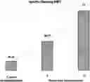
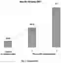
Sources:
- United States Patent and Trademark Office - verify current appl. status at the USPTO↗
Recent applications in this class:
- » 20210196772 2021-07-01
CONFECTIONS FOR SKIN PROTECTION: COMPOSITIONS, METHODS FOR MAKING, AND APPLICATIONS THEREOF - » 20200046786 2020-02-13
COMPOSITIONS AND METHODS OF USE - » 20190255129 2019-08-22
Lipids with odd number of carbon atoms and their use as pharmaceutical composition or nutritional supplement - » 20160008414 2016-01-14
Pharmaceutical composition containing extract for preventing or treating stroke or degenerative brain diseases - » 20130330372 2013-12-12
Formulations comprising extracts from primitive plant species (mosses, ferns and lichens) to treat and prevent cancers - » 20110286953 2011-11-24
WHITENING AGENT, ANTI-AGING AGENT, AND ANTI-OXIDANT AGENT - » 20110071225 2011-03-24
METHOD FOR OBTAINING A SEQUOYITOL-CONTAINING EXTRACT FROM GENUS NEPHROLEPIS AND USES THEREOF - » 20100209543 2010-08-19
Compositions and methods of use - » 20100081832 2010-04-01
Composition for treating cancer cells and preparation method for the same - » 20100056812 2010-03-04
Composition for treating cancer cells and preparation method for the same