SYSTEM AND METHOD FOR CORRELATING OXIMETER MEASUREMENTS WITH BLOOD PRESSURE
US20230329645A1
2023-10-19
18/336,987
2023-06-17
Abstract:
A system and method for using an oximeter that monitors a patient's blood pressure over an extended time duration requires creating a line graph. In detail, the line graph is created to provide a steady state correlation between blood flow measurements taken by the oximeter and blood pressure measurements taken by a sphygmomanometer. To create this graph, blood pressure measurements (sphygmomanometer) and blood flow measurements (oximeter) are recorded together and collated during a heart muscle cycle of the patient. Specifically, these measurements are considered together during the same heart muscle cycle while the patient is either standing, sitting, or reclining. This establishes three respective data sets which are then used as reference points to create the line-graph. Thereafter, blood flow measurements with the oximeter can be referenced to the line-graph for direct indications of blood pressure.
Interested in similar patents?
Get notified when new applications in this technology area are published.
Classification:
A61B5/7246 » CPC main
Measuring for diagnostic purposes ; Identification of persons; Signal processing specially adapted for physiological signals or for diagnostic purposes; Details of waveform analysis using correlation, e.g. template matching or determination of similarity
A61B5/14551 » CPC further
Measuring for diagnostic purposes ; Identification of persons; Measuring characteristics of blood , e.g. gas concentration, pH value; Measuring characteristics of body fluids or tissues, e.g. interstitial fluid, cerebral tissue using optical sensors, e.g. spectral photometrical oximeters for measuring blood gases
A61B5/0261 » CPC further
Measuring for diagnostic purposes ; Identification of persons; Detecting, measuring or recording pulse, heart rate, blood pressure or blood flow; Combined pulse/heart-rate/blood pressure determination; Evaluating a cardiovascular condition not otherwise provided for, e.g. using combinations of techniques provided for in this group with electrocardiography or electroauscultation; Heart catheters for measuring blood pressure; Measuring blood flow using optical means, e.g. infra-red light
A61B2560/0223 » CPC further
Constructional details of operational features of apparatus; Accessories for medical measuring apparatus; Operational features of calibration, e.g. protocols for calibrating sensors
A61B5/00 IPC
Measuring for diagnostic purposes ; Identification of persons
A61B5/022 » CPC further
Measuring for diagnostic purposes ; Identification of persons; Detecting, measuring or recording pulse, heart rate, blood pressure or blood flow; Combined pulse/heart-rate/blood pressure determination; Evaluating a cardiovascular condition not otherwise provided for, e.g. using combinations of techniques provided for in this group with electrocardiography or electroauscultation; Heart catheters for measuring blood pressure; Measuring pressure in heart or blood vessels by applying pressure to close blood vessels, e.g. against the skin; Ophthalmodynamometers
A61B5/0205 » CPC further
Measuring for diagnostic purposes ; Identification of persons; Detecting, measuring or recording pulse, heart rate, blood pressure or blood flow; Combined pulse/heart-rate/blood pressure determination; Evaluating a cardiovascular condition not otherwise provided for, e.g. using combinations of techniques provided for in this group with electrocardiography or electroauscultation; Heart catheters for measuring blood pressure Simultaneously evaluating both cardiovascular conditions and different types of body conditions, e.g. heart and respiratory condition
A61B5/1455 IPC
Measuring for diagnostic purposes ; Identification of persons; Measuring characteristics of blood , e.g. gas concentration, pH value; Measuring characteristics of body fluids or tissues, e.g. interstitial fluid, cerebral tissue using optical sensors, e.g. spectral photometrical oximeters
A61B5/026 IPC
Measuring for diagnostic purposes ; Identification of persons; Detecting, measuring or recording pulse, heart rate, blood pressure or blood flow; Combined pulse/heart-rate/blood pressure determination; Evaluating a cardiovascular condition not otherwise provided for, e.g. using combinations of techniques provided for in this group with electrocardiography or electroauscultation; Heart catheters for measuring blood pressure Measuring blood flow
Description
This continuation-in-part application claims the benefit of U.S. Patent Application Publication No. US 2022/0328178A1, filed Oct. 7, 2021. The entire contents of application Ser. No. 17/496,052 are hereby incorporated by reference herein.
FIELD OF THE INVENTION
The present invention pertains to systems and methods for continuously monitoring a patient's blood pressure over an extended time duration. More specifically, the present invention pertains to systems and methods that use a line graph to calibrate blood flow measurements from an oximeter with correlated blood pressure measurements from a sphygmomanometer. The present invention is particularly, but not exclusively, useful for providing continuous blood pressure information using only blood flow measurements from an oximeter.
BACKGROUND OF THE INVENTION
For any health care situations there are many possible reasons why there is a need to measure and monitor a patient's blood pressure. Typically, this is done by intermittently using a sphygmomanometer. Although it is well known that a sphygmomanometer is a useful and reliable medical device for measuring a patient's blood pressure, its repetitive use may not be practical when continuous monitoring of a patient's blood pressure for extended time durations is necessary or preferable.
It is also well known in the medical arts that an oximeter is capable of continuously measuring blood flow. Specifically, oximeters provide measurements of blood oxygen saturation levels that are indicative of the volume of blood being measured. Oximeter measurements, however, like blood pressure measurements, are pulsatile. The respective pulses, however, have different dimensional characteristics. In the context of the present invention, the similarities and differences in the relationship between blood flow and blood pressure are important for several reasons. The similarities include:
-
- Blood flow is a direct temporal consequence of blood pressure variations produced during a heart muscle cycle (i.e. pulse).
- The dimensional characteristics of blood flow and blood pressure have concurrence in that blood pressure affects blood flow repetitively during each heart muscle cycle.
On the other hand, for measurement purposes, there are distinctive differences between blood pressure and blood flow that must be reconciled. These differences include: - The maximum amplitude of a blood pressure measurement, “Pmax”, and the maximum amplitude of a flood flow measurement “Fmax” during a heart muscle cycle are inversely proportional.
- “Pmax” and “Fmax” occur at different times during a heart muscle cycle.
- The respective rates of change for “ΔPmax” and “ΔFmax” from pulse to pulse may be different, i.e. “ΔPmax”≠“ΔFmax”.
- A sphygmomanometer does not measure both systolic and diastolic pressures during the same heart muscle cycle.
For reasons set for the above it is an object of the present invention to continuously recalibrate blood pressure measurements with corresponding blood flow measurements. This is done so an oximeter can be used alone, to continuously monitor blood pressure trends for successive heart muscle functions over a predetermined time duration. Another object of the present invention is to incorporate a line graph in a device which can be used to calibrate blood pressure trends with an oximeter. Yet another object of the present invention is to provide a device for measuring blood pressure with an oximeter that is easy to manufacture, is simple to use and is cost effective.
SUMMARY OF THE INVENTION
In accordance with the present invention, a system and method are disclosed for using blood flow measurements from a patient as indications of the patient's blood pressure trends. Structurally, the system of the present invention is a combination of components that includes a sphygmomanometer, an oximeter, and a collator. Functionally, the sphygmomanometer is used to periodically measure a patient's blood pressure “P”. On the other hand, the oximeter is used continuously to measure the patient's blood oxygen saturation levels as indicators of his/her blood flow “F”. The collator then collects the blood pressure measurements and the blood flow measurements and combines selected dimensional aspects of these measurements into data sets.
For each data set, the sphygmomanometer measures a maximum blood pressure measurement “Psystolic” near the beginning of each heart muscle cycle. The sphygmomanometer also measures a “Pdiastolic” during the same heart muscle cycle. In a separate operation, the oximeter measures blood flow variations that include a maximum amplitude “Fmax” near the end of each heart muscle cycle. Further, the sphygmomanometer can also be used to measure a heart pulse rate from the patient to establish the duration for a heart muscle cycle.
As appreciated by the present invention, “P” (blood pressure) and “Fmax” (blood flow) have an inverse relationship that must be considered in the context of a heart muscle cycle. To account for this fact, the collator collects “Psystolic”, “Pdiastolic” and “Fmax” measurements during each heart muscle cycle. From these measurements, “Psystolic” and “Pdiastolic” are combined to establish a data set that can then be used as a steady state quantified “Δss”. Mathematically, “Δss”=“Psystolic”−“Pdiastolic” and it is constant. Although, blood pressure changes “ΔPsystolic” and “Pdiastolic” will not be directly equal to the blood flow changes “ΔFmax”, the quantified “ΔSS” for “Psystolic” and “Pdiastolic” for blood pressures can be considered concurrent with “ΔFmax”.
It happens that in a sequence of heart muscle cycles, from one heart muscle cycle to the next, “Pdiastolic” is more reliable for use as a reference point than is “Psystolic”. Thus, for an operation of the present invention “Pdiastolic” values are preferably used as reference points along a line-graph for a sequence of respective “Δss”. Functionally, the resultant line-graph can then be used for an extended time period to calibrate “Fmax” measurement from the oximeter with a blood pressure “P” from “Δss”.
In detail, a line-graph is created for the present invention using at least two reference points. Importantly, each reference point is separately created with a “Psystolic”, a “Pdiastolic” measurement which are combined in a data set for use as a quantified “Δss” at a location on the line-graph. Further, each quantified “Δss” is individually established when the patient is posed in different positions. Consequently, the plurality of quantified “Δss” can create the line-graph, with each location along the line graph providing a unique comparison “Δss”. Thus, the line graph calibrates an “F” measurement from the oximeter with a correlated “P”. With this calibration, the result is that the correlated “P” can be shown on a display as an indication of blood pressure.
As noted above, several factors must be considered during the creation of a line graph. For instance, “Pmax” and “Fmax” are measured separately, and they have an inverse relationship. Furthermore, between different quantified “Δss”, the rate of change “ΔPmax” is not equal to the rate of change in “ΔFmax”. Thus, each unique steady state comparison Δss along the line graph will change and have a new value that accounts for the fact that in a sequence each “Δss”=(P±ΔP) and (F±ΔF).
A methodology for the present invention requires a sequence of steps for obtaining blood flow measurements from a patient which can be continuously monitored and used as real time indications of the patient's blood pressure. Further, the methodology provides instructions that are useful for manufacturing a device in accordance with the present invention. It is also useful for subsequently monitoring a patient's blood pressure with the device.
In use, a sphygmomanometer is positioned on a patient to measure his/her blood pressure “P”. At the same time, an oximeter is also positioned on the patient to measure blood flow “F”. With this oximeter/sphygmomanometer combination a maximum blood pressure “Pmax” is measured by the sphygmomanometer, and a contemporary maximum blood flow “Fmax” is measured by the oximeter. A pulse rate measurement can also be obtained from the sphygmomanometer and be used to determine the time duration for the patient's heart muscle cycle.
Because “Pmax” and “Fmax” have concurrence in the same heart muscle cycle, the measured values for “Pmax” and “Fmax” can be collated together as components for use as a same data set. Each collated data set is thereby combined into a steady state quantified comparison “Δss”. Importantly, each quantified comparison “Δss” is unique with blood pressure and blood flow measurements. More specifically, each quantified comparison “Δss” includes measurements that are taken from the patient while he/she is posed in different positions, such as standing, sitting, or lying down.
A line graph for the present invention is created using the “Pmax” and “Fmax” values taken for successive quantified comparisons “Δss”. Specifically, “F” will establish the horizontal axis of the line graph, while “P” will establish the vertical axis. Because “Pmax” and “Fmax” have an inverse relationship, the horizontal axis of the line graph will show a decreasing value for “F”. On the other hand, the vertical axis of the line graph will show an increasing value for “P”. With this inverse relationship, each location on the resulting line graph, between quantified comparisons “Δss”, will represent a specific comparison “Δss” having unique values for “P” and “F”.
It is important to note that between any two quantified comparisons “Δss”, at each location on the line graph, the rate of change “ΔP” is not equal to the rate of change in “ΔF”. Consequently, they must be considered separately for each successive comparison “Δss”. Accordingly, values for a successive “Δss”, using values from its predecessor “Δss”, will equal (P±ΔP) and (F±ΔF). When using a line graph as disclosed here, values for “F” which are being continuously measured by an oximeter, can be directly correlated at every location along the line graph with a corresponding “P” from the same comparison “Δss”.
Additional considerations for using the methodology of the present invention include the fact that a quantified comparison “Δss” can be periodically recalibrated with updated “Pmax” measurements taken by the sphygmomanometer (e.g. every 30 minutes). Furthermore, depending on the number of multiple quantified comparisons “Δss” that are measured, they can all be collectively used as different reference points to create a continuous line graph with differently oriented line segments (e.g. a 3-point line graph). For example, a 3-point line graph can be created having two different line segments. In this case, each line segment will be established between only two different quantified comparisons “Δss”.
DESCRIPTION OF THE DRAWINGS
The novel features of this invention, as well as the invention itself, both as to its structure and its operation, will be best understood from the accompanying drawings, taken in conjunction with the accompanying description, in which similar reference characters refer to similar parts, and in which:
FIG. 1 shows the structural elements of a system for the present invention being used in an intended operational environment;
FIG. 2 shows respective traces of blood pressure and blood flow measurements taken during a sequence of consecutive time durations;
FIG. 3 shows the inverse relationship between blood pressure and blood flow measurements that are used to create a quantified comparison “Δss”; and
FIG. 4 is a 3-point line graph created using three distinctively different quantified comparisons “Δss”.
DESCRIPTION OF THE PREFERRED EMBODIMENTS
Referring initially to FIG. 1, a system for continuously using blood flow measurements “F” from a patient as indications of the patient's blood pressure “P” is shown and is generally designated 10. As shown in FIG. 1, system 10 includes an electronic device 12 that is connected directly with the patient 14. Specifically, a sphygmomanometer 16 is fitted onto the patient 14 to obtain blood pressure measurements “P” from the patient 14. Similarly, an oximeter 18 is fitted onto the patient 14 to obtain blood flow measurements “F” from the patient 14. Both measurements, “P” and “F” are taken during a same quantified comparison “Δss”.
In detail, FIG. 1 shows that the sphygmomanometer 16 is connected directly with the collator 20 of the device 12. This connection is shown in FIG. 1 as a dot-dash line 22 to signify that blood pressure measurements “P” are taken intermittently. The minor interruptions between “P” measurements are primarily due to time limitations and the labor-intensive nature for an operation of the sphygmomanometer 16. On the other hand, FIG. 1 also shows that the connection between the oximeter 18 and the collator 20 of the device 12 is a solid line 24. In this case, the solid line 24 signifies that blood flow measurements “F” from the oximeter 18 can be taken continuously with minimal, if any, interruption. In this operation, it is to be appreciated that the collator 20 functions to collect blood pressure measurements “P” along with blood flow measurements “F”. During this operation, collator 20 also functions to then combine the “P” and “F” measurements for comparison purposes.
In accordance with the present invention, the combination of a single “P” measurement and a single “F” measurement constitute a data set which is then quantified. For the present invention, quantification of the data set specifically requires creation of a steady state quantified comparison “Δss” for the measurements. As disclosed below in detail, a plurality of quantified comparisons “Δss” are required for an operation of the system 10.
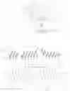
Referring now to FIG. 2, the data required to establish a quantified comparison “Δss” requires “Psystolic” and “Pdiastolic” that are respectively shown in a blood pressure data trace generally designate 26. Also important is the value for “Fmax” as shown in a blood flow data trace generally designated 28. For the system 10, a common nexus between “Pdiastolic” and “Fmax” is shown for an exemplary episode 30 from traces 26 and 28 which last for the same time duration “Δt” which is the time required for a sphygmomanometer to record “Δss”. Specifically, “Δt” of episode 30 involves the time required for a sphygmomanometer 16 to measure at least one “Pdiastolic” in a sequence of heart muscle cycles. Importantly, it has been determined that “Pdiastolic” is more reliable than “Psystolic” as a reference for identifying “P” over extended time periods.
As shown in the blood flow trace 28 of FIG. 2, an episode 32 can be selected from within an episode 30. Specifically, a measured “Pdiastolic” and a single a “Fmax” will occur together only once during “Δt” of the episode 30. On the other hand, occurrences of “Fmax” occur continuously for each heart muscle cycle during “Δt”.
In detail it happens that “Δt” will typically extend through several heart muscle cycles. The consequence here is that because of the operational requirements of a sphygmomanometer 16, the time interval between the “Pdiastolic” measured in one heart cycle and “Pdiastolic” that can be measured for the next heart muscle cycle will necessarily be delayed “Δt”. Although “Δt” will last for a few heart muscle cycles, there is only one “Pdiastolic” that can be measured during an episode 30.
As more specifically shown in the flow data trace 28 of FIG. 2, it happens during any episode 30 for the sphygmomanometer 16, several separate episodes 32 will occur sequentially for the oximeter 18. Importantly, within the time duration “Δtox” of each episode 32 there will always be both a “Pmax” and an “Fmax”.
FIG. 2 also shows that although only one “Pdiastolic” can be measured somewhere within the time duration “Δt” both this “Pmax” and an “Fmax” will occur at least once in a same episode 30 during “Δt”. Thus, for purposes of system 10, the measurements of “Pmax” and “Fmax” can be effectively considered to be concurrent. Accordingly, they can be used as components for establishing a quantified “Δss”.
FIG. 3 shows blood pressure variations 31 for “P”, and blood flow variations 33 for “F” during a quantified “Δss”. Note: in FIG. 3 the value of “P” variations 31 increases in an upward direction. At the same time, the value of “F” variations 33 increases in a downward direction. This happens because, with an increased volume of blood flow “F”, light absorption also increases. However, with increased light absorption, the magnitude of light signals measured by an oximeter 18 are decreased. Thus, the inverse relationship. A compensation of this inverse relationship by the collator 20, which uses any “F” and only a measured “Pdiastolic” during an episode 30, is referred to here as a quantified comparison “Δss”. For purposes of the present invention, quantified comparisons “Δss” are essential for creating a line graph 34 such as disclosed below with reference to FIG. 4.
As seen in FIG. 4, a line graph 34 is shown which constitutes a continuous sequence of comparisons “Δss”. In detail, the line graph 34 is established between quantified comparisons “Δss” which are respectively located at reference points 36, 38 and 40. All locations along the line graph 34, as well as locations on extensions therefrom beyond the points 36 and 40, each identify a unique “F” and “P” relationship for a unique “Δss”. For example, consider a measured value for “F” from the oximeter 18 which is shown at point 42. This point 42 references a point 44 on graph line 34 that calibrates “F” to a value for “P”. It is this value for “P” that corresponds with a unique “Δss” is observed by a patient 14 as his/her blood pressure.
While the particular System and Method for Correlating Oximeter Measurements with Blood Pressure as herein shown and disclosed in detail is fully capable of obtaining the objects and providing the advantages herein before stated, it is to be understood that it is merely illustrative of the presently preferred embodiments of the invention and that no limitations are intended to the details of construction or design herein shown other than as described in the appended claims.
Claims
What is claimed is:1. A system for continuously using blood flow measurements “F” from a patient as indications of the patient's blood pressure “P”, which comprises:
a sphygmomanometer for measuring blood pressure variations in a patent's vasculature including a maximum blood pressure measurement “Psystolic” near the beginning of each heart muscle cycle and a pressure measurement “Pdiastolic” near the end of each heart muscle cycle;
an oximeter for measuring blood flow variations commensurate with the blood flow variations “F” including a maximum amplitude “Fmax” near the end of each heart muscle cycle; and
a collator connected with the sphygmomanometer and with the oximeter to establish a steady state quantified comparison “Δss” between “Psystolic” and “Pdiastolic”; and
a line-graph created by a plurality of quantified comparison “Δss” for calibrating the use of measured “F” from the oximeter as an indicator of blood pressure “P” for the patient.
2. The system of claim 1 wherein the collator is preprogrammed with input information, including a heart pulse rate from the patient for identifying a duration for a heart muscle cycle, and wherein the collator collects a value for “P” relative to a value of “F” during a same heart muscle cycle to establish a data set for use in providing quantified comparison “Δss”.
3. The system of claim 2 wherein the line-graph is created with at least two reference points, wherein each reference point is identified by a separate quantified comparison “Δss”, wherein each quantified comparison “Δss” is established when the patient is respectively posed in different positions, wherein the plurality of quantified comparison “Δss” collectively establish the line-graph, and wherein each location along the line graph between quantified comparison “Δss” provides a unique “Δss” to correlate a measured “Fmax” along the line graph, with a corresponding “P” to be indicated by a display as an indication of blood pressure.
4. The system of claim 3 wherein “Pmax” and “Fmax” have an inverse relationship, and further wherein between successive comparison “Δss” on the line-graph remains constant but the rate of change “ΔP” is not equal to the rate of change in “ΔF”, with a new steady state comparison “Δss” for the subsequent data set having a new value wherewith “Δss”=(P±ΔP) and (F±ΔF).
5. The system of claim 4 wherein a “Psystolic”, a “Pdiastolic” and an “Fmax” are periodically re-measured for each quantified comparison “Δss”, and wherein a re-measurement is accomplished at least every thirty minutes to reconfigure the line-graph.
6. The system of claim 4 wherein data sets are created with the patient posed standing, sitting, and lying down to respectively create the quantified comparisons “Δss” needed for a 3-point line graph.
7. The system of claim 1 wherein the duration of a heart muscle cycle is determined using blood pressure variations measured by the sphygmomanometer.
8. The system of claim 1 wherein the line graph is created using the “P” and “Fmax” values taken for successive quantified comparisons “Δss”, and wherein to account for “P” and “Fmax” having an inverse relationship, a horizontal axis for the graph will show a decreasing value for “Fmax” while a vertical axis for the graph will show an increasing value for “P”, with each location on the resulting line graph between any two quantified comparisons “Δss” representing a specific comparison “Δss” having unique values for “P” relative to “Fmax”.
9. A method for using blood flow measurements from a patient as indications of blood pressure, which comprises the steps of:
positioning a sphygmomanometer on a patient to measure blood pressure “P” of the patient, wherein “P” includes a “Psystolic” and a “Pdiastolic”;
positioning an oximeter on a patient to measure blood flow “F” of the patient including an “Fmax”;
obtaining a pulse rate measurement from the sphygmomanometer;
using the pulse rate to determine a time duration for a heart muscle cycle;
taking “P” and “Fmax” from the measuring step for use as components in a data set wherein “P” and “Fmax have concurrence in the same heart muscle cycle;
establishing different data sets, wherein each data set is specific with the patient posed in different positions for each data set;
quantifying each data set as an individually specific steady state quantified comparison “Δss”, wherein “P” and “Fmax” are taken with the patient posed in different positions during the establishing step, and wherein “P” and “Fmax” have an inverse relationship;
creating a line graph with a plurality of steady state quantified comparisons “Δss”, wherein each location on the line graph between quantified “Δss” is a unique comparison “Δss”, and further wherein between successive quantified comparisons “Δss” is constant but the rate of change “ΔP” is not equal to the rate of change in “ΔF” with a new value for each unique comparison “Δss”=(P±ΔP) and (F±ΔF);
calibrating a measured “F” with a corresponding “P” in a comparison “Δss” for every location along the line graph;
displaying “P” as an indication of blood pressure based on the graph line location for “Δss” fixed by the measured “Fmax”.
10. The method of claim 9 wherein the data sets are periodically remeasured with updated “Pmax” measurements taken by the sphygmomanometer and updated “Fmax” measurements taken by the oximeter.
11. The method of claim 9 wherein “P” is measured during the heart muscle cycle, and “Fmax” is measured near the end of the heart muscle cycle.
12. The method of claim 11 wherein “P” and “Fmax” have concurrence within a same heart muscle cycle.
13. The method of claim 9 wherein different data sets are established with the patient respectively sitting, standing, and lying down.
14. The method of claim 13 wherein the different data sets establish a 3-point line graph.
15. The method of claim 9 wherein the line graph is created using the “P” and “Fmax” values taken for successive quantified comparison “Δss”, and wherein to account for “P” and “F” having an inverse relationship, a horizontal axis for the line graph will show a decreasing value for “F” while a vertical axis for the graph will show an increasing value for “P”, with each location on the line graph representing a comparison “Δss” having unique values for “P” and “F” between any two quantified comparisons “Δss”.
16. A method for using blood flow measurements “F” from a patient as indications of blood pressure “P” for the patient which comprises the steps of:
measuring a blood pressure “P1”, wherein “P1” includes “Psystolic1” and “Pdiastolic1”, and a maximum blood flow value “Fmax1” during a same heart muscle cycle to establish a data set therewith, wherein the data set is a first steady state quantified comparison “Δss1”=“Pmax1” and “Fmax1”;
measuring a blood pressure “P2”, wherein “P2” includes “Psystolic2” and “Pdiastolic2”, and a maximum blood flow value “Fmax2” during a same heart muscle cycle to establish a data set therewith, wherein the data set is a second steady state quantified comparison “Δss2”=“Pmax2” and “Fmax2”;
creating a line graph using “Δss1” and “Δss2” as separate reference points, wherein each location on the line graph between these reference points is representative of an independent unique comparison “Δss”; and
referencing an observed blood flow measurement “F” to a location on the line graph with a “Pdiastolic” to identify a “P” as an indication of the patient's blood pressure.
17. The method of claim 16 wherein there is a unique “Δss” at each location on the line graph between the different quantified “Δss”, and further wherein between successive “Δss” on the line graph the rate of change “ΔP” is not equal to the rate of change in “ΔF” with a new value for each unique “Δss”=(P±ΔP) and (F±ΔF).
18. The method of claim 16 wherein “P1” and “P2” are measured using a sphygmomanometer, and “Fmax1” and “Fmax2” are measured using an oximeter.
19. The method of claim 18 wherein “Psystolic” is measured near the beginning of the heart muscle cycle, while “Pdiastolic” and “Fmax” is measured near the end of the heart muscle cycle, wherein “P” and “Fmax” have concurrence within a same heart muscle cycle, and further wherein different data sets are established with the patient respectively sitting, standing, and lying down.
20. The method of claim 19 wherein the line graph is created using the “P” and “Fmax” values taken for successive quantified “Δss”, and wherein to account for “P” and “Fmax” having an inverse relationship, a horizontal axis for the graph will show a decreasing value for “F” while a vertical axis for the graph will show an increasing value for “P”, and wherein each location on the resulting line graph represents a comparison “Δss” having unique values for “P” and “F”.
Images & Drawings included:


Sources:
- United States Patent and Trademark Office - verify current appl. status at the USPTO↗
Recent applications in this class:
- » 20250057483 2025-02-20
SYSTEM AND METHOD FOR TASK-LESS MAPPING OF BRAIN ACTIVITY - » 20240423548 2024-12-26
Method and Medical Device for Determining a Periodicity of a Physiological Signal - » 20240407732 2024-12-12
METHOD AND DEVICE FOR DETECTING A NEURAL RESPONSE IN A NEURAL MEASUREMENT - » 20240341694 2024-10-17
COMPARING MEASUREMENTS OF A HEART OF A PATIENT BEFORE AND AFTER ONE OR MORE SURGICAL PROCEDURES - » 20240293085 2024-09-05
SYSTEM, DEVICE AND METHOD OF DYNAMIC GLUCOSE PROFILE RESPONSE TO PHYSIOLOGICAL PARAMETERS - » 20240268766 2024-08-15
BLOOD-PRESSURE-MEASURING DEVICE AND BLOOD-PRESSURE-MEASURING SYSTEM - » 20240180496 2024-06-06
Method and device for detecting a neural response in a neural measurement - » 20240172998 2024-05-30
CONTINUOUS LONG-TERM MONITORING OF A SUBJECT - » 20230371903 2023-11-23
BIOLOGICAL INFORMATION OBTAINMENT DEVICE, BIOLOGICAL INFORMATION OBTAINMENT SYSTEM, AND BIOLOGICAL INFORMATION OBTAINMENT METHOD - » 20230293112 2023-09-21
Physiological Signal Feature Extraction Method and Physiological Signal Feature Extraction Device Thereof