ASSISTED SURGICAL PLANNING SYSTEM AND OPERATION METHOD THEREOF
US20260053583A1
2026-02-26
19/298,138
2025-08-12
Smart Summary: An assisted surgical planning system helps doctors plan surgeries more effectively. It uses a computer with special AI tools to create images of organs from CT scans. A user interface shows these images along with different paths for inserting a needle. The system suggests the best needle insertion paths based on the images and an initial path provided by the doctor. This technology aims to improve the accuracy and safety of surgical procedures. 🚀 TL;DR
Abstract:
An assisted surgical planning system includes a host computer, a discriminative AI module, a user navigation interface, and a generative AI module. The discriminative AI module is electrically connected to the host computer, and is configured to form plural organ segmentation images based on plural CT images. The user navigation interface is electrically connected to the host computer, and is configured to display the organ segmentation images, an initial needle insertion path, plural suggested needle insertion paths, and a final needle insertion path. The generative AI module is electrically connected to the host computer, such that the host computer connects the user navigation interface to the generative AI module. The generative AI module is configured to generate the suggested needle insertion paths based on the organ segmentation images and the initial needle insertion path, and display the suggested needle insertion paths in the user navigation interface.
Inventors:
- PING-LANG YEN 7 🇹🇼 Taipei, Taiwan
- Hao-Cheng ZUO 2 🇹🇼 Taipei, Taiwan
- Cheng-Yen CHUNG 2 🇹🇼 Taipei, Taiwan
Applicant:
Interested in similar patents?
Get notified when new applications in this technology area are published.
Classification:
A61B34/32 » CPC main
Computer-aided surgery; Manipulators or robots specially adapted for use in surgery; Surgical robots operating autonomously
A61B17/3403 » CPC further
Surgical instruments, devices or methods, e.g. tourniquets; Trocars; Puncturing needles Needle locating or guiding means
G16H40/67 » CPC further
ICT specially adapted for the management or administration of healthcare resources or facilities; ICT specially adapted for the management or operation of medical equipment or devices for the operation of medical equipment or devices for remote operation
A61B2034/107 » CPC further
Computer-aided surgery; Manipulators or robots specially adapted for use in surgery; Computer-aided planning, simulation or modelling of surgical operations Visualisation of planned trajectories or target regions
A61B2034/2065 » CPC further
Computer-aided surgery; Manipulators or robots specially adapted for use in surgery; Surgical navigation systems; Devices for tracking or guiding surgical instruments, e.g. for frameless stereotaxis; Tracking techniques Tracking using image or pattern recognition
A61B2034/252 » CPC further
Computer-aided surgery; Manipulators or robots specially adapted for use in surgery; User interfaces for surgical systems indicating steps of a surgical procedure
A61B2090/3762 » CPC further
Instruments, implements or accessories specially adapted for surgery or diagnosis and not covered by any of the groups - , e.g. for luxation treatment or for protecting wound edges; Image-producing devices or illumination devices not otherwise provided for; Surgical systems with images on a monitor during operation using X-rays, e.g. fluoroscopy using computed tomography systems [CT]
A61B17/34 IPC
Surgical instruments, devices or methods, e.g. tourniquets Trocars; Puncturing needles
A61B34/00 IPC
Computer-aided surgery; Manipulators or robots specially adapted for use in surgery
A61B34/10 IPC
Computer-aided surgery; Manipulators or robots specially adapted for use in surgery Computer-aided planning, simulation or modelling of surgical operations
A61B34/20 IPC
Computer-aided surgery; Manipulators or robots specially adapted for use in surgery Surgical navigation systems; Devices for tracking or guiding surgical instruments, e.g. for frameless stereotaxis
A61B90/00 IPC
Instruments, implements or accessories specially adapted for surgery or diagnosis and not covered by any of the groups - , e.g. for luxation treatment or for protecting wound edges
Description
CROSS-REFERENCE TO RELATED APPLICATION
This application claims priority to U.S. Provisional Application Ser. No. 63/684,875, filed Aug. 20, 2024, and Taiwan Application Serial Number 113141733, filed Oct. 30, 2024, which are herein incorporated by reference.
BACKGROUND
Technical Field
The present disclosure relates to an assisted surgical planning system and an operation method of the assisted surgical planning system.
Description of Related Art
When preoperative needle insertion path planning for a puncture (e.g., a tumor puncture) is performed, doctors need to observe vital organs, hard-to-identify nerves, and small blood vessels, etc. on two-dimensional (2D) computed tomography (CT) images, and select appropriate image slices for path planning after repeated checks. The doctors' biggest concerns in path planning are risks of damaging blood vessels and causing complications, but such information is often not easily noticed in CT scans. The doctors may have to guess the locations of the vessels, or use a developer to take CT images, or use a contrast-enhanced computed tomography (CECT) method.
Finding access to safe needle insertion paths is challenging, highly dependent on the experience and expertise of the doctors, time-consuming and subject to subjective opinions. Additionally, since the doctors are not used to relying on three-dimensional (3D) image reconstruction when performing needle insertion path planning, they will mostly perform path planning in an axial view of a CT image. A slice plane of the axial view is easier for the doctors to imagine and more convenient for path planning. However, cross-plane needle insertion path planning is not easy for the doctors, and the doctors need to spend a lot of time before a surgery to adjust needle insertion paths and angles, and perform a CT scan after each stage of needle insertion. If it is found that the needle insertion path is not suitable or deviates from the original expectation during the surgery, it is necessary to make timely adjustment or even perform needle re-insertion to reduce the surgical risk.
SUMMARY
According to some embodiments of the present disclosure, an assisted surgical planning system comprises a host computer, a discriminative artificial intelligence (AI) module, a user navigation interface and a generative AI module. The discriminative artificial intelligence (AI) module is electrically connected to the host computer, and is configured to form plural organ segmentation images based on plural computed tomography (CT) images. The user navigation interface is electrically connected to the host computer, and is configured to display the organ segmentation images, an initial needle insertion path, plural suggested needle insertion paths, and a final needle insertion path. The generative AI module is electrically connected to the host computer, such that the host computer connects the user navigation interface to the generative AI module. The generative AI module is configured to generate the suggested needle insertion paths based on the organ segmentation images and the initial needle insertion path, and display the suggested needle insertion paths in the user navigation interface.
In some embodiments, the generative AI module is configured for selection and adjustment in the user navigation interface based on the suggested needle insertion paths to generate the final needle insertion path.
In some embodiments, the generative AI module comprises a user input unit configured to input an entry point, a target point, plural danger zones and a tumor into the organ segmentation images.
In some embodiments, the generative AI module further comprises a task unit configured to adjust the initial needle insertion path to generate the suggested needle insertion paths.
In some embodiments, the generative AI module further comprises a judgment unit configured to steer the suggested needle insertion paths away from the danger zones and adjust the target point to the center of the tumor.
In some embodiments, the generative AI module further comprises an output unit configured to generate coordinates and images of the suggested needle insertion paths.
In some embodiments, the assisted surgical planning system further comprises a visualization module. The visualization module is electrically connected to the generative AI module, and is configured to receive the CT images, the coordinates of the suggested needle insertion paths and coordinates of the final needle insertion path.
In some embodiments, the assisted surgical planning system further comprises a CT reslice module. The CT reslice module is electrically connected to the generative AI module, and is configured to receive the CT images and transmit the organ segmentation images to the generative AI module.
In some embodiments, the assisted surgical planning system further comprises a message module. The message module is electrically connected to the generative AI module, and is configured to transmit voice messages and type messages to the generative AI module.
In some embodiments, the assisted surgical planning system further comprises a robot module. The robot module is electrically connected to the generative AI module, and is configured to receive a command from the generative AI module to operate a robot having a puncture needle.
According to some embodiments of the present disclosure, an operation method of an assisted surgical planning system comprises: obtaining, by a discriminative AI module, plural CT images; setting an initial needle insertion path in a user navigation interface based on the CT images; forming, by the discriminative AI module, plural organ segmentation images based on the CT images; generating, by a generative AI module, plural suggested needle insertion paths based on the initial needle insertion path and the organ segmentation images, wherein the discriminative AI module, the generative AI module and the user navigation interface are electrically connected to a host computer; displaying the suggested needle insertion paths in the user navigation interface; and selecting and adjusting the suggested needle insertion paths from the user navigation interface, such that the generative AI module generates a final needle insertion path.
In some embodiments, the operation method of an assisted surgical planning system further comprises: selecting and adjusting the suggested needle insertion paths from the user navigation interface to form a modified path; and generating, by the generative AI module, plural other suggested needle insertion paths based on the modified path and the organ segmentation images.
In some embodiments, the operation method of an assisted surgical planning system further comprises: contextually training the generative AI module.
In some embodiments, contextually training the generative AI module comprises: inputting an entry point, a target point, plural danger zones and a tumor into the organ segmentation images, wherein the danger zones comprise arteries, veins, and blood vessels.
In some embodiments, contextually training the generative AI module further comprises: adjusting the initial needle insertion path to generate the suggested needle insertion paths; and generating coordinates and images of the suggested needle insertion paths.
In some embodiments, adjusting the initial needle insertion path to generate the suggested needle insertion paths comprises: steering the suggested needle insertion paths away from the danger zones; and adjusting the target point to the center of the tumor.
In some embodiments, the operation method of an assisted surgical planning system further comprises: receiving, by a visualization module electrically connected to the generative AI module, the CT images, the coordinates of the suggested needle insertion paths and coordinates of the final needle insertion path.
In some embodiments, the operation method of an assisted surgical planning system further comprises: receiving the CT images and transmitting the organ segmentation images to the generative AI module by a CT reslice module electrically connected to the generative AI module.
In some embodiments, the operation method of an assisted surgical planning system further comprises: transmitting, by a message module electrically connected to the generative AI module, voice messages and type messages to the generative AI module.
In some embodiments, the operation method of an assisted surgical planning system further comprises: receiving, by a robot module electrically connected to the generative AI module, a command from the generative AI module to operate a robot having a puncture needle.
In the above embodiments of the present disclosure, since the assisted surgical planning system includes the generative AI module and the host computer connects the user navigation interface to the generative AI module, the generative AI module can generate the suggested needle insertion paths based on the initial needle insertion path and the organ segmentation images, and the user navigation interface displays the suggested needle insertion paths for doctors' reference. The doctors can interact with the generative AI module through the user navigation interface, select and adjust the suggested needle insertion paths from the user navigation interface, such that the generative AI module generates the final needle insertion path. In this way, the assisted surgical planning system and the operation method thereof can effectively save the time of preoperative needle insertion path planning and reduce the influence of subjective opinions. In addition, the doctors can check, confirm, interact and adjust back and forth on customary two-dimensional (2D) images (e.g., organ segmentation images) when the assisted surgical planning system performs needle insertion path planning, so that the correctness of the needle insertion paths is improved, and the surgical risk can be further reduced.
BRIEF DESCRIPTION OF THE DRAWINGS
The patent or application file contains at least one drawing executed in color. Copies of this patent or patent application publication with color drawing(s) will be provided by the Office upon request and payment of the necessary fee.
Aspects of the present disclosure may be best understood from subsequent embodiments when read in conjunction with the drawings. Note that, in accordance with standard practices in this industry, various features are not drawn to scale. In fact, the dimensions of the various features may be arbitrarily increased or decreased for clarity of argument.
FIG. 1 illustrates a schematic diagram of an assisted surgical planning system according to one embodiment of the present disclosure;
FIG. 2 illustrates a sketch flow diagram of an operation method of the assisted surgical planning system according to one embodiment of the present disclosure;
FIG. 3 illustrates an image of a user navigation interface in FIG. 1 when a doctor performs initial path planning;
FIG. 4 illustrates an image of the user navigation interface in FIG. 1 when a final path is generated;
FIG. 5 illustrates a flow diagram of an operation method of an assisted surgical planning system according to one embodiment of the present disclosure;
FIG. 6 illustrates a block diagram of a generative AI module in FIG. 1;
FIG. 7 illustrates an image of the generative AI module in FIG. 6 when being contextually trained;
FIG. 8 illustrates images of the generative AI module when generating suggested needle insertion paths based on an initial needle insertion path and organ segmentation images according to one embodiment of the present disclosure;
FIG. 9 illustrates a schematic diagram of interaction between a doctor and the generative AI module according to one embodiment of the present disclosure;
FIG. 10 illustrates images when the initial needle insertion path is set according to one embodiment of the present disclosure;
FIG. 11 illustrates images when the organ segmentation images are formed according to one embodiment of the present disclosure;
FIG. 12 illustrates images when K-degree image reslicing is performed along the initial needle insertion path according to one embodiment of the present disclosure;
FIG. 13 illustrates images when the suggested needle insertion paths are generated according to one embodiment of the present disclosure;
FIG. 14 illustrates images when a final needle insertion path is generated according to one embodiment of the present disclosure;
FIG. 15 illustrates a flow diagram of an operation method of an assisted surgical planning system according to another embodiment of the present disclosure;
FIG. 16 illustrates a block diagram of the assisted surgical planning system according to one embodiment of the present disclosure; and
FIG. 17 illustrates a flow diagram of an operation method of an assisted surgical planning system according to yet another embodiment of the present disclosure.
DETAILED DESCRIPTION
The following embodiments of the present disclosure provide a number of different embodiments, or examples, for implementing different characteristics of the subject matter provided. Specific examples of components and arrangements are described below to simplify the case. Obviously, these examples are examples only and are not intended as limitations. In addition, component symbols and/or letters may be repeated in each example of the case. Such repetition is intended for the purpose of simplicity and clarity, and does not itself specify the relationship between the various embodiments and/or configurations discussed.
Spatial relative terms such as “below”, “under”, “lower”, “above” and “upper” may be used for descriptive purposes herein to describe the relation of one element or feature to another as shown in the drawings. The spatial relative terms are intended to encompass different orientations of devices in use or operation other than those shown in the drawings. The devices may be oriented in other ways (to rotate 90 degrees or otherwise) and spatial relative descriptors used herein may be interpreted accordingly.
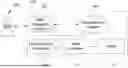
FIG. 1 illustrates a flow diagram of an assisted surgical planning system 100 according to one embodiment of the present disclosure. The assisted surgical planning system 100 includes a host computer 110, a discriminative AI module 120, a user navigation interface 130, a generative AI module 140, and a robot 150. The discriminative AI module 120 is electrically connected to the host computer 110. In some embodiments, the host computer 110 can be a Host computer. The user navigation interface 130 is electrically connected to the host computer 110, and may include a display screen. The generative AI module 140 is electrically connected to the host computer 110, such that the host computer 110 connects the user navigation interface 130 to the generative AI module 140. The robot 150 is electrically connected to the host computer 110 and can be a robotic arm having a puncture needle.
When in use, the discriminative AI module 120 can form plural organ segmentation images based on plural computed tomography (CT) images. The generative AI module 140 can generate plural suggested needle insertion paths based on the organ segmentation images and an initial needle insertion path provided by a user 200, and display the suggested needle insertion paths in the user navigation interface 130. The user 200 can be a doctor, e.g., a surgeon. The generative AI module 140 can perform selection and adjustment by the user 200 in the user navigation interface 130 based on the suggested needle insertion paths to generate a final needle insertion path through interaction. The user navigation interface 130 is configured to display the organ segmentation images, the initial needle insertion path, the suggested needle insertion paths, and the final needle insertion path. In some embodiments, the various needle insertion paths described above are, for example, needle insertion paths used in tumor punctures.
With the above configuration, the user 200 can observe the paths in the user navigation interface 130 and interact with the assisted surgical planning system 100. At the same time, the user navigation interface 130 is connected to the generative AI module 140 using the host computer 110. In this way, the user 200 can interact directly with the generative AI module 140 through the user navigation interface 130 and automatically visualize answers of the generative AI module 140 into the user navigation interface 130. After back-and-forth interaction and visualization, the assisted surgical planning system 100 generates a needle insertion path as the final insertion path which is transmitted to the robot 150 for subsequent path tracking.
Herein, “modules” and “units” can be implemented by software, hardware, or a combination of both. Plural different modules can be implemented in a same software or hardware structure, or one module can be implemented by plural different software or hardware structures. Hardware may include a processor, a memory, a hard disk, a sensor, or combinations thereof.
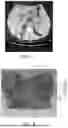
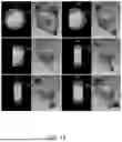
FIG. 2 illustrates a sketch flow diagram of an operation method of the assisted surgical planning system 100 according to one embodiment of the present disclosure. FIG. 3 illustrates an image of the user navigation interface in FIG. 1 when a doctor performs initial path planning. Referring also to FIGS. 2 and 3, first of all, in step S1, the doctor performs initial path planning. The doctor can perform initial path planning in CT images of a patient, for example, a brown path in FIG. 3 is an initial needle insertion path 102, and this step can provide a possible path range. Then, in step S2, the discriminative AI performs organ segmentation. Thus, the initial needle insertion path 102 and information about completion of the organ segmentation can be transmitted to the generative AI.
FIG. 4 illustrates an image of the user navigation interface 130 in FIG. 1 when the final path is generated. Reference is made also to FIG. 2 and FIG. 4, and then in step S3, the generative AI performs a path suggestion. In steps S2 and S3, the generative AI performing a cross-plane path suggestion can be implemented to generate light blue suggested needle insertion paths 104 as shown in FIG. 4. Then, in step S4, the suggested needle insertion paths 104 can be selected and adjusted by the doctor. In steps S3 and S4, the generative AI can interact with the doctor and adjust constantly. Then, in step S5, a needle insertion path is generated finally as a final path, e.g., a bright green final needle insertion path 106 as shown in FIG. 4.
In the following description, the assisted surgical planning system 100 in FIG. 1 is used as an example to describe its operation method.
FIG. 5 illustrates a flow diagram of the operation method of the assisted surgical planning system according to one embodiment of the present disclosure. The operation method of the assisted surgical planning system includes the following steps. Referring also to FIGS. 1 and 5, in step S11, the discriminative AI module 120 obtains plural CT images (as shown in FIG. 3). Then, in step S12, an initial needle insertion path 102 is set in the user navigation interface 130 based on the CT image (see FIG. 3). Next, in step S13, the discriminative AI module 120 forms plural organ segmentation images based on the CT images. Then, in step S14, the generative AI module 140 generates plural suggested needle insertion paths 104 based on the initial needle insertion path 102 and the organ segmentation images (see FIG. 4). Subsequently, in step S15, the suggested needle insertion paths 104 are displayed in the user navigation interface 130. Finally, in step S16, the suggested needle insertion paths 104 are selected and adjusted from the user navigation interface 130, such that the generative AI module 140 generates a final needle insertion path 106 (see FIG. 4). In addition, the doctor can select and adjust the suggested needle insertion paths from the user navigation interface 130 to form a modified path. Then, the generative AI module 140 can generate plural other suggested needle insertion paths based on the modified path and the organ segmentation images for the doctor's reference.
Specifically, since the assisted surgical planning system 100 includes the generative AI module 140 and the host computer 110 connects the user navigation interface 130 to the generative AI module 140, the generative AI module 140 can generate the suggested needle insertion paths 104 (see FIG. 4) based on the initial needle insertion path 102 (see FIG. 3) and the organ segmentation images, and the user navigation interface 130 displays the suggested needle insertion paths 104 for the doctor's reference. The doctor can interact with the generative AI module 140 through the user navigation interface 130, and select and adjust the suggested needle insertion paths 104 from the user navigation interface 130, such that the generative AI module 140 generates the final needle insertion path 106 (see FIG. 4). In this way, the assisted surgical planning system 100 and the operation method thereof can effectively save the time of preoperative needle insertion path planning and reduce the influence of subjective opinions. In addition, the doctor can check, confirm, interact and adjust back and forth on customary two-dimensional (2D) images (e.g., organ segmentation images) when the assisted surgical planning system 100 performs needle insertion path planning, so that the correctness of the needle insertion paths is improved, and the surgical risk can be further reduced.
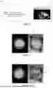
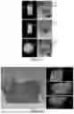
FIG. 6 illustrates a block diagram of the generative AI module 140 in FIG. 1. FIG. 7 illustrates an image of the generative AI module 140 in FIG. 6 when being contextually trained. Referring also to FIGS. 6 and 7, the generative AI module 140 includes a user input unit 142, a task unit 144, a judgment unit 146, and an output unit 148. In some embodiments, the generative AI module 140 can be contextually trained, that is, the generative AI module 140 is contextually learned and trained in a customized manner, such that the generative AI module 140 learns about the surgical context and its tasks, and provides a more clinically appropriate response, e.g., provides suggested needle insertion paths for the doctor to choose and adjust. Contextual training includes inputting an entry point P1 (e.g., a green point in FIG. 7), a target point P2 (e.g., a yellow point in FIG. 7), plural danger zones 103 (e.g., pink and dark blue zones in FIG. 7), and a tumor 101 (e.g., a red zone in FIG. 7) in a medical image (e.g., organ segmentation image) through the user input unit 142. In some embodiments, the danger zones 103 may include arteries, veins, and blood vessels.
FIG. 8 illustrates images of the generative AI module 140 when generating the suggested needle insertion paths 104 based on the initial needle insertion path 102 and the organ segmentation images according to one embodiment of the present disclosure. Then, the initial needle insertion path 102 (see FIG. 7) is adjusted through the task unit 144 to generate the suggested needle insertion paths 104. The task unit 144 is designed to perform fine adjustment of the paths, while the judgment unit 146 is designed to find a safer and more effective path (e.g., away from the danger zones), and can steer the suggested needle insertion paths 104 away from the danger zones 103 and adjust the target point P2 to the center of the tumor 101. In addition, there are more clinical considerations such as fat thickness in different zones, variables such as organ displacement caused by water pumping, etc. The contextual learning only enables the generative AI module 140 to learn the doctor's experience and then to perform better clinical path planning. Coordinates and images of the suggested needle insertion paths 104 can then be generated through the output unit 148. For example, the assisted surgical planning system 100 in FIG. 1 can capture the coordinates and display the coordinates in the user navigation interface 130 (see FIG. 1) for interaction with the doctor.
After the doctor has completed the planning of the initial needle insertion path 102 in FIG. 7, the assisted surgical planning system 100 (see FIG. 1) performs K-degree (e.g. 360-degree) rotation and reslicing along the path to provide more path possibilities in the space. In this way, n (e.g., six) resliced images of 0°, 30°, 60°, 90°, 120°, and 150° in FIG. 8 can be obtained after the rotation. The generative AI module 140 performs m path suggestions on the above n image slices, e.g., five suggested needle insertion paths 104 on each slice in FIG. 8. The doctor can then perform interaction and adjustment to the desired slices and paths.
FIG. 9 illustrates a schematic diagram of interaction between the doctor and the generative AI module 140 (see FIG. 1) according to one embodiment of the present disclosure. A screen in FIG. 9 can be displayed in the user navigation interface 130 of FIG. 1. For example, the doctor considers one of a plurality of slices (e.g., 30° slice in FIG. 8) to be the most appropriate needle insertion slice, but in several suggestions provided by the generative AI module 140, a path 5 needs to be finely adjusted and asked to generate another path. After being instructed by the doctor's requirements, the generative AI module 140 can finely adjust the path 5 to generate a path 6 in response to the suggestion to meet the doctor's request.
FIG. 10 illustrates images when the initial needle insertion path 102 is set according to one embodiment of the present disclosure. FIG. 11 illustrates images when the organ segmentation images are formed according to one embodiment of the present disclosure. Referring also to FIGS. 10 and 11, the doctor can perform initial path planning in the CT images of FIG. 10, for example, providing the initial needle insertion path 102, and then the assisted surgical planning system 100 (see FIG. 1) can perform automated organ segmentation by the discriminative AI module 120 to obtain the organ segmentation images of FIG. 11.
FIG. 12 illustrates images when K-degree image reslicing is performed along the initial needle insertion path 102 according to one embodiment of the present disclosure. Then, K-degree (e.g., 360 degree) image reslicing is performed along the initial needle insertion path 102 planned by the doctor.
FIG. 13 illustrates images when the suggested needle insertion paths 104 are generated according to one embodiment of the present disclosure. Then, the generative AI module 140 (see FIG. 1) generates a plurality of suggested needle insertion paths 104 on each image cut.
FIG. 14 illustrates images when the final needle insertion path 106 is generated according to one embodiment of the present disclosure. Finally, the suggested needle insertion paths 104 generated by the generative AI module 140 (see FIG. 1) can be displayed in 3D (see the left half of FIG. 14) and 2D (see the right half of FIG. 14) images in the user navigation interface 130, so that the doctor can check and confirm back and forth on the customary CT images, and make interaction and adjustment. All information is visualized in 3D and 2D interfaces to facilitate the doctor to observe and finely adjust, where the 2D image provides a multi-planar reconstruction (MPR) view of the needle insertion paths in three directions. The 3D and 2D interfaces include the tumor 101 (e.g., red zones) and the danger zones 103 (e.g., pink and blue zones) automatically segmented. At the same time, the needle insertion paths are displayed, including the initial needle insertion path 102 (e.g., brown path) of the doctor, the suggested needle insertion paths 104 (e.g., blue paths) generated by the system, and the final needle insertion path 106 (e.g., bright green path) selected after interaction.
After the interaction between the doctor and the system to select the final needle insertion path 106 is completed, since the images read by the generative AI module 140 (see FIG. 1) are a plurality of two-dimensional image slices (i.e., CT images), the coordinates of the path when a path suggestion is made are also two-dimensional image coordinates. However, the system displays the location of a path in a 3D space in the user navigation interface 130 (see FIG. 1) and transmits the path to the robot 150 (see FIG. 1) for path tracking, so 2D path coordinates transmitted back by the generative AI module 140 can be converted to path coordinates in a 3D space. In some embodiments, when multi-angle reslicing of a two-dimensional image is performed, the system records the coordinates of the image in the space at each angle
( i . e . , CT ( n degree ) 3 D T ) ,
so that the path coordinates
( i . e . , □ CT ( n degree ) P entry , □ CT ( n degree ) P target )
in any two-dimensional image (i.e.,) can be converted to path coordinates
( i . e . , □ 3 D P entry , □ 3 D P target )
in the 3D space. A 2D-to-3D coordinate conversion formula is as follows:
3 D P entry = CT 3 D T CT P entry 3 D P target = CT 3 D T CT P target .
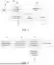
FIG. 15 illustrates a flow diagram of an operation method of an assisted surgical planning system according to another embodiment of the present disclosure. In step S21, data acquisition is performed, for example, CT images of a patient are obtained and displayed in an interface (e.g., the user navigation interface described above). Then, in step S22, a doctor sets an initial needle insertion path. Selection of an initial path by the doctor is performed on the CT images of the patient to provide an expected needle insertion zone. Then in step S23, segmentation of organs and blood vessels is performed. The assisted surgical planning system performs automatic organ segmentation to segment danger zones, tumors, etc., so that subsequently the generative AI module can identify the zones. In addition, the system then performs k-degree (e.g., 360-degree) image reslicing along the initial path. Then in step S24, check for the needle insertion path is performed. For example, a path safety check is performed to ensure the safety of the initial path and ensure that the initial path does not pass through the danger zones. Then, in step S25, alternative needle insertion paths (e.g., the suggested needle insertion paths described above) are provided. Then, in step S26, from feedback from doctor (e.g., adjustment and selection of the suggested needle insertion paths), fine adjustment of the needle insertion paths is performed through interaction with the doctor. Then, in step S27, coordinates (e.g., the coordinates of the final needle insertion path described above) are obtained. In addition, in steps S24-S27, a repeat for all CT image slices rotated in n degree angular increments can be performed. In step S28, 2D and 3D visualization (as displayed in the user navigation interface 130 described above) is performed. In other words, the coordinate position of the path finally selected after the interaction (the final needle insertion path described above) can be calculated by the system and displayed synchronously in the 2D and 3D interfaces (as shown in FIG. 14).
FIG. 16 illustrates a flow diagram of the assisted surgical planning system according to one embodiment of the present disclosure. The assisted surgical planning system 100 further includes a visualization module 162, a CT reslice module 164, a message module 166, a robot module 168, a robot 150, and a screen 180. The visualization module 162, the CT reslice module 164, the message module 166 and the robot module 168 are electrically connected to the generative AI module 140. The generative AI module 140 can receive prompts (e.g., files). The visualization module 162 can receive the CT images and the coordinates of the paths generated by the generative AI module 140, e.g., the coordinates of the suggested needle insertion paths and the coordinates of the final needle insertion path, and displays the paths on the screen 180. The CT reslice module 164 can receive the CT images and transmit the organ segmentation images to the generative AI module 140. The message module 166 can receive voice messages and type messages and transmits the messages to the generative AI module 140. The assisted surgical planning system 100 can transmit both audio messages and text messages. The robot module 168 can receive a command from the generative AI module 140 to operate the robot 150 having a puncture needle.
The generative AI module 140 can communicate with an application program interface (API) of a large language model (LLM), including sending images, commands and messages, and after receiving messages transmitted back, interpreting the messages, extracting the coordinates in the messages, and displaying the messages to the user, etc. The application program interface has a vector storage 170. In addition, whether an answer matches the user's question is checked. We write the application program interface into the back end of the system interface to interface with the generative AI module 140, so that the user can use the system and the back-end generative AI to make voice interaction, and the response and the generated paths of the generative AI can be automatically captured by the system and displayed in the system interface (e.g., the screen 180).
FIG. 17 illustrates a flow diagram of an operation method of an assisted surgical planning system according to yet another embodiment of the present disclosure. A process of interaction between a doctor and the assisted surgical planning system is as follows. In step S31, Doctor: Please provide several alternative paths. (audio input). Then in step S32, AI: I have provided you several paths and numbered, please choose your favorite one. (audio output; Screen shows these numbered paths). Then in step S33, Doctor: I choose number #(audio input). Then in step S34, AI: I highlight the chosen path, is this the path you have chosen? (audio output; Screen shows these paths, with the chosen highlighted path). Then in step S35, Doctor: that is correct. (audio input). Then in step S36, AI: Selected path number #. Ready for needle insertion. (audio output). Then in step S37, Doctor: Please move the robot to the ready position. (audio input). Finally, in step S38, AI: Send the command to move the end-effector of a robot arm to the target position. (Action).
The foregoing outlines the features of several embodiments so that those skilled in the art may better understand the aspects of the present disclosure. Those skilled in the art should understand that they can easily use the present disclosure as a basis for designing or modifying other processes and structures to achieve the same purposes and/or to achieve the same advantages as the embodiments described herein. Those skilled in the art should also be aware that such equivalent constructions are not divorced from the spirit and scope of the present disclosure, and that, without deviating from the spirit and scope of the present disclosure, they may be subject here to various alterations, substitutions and alterations.
Claims
What is claimed is:1. An assisted surgical planning system, comprising:
a host computer;
a discriminative artificial intelligence (AI) module electrically connected to the host computer, and configured to form a plurality of organ segmentation images based on a plurality of computed tomography (CT) images;
a user navigation interface electrically connected to the host computer, and configured to display the organ segmentation images, an initial needle insertion path, a plurality of suggested needle insertion paths, and a final needle insertion path; and
a generative AI module electrically connected to the host computer, such that the host computer connects the user navigation interface to the generative AI module, wherein the generative AI module is configured to generate the suggested needle insertion paths based on the organ segmentation images and the initial needle insertion path, and display the suggested needle insertion paths in the user navigation interface.
2. The assisted surgical planning system according to claim 1, wherein the generative AI module is configured for selection and adjustment in the user navigation interface based on the suggested needle insertion paths to generate the final needle insertion path.
3. The assisted surgical planning system according to claim 1, wherein the generative AI module comprises a user input unit configured to input an entry point, a target point, a plurality of danger zones and a tumor into the organ segmentation images.
4. The assisted surgical planning system according to claim 3, wherein the generative AI module further comprises a task unit configured to adjust the initial needle insertion path to generate the suggested needle insertion paths.
5. The assisted surgical planning system according to claim 4, wherein the generative AI module further comprises a judgment unit configured to steer the suggested needle insertion paths away from the danger zones and adjust the target point to a center of the tumor.
6. The assisted surgical planning system according to claim 5, wherein the generative AI module further comprises an output unit configured to generate coordinates and images of the suggested needle insertion paths.
7. The assisted surgical planning system according to claim 1, further comprising:
a visualization module electrically connected to the generative AI module, and configured to receive the CT images, coordinates of the suggested needle insertion paths and coordinates of the final needle insertion path.
8. The assisted surgical planning system according to claim 1, further comprising:
a CT reslice module electrically connected to the generative AI module, and configured to receive the CT images and transmit the organ segmentation images to the generative AI module.
9. The assisted surgical planning system according to claim 1, further comprising:
a message module electrically connected to the generative AI module, and configured to transmit voice messages and type messages to the generative AI module.
10. The assisted surgical planning system according to claim 1, further comprising:
a robot module electrically connected to the generative AI module, and configured to receive a command from the generative AI module to operate a robot having a puncture needle.
11. An operation method of an assisted surgical planning system, comprising:
obtaining, by a discriminative AI module, a plurality of CT images;
setting an initial needle insertion path in a user navigation interface based on the CT images;
forming, by the discriminative AI module, a plurality of organ segmentation images based on the CT images;
generating, by a generative AI module, a plurality of suggested needle insertion paths based on the initial needle insertion path and the organ segmentation images, wherein the discriminative AI module, the generative AI module and the user navigation interface are electrically connected to a host computer;
displaying the suggested needle insertion paths in the user navigation interface; and
selecting and adjusting the suggested needle insertion paths from the user navigation interface, such that the generative AI module generates a final needle insertion path.
12. The operation method of an assisted surgical planning system according to claim 11, further comprising:
selecting and adjusting the suggested needle insertion paths from the user navigation interface to form a modified path; and
generating, by the generative AI module, a plurality of other suggested needle insertion paths based on the modified path and the organ segmentation images.
13. The operation method of an assisted surgical planning system according to claim 11, further comprising:
contextually training the generative AI module.
14. The operation method of an assisted surgical planning system according to claim 13, wherein contextually training the generative AI module comprises:
inputting an entry point, a target point, a plurality of danger zones and a tumor into the organ segmentation images, wherein the danger zones comprise arteries, veins, and blood vessels.
15. The operation method of an assisted surgical planning system according to claim 14, wherein contextually training the generative AI module further comprises:
adjusting the initial needle insertion path to generate the suggested needle insertion paths; and
generating coordinates and images of the suggested needle insertion paths.
16. The operation method of an assisted surgical planning system according to claim 15, wherein adjusting the initial needle insertion path to generate the suggested needle insertion paths comprises:
steering the suggested needle insertion paths away from the danger zones; and
adjusting the target point to a center of the tumor.
17. The operation method of an assisted surgical planning system according to claim 11, further comprising:
receiving, by a visualization module electrically connected to the generative AI module, the CT images, coordinates of the suggested needle insertion paths and coordinates of the final needle insertion path.
18. The operation method of an assisted surgical planning system according to claim 11, further comprising:
receiving the CT images and transmitting the organ segmentation images to the generative AI module by a CT reslice module electrically connected to the generative AI module.
19. The operation method of an assisted surgical planning system according to claim 11, further comprising:
transmitting, by a message module electrically connected to the generative AI module, voice messages and type messages to the generative AI module.
20. The operation method of an assisted surgical planning system according to claim 11, further comprising:
receiving, by a robot module electrically connected to the generative AI module, a command from the generative AI module to operate a robot having a puncture needle.
Images & Drawings included:


Sources:
- United States Patent and Trademark Office - verify current appl. status at the USPTO↗
Recent applications in this class:
- » 20260041509 2026-02-12
TRAINING SYSTEM FOR A NEURAL NETWORK TO GUIDE A ROBOTIC ARM TO OPERATE A CATHETER - » 20260033901 2026-02-05
SYNCHRONIZED ROBOTIC BONE MILLING - » 20260013959 2026-01-15
Delivery System And Method For Delivering Material To A Target Site During A Medical Procedure - » 20260013958 2026-01-15
AUTOMATED TISSUE TREATMENT DEVICES, SYSTEMS, AND METHODS - » 20260000474 2026-01-01
ROBOTIC SURGERY SYSTEM LAYOUTS AND RELATED METHODS - » 20250359953 2025-11-27
CLOSED-LOOP FEEDBACK BASED ON MIXED DIMENSIONALITY IMAGING - » 20250352287 2025-11-20
SYSTEMS AND METHODS FOR ROBOTIC MEDICAL SYSTEM INTEGRATION WITH EXTERNAL IMAGING - » 20250352286 2025-11-20
APPARATUS FOR DRIVING SURGICAL ROBOT SYSTEM AND METHOD THEREOF - » 20250331937 2025-10-30
System And Method For Aligning An End Effector To A Haptic Object - » 20250331936 2025-10-30
SYSTEMS AND TECHNIQUES FOR MINIMALLY-INVASIVE PROCEDURES