PLURIPOTENT STEM CELL-DERIVED MEGAKARYOCYTES AND PLATELETS
US20260103680A1
2026-04-16
19/117,857
2023-10-05
Smart Summary: Researchers have developed a way to create megakaryocytes and platelets from human induced pluripotent stem cells (iPSCs) outside the body. These cells and platelets are functional and closely mimic those found in human blood and bone marrow. The process is efficient and can produce large quantities of these important blood components. Additionally, the invention includes isolated cells and platelets, as well as potential treatments using these products. Overall, this advancement could improve therapies related to blood disorders and enhance our understanding of blood cell development. 🚀 TL;DR
Abstract:
The present disclosure provides for efficient ex vivo processes for generating megakaryocytes and/or platelets from human induced pluripotent stem cells (iPSCs). Cells and platelets generated according to the disclosure in various embodiments are functional and/or more closely resemble the corresponding lineage isolated from peripheral blood, bone marrow, or other tissues. The present invention in some aspects provides isolated cells/platelets and compositions produced by the methods disclosed herein, as well as methods for therapy.
Applicant:
Interested in similar patents?
Get notified when new applications in this technology area are published.
Classification:
C12N5/0644 » CPC main
Undifferentiated human, animal or plant cells, e.g. cell lines; Tissues; Cultivation or maintenance thereof; Culture media therefor; Animal cells or tissues; Human cells or tissues; Vertebrate cells; Cells from the blood or the immune system Platelets; Megakaryocytes
A61K35/19 » CPC further
Medicinal preparations containing materials or reaction products thereof with undetermined constitution; Materials from mammals; Compositions comprising non-specified tissues or cells; Compositions comprising non-embryonic stem cells; Genetically modified cells; Blood; Artificial blood Platelets; Megacaryocytes
C12N2501/145 » CPC further
Active agents used in cell culture processes, e.g. differentation; Growth factors Thrombopoietin [TPO]
C12N2506/45 » CPC further
Differentiation of animal cells from one lineage to another; Differentiation of pluripotent cells from artificially induced pluripotent stem cells
Description
CROSS-REFERENCE TO RELATED APPLICATIONS
This application claims the benefit of and priority to U.S. Provisional Patent Application No. 63/413,337 filed Oct. 5, 2022, the contents of which are hereby incorporated by reference in their entirety.
SEQUENCE LISTING
The instant application contains a Sequence Listing which has been submitted in XML format via EFS-Web and is hereby incorporated by reference in its entirety. Said XML copy, created on Sep. 26, 2023, is named GRU-013PC_Sequence_Listing.xml and is 30,048 bytes in size.
BACKGROUND
Besides playing a major role in physiological hemostasis, thrombosis and wound healing, platelets can also make great contributions to host inflammation and immune responses to infection and injury. Many patients with hematopoietic disease or undergoing aggressive chemotherapy require platelet transfusions using platelet concentrates obtained through blood/platelet donation. However, platelets for transfusion obtained from volunteer blood donors are a limited resource because, for instance, the blood donation industry is facing an ongoing crisis as the demand for blood products, particularly platelets, frequently outstrips supply. Therefore, development of large scale, off-the-shelf, megakaryocytes and/or platelets, such as those from hematopoietic stem cells (HSCs), is an attractive tool in regenerative medicine.
SUMMARY OF THE DISCLOSURE
The present disclosure, in various aspects and embodiments, provides methods for generating hematopoietic lineages for cell therapy, including a megakaryocyte lineage, such as megakaryocyte-erythroid progenitors (MEPs), CFU-Me (pluripotential hemopoietic stem cell or hemocytoblast), megakaryoblast, promegakaryocyte, and megakaryocyte, as well as proplatelets, preplatelets or platelets derived therefrom. In various embodiments, the invention provides for efficient ex vivo processes for developing megakaryocytes and/or platelets from human induced pluripotent stem cells (iPSCs), including gene edited iPSCs. The megakaryocytes and/or proplatelets, preplatelets or platelets generated according to the present disclosure in various embodiments are functional and/or more closely resemble the corresponding natural lineages isolated from bone marrow or blood or the corresponding natural platelets isolated from blood. The present invention also provides isolated cells and compositions produced by the methods disclosed herein, as well as methods for therapy.
In other aspects and embodiments, this disclosure provides HSCs that are derived from iPSCs that are gene edited to be immune compatible with a significant portion of the population. These HSC populations can be used for more efficient ex vivo platelet production, or in other aspects, can be used to deliver HSCs or megakaryocytes, platelets, or their progenitors to patients in need to reduce or eliminate requirements for periodic transfusions.
In one aspect, the disclosure provides a method for preparing a cell population comprising megakaryocytes. The megakaryocytes of the present disclosure are capable of further generating functional platelets. The method of the present disclosure comprises preparing a pluripotent stem cell (PSC) population, such as an induced pluripotent stem cell (iPSC) population differentiated to embryoid bodies, and enriching for CD34+ cells to thereby prepare a CD34+-enriched population. Endothelial-to-hematopoietic transition (EHT) is induced in the CD34+-enriched population to thereby prepare a population comprising hematopoietic stem cells (HSCs) and/or hematopoietic stem progenitor cells (HSPCs). The resulting cell population (or fraction thereof) can be differentiated to megakaryocytes and optionally used to produce platelets.
In various embodiments, the iPSCs are prepared by reprogramming somatic cells. In some embodiments, iPSCs are derived from CD34+ cells isolated from peripheral blood. In various embodiments, the iPSCs are autologous or allogenic (e.g., HLA-matched at one or more loci) with respect to a recipient (a subject in need of treatment as described herein). In various embodiments, the iPSCs can be gene edited to assist in HLA matching. For example, iPSCs can be gene edited to delete one or more of HLA-A, HLA-B, and HLA-C, and to delete one or more of HLA-DP, HLA-DQ, and HLA-DR. In certain embodiments, the iPSCs retain expression of at least one HLA Class I and at least one HLA Class II complex. In certain embodiments, iPSCs are homozygous for at least one retained Class I and Class II loci. In some embodiments, iPSCs are gene edited to be HLA-Aneg, homozygous for both HLA-B and HLA-C, and HLA-DPB1neg and HLA-DQB1neg. In some embodiments, the iPSCs are further homozygous for HLA-DRB1.
In various embodiments, iPSCs are prepared, and expanded using a culture system. Expanded iPSCs can be recovered from the culture and used for generating embryoid bodies (EBs). EBs, created by differentiation of iPSCs, are three-dimensional aggregates of iPSCs and comprise the three (or alternatively two or one) embryonic germ layer(s) based on the differentiation method(s). In some embodiments, the process according to each aspect can comprise generating CD34+ cells from the pluripotent stem cells (e.g., EBs) and inducing endothelial-to-hematopoietic differentiation. HSCs can be generated from the cell populations using various stimuli or factors, including mechanical, biochemical, metabolic, and/or topographical stimuli, as well as factors such as extracellular matrix, niche factors, cell-extrinsic factors, induction of cell-intrinsic properties; and including pharmacological and/or genetic means.
In some embodiments, iPSC differentiation proceeds until cells are at least about 20% CD34+ or at least about 30% CD34+. In some embodiments, CD34 enrichment and EHT may be induced at Day 7 to Day 14 of iPSC differentiation. Differentiation of iPSCs can be according to known techniques. In some embodiments, iPSC differentiation involves factors such as, but not limited to, combinations of bFGF, Y27632, BMP4, VEGF, SCF, EPO, TPO, IL-6, IL-11, and/or IGF-1.
Induction of EHT can be with any known process. In some embodiments, induction of EHT generates a hematopoietic stem cell (HSC) population comprising LT-HSCs. In some embodiments, EHT generates HSCs through endothelial or hemogenic endothelial cell (HEC) precursors using mechanical, biochemical, pharmacological and/or genetic means (e.g., via stimulation, inhibition, and/or genetic modifications). In some embodiments, the EHT generates a stem cell population comprising one or more of long-term hematopoietic stem cells (LT-HSCs), short-term hematopoietic stem cells (ST-HSCs), and hematopoietic stem progenitor cells. In some embodiments, EHT is induced in the culture from about 5 days to about 7 days. In embodiments, EHT takes place using a medium comprising one or more growth factors and cytokines selected from TPO, SCF, Flt3L, IL3, IL-6, IL7, IL-11, IGF, bFGF, and IL15. The medium may optionally comprise one or more of VEGF, bFGF, a BMP activator, a Wnt pathway activator, or ROCK inhibitors (e.g., thiazovivin or Y27632). In some embodiments, the HSC and/or HSPC population or fraction thereof is differentiated independent of the use of an agonist of a mechanosensitive receptor or a mechanosensitive channel, such as Yoda1. In some embodiments, the use of an agonist of a mechanosensitive receptor or a mechanosensitive channel (e.g., Yoda1) is optional.
In various embodiments, pharmacological Piezol activation is applied to CD34+ cells (i.e., CD34+-enriched cells). In certain embodiments, pharmacological Piezol activation may further be applied to iPSCs, embryoid bodies, ECs, hemogenic endothelial cells (HECs), HSCs, hematopoietic progenitors, as well as hematopoietic lineage(s), including megakaryocyte lineage that is responsible for the generation of platelets. In certain embodiments, Piezol activation is applied at least to EBs generated from iPSCs, and/or CD34+ cells isolated from EBs, which in accordance with various embodiments, allows for superior generation of megakaryocytes as compared to other methods for inducing EHT.
In various embodiments, CD34+ cells (e.g., the floater and/or adherent cells) are harvested from the culture undergoing endothelial-to-hematopoietic transition between Day to Day 20 of iPSC differentiation, such as from Day 12 to Day 17. In some embodiments, the CD34+ cells comprise non-adherent cells. In various embodiments, the HSCs or CD34+-enriched cells are further expanded.
HSCs and/or HSPCs which give rise to megakaryocytes and/or platelets can be identified based on the expression of CD34 and the absence of lineage specific markers (termed Lin−). In some embodiments, the stem cell population for differentiation to a hematopoietic lineage is at least about 80% CD34+, or at least about 90% CD34+, or at least about 95% CD34+.
In various embodiments, the HSC and/or HSPC population or fraction thereof is differentiated to megakaryocytes ex vivo, from which platelets can be generated.
In some embodiments, the step of differentiating the population comprising HSCs and/or HSPCs into megakaryocytes comprises culturing with thrombopoietin (TPO). The culture may further comprise one or more additional cytokines or growth factors, such as those selected from IL-1, IL-3, IL-6, IL-9, IL-11, SCF, SDF-1, and PDGF-BB. The cytokines and growth factors, including TPO, can be selected to further expand megakaryocytes. In some embodiments, such additional cytokines or growth factors for expanding megakaryocytes may be selected from stem cell factor (SCF), FMS-like tyrosine kinase 3 ligand (Flt3L), IL-6, IL-9, and erythropoietin (EPO).
The mature megakaryocytes can form proplatelets and platelets as known in the art, such as by culture in the presence of fibroblast growth factor 4 (FGF4) and stromal cell-derived factor 1 (SDF 1). In some embodiments, the megakaryocytes or proplatelets are cultured in a bioreactor subjecting the cells to hydrodynamic shear stress. For example, platelets can be produced in static 2D, serum-free, cytokine-dependent conditions. Alternatively, platelets can be produced in a three-dimensional (3D) microenvironment. In various embodiments, the platelets will have a phenotype that is CD41+CD42b+. Recovered platelets are activatable by thrombin. The platelets can be recovered and subjected to gamma irradiation before transfusion therapy.
The composition of this disclosure (e.g., comprising platelets prepared according to this disclosure) may further comprise a pharmaceutically acceptable carrier. Such carrier solutions also can contain buffers, diluents, and other suitable additives. Cell or platelet compositions may be provided in implantable devices (e.g., scaffolds) or in bags or in vials, tubes or a container in an appropriate volume and stored frozen until use. In various embodiments, the composition comprises at least about 109 platelets, or at least about 1010 platelets, or at least about 1011 platelets, or at least about 1012 platelets per 50 or 100 mL volume.
In other aspects, the invention provides a method for platelet therapy, comprising administering platelets (prepared as described herein), or pharmaceutically acceptable composition thereof, to a human subject in need thereof. In various embodiments, the methods described herein are used to treat subject who has thrombocytopenia, such as ACTN1-related thrombocytopenia, amegakaryocytic thrombocytopenia with radio-ulnar synostosis, ANKRD26 related thrombocytopenia, autosomal dominant thrombocytopenia, congenital amegakaryocytic thrombocytopenia, CYCS-related thrombocytopenia, FYB related thrombocytopenia, idiopathic thrombocytopenic purpura, or X-linked thrombocytopenia. In some embodiments, platelets are administered to a subject suffering from hemorrhage.
Other aspects and embodiments of this disclosure will be apparent from the following detailed disclosure and working examples.
BRIEF DESCRIPTION OF DRAWINGS
FIG. 1 shows that ETV2 over-expression (OE) does not affect pluripotency. FIG. 1 shows FACS plots representative of transduction efficiency of iPSC with an adenoviral vector to overexpress ETV2 and GFP sequences. ETV2 overexpression does not affect the iPSC stemness as shown by the expression of the TRA-1-60 stemness marker.
FIG. 2 shows that ETV2 over-expression (OE) increases the yield of hemogenic endothelial cells. Representative flow cytometric analysis of hemogenic endothelial cells (described as CD235a-CD34+CD31+) and relative quantification demonstrates that ETV2-OE enhances the formation of hemogenic endothelial cells.
FIG. 3 shows that ETV2 over-expression (OE) enhances CD34+ cell formation during iPSC differentiation. Representative flow cytometric analysis of CD34+ cells and relative quantification demonstrates that ETV2-OE enhances the CD34+ cell formation.
FIG. 4 shows FACS analysis of megakaryocyte differentiation indicating the commitment of HSCs to megakaryocyte lineage.
FIG. 5 shows that megakaryocyte cells derived from iPSC-derived HSCs are capable of expansion ex vivo.
FIG. 6 shows immunofluorescence analysis of platelet differentiation of iPSC-derived HSCs, indicating that derived megakaryocytes are phenotypically similar to BM CD34+ derived megakaryocytes and are able to release platelets.
FIG. 7 shows thrombus formation with platelets produced from iPSC-derived HSCs, indicating that these platelets are able to clot and promote the thrombus formation once activated.
FIG. 8A and FIG. 8B show the phenotype analysis of HLA edited (e.g., triple knockout) cells performed by FACS and immunofluorescence. FIG. 8A shows the overall expression of HLA class-I molecules (HLA-A, HLA-B, and HLA-C) on the cell surface, where the HLA edited cells are positive for overall HLA class-I expression to a similar degree as wild-type cells. FIG. 8B shows cell expression of HLA-A via immunofluorescence, where HLA-A is not expressed in the HLA edited clone.
FIG. 9 shows that the HLA edited clones preserve their pluripotency (maintain trilineage differentiation), as illustrated by immunofluorescence, with ectoderm differentiation indicated by NESTIN-488 and PAX6-594 staining, mesoderm differentiation indicated by GATA-488 staining, and endoderm differentiation indicated by CXCR4-488 and FOX2A-594 staining.
FIG. 10 shows the immune compatibility of the HLA edited HSCs. HLA edited HSCs and control HSCs (WT, B2M KO, and HLA Class II null) were co-cultured with peripheral blood mononuclear cells (PBMCs) matching HLA-B and HLA-C, but with mismatched HLA-A, and PBMC-mediated cytotoxicity was measured by an annexin V staining assay.
FIG. 11 shows in vivo engrafting potential of HLA edited HSCs. Equal proportions of mCherry HLA edited HSCs and wild-type HSCs (gHSCs) were mixed for a competitive transplant into mice, where bone marrow (BM) and peripheral blood samples were evaluated by FACS to compare the relative amounts of each cell type present in the samples.
FIG. 12 shows that HLA-edited HSCs can differentiate into megakaryocytes (MK) which can further differentiate into platelets. Images at the left show increased proportion of platelets in HSCs by light microscopy at 1000× magnification. The graph at the right shows a statistically significant increase in the proportion of platelets differentiated from HLA-edited HSCs compared to those differentiated from bone marrow (BM) CD34+ cells and CD34+ cell populations isolated from differentiated iPSCs (e.g., EBs).
The term “gHSC” is used herein to refer to the iPSC-derived hematopoietic stem cells of the present disclosure.
The terms “wild type” (WT), “unedited”, “non-HLA-edited” are used interchangeability herein to refer to the non-gene edited cells of the present disclosure.
EB34+ cells refer to Embryonic body derived CD34+ cells. These comprise hemogenic endothelial cells.
DESCRIPTION OF THE INVENTION
The present disclosure, in various aspects and embodiments, provides methods for generating hematopoietic lineages for cell therapy, including a megakaryocyte lineage, such as megakaryocyte-erythroid progenitors (MEPs), CFU-Me (pluripotential hemopoietic stem cell or hemocytoblast), megakaryoblast, promegakaryocyte, and megakaryocyte, as well as proplatelets, preplatelets or platelets derived therefrom. In various embodiments, the invention provides for efficient ex vivo processes for developing megakaryocytes and/or platelets from human induced pluripotent stem cells (iPSCs), including gene edited iPSCs. The megakaryocytes and/or proplatelets, preplatelets or platelets generated according to the present disclosure in various embodiments are functional and/or more closely resemble the corresponding natural lineages isolated from bone marrow or blood or the corresponding natural platelets isolated from blood. The present invention also provides isolated cells and compositions produced by the methods disclosed herein, as well as methods for therapy.
In other aspects and embodiments, this disclosure provides HSCs that are derived from iPSCs that are gene edited to be immune compatible with a significant portion of the population. These HSC populations can be used for more efficient ex vivo platelet production, or in other aspects, can be used to deliver HSCs or megakaryocytes, platelets, or their progenitors to patients in need to reduce or eliminate requirements for periodic transfusions.
In accordance with aspects and embodiments of this disclosure, the ability of human induced pluripotent stem cells (hiPSCs) to produce essentially limitless pluripotent stem cells (PSCs) is leveraged to generate boundless supply of hematopoietic cells, including but not limited to therapeutic lineages giving rise to megakaryocytes, which, in turn, release proplatelets and platelets. Use of platelets in therapy has been limited because volunteer blood donors are a limited resource. Since donor-derived platelets must be stored at room temperature, their shelf life are limited to about 5 days, they are prone to bacterial growth during storage and lack consistency between donor derived batches. Moreover, platelet transfusion is often associated with several risks to the recipient including allergic reactions and febrile nonhemolytic reactions. See, for example, Kaufman, Richard M., et al. “Platelet transfusion: a clinical practice guideline from the AABB.” Annals of Internal Medicine 162, no. 3 (2015): 205-213. Moreover, compared to primary cells, hiPSCs can more readily undergo genetic modifications in vitro, thereby offering opportunities to improve cell/platelet numbers, as well as bypassing HLA-matching issues for example. Additionally, fully engineered hiPSC clones, as compared to primary cells, can serve as a stable and safe source. Further, because hiPSCs, unlike human Embryonic Stem Cells (hESCs), are of non-embryonic origin, they are also free of ethical concerns and are consistent in quality. Accordingly, use of hiPSCs according to this disclosure confers several advantages over primary cells to generate therapeutic hematopoietic lineages, such as a megakaryocyte lineage, including as megakaryocytes and platelets (including progenitors thereof).
In one aspect, the disclosure provides a method for preparing a cell population comprising megakaryocytes. The megakaryocytes of the present disclosure are capable of further generating functional platelets. The method of the present disclosure comprises preparing a pluripotent stem cell (PSC) population, such as an induced pluripotent stem cell (iPSC) population differentiated to embryoid bodies, and enriching for CD34+ cells to thereby prepare a CD34+-enriched population. Endothelial-to-hematopoietic transition (EHT) is induced in the CD34+-enriched population to thereby prepare a population comprising hematopoietic stem cells (HSCs) and/or hematopoietic stem progenitor cells (HSPCs). In some embodiments, CD34+ cells are enriched from the population comprising HSCs and/or HSPCs. In various embodiments, EHT is induced for at least 2 days, and up to 12 days. The resulting cell population (or fraction thereof) can be differentiated to megakaryocytes and optionally used to produce platelets.
Conventionally, hematopoietic lineages are prepared by differentiation of iPSCs to embryoid bodies up to day 8 to harvest CD34+ cells. CD34 is commonly used as a marker of hemogenic endothelial cells, hematopoietic stem cells, and hematopoietic progenitor cells. In accordance with aspects and embodiments of this disclosure, it is discovered that inducing endothelial-to-hematopoietic transition (EHT) of a CD34+ cell population, and which can be derived from iPSCs-embryoid bodies, can be used for the ex vivo generation of superior hematopoietic stem cells and hematopoietic lineages, such as megakaryocyte lineages, which are further capable of generating functional platelets.
In some embodiments, CD34+ cells (i.e., recovered from EB dissociation) are contacted with an effective amount of an agonist of a mechanosensitive receptor or a mechanosensitive channel that increases the activity or expression of Dnmt3b. In some embodiments, the mechanosensitive receptor is Piezol. Exemplary Piezol agonists include Yoda1, Jedi1, and Jedi2 or analogues thereof. In some embodiments, the mechanosensitive receptor is Trpv4. An exemplary Trpv4 agonist is GSK1016790A. Other manners for inducing EHT (alternatively or in addition) can be used and are described herein. In some embodiments, after inducing EHT, the cells (the population comprising HSCs and/or HSPCs) are differentiated to a megakaryocyte lineage, e.g., a cell population comprising megakaryocytes which are capable of generating or secreting platelets.
In various embodiments, the iPSCs are prepared by reprogramming somatic cells. The term “induced pluripotent stem cell” or “iPSC” refers to cells derived from somatic cells, such as skin or blood cells that have been reprogrammed back into an embryonic-like pluripotent state. In some embodiments, iPSCs are generated from somatic cells such as (but not limited to) fibroblasts or PBMCs (or cells isolated therefrom). In some embodiments, the iPSCs are derived from lymphocytes, granulocyte/macrophage lineage-restricted progenitors (GMPs), cord blood cells, PBMCs, CD34+ cells, or other human primary tissues. In some embodiments, iPSCs are derived from CD34+ cells isolated from peripheral blood. In various embodiments, the iPSCs are autologous or allogenic (e.g., HLA-matched at one or more loci) with respect to a recipient (a subject in need of treatment as described herein). In various embodiments, the iPSCs can be gene edited to assist in HLA matching (such as deletion of one or more HLA Class I and/or Class II alleles or their master regulators, including but not limited beta-2-microglobulin (B2M), CIITA, etc.), or gene edited to delete or express other functionalities. For example, iPSCs can be gene edited to delete one or more of HLA-A, HLA-B, and HLA-C, and to delete one or more of ILA-DP, ILA-DQ, and HLA-DR. In certain embodiments, the iPSCs retain expression of at least one HLA Class I and at least one HLA Class II complex. In certain embodiments, iPSCs are homozygous for at least one retained Class I and Class II loci.
In various embodiments, HSCs and megakaryocyte lineage cells are derived from iPSCs which are gene edited to be one of: (i) HLA-A−B+C+DP−DR+DQ+, (ii) HLA-A−B+C+DP+DR+DQ−, (iii) HLA-A−B+C+DP−DR+DQ−, (iv) HLA-A−B−C+DP-DR+DQ+; (v) HLA-A−B−C+DP+DR+DQ−, (vi) HLA-A−B−C+DP−DR+DQ−. For retained HLA (for example HLA-B, HLA-C, and HLA-DR), cells can be homozygous or retain only a single copy of the gene. For example, the modified cells are identified at least as (a) HLA-C+ and ILA-DR+, and optionally identified as one or more of (b) HLA-B−, (c) HLA-DP−, and (d) HLA-DQ−. In exemplary embodiments, the modified cells are HLA-B+, HLA-DP−, and HLA-DQ−.
In some embodiments, iPSCs are gene edited to be HLA-Aneg, homozygous for both HLA-B and HLA-C, and HLA-DPB1neg and HLA-DQB1neg. In some embodiments, the iPSCs are further homozygous for HLA-DRB1.
As used herein, the term “neg,” (−), or “negative,” with respect to a particular HLA Class I or Class II molecule indicates that both copies of the gene have been disrupted in the cell line or population, and thus the cell line or population does not display significant functional expression of the gene. Such cells can be generated by full or partial gene deletions or disruptions, or alternatively with other technologies such as siRNA. As used herein, the term “delete” in the context of a genetic modification of a target gene (i.e., gene edit) refers to abrogation of functional expression of the corresponding gene product (i.e., the corresponding polypeptide). Such gene edits include full or partial gene deletions or disruptions of the coding sequence, or deletions of critical cis-acting expression control sequences.
In some embodiments, the iPSCs are gene edited using gRNAs that are 16, 17, 18, 19, 20, 21, 22, 23, 24, 25, 26 or more nucleotides in length. In some embodiments, the gRNAs comprise a modification at or near the 5′ end (e.g., within 1-10, 1-5, or 1-2 nucleotides of the 5′ end) and/or a modification at or near the 3′ end (e.g., within 1-10, 1-5, or 1-2 nucleotides of the 3′ end). In some embodiments, the modified gRNAs exhibit increased resistance to nucleases. In some embodiments, a gRNA comprises two separate RNA molecules (i.e., a “dual gRNA”). A dual gRNA comprises two separate RNA molecules: a “crispr RNA” (or “crRNA”) and a “tracr RNA” and is well known to one of skill in the art.
Generally, various gene editing technologies are known, which can be applied according to various embodiments of this disclosure. Gene editing technologies include but are not limited to zinc fingers (ZFs), transcription activator-like effectors (TALEs), etc. Fusion proteins containing one or more of these DNA-binding domains and the cleavage domain of Fokl endonuclease can be used to create a double-strand break in a desired region of DNA in a cell (See, e.g., US Patent Appl. Pub. No. US 2012/0064620, US Patent Appl. Pub. No. US 2011/0239315, U.S. Pat. No. 8,470,973, US Patent Appl. Pub. No. US 2013/0217119, U.S. Pat. No. 8,420,782, US Patent Appl. Pub. No. US 2011/0301073, US Patent Appl. Pub. No. US 2011/0145940, U.S. Pat. Nos. 8,450,471, 8,440,431, 8,440,432, and US Patent Appl. Pub. No. 2013/0122581, the contents of all of which are hereby incorporated by reference). In some embodiments, gene editing is conducted using CRISPR associated Cas system (e.g., CRISPR-Cas9), as known in the art. See, for example, U.S. Pat. Nos. 8,697,359, 8,906,616, and 8,999,641, each of which is hereby incorporated by reference in its entirety. In various embodiments, the gene editing employs a Type II Cas endonuclease (such as Cas9) or employs a Type V Cas endonuclease (such as Cas12a). Type II and Type V Cas endonucleases are guide RNA directed. Design of gRNAs to guide the desired gene edit (while limiting or avoiding off target edits) is known in the art. See, for example, Mohr S E, et al., CRISPR guide RNA design for research applications, FEBS J. 2016 September; 283(17): 3232-3238. In still other embodiments, non-canonical Type II or Type V Cas endonucleases having homology (albeit low primary sequence homology) to S. pyogenes Cas9 or Prevotella and Francisella1 (Cpf1 or Casl2a) can be employed. Numerous such non-canonical Cas endonucleases are known in the art. Nidhi S, et al. Novel CRISPR-Cas Systems: An Updated Review of the Current Achievements, Applications, and Future Research Perspectives, Int J Mol Sci. 2021 April; 22(7): 3327. In still other embodiments, the gene editing employs base editing or prime editing to incorporate mutations without instituting double strand breaks. See, for example, Antoniou P, et al., Base and Prime Editing Technologies for Blood Disorders, Front. Genome Ed., 28 Jan. 2021; Matsuokas I G, Prime Editing: Genome Editing for Rare Genetic Diseases Without Double-Strand Breaks or Donor DNA, Front. Genet., 9 Jun. 2020. Various other gene editing processes are known, including use of dead Cas (dCas) systems (e.g., Cas fusion proteins) to target DNA modifying enzymes to desired targets using the dCas as a guide RNA-directed system. Brezgin S, Dead Cas Systems: Types, Principles, and Applications, Int J Mol Sci. 2019 December; 20(23): 6041.
Base editors that can install precise genomic alterations without creating double-strand DNA breaks can also be used in gene editing (e.g., designing gene therapy vectors) in the cells (e.g., iPSCs). Base editors essentially comprise a catalytically disabled nuclease, such as Cas9 nickase (nCas9), which is incapable of making DSBs and is fused to a nucleobase deaminase enzyme and, in some cases, a DNA glycosylase inhibitor. Currently, there are 2 major categories of base editors, cytidine base editors (CBEs) and adenine base editors (ABEs), which catalyze C>T and A>G transitions. Base editors can be delivered, for example, via HDAd5/35++ vectors to efficiently edit promoters and enhancers to active or inactivate a gene. Exemplary methods are described in U.S. Pat. Nos. 9,840,699; 10,167,457; 10,113,163; 11,306,324; 11,268,082; 11,319,532; and 11,155,803. Also contemplated are prime editors that comprise a reverse transcriptase conjugated to (e.g., fused with) a Cas endonuclease and a polynucleotide useful as a DNA synthesis template conjugated to (e.g., fused with) a guide RNA, as described in WO 2020/191153.
Exemplary vectors that can be used for the genome editing applications include, but are not limited to, plasmids, retroviral vectors, lentiviral vectors, adenovirus vectors (e.g., Ad5/35, Ad5, Ad26, Ad34, Ad35, Ad48, parvovirus (e.g., adeno-associated virus (AAV) vectors, herpes simplex virus vectors, baculoviral vectors, coronavirus, negative strand RNA viruses such as orthomyxovirus (e.g., influenza virus), rhabdovirus (e.g., rabies and vesicular stomatitis virus), paramyxovirus (e.g. measles and Sendai), positive strand RNA viruses, such as picornavirus and alphavirus, and double stranded DNA viruses including herpes virus (e.g., Herpes Simplex virus types 1 and 2, Epstein-Barr virus, cytomegalovirus), and poxvirus (e.g., canarypox, vaccinia or modified vaccinia virus. The vector comprising the nucleic acid molecule of interest may be delivered to the cell (e.g., iPS cells, endothelial cells, hemogenic endothelial cells, HSCs (ST-HSCs or LT-HSCs) via any method known in the art, including but not limited to transduction, transfection, infection, and electroporation. Any of these vectors may include transposable element (such as a piggyback transposon or sleeping beauty transposon). Transposons insert specific sequences of DNA into genomes of vertebrate animals. The gene of interest can be integrated into the genome of a mammalian cell by transposase-catalyzed cleavage of similar excision sites that exist within the nuclear genome of the cell.
For increased efficiency, in some embodiments, the Cas and the gRNA can be combined before being delivered into the cells. The Cas-gRNA complex is known as a ribonucleoprotein (RNP). A number of methods have been developed for direct delivery of RNPs to cells. For example, RNP can be delivered into cells in culture by lipofection or electroporation. Electroporation using a nucleofection protocol can be employed, and this procedure allows the RNP to enter the nucleus of cells quickly, so it can immediately start cutting the genome. See, for example, Zhang S, Shen J, Li D, Cheng Y. Strategies in the delivery of Cas9 ribonucleoprotein for CRISPR/Cas9 genome editing. Theranostics. 2021 Jan. 1; 11(2):614-648, hereby incorporated by reference in its entirety. In some embodiments, Cas9 and gRNA are electroporated as RNP into the donor iPSCs and/or HSCs.
Generally, a protospacer adjacent motif (PAM) is required for a Cas nuclease to cut and is generally found 3-4 nucleotides downstream from the cut site. The PAM is a short DNA sequence (usually 2-6 base pairs in length) that follows the DNA region targeted for cleavage by the CRISPR system, such as CRISPR-Cas9. In some embodiments, the PAM sequences, sgRNAs, or base editing tools targeting haplotypes or polymorphs of HLA loci does not include four Gs, four Cs, GC repeats, or combinations thereof.
In some embodiments, a CRISPR/Cas9 system specific to a unique HLA haplotype can be developed by designing singular gRNAs targeting each of the donor-specific HLA-A, HLA-DPB1, and HLA-DQB1 genes (for example), using the gRNAs as described herein. To perform genetic knockout, the gRNA targets the Cas9 protein to the appropriate site to edit. Next, the Cas9 protein can perform a double strand break (DSB), where the DNA repairs through a non-homologous end joining (NHEJ) mechanism which generates indels resulting in a frameshift mutation and terminates the resulting protein's function. However, off-target genetic modifications can occur and alter the function of otherwise intact genes. For example, the Cas9 endonuclease can create DSBs at undesired off-target locations, even in the presence of some degree of mismatch. This off-target activity can create genome instability events, such as point mutations and genomic structural variations. In various embodiments, a sgRNA targeting HLA-A can target a region of chromosome 6 defined as 29942532-29942626. In various embodiments, a sgRNA targeting HLA-DQB1 can target a region of chromosome 6 defined as 32665067-32664798. In various embodiments, a sgRNA targeting HLA-DPB1 can target a region of chromosome 6 defined as 33080672-33080935.
gRNAs can be used to develop clonal iPSCs. Such iPSC lines can be evaluated for (i) ON-target edits, (ii) OFF-target edits, and (iii) Translocation edits, for example using sequencing, as described herein. Specifically, such assays can be performed by multiplex PCR with primers designed to target and enrich regions of interest followed by next-generation sequencing (e.g., Amplicon sequencing, AMP-seq). The ON-target panel and the translocation panel can amplify the intended edited region, allowing for selection of iPSC clones with the expected edits which are free from chromosomal translocation arising from unintended DSB cut-site fusion. The OFF-target panel can enrich any potential off-target regions identified via sequencing and allows for selection of iPSC clones with negligible off-target mutations. Together, these assays enable a screen of the iPSC clones to select the clones with the desired edits, while excluding potential CRISPR/Cas9-related genome integrity issues.
In some embodiments, to further ensure the genomic stability and integrity of reprogrammed and edited iPSCs, genetic and genomic assays can be performed to select for clones which, for example, did not undergo translocation and mutation events, and that did not integrate the episomal vectors. For example, whole-genome sequencing (WGS) is performed on CD34+ cells and on iPSC clones after reprogramming, where the genomes are compared for differences arising from editing. These analyses provide an assessment of which iPSC clone genomes differ from the CD34+ starting material, enabling informed selection iPSC clones which did not accrue mutations during the reprogramming.
In some embodiments, karyotyping analyses using systems such as KARYOSTAT assays is used to select iPSC clones which did not accrue indels and translocation during the reprogramming, for example as described in Ramme A P, et al, “Supporting dataset of two integration-free induced pluripotent stem cell lines from related human donors,” Data Brief. 2021 May 15; 37:107140, hereby incorporated by reference in its entirety. KARYOSTAT assays allow for visualization of chromosome aberrations with a resolution similar to G-banding karyotyping. The size of structural aberration that can be detected is >2 Mb for chromosomal gains and >1 Mb for chromosomal losses. The KARYOSTAT array is functionalized for balanced whole-genome coverage with a low-resolution DNA copy number analysis, where the assay covers all 36,000 RefSeq genes, including 14,000 OMIM targets. The assay enables the detection of aneuploidies, submicroscopic aberrations, and mosaic events.
In some embodiments, Array Comparative Genomic Hybridization (aCGH) analyses is used to select iPSC clones which did not accrue copy number aberrations (CNA) during reprogramming, for example as described in Wiesner et al. “Molecular Techniques,” Editor(s): Klaus J. Busam, Pedram Gerami, Richard A. Scolyer, “Pathology of Melanocytic Tumors,” Elsevier, 2019, pp. 364-373, ISBN 9780323374576; and Hussein S M, et al. “Copy number variation and selection during reprogramming to pluripotency,” Nature. 2011 Mar. 3; 471(7336):58-62, hereby incorporated by reference in its entirety. aCGH is a technique that analyzes the entire genome for CNA by comparing the sample DNA to reference DNA.
In some embodiments, targeted heme malignancy NGS panel analyses is used to select iPSC clones which did not accrue hematologic malignancy mutations during reprogramming. For example, targeted heme malignancy NGS panels can focus on myeloid leukemia, lymphoma, and/or other hematologic malignancy-associated genes to generate a smaller, more manageable data set than broader methods. Targeted heme malignancy NGS panel analysis includes the use of highly multiplexed PCR to amplify regions associated with hematologic malignancies followed by next-generation sequencing.
In some embodiments, Droplet Digital PCR (ddPCR) is used to select iPSC clones which did not integrate episomal vectors and that have been passaged enough for episomal vector clearance. As discussed herein, iPSC reprogramming of CD34+ cells can be achieved by delivering episomal vectors encoding reprogramming factors. However, episomal vectors can, albeit rarely, randomly integrate into the cellular genome, which could disrupt developmental processes, homeostasis, etc. Therefore, ddPCR methods can be used to detect residual episomal vector in the iPSC cultures and enable selection of iPSC clones which did not integrate episomal vectors.
In some embodiments, after assessing that the selected clones are free from genomic aberrations related to editing, the clones can be additionally tested for spontaneous mutations that might arise during expansion. For example, mutations affecting hematologic malignancy genes, indel, translocations, number aberrations, e.g., as described for the pre-edited reprogrammed clones. Analyses for spontaneous mutations can include whole-genome sequencing (WGS), KARYOSTAT analysis, Array Comparative Genomic Hybridization (aCGH) analysis, targeted heme malignancy NGS panel AMP-Seq analysis, and/or Droplet Digital PCR (ddPCR).
Somatic cells may be reprogrammed by expression of reprogramming factors selected from Sox2, Oct3/4, c-Myc, Nanog, Lin28, and klf4. In some embodiments, the reprogramming factors are Sox2, Oct3/4, c-Myc, Nanog, Lin28, and klf4. In some embodiments, the reprogramming factors are Sox2, Oct3/4, c-Myc, and klf4. Methods for preparing iPSCs are described, for example, in U.S. Pat. Nos. 10,676,165; 9,580,689; and 9,376,664, which are hereby incorporated by reference in their entireties. In various embodiments, reprogramming factors are expressed using well known viral vector systems, such as lentiviral, Sendai, or measles viral systems. Alternatively, reprogramming factors can be expressed by introducing mRNA(s) encoding the reprogramming factors into the somatic cells. Further still, iPSCs may be created by introducing a non-integrating episomal plasmid expressing the reprogramming factors, i.e., for the creation of transgene-free and virus-free iPSCs. Known episomal plasmids can be employed with limited replication capabilities and which are therefore lost over several cell generations.
In some embodiments, the human pluripotent stem cells (e.g., iPSCs) are gene-edited. Gene-editing can include, but is not limited to, modification of HLA genes (e.g., deletion of one or more HLA Class I and/or Class II genes), deletion of β2 microglobulin (β2M), deletion of CIITA, deletion or addition of receptor genes. Alternatively, engineered iPSCs with one or more HLA knockouts and TCR knockouts can be placed in a bioreactor for a feeder-and-serum-free differentiation, under GMP-grade conditions, to generate fully functional megakaryocytes and resulting platelets.
In various embodiments, iPSCs are prepared, and expanded using a culture system. Expanded iPSCs can be recovered from the culture for generating embryoid bodies (EBs). EBs, created by differentiation of iPSCs, are three-dimensional aggregates of iPSCs and comprise the three (or alternatively two or one) embryonic germ layer(s) based on the differentiation method(s). Preparation of EBs is described, for example, in US 2019/0177695, which is hereby incorporated by reference in its entirety. In some embodiments, EBs prepared by differentiation of the iPSCs, are expanded in a bioreactor as described, for example, in Abecasis B. et al., Expansion of 3D human induced pluripotent stem cell aggregates in bioreactors: Bioprocess intensification and scaling-up approaches. J of Biotechnol. 246 (2017) 81-93. EBs can be used to generate any desired cell type. Other methods, including a 3D suspension culture, for expansion or differentiation of EBs is described in WO 2020/086889, which is hereby incorporated by reference in its entirety.
In some embodiments, the process according to each aspect can comprise generating CD34+ cells from the pluripotent stem cells (e.g., EBs) and inducing endothelial-to-hematopoietic differentiation. HSCs comprising relatively high frequency of LT-HSCs can be generated from the cell populations using various stimuli or factors, including mechanical, biochemical, metabolic, and/or topographical stimuli, as well as factors such as extracellular matrix, niche factors, cell-extrinsic factors, induction of cell-intrinsic properties, and including pharmacological and/or genetic means.
In some embodiments, the method comprises preparing endothelial cells with hemogenic potential from pluripotent stem cells, prior to induction of EHT. In some embodiments, the combined over-expression of GATA2/ETV2, GATA2/TAL1, or ER71/GATA2/SCL can lead to the formation of endothelial cells with hemogenic potential from PSC sources. In some embodiments, the method comprises overexpression of E26 transformation-specific variant 2 (ETV2) transcription factor in the iPSCs. ETV2 can be expressed by introduction of an encoding non-integrating episomal plasmid, for constitutive or inducible expression of ETV2, and for production of transgene-free hemogenic ECs. In some embodiments, ETV2 is expressed from an mRNA introduced into the iPSCs. mRNA can be introduced using any available method, including electroporation or lipofection. Differentiation of cells expressing ETV2 can comprise addition of VEGF-A. See Wang K, et al., Robust differentiation of human pluripotent stem cells into endothelial cells via temporal modulation of ETV2 with mRNA. Sci. Adiv Vol. 6 (2020). Cells generated in this manner may be used for producing CD34+ cells and inducing EHT according to embodiments of this disclosure.
Following CD34+ enrichment, HSCs and/or HSPCs are then generated from the endothelial cells using mechanical, biochemical, pharmacological and/or genetic stimulation or modification.
In some embodiments, iPSC differentiation proceeds until cells are at least about 10% CD34+, or at least about 20% CD34+, or at least about 25% CD34+, or at least about 30% CD34+. In some embodiments, CD34 enrichment and EHT may be induced at Day 7 to Day 14 of iPSC differentiation, such as for example, Day 8, Day 9, Day 10, Day 11, Day 12, Day 13, or Day 14. Differentiation of iPSCs can be according to known techniques. In some embodiments, iPSC differentiation involves factors such as, but not limited to, combinations of bFGF, Y27632, BMP4, VEGF, SCF, EPO, TPO, IL-6, IL-11, and/or IGF-1. In some embodiments, hPSCs are differentiated using feeder-free, serum-free, and/or GMP-compatible materials. Serum free culture generally comprises a cocktail of cytokines/growth factors/small molecules.
In some embodiments, hPSCs are co-cultured with murine bone marrow-derived feeder cells such as OP9, a feeder layer of STO mouse fibroblasts or blood-derived peripheral blood mononuclear cells (PBMCs) or cord blood-derived mesenchymal stem cells or lymphocyte-derived cancer cell lines cells in serum-containing medium. The culture can contain growth factors and cytokines to support differentiation of embryoid bodies or monolayer system. The feeder cell co-culture system can be used to generate multipotent HSPCs, which can be differentiated further to several hematopoietic lineages including monocytes or macrophages, dendritic cells, neutrophils, NK cells, T lymphocytes, B lymphocytes, megakaryocytes, and erythrocytes. See Netsrithong R. et al., Multilineage differentiation potential of hematoendothelial progenitors derived from human induced pluripotent stem cells, Stem Cell Research & Therapy Vol. 11 Art. 481 (2020). Alternatively, a stepwise process using defined conditions with specific signals can be used. For example, the expression of HOXA9, ERG, RORA, SOX4, and MYB in human PSCs favors the direct differentiation into CD34+/CD45+ progenitors with multilineage potential. Further, expression of factors such as HOXB4, CDX4, SCL/TAL1, or RUNXIa support the hematopoietic program in human PSCs. See Doulatov S. et al., Induction of multipotential hematopoietic progenitors from human pluripotent stem cells via re-specification of lineage-restricted precursors, Cell Stem Cell. 2013 Oct. 3; 13(4).
Differentiation of iPSCs (e.g., to EBs) may employ a WNT agonist, such as CHIR99021. A WNT agonist is a molecule that mimics or increases WNT signaling. Non-limiting examples of WNT agonists include small molecules CHIR-99021 (CAS 252917-06-9), a 2-amino-4,6-disubstituted pyrimidine, e.g., BML 284 (CAS 853220-52-7), SKL 2001 (CAS 909089-13-0), WAY 262611 (CAS 1123231-07-1), WAY 316606 (CAS 915759-45-4), SB 216763 (CAS 280744-09-4), IQ 1 (CAS 331001-62-8), QS 11 (CAS 944328-88-5), deoxycholic acid (CAS 83-44-3), BIO (CAS 667463-62-9), kenpaullone (CAS 142273-20-9), or a (hetero) arylpyrimidine. In some embodiments, a WNT agonist is an agonist antibody or functional fragment thereof or an antibody-like polypeptide.
Differentiation of iPSCs (e.g., to EBs) may employ a ROCK inhibitor. Exemplary ROCK inhibitors that find use for establishing and differentiation iPSCs include but are not limited to: thiazovivin, Y27632, Fasudil, AR122-86, RevitaCell™ Supplement, H-1152, Y-30141, Wf-536, HA-1077, hydroxyl-HA-1077, GSK269962A, SB-772077-B, N-(4-Pyridyl)-N′-(2,4,6-trichlorophenyl)urea, 3-(4-Pyridyl)-1H-indole, and (R)-(+)-trans-N-(4-Pyridyl)-4-(1-aminoethyl)-cyclohexanecarboxamide, H-100, and ROCK inhibitors disclosed in U.S. Pat. No. 8,044,201, which is hereby incorporated by reference in its entirety.
Induction of EHT can be with any known process. In some embodiments, induction of EHT generates a hematopoietic stem cell (HSC) population comprising LT-HSCs. In some embodiments, EHT generates HSCs through endothelial or hemogenic endothelial cell (HEC) precursors using mechanical, biochemical, pharmacological and/or genetic means (e.g., via stimulation, inhibition, and/or genetic modifications). In some embodiments, the EHT generates a stem cell population comprising one or more of long-term hematopoietic stem cells (LT-HSCs), short-term hematopoietic stem cells (ST-HSCs), and hematopoietic stem progenitor cells. In various embodiments, EHT can be induced in the culture for from 2 days to 12 days, such as about 4 days to about 8 days (e.g., about 4 days, about 5 days, about 6 days, about 7 days, or about 8 days). In some embodiments, EHT is induced in the culture from about 5 days to about 7 days. In embodiments, EHT takes place using a medium comprising one or more growth factors and cytokines selected from TPO, SCF, Flt3L, IL3, IL-6, IL7, IL-11, IGF, bFGF, and IL15. The medium may optionally comprise one or more of VEGF, bFGF, a BMP activator, a Wnt pathway activator, or ROCK inhibitors (e.g., thiazovivin or Y27632).
In some embodiments, the HSC and/or HSPC population or fraction thereof is differentiated independent of the use of an agonist of a mechanosensitive receptor or a mechanosensitive channel, such as Yoda1. In some embodiments, the use of an agonist of a mechanosensitive receptor or a mechanosensitive channel (e.g., Yoda1) is optional. Thus, in some embodiments, CD34+ cells are enriched from a differentiated pluripotent stem cell population to prepare a CD34+-enriched population. Endothelial-to-hematopoietic transition of the CD34+-enriched cell population is induced for at least two days, but no more than 12 days in which the use of an agonist of a mechanosensitive receptor or a mechanosensitive channel such as yoda1, jedi1, jedi2, or ssRNA40 is optional. In some embodiments, the endothelial-to-hematopoietic transition of the CD34+-enriched cell population is induced for at least for two days, and further for about 4 hours, or about 8 hours, or about 12 hours, or about 16 hours, or about 20 hours, or about 24 hours, or about 2 days, or about 3 days, or about 4 days, or about 5 days, or about 6 days, or about 7 days, or about 8 days, or about 9 days, or about 10 days. The total EHT differentiation proceeds for no more than 12 days.
In some embodiments, the method comprises increasing the expression or activity of dnmt3b in PSCs, embryoid bodies, CD34+-enriched cells, ECs, HECs or HSCs, which can be by mechanical, genetic, biochemical, or pharmacological means. In some embodiments, the method comprises increasing activity or expression of DNA (cytosine-5-)-methyltransferase 3 beta (Dnmt3b) and/or GTPase IMAP Family Member 6 (Gimap6) in the cells. See WO 2019/236943 and WO 2021/119061, which are hereby incorporated by reference in their entirety. In some embodiments, the induction of EHT comprises increasing the expression or activity of dnmt3b.
In some embodiments, cells are contacted with an effective amount of an agonist of a mechanosensitive receptor or a mechanosensitive channel that increases the activity or expression of Dnmt3b. In some embodiments, the mechanosensitive receptor is Piezol. An exemplary Piezol agonist is yoda1. In some embodiments, the mechanosensitive receptor is Trpv4. An exemplary Trpv4 agonist is GSK1016790A. Yoda1 (2-[5-[[(2,6-Dichlorophenyl)methyl]thio]-1,3,4-thiadiazol-2-yl]-pyrazine) is a small molecule agonist developed for the mechanosensitive ion channel Piezol. Syeda R, Chemical activation of the mechanotransduction channel Piezol. eLife (2015).
Derivatives of Yoda1 can be employed in various embodiments. For example, derivatives comprising a 2,6-dichlorophenyl core are employed in some embodiments. Exemplary agonists are disclosed in Evans E L, et al., Yoda1 analogue (Dookul) which antagonizes Yoda1-evoked activation of Piezol and aortic relaxation, British J of Pharmacology 175(1744-1759): 2018. Still other Piezol agonist include Jedi1, Jedi2, single-stranded (ss) RNA (e.g., ssRNA40) and derivatives and analogues thereof. See Wang Y., et al., A lever-like transduction pathway for long-distance chemical-and mechano-gating of the mechanosensitive Piezol channel. Nature Communications (2018) 9:1300; Sugisawa, et al., RNA Sensing by Gut Piezol Is Essential for Systemic Serotonin Synthesis, Cell, Volume 182, Issue 3, 2020, Pages 609-624, which are hereby incorporated by reference in their entireties. These Piezol agonists are commercially available. In various embodiments, the effective amount of the Piezol agonist or derivative is in the range of about 1 μM to about 500 μM, or about 5 μM to about 200 μM, or about 5 μM to about 100 μM, or in some embodiments, in the range of about 25 μM to about 150 μM, or about 25 μM to about 100 μM, or about 25 μM to about 50 μM. Alternatively, single-stranded (ss) RNA (e.g., ssRNA40), and derivatives and analogues thereof, can be used for Piezol activation.
In various embodiments, pharmacological Piezol activation is applied to CD34+ cells (i.e., CD34+-enriched cells). In certain embodiments, pharmacological Piezol activation may further be applied to iPSCs, embryoid bodies, ECs, hemogenic endothelial cells (HECs), HSCs, hematopoietic progenitors, as well as hematopoietic lineage(s), including megakaryocyte lineage that is responsible for the generation of platelets. In certain embodiments, Piezol activation is applied at least to EBs generated from iPSCs, and/or CD34+ cells isolated from EBs, which in accordance with various embodiments, allows for superior generation of megakaryocytes as compared to other methods for inducing EHT.
Alternatively or in addition, the activity or expression of Dnmt3b can be increased directly in the cells, e.g., in CD34+-enriched cells. For example, mRNA expression of Dnmt3b can be increased by delivering Dnmt3b-encoding transcripts to the cells, or by introducing a Dnmt3b-encoding transgene, or a transgene-free method, not limited to introducing a non-integrating episome to the cells. In some embodiments, gene editing is employed to introduce a genetic modification to Dnmt3b expression elements in the cells, such as, but not limited to, to increase promoter strength, ribosome binding, RNA stability, and/or impact RNA splicing.
In some embodiments, the method comprises increasing the activity or expression of Gimap6 in the cells, alone or in combination with Dnmt3b and/or other genes that are up- or down regulated upon cyclic strain or Piezol activation. To increase activity or expression of Gimap6, Gimap6-encoding mRNA transcripts can be introduced to the cells, transgene-free approaches can also be employed, including but not limited, to introducing an episome to the cells; or alternatively a Gimap6-encoding transgene. In some embodiments, gene editing is employed to introduce a genetic modification to Gimap6 expression elements in the cells (such as one or more modifications to increase promoter strength, ribosome binding, RNA stability, or to impact RNA splicing).
In embodiments of this disclosure employing mRNA delivery to cells, known chemical modifications can be used to avoid the innate-immune response in the cells. For example, synthetic RNA comprising only canonical nucleotides can bind to pattern recognition receptors, and can trigger a potent immune response in cells. This response can result in translation block, the secretion of inflammatory cytokines, and cell death. RNA comprising certain non-canonical nucleotides can evade detection by the innate immune system, and can be translated at high efficiency into protein. See U.S. Pat. No. 9,181,319, which is hereby incorporated by reference, particularly with regard to nucleotide modification to avoid an innate immune response.
In some embodiments, expression of Dnmt3b and/or Gimap6 is increased by introducing a transgene into the cells, which can direct a desired level of overexpression (with various promoter strengths or other selection of expression control elements). Transgenes can be introduced using various viral vectors or transfection reagents (including Lipid Nanoparticles) as are known in the art. In some embodiments, expression of Dnmt3b and/or Gimap6 is increased by a transgene-free method (e.g., episome delivery). In some embodiments, expression or activity of Dnmt3b and/or Gimap6 or other genes disclosed herein are increased using a gene editing technology, for example, to introduce one or more modifications to increase promoter strength, ribosome binding, or RNA stability.
In some embodiments, the method comprises applying cyclic 2D, 3D, or 4D stretch to cells. In various embodiments, the cells subjected to cyclic 2D, 3D, or 4D stretch are selected from one or more of CD34+-enriched cells, iPSCs, ECs, and HECs. For example, a cell population is introduced to a bioreactor that provides a cyclic-strain biomechanical stretching, as described in WO 2017/096215, which is hereby incorporated by reference in its entirety. The cyclic-strain biomechanical stretching can increase the activity or expression of Dnmt3b and/or Gimap6. In these embodiments, mechanical means apply stretching forces to the cells, or to a cell culture surface having the cells (e.g., ECs or HECs) cultured thereon. For example, a computer controlled vacuum pump system or other means for providing a stretching force (e.g., the FlexCell™ Tension System, the Cytostretcher System) attached to flexible biocompatible and/or biomimetic surface can be used to apply cyclic 2D, 3D, or 4D stretch ex vivo to cells under defined and controlled cyclic strain conditions. For example, the applied cyclic stretch can be from about 1% to about 20% cyclic strain (e.g., about 6% cyclic strain) for several hours or days (e.g., about 7 days). In various embodiments, cyclic strain is applied for at least about one hour, at least about two hours, at least about six hours, at least about eight hours, at least about 12 hours, at least about 24 hours, at least about 48 hrs, at least about 72 hrs, at least about 96 hrs, at least about 120 hrs, at least about 144 hrs, or at least about 168 hrs.
Alternatively or in addition, EHT is stimulated by Trpv4 activation. The Trpv4 activation can be by contacting cells (e.g., CD34+-enriched cells, ECs, or HECs) with one or more Trpv4 agonists, which are optionally selected from GSK1016790A, 4alpha-PDD, or analogues and/or derivatives thereof.
Where cell populations are described herein as having a certain phenotype it is understood that the phenotype represents a significant portion of the cell population, such as at least 25%, at least 40%, or at least about 50%, or at least about 60%, or at least about 75%, or at least about 80%, or at least about 90% of the cell population. Further, at various steps, cell populations can be enriched for cells of a desired phenotype, and/or depleted of cells of an undesired phenotype, such that cell population comprise at least about 75%, or at least about 80%, or at least about 90% of the desired phenotype. Such positive and negative selection methods are known in the art. For example, cells can be sorted based on cell surface antigens (including those described herein) using a fluorescence activated cell sorter, or magnetic beads which bind cells with certain cell surface antigens. Negative selection columns can be used to remove cells expressing undesired cell-surface markers. In some embodiments, cells are enriched for CD34+ cells (prior to and/or after undergoing EHT). In some embodiments, the cell population is cultured under conditions that promote expansion of CD34+ cells to thereby produce an expanded population of stem cells.
In various embodiments, CD34+ cells (e.g., the floater and/or adherent cells) are harvested from the culture undergoing endothelial-to-hematopoietic transition between Day to Day 20 of iPSC differentiation, such as from Day 12 to Day 17. In some embodiments, the CD34+ cells comprise non-adherent cells.
In various embodiments, the HSCs or CD34+-enriched cells are further expanded. For example, the HSCs or CD34+-enriched cells can be expanded according to methods disclosed in U.S. Pat. Nos. 8,168,428; 9,028,811; 10,272,110; and 10,278,990, which are hereby incorporated by reference in their entireties. In some embodiments, ex vivo expansion of HSCs or CD34+-enriched cells employs prostaglandin E2 (PGE2) or a PGE2 derivative. In some embodiments of this disclosure, the HSCs comprise at least about 0.01% LT-HSCs, or at least about 0.05% LT-HSCs, or at least about 0.1% LT-HSCs, or at least about 0.5% LT-HSCs, or at least about 1% LT-HSCs.
HSCs and/or HSPCs which give rise to megakaryocytes and/or platelets can be identified based on the expression of CD34 and the absence of lineage specific markers (termed Lin−). In some embodiments, a population of stem cells comprising HSCs and/or HSPCs are enriched, for example, as described in U.S. Pat. No. 9,834,754, which is hereby incorporated by reference in its entirety. For example, this process can comprise sorting a cell population based on expression of one or more of CD34, CD90, CD38, and CD43. A fraction can be selected for further differentiation that is one or more of CD34+, CD90−, CD38−, and CD43−. In some embodiments, the stem cell population for differentiation to a hematopoietic lineage is at least about 80% CD34+, or at least about 90% CD34+, or at least about 95% CD34+.
In some embodiments, the stem cell population, or CD34+-enriched cells or fraction thereof, or derivative population are expanded as described in US 2020/0308540, which is hereby incorporated by reference in its entirety. For example, the cells are expanded by exposing the cells to an aryl hydrocarbon receptor antagonist including, for example, SR1 or an SR1-derivative. See also, Wagner et al., Cell Stem Cell 2016; 18(1):144-55 and Boitano A., et al., Aryl Hydrocarbon Receptor Antagonists Promote the Expansion of Human Hematopoietic Stem Cells. Science 2010 Sep. 10; 329(5997): 1345-1348.
In some embodiments, the compound that promotes expansion of CD34+ cells includes a pyrimidoindole derivative including, for example, UM171 or UM729 (see US 2020/0308540, which is hereby incorporated by reference).
In some embodiments, the stem cell population or CD34+-enriched cells are further enriched for cells that express Periostin and/or Platelet Derived Growth Factor Receptor Alpha (pdgfra) or are modified to express Periostin and/or pdgfra, as described in WO 2020/205969 (which is hereby incorporated by reference in its entirety). Such expression can be by delivering encoding transcripts to the cells, or by introducing an encoding transgene, or a transgene-free method, not limited to introducing a non-integrating episome to the cells. In some embodiments, gene editing is employed to introduce a genetic modification to expression elements in the cells, such as to modify promoter activity or strength, ribosome binding, RNA stability, or impact RNA splicing.
In still other embodiments, the stem cell population or CD34+-enriched cells are cultured with an inhibitor of histone methyltransferase EZH1. Alternatively, EZH1 is partially or completely deleted or inactivated or is transiently silenced in the stem cell population. Inhibition of EZH1 can direct myeloid progenitor cells (e.g., CD34+CD45+) to lymphoid lineages. See WO 2018/048828, which is hereby incorporated by reference in its entirety. In still other embodiments, EZH1 is overexpressed in the stem cell population.
In various embodiments, the HSC and/or HSPC population or fraction thereof is differentiated to megakaryocytes ex vivo, from which platelets can be generated.
In some embodiments, population comprising HSCs and/or HSPCs or their progeny can be cultured with a Notch ligand, partial or full, SHH, extracellular matrix component(s), and/or combinations thereof, ex vivo, to differentiate the cells. Further, according to known processes, xenogenic OP9-DL1 or a feeder layer of STO mouse fibroblasts or blood-derived peripheral blood mononuclear cells (PBMCs) or cord blood-derived mesenchymal stem cells or lymphocyte-derived cancer cell lines cells are often employed for differentiation of hematopoietic cells. The OP9-DL1 co-culture system uses a bone marrow stromal cell line (OP9) transduced with the Notch ligand delta-like-1 (DLL1) to support T cell development from stem cell sources. The OP9-DL1 system limits the potential of the cells for clinical application. There is a need for feeder-cell-free systems that can generate hematopoietic cells from the hiPSCs for clinical use, and in some embodiments the present invention meets this objective. In a non-limiting example, to generate megakaryocytes, iPSC expansion is performed for 6 days, followed by embryoid body formation, which takes about 8 days. The cells are further cultured for about 5 days to enable the development of CD34+ hemogenic endothelial cells, from which HSCs are derived. The HCS are then cultured in a specific media for differentiation to megakaryocytes and/or further into platelets.
The term “Notch ligand” as used herein refers to a ligand capable of binding to a Notch receptor polypeptide present in the membrane of a hematopoietic stem cell or progenitor T cell. The Notch receptors include Notch-1, Notch-2, Notch-3, and Notch-4. Notch ligands typically have a DSL domain (D-Delta, S-Serrate, and L-Lag2) comprising 20-22 amino acids at the amino terminus, and from 3 to 8 EGF repeats on the extracellular surface. In various embodiments, the Notch ligand comprises at least one of Delta-Like-1 (DLL1), Delta-Like-4 (DLL4), SFIP3, DeltaMax (disclosed in PCT/US2020/041765 and PCT/US2020/030977, which are incorporated herein in their entirety by reference), Jagged 1 (JAG1), Jagged 2 (JAG2), Delta-like ligand 3 (DLL3), and X-delta 2, or a functional portion thereof.
“Notch ligand” as used herein also includes intact (full-length), partial (a truncated form), or modified (comprising one or more mutations, such as conservative mutations) notch ligands as well as Notch ligands from any species or fragments thereof that retain at least one activity or function of a full-length Notch ligand. Also included are peptides that mimic notch ligands. Notch ligands can be “canonical notch ligands” or “non-canonical notch ligands.” Canonical notch ligands are characterized by extracellular domains typically comprising an N-terminal (NT) domain followed by a Delta/Serrate/LAG-2 (DSL) domain and multiple tandemly arranged Epidermal Growth Factor (EGF)-like repeats. The DSL domain together with the flanking NT domain and the first two EGF repeats containing the Delta and OSM-11-like proteins (DOS) motif are typically required for canonical ligands to bind Notch. The intracellular domains of some canonical ligands contain a carboxy-terminal PSD-95/Dlg/ZO-1-ligand (PDZL) motif that plays a role independent of Notch signaling.
In some embodiments, the Notch ligand is an anti-Notch (agonistic) antibody that can bind and engage Notch signaling. In some embodiments, the antibody is a monoclonal antibody (including a human or humanized antibody), a single chain antibody (scFv), a nanobody, or other antibody fragment or antigen-binding molecule capable of activating the Notch signaling pathway.
In some embodiments, the Notch ligand is a Delta family Notch ligand. The Delta family ligand in some embodiments is Delta-1 (Genbank Accession No. AF003522, Homo sapiens), Delta-like 1 (DLL1, Genbank Accession No. NM_005618 and NP_005609, Homo sapiens; Genbank Accession No. X80903, 148324, M. musculus), Delta-4 (Genbank Accession No. AF273454, BAB18580, Mus musculus; Genbank Accession No. AF279305, AAF81912, Homo sapiens), and/or Delta-like 4 (DLL4; Genbank Accession. No. Q9NR61, AAF76427, AF253468, NM_019074, Homo sapiens; Genbank Accession No. NM 019454, Mus musculus). Notch ligands are commercially available or can be produced, for example, by recombinant DNA techniques.
In some embodiments, the Notch ligand comprises an amino acid sequence that is at least about 70%, or at least about 80%, or at least about 90%, or at least about 95%, or at least about 97% identical (e.g., about 100% identical) to human DLL1 or DLL4 Notch ligand. Functional derivatives of Notch ligands (including fragments or portions thereof) will be capable of binding to and activating a Notch receptor. Binding to a Notch receptor may be determined by a variety of methods known in the art including in vitro binding assays and receptor activation/cell signaling assays.
In some embodiments, the Notch ligand is a DLL4 having one or more affinity enhancing mutations, such as one or more (or all) of: G28S, F107L, I143F, H194Y, L206P, N257P, T271L, F280Y, S301R and Q305P, with respect to hDLL4. See Gonzalez-Perez, et al., Affinity-matured DLL4 ligands as broad-spectrum modulators of Notch signaling, Nature Chemical Biology (2022).
In various embodiments, the Notch ligands are soluble, and are optionally immobilized on microparticles or nanoparticles, which are optionally paramagnetic to allow for magnetic enrichment or concentration processes. In still other embodiments, the Notch ligands are immobilized on a 2D or 3D culture surface, optionally with other adhesion molecules such as VCAM-1. See US 2020/0399599, which is hereby incorporated by reference in its entirety. In other embodiments, the beads or particles are polymeric (e.g., polystyrene or PLGA), gold, iron dextran, or constructed of biological materials, such as particles formed from lipids and/or proteins. In various embodiments, the particle has a diameter or largest dimension of from about 0.01 μm (10 nm) to about 500 μm (e.g., from about 1 μm to about 7 μm). In still other embodiments, polymeric scaffolds with conjugated ligands can be employed, as described in WO 2020/131582, which is hereby incorporated by reference in its entirety. For example, scaffold can be constructed of polylactic acid, polyglycolic acid, PLGA, alginate or an alginate derivative, gelatin, collagen, agarose, hyaluronic acid, poly(lysine), polyhydroxybutyrate, poly-epsilon-caprolactone, polyphosphazines, poly(vinyl alcohol), poly(alkylene oxide), poly(ethylene oxide), poly(allylamine), poly(acrylate), poly(4-aminomethylstyrene), pluronic polyol, polyoxamer, poly(uronic acid), poly(anhydride), poly(vinylpyrrolidone), and any combination thereof. In some embodiments, the scaffold comprises pores having a diameter between about 1 pm and 100 pm.
In some embodiments, the C-terminus of the Notch ligand is conjugated to the selected support. In some embodiments, this can include adding a sequence at the C-terminal end of the Notch ligand that can be enzymatically conjugated to the support, for example, through a biotin molecule. In another embodiment, a Notch ligand-Fc fusion is prepared, such that the Fc segment can be immobilized by binding to protein A or protein G that is conjugated to the support. Of course, any of the known protein conjugation methods can be employed.
Thus, in various embodiments, the Notch ligand is immobilized, functionalized, and/or embedded in 2D or 3D culture system. The Notch ligand may be incorporated along with a component of extracellular matrix, such as one or more selected from fibronectin, RetroNectin, and laminin. In some embodiments, the Notch ligand and/or component of extracellular matrix are embedded in inert materials providing 3D culture conditions. Exemplary materials include, but are not limited to, cellulose, alginate, and combinations thereof. In some embodiments, the Notch ligand, a component of extracellular matrix, or combinations thereof, are in contact with culture conditions providing topographical patterns and/or textures (e.g., roughness) to cells conducive to differentiation and/or expansion.
In some embodiments, the step of differentiating the population comprising HSCs and/or HSPCs into megakaryocytes comprises culturing with thrombopoietin (TPO). The culture may further comprise one or more additional cytokines or growth factors, such as those selected from IL-1, IL-3, IL-6, IL-9, IL-11, SCF, SDF-1, and PDGF-BB. The cytokines and growth factors, including TPO, can be selected to further expand megakaryocytes. In some embodiments, such additional cytokines or growth factors for expanding megakaryocytes may be selected from stem cell factor (SCF), FMS-like tyrosine kinase 3 ligand (Flt3L), IL-6, IL-9, and erythropoietin (EPO).
In some embodiments, immature megakaryocytes are generated, and which can be identified as CD34−CD41+CD61+CD42b−. Maturation of the megakaryocytes can be promoted by culturing with one or more of cytokines and growth factors selected from stem cell factor (SCF), IL-6, and IL-9. In some embodiments, erythropoietin (EPO) and/or IL-8 are excluded. Megakaryocyte maturation involves an increase in cytoplasmic volume, an increase in the number of alpha and dense granules, the formation of a dense tubular network, and/or the formation of an open canalicular system for granule release. In some embodiments, the mature megakaryocytes contain granules. In some embodiments, the mature megakaryocytes contain no granules.
The megakaryocyte ploidy can correlate with platelet production. For example, in vivo, bone marrow with megakaryocytes of higher ploidy produces platelets that are both larger and more heterogeneous. In some embodiments, the mature megakaryocytes produced according to this disclosure comprise a ploidy of at least 8N. In some embodiments, the mature megakaryocytes comprise a mean ploidy of at least about 8N, about 16N, about 32N, or about 64N.
The mature megakaryocytes can form proplatelets and platelets as known in the art, such as by culture in the presence of fibroblast growth factor 4 (FGF4) and stromal cell-derived factor 1 (SDF 1). In some embodiments, the megakaryocytes or proplatelets are cultured in a bioreactor subjecting the cells to hydrodynamic shear stress. For example, platelets can be produced in static 2D, serum-free, cytokine-dependent conditions. Alternatively, platelets can be produced in a three-dimensional (3D) microenvironment. In various embodiments, the platelets will have a phenotype that is CD41+CD42b+. Recovered platelets are activatable by thrombin. The platelets can be recovered and subjected to gamma irradiation before transfusion therapy.
The composition of this disclosure (e.g., comprising platelets prepared according to this disclosure) may further comprise a pharmaceutically acceptable carrier. Such carrier solutions also can contain buffers, diluents, and other suitable additives. A buffer refers to a solution or liquid whose chemical makeup neutralizes acids or bases without a significant change in pH. Examples of buffers envisioned by the invention include, but are not limited to, normal/physiologic saline (0.9% NaCl), 5% dextrose in water (D5W), Dulbecco's phosphate buffered saline (PBS), Ringer's solution. The composition may comprise a vehicle suitable for intravenous infusion or other administration route, and the composition may include a suitable cryoprotectant. An exemplary carrier is DMSO (e.g., about 10% DMSO). Other carriers may include dimethoxy ethane (DME), N,N-dimethylformamide (DMF), or dimethylacetamide, including mixtures or combinations thereof. Cell or platelet compositions may be provided in implantable devices (e.g., scaffolds) or in bags or in vials, tubes or a container in an appropriate volume and stored frozen until use.
In various embodiments, the composition comprises at least about 109 platelets, or at least about 1010 platelets, or at least about 1011 platelets, or at least about 1012 platelets per 50 or 100 mL volume.
The pharmaceutical compositions for use in the disclosed methods may also contain additional therapeutic agents for treatment of the particular targeted disorder. For example, a pharmaceutical composition may also include cytokines and growth factors (interleukins, interferons, FGF, VEGF, PDGF, PIGF, STAT, etc.). Such additional factors and/or agents may be included in the pharmaceutical composition to produce advantages of the therapeutic approaches disclosed herein, i.e., provide improved therapeutic efficacy with reduced systemic toxicity.
In other aspects, the platelet are used to enrich platelet rich plasma (PRP) from a subject in need of the PRP treatment. Here, a combination of intact platelets or lysates extracted therefrom can be used to enrich other source of platelet-enriched plasma, such as a donor-derived PRP. Optionally, it could include growth factors, cytokines or other agents from other sources, which complement the PRP-based therapeutic applications in a patient.
In other aspects, the invention provides a method for platelet therapy, comprising administering platelets (prepared as described herein), or pharmaceutically acceptable composition thereof, to a human subject in need thereof. In various embodiments, the methods described herein are used to treat subject who has thrombocytopenia, such as ACTN1-related thrombocytopenia, amegakaryocytic thrombocytopenia with radio-ulnar synostosis, ANKRD26 related thrombocytopenia, autosomal dominant thrombocytopenia, congenital amegakaryocytic thrombocytopenia, CYCS-related thrombocytopenia, FYB related thrombocytopenia, idiopathic thrombocytopenic purpura, or X-linked thrombocytopenia. In some embodiments, platelets are administered to a subject suffering from hemorrhage.
As used herein, the term “about” means 10% of the associated numerical value.
Certain aspects and embodiments of this disclosure are further described with reference to the following examples.
EXAMPLES
Example 1—ETV2 Over-Expression Increases the Yield of Hemogenic Endothelial Cells and Enhances the CD34+ Cell Formulation During iPSC Differentiation but does not Affect Pluripotency
Methods
iPSCs were developed from hCD34+ cells by episomal reprogramming as known in the art and essentially as described in Yu, et al. Induced pluripotent stem cell lines derived from human somatic cells, Science 318, 1917-1920, (2007); and J. Yu, et al. Human induced pluripotent stem cells free of vector and transgene sequences. Science 324, 797-801, (2009). Embryoid Bodies and hemogenic endothelium differentiation was performed essentially as described in: R. Sugimura, et al., Haematopoietic stem and progenitor cells from human pluripotent stem cells. Nature 545, 432-438, (2017); C. M. Sturgeon, et al, Wnt signaling controls the specification of definitive and primitive hematopoiesis from human pluripotent stem cells. Nat Biotechnol 32, 554-561, (2014); J. Yu, et al. Induced pluripotent stem cell lines derived from human somatic cells. Science 318, 1917-1920, (2007); and J. Yu, et al. Human induced pluripotent stem cells free of vector and transgene sequences. Science 324, 797-801, (2009).
Briefly, hiPSC were dissociated and resuspended in media supplemented with L-glutamine, penicillin/streptomycin, ascorbic acid, human holo-Transferrin, monothioglycerol, BMP4, and Y-27632. Next, cells were seeded in 10 cm dishes (EZSPHERE or low attachment plate) for the EB formation. On Day 1, bFGF and BMP4 were added to the medium. On Day 2, the media was replaced with a media containing SB431542, CHIR99021, bFGF, and BMP4. On Day 4, the cell media was replaced with a media supplemented with VEGF and bFGF. On day 6, the cell media was replaced with a media supplemented with bFGF, VEGF, interleukin (IL)-6, IGF-1, IL-11, SCF, and EPO. Cells were maintained in a 5% CO2, 5% O2, and 95% humidity incubator. To harvest the CD34+ cells, the EBs were dissociated on day 8, cells were filtered through a 70 m strainer, and CD34+ cells were isolated by CD34 magnetic bead staining.
Results
An adenoviral vector containing both ETV2 and GFP sequences under the control of the EF1A promoter was used to transduce induced pluripotent stem cells (iPSCs). After the transduction, about 45% of the iPSC culture was observed to be GFP positive, thus confirming ETV2 overexpression (ETV2-OE). It was further observed that ETV2-OE in iPSC cells preserves the pluripotency properties of iPSCs as shown by the stemness marker expression TRA-1-60 (FIG. 1). FIG. 1 shows FACS plots representative of transduction efficiency of iPSC with an adenoviral vector to overexpress the ETV2 and the GFP sequences.
Next, the ETV2-OE-iPSCs were differentiated (along with control iPSCs transduced with a vector bearing the GFP sequence without ETV2) to embryoid bodies and subsequently to hemogenic endothelial cells (Strugeon et al., 2014). The results suggest that the overexpression of ETV2 boosts the formation of hemogenic endothelial cells as demonstrated by the expression of the CD34+ and CD31+ markers within the CD235a− population (FIG. 2). Specifically, FIG. 2 shows representative flow cytometric analysis of hemogenic endothelial cells (defined here as CD235a-CD34+CD31+) and relative quantification demonstrates that ETV2-OE enhances the formation of hemogenic endothelial cells as compared to controls.
Moreover, the results suggest that ETV2-OE enhances the formation of the CD34+ cells (FIG. 3). FIG. 3 shows representative flow cytometric analysis of CD34+ cells and relative quantification demonstrates that ETV2-OE enhances the CD34+ cell formation.
Overall, these data indicate that ETV2 overexpression in iPSCs does not affect their pluripotency properties and facilitates their ability to undergo the hemogenic endothelial and hematopoietic differentiations.
Example 2—iPSC-Derived HSCs Generated with Piezol Activation Undergo T Cell Differentiation Similar to Bone Marrow-Derived HSCs
Methods
To analyze the EHT, EB-derived CD34+ cells were suspended in medium containing Y-27632, TPO, IL-3, SCF, IL-6, IL-11, IGF-1, VEGF, bFGF, BMP4, and FLT3. After the cells had adhered to the bottom of the wells for approximately 4-18 hours (by visual inspection), Yoda1 was added to the cultures for some experiments. After 4-7 days, the cells were collected for analysis.
iPSCs were differentiated to embryoid bodies for 8 days. At day 8, CD34+ cells from iPSC-derived embryoid bodies were harvested and cultured for additional 5 to 7 days to induce endothelial-to-hematopoietic (EHT) transition (with or without Yoda1). Then, CD34+ cells were harvested from the EHT culture between day 5 to day 7 for further hematopoietic lineage differentiation.
Results
FIG. 4 shows FACS analysis of megakaryocyte differentiation indicating the commitment of the cells to megakaryocyte lineage. An HSC population produced by inducing EHT in CD34+ cells differentiated from iPSCs (D8+7 iPSC-CD34+, with or without Yoda 1 “Y”) outperform CD34+ cells isolated from differentiated iPSCs (D8 iPSC-CD34+), and bone marrow (BM) CD34+ cells, in terms of supporting differentiation to megakaryocytes. FIG. 5 shows that megakaryocyte cells derived from D8+7 iPSC-CD34+ cells (with or without Y) are capable of expansion ex vivo, and outperform megakaryocytes differentiated from D8 iPSC-CD34+ cells, and similar to megakaryocytes differentiated from BM CD34+ cells.
FIG. 6 shows immunofluorescence analysis of platelet differentiation of megakaryocytes derived from D8+7 iPSC-CD34+ cells (with or without Y), and CD34+ cells from bone marrow, indicating that D8+7 iPSC-CD34+-derived megakaryocytes are phenotypically similar to BM CD34+-derived megakaryocyte and are able to release platelets.
FIG. 7 shows thrombus formation from D8+7 iPSC-CD34 (+Y)-derived platelets, indicating that these platelets are able to clot and promote the thrombus formation once activated.
Example 3 Evaluating Off-Target Editing in HLA Knockout HSCs
HLA typing of the triple knockout (HLA edited) HSC clones was performed to check for unwanted editing and to ensure that no major editing events, e.g., deletion(s), occurred within other regions of chromosome 6. Sequencing methods and analyses were performed to evaluate the degree of gRNA off-target activity and to select gRNAs that represent a low risk of affecting non-target HLA genes.
Sequencing was performed by using in situ break labelling in fixed and permeabilized cells by ligating a full-length P5 sequencing adapter to end-prepared DSBs. Genomic DNA was extracted, fragmented, end-prepared, and ligated using a chemically modified half-functional P7 adapter. The resulting DNA libraries contained a mixture of functional DSB-labelled fragments (P5:P7) and non-functional genomic DNA fragments (P7:P7). Subsequent DNA sequencing of the DNA libraries enriched for DNA-labelled fragments, eliminating all extraneous, non-functional DNA. As the library preparation is PCR-free, each sequencing read obtained was equivalent to a single labelled DSB-end from a cell. This generated a DNA break readout, enabling the direct detection and quantification of genomic DSBs by sequencing without the need for error-correction and enabled mapping a clear list of off-target mutations.
Table 1 below summarizes the results of the editing strategy in two representative clones relative to a wild-type cell.
| TABLE 1 |
| Clonal HSC HLA knockouts. |
| Sample | ||||
| ID | Locus | Allele 1 | Allele 2 | Comments |
| WT | A | A*01:01:01 | A*01:01:01 | Not affected |
| (gHSC) | B | B*08:01:01 | B*08:01:01 | Not affected |
| C | C*07:01:01 | C*07:01:01 | Not affected | |
| DPA1 | DPA1*01:03:01 | DPA1*02:01:02 | Not affected | |
| DPB1 | DPB1*01:01:01 | DPB1*04:01:01 | Not affected | |
| DQA1 | DQA1*05:01:01 | DQA1*05:01:01 | Not affected | |
| DQB1 | DQB1*02:01:01 | DQB1*02:01:01 | Not affected | |
| DRB1 | DRB1*03:01:01 | DRB1*03:01:01 | Not affected | |
| DRB345 | DRB3*01:01:02 | DRB3*01:01:02 | Not affected | |
| Clone | A | xxx | xxx | Deletion in |
| #1 | Exon 2 | |||
| B | B*08:01:01 | B*08:01:01 | Not affected | |
| C | C*07:01:01 | C*07:01:01 | Not affected | |
| DPA1 | DPA1*01:03:01 | DPA1*02:01:02 | Not affected | |
| DPB1 | xxx | xxx | Deletion in | |
| Exon 2 | ||||
| DQA1 | DQA1*05:01:01 | DQA1*05:01:01 | Not affected | |
| DQB1 | xxx | xxx | Delction in | |
| Exon 2 | ||||
| DRB1 | DRB1*03:01:01 | DRB1*03:01:01 | Not affected | |
| DRB345 | DRB3*01:01:02 | DRB3*01:01:02 | Not affected | |
| Clone | A | xxx | xxx | Deletion in |
| #2 | Exon 2 | |||
| B | B*08:01:01 | B*08:01:01 | Not affected | |
| C | C*07:01:01 | C*07:01:01 | Not affected | |
| DPA1 | DPA1*01:03:01 | DPA1*02:01:02 | Not affected | |
| DPB1 | xxx | xxx | Deletion in | |
| Exon 2 | ||||
| DQA1 | DQA1*05:01:01 | DQA1*05:01:01 | Not affected | |
| DQB1 | xxx | xxx | Deletion in | |
| Exon 2 | ||||
| DRB1 | DRB1*03:01:01 | DRB1*03:01:01 | Not affected | |
| DRB345 | DRB3*01:01:02 | DRB3*01:01:02 | Not affected | |
Table 2 provides a non-limiting example of gRNAs used in the experiments which can be used to knock out expression of indicated HLA genes.
| TABLE 2 |
| Exemplary gRNA sequences |
| gRNA ID | Spacer sequence | |
| HLA-A | CGTAGCCCACGGCGATGAAG (SEQ ID NO: 1) | |
| ATTTCTTCACATCCGTGTCC (SEQ ID NO: 2) | ||
| GTCTCCTGGTCCCAATACTC (SEQ ID NO: 3) | ||
| GTAGCCCACGGCGATGAAGC (SEQ ID NO: 4) | ||
| TAGCCCACGGCGATGAAGCG (SEQ ID NO: 5) | ||
| GAGGGTTCGGGGCGCCATGA (SEQ ID NO: 6) | ||
| CCGGAACACACGGAATGTGA (SEQ ID NO: 7) | ||
| ACAGCGACGCCGCGAGCCAG (SEQ ID NO: 8) | ||
| CCACTCACAGATTGACCGAG (SEQ ID NO: 9) | ||
| CGACGCCGCGAGCCAGAGGA (SEQ ID NO: 10) | ||
| CAGATTGACCGAGTGGACCT (SEQ ID NO: 11) | ||
| CAGACTGACCGAGTGGACCT (SEQ ID NO: 12) | ||
| ACAGACTGACCGAGTGGACC (SEQ ID NO: 13) | ||
| GCCGGGCCGGGACACGGATG (SEQ ID NO: 14) | ||
| CCAGGAGACACGGAATGTGA (SEQ ID NO: 15) | ||
| CCAGTCACAGACTGACCGAG (SEQ ID NO: 16) | ||
| TCGACAGCGACGCCGCGAGC (SEQ ID NO: 17) | ||
| HLA- | GAGATACATCTACAACCGGG (SEQ ID NO: 18) | |
| DPB1 | TGGAGAGATACATCTACAAC (SEQ ID NO: 19) | |
| GGTTGTAGATGTATCTCTCC (SEQ ID NO: 20) | ||
| GGAGAGATACATCTACAACC (SEQ ID NO: 21) | ||
| AGGAATGCTACGCGTTTAAT (SEQ ID NO: 22) | ||
| HLA- | CAGATACATCTATAACCGAG (SEQ ID NO: 23) | |
| DQB1 | CGAGTACTGGAACAGCCAGA (SEQ ID NO: 24) | |
| CCCGTTGGTGAAGTAGCACA (SEQ ID NO: 25) | ||
| GTGCTACTTCACCAACGGGA (SEQ ID NO: 26) | ||
| AGGTCGTGCGGAGCTCCAAC (SEQ ID NO: 27) | ||
| AGGTGACAGTTGCTACCCGA (SEQ ID NO: 28) | ||
| TCCCGTTGGTGAAGTAGCAC (SEQ ID NO: 29) | ||
| AACTACGAGGTGGCGTACCG (SEQ ID NO: 30) | ||
| CTGTTCCAGTACTCGGCAAC (SEQ ID NO: 31) | ||
| ACTACGAGGTGGCGTACCGC (SEQ ID NO: 32) | ||
| CGAAGCGCACGTACTCCTCT (SEQ ID NO: 33) | ||
The results show that the editing strategy was successful in selectively targeting the HLA-A, DPB1, and DQB1 genes without affecting the other HLA genes or introducing major deletion elsewhere.
These results were confirmed by a phenotypic analysis of the HLA edited clones by FACS and immunofluorescence. As shown in FIGS. 8A and B, HLA edited cells tested positive for overall expression of HLA class-I molecules, comparable to the overall expression of HLA class-I molecules of wild-type cells. Specific expression of HLA-A via immunofluorescence confirmed that HLA-A was not expressed in the HLA edited cells, corroborating the finding that the gene editing strategy was successful in deleting only the HLA-A gene. Specifically, FIG. 8A, shows that the HLA edited cells were all positive for class-I like HLA to the same extent as the wild type (WT) (i.e., non-HLA-edited) cells. This result indicates that despite the deletion of HLA-A, other class-I molecules like HLA-B and C were expressed and not affected by the gene editing strategy.
To confirm that the HLA-A gene was deleted, specific expression of the HLA-A was analyzed with immunofluorescence. As can be seen in FIG. 8B, HLA-A was not expressed in the HLA edited clone indicating that the gene editing strategy was efficient in specifically deleting the HLA-A gene only. Such preservation of overall class-I expression with deletion of HLA-A will facilitate patient matching while avoiding NK-cell mediated rejection.
Example 4 Evaluating Pluripotency and Immunocompatibility of HLA Edited HSCs
The ability of HLA edited cells to preserve pluripotency was evaluated. As shown in FIG. 9, immunofluorescence evaluation of the HLA edited iPSC clones indicated that they maintained trilineage differentiation, with ectoderm differentiation indicated by NESTIN-488 and PAX6-594 staining, mesoderm differentiation indicated by GATA-488 staining, and endoderm differentiation indicated by CXCR4-488 and FOX2A-594 staining.
HLA class I molecules are expressed on the surface of all nucleated cells and if the HLA class I molecules are mismatched between donor and recipient, then the cells could be recognized and killed by CD8+ T cells. Additionally, HLA mismatching could lead to cytokine release syndrome (CRS) and graft-versus-host disease (GVHD). Conversely, the complete deletion of HLA-I molecules, via B2M KO, would make the cell a target of NK cell-mediated cytotoxicity. The preservation of overall class-I expression with deletion of HLA-A can facilitate patient matching while preventing the NK-cell mediated rejection. Thus, the immunocompatibility of the HLA edited HSCs was tested by co-culture with peripheral blood mononuclear cells (PBMCs) to evaluate if the immune cells would reject a graft of the HLA edited and wild type HSCs (gHSCs).
Wild-type (gHSCs) and HLA edited HSCs were co-cultured with PBMCs matching the HLA-B and HLA-C markers, but with mismatched HLA-A. B2M KO HSCs lacking expression of HLA class-I molecules and CIITA KO HSCs lacking expression of class-II molecules were used as controls to compare the degree of PBMC-mediated cytotoxicity for HLA-null and mismatched HLA, respectively. FIG. 10 shows the results of the PBMC-mediated cytotoxicity assay in the co-cultures as measured by an annexin V staining. The results show that deletion of HLA-A in the HLA edited HSCs protects the cells from PBMC-mediated cytotoxicity, while WT, B2M KO, and CIITA KO were susceptible to PBMC-mediated cytotoxicity. Co-cultured HSCs with sorted CD8+ T cells from the same PBMC donor protected HLA edited and B2M KO HSCs from CD8+ T cell cytotoxicity. Conversely, co-cultured HSCs with sorted NK cells only protected the WT and HLA edited cells from the NK cell-mediated cytotoxicity.
In summary, the immune compatibility results show that the CD8+ T cells present in the PBMC samples were responsible for killing the cells with mismatched HLA molecules (WT) and CIITA KO, while the NK cells present in the PBMCs were responsible for killing the HLA-null cells (B2M KO). However, HLA edited HSCs are protected from CD8+ T cell-mediated cytotoxicity (because the mismatched HLA-A had been knocked out), and protected from NK cell-mediated cytotoxicity (because HLA class I molecule expression was largely preserved).
Example 5 Evaluating the In Vivo Engraftment Potential of HLA Edited HSCs
To evaluate the engrafting potential of HLA edited HSCs, the cells' ability to engraft in vivo was evaluated by a competitive transplant against WT HSCs. Equal proportions of mCherry HLA edited HSCs and wild-type HSCs (gHSCs) were admixed and transplanted into mice, where bone marrow (BM) and peripheral blood samples were recovered and evaluated by FACS to compare the relative amounts of each cell type present in the samples. As shown in FIG. 11, both the HLA edited HSCs and the WT HSCs contributed to approximately equal engraftment in the BM and peripheral blood samples. These results confirm that HLA edited HSCs (prepared according to this disclosure) are comparable to WT HSCs in their engraftment and reconstitution potential. Hence, it is expected that properties of the WT (unedited, parent) HSCs are consistent with that of the HLA edited HSCs of the present disclosure, for generating T cell lineages.
Example 6: Differentiation of HLA Edited HSCs to Pro-Megakaryocytes Pro-Platelets
It was determined that the HLA-edited HSCs were able to differentiate into megakaryocytes (MK) and further into platelets. Differentiation was compared against bone marrow (BM)-derived CD34+ cells and iPSC-CD34+ cells. As shown in FIG. 12, the HLA-edited HSCs exhibited a statistically significant increase in platelet content compared to the BM CD34+ and iPSC-34+ cell populations. Thus, HLA-edited HSCs can differentiate into megakaryocytes (MK) which can further support differentiation into platelets.
REFERENCES
- 1. Nianias, A. & Themeli, M. Induced Pluripotent Stem Cell (iPSC)-Derived Lymphocytes for Adoptive Cell Immunotherapy: Recent Advances and Challenges. Curr Hematol Malig Rep 14, 261-268 (2019).
- 2. Brauer, P. M., Singh, J., Xhiku, S. & Zúñiga-Pflücker, J. C. T Cell Genesis: In Vitro Veritas Est? Trends Immunol 37, 889-901 (2016).
- 3. Kennedy, M. et al. T Lymphocyte Potential Marks the Emergence of Definitive Hematopoietic Progenitors in Human Pluripotent Stem Cell Differentiation Cultures. Cell Reports 2, 1722-1735 (2012).
- 4. Sturgeon, C. M., Ditadi, A., Awong, G., Kennedy, M. & Keller, G. Wnt Signaling Controls the Specification of Definitive and Primitive Hematopoiesis From Human Pluripotent Stem Cells. Nat Biotechnol 32, 554-561 (2014).
- 5. Chang, C.-W., Lai, Y.-S., Lamb, L. S. & Townes, T. M. Broad T-Cell Receptor Repertoire in T-Lymphocytes Derived from Human Induced Pluripotent Stem Cells. PLoS One 9, (2014).
- 6. Nishimura, T. et al. Generation of Rejuvenated Antigen-Specific T Cells by Reprogramming to Pluripotency and Redifferentiation. Cell Stem Cell 12, 114-126 (2013).
- 7. Themeli, M. et al. Generation of tumor-targeted human T lymphocytes from induced pluripotent stem cells for cancer therapy. Nat Biotechnol 31, 928-933 (2013).
- 8. Vizcardo, R. et al. Regeneration of Human Tumor Antigen-Specific T Cells from iPSCs Derived from Mature CD8+ T Cells. Cell Stem Cell 12, 31-36 (2013).
- 9. Montel-Hagen, A. et al. Organoid-induced differentiation of conventional T cells from human pluripotent stem cells. Cell Stem Cell 24, 376-389.e8 (2019).
- 10. Guo, R. et al. Guiding T lymphopoiesis from pluripotent stem cells by defined transcription factors. Cell Research 30, 21-33 (2020).
- 11. Nagano, S. et al. High Frequency Production of T Cell-Derived iPSC Clones Capable of Generating Potent Cytotoxic T Cells. Molecular Therapy—Methods & Clinical Development 16, 126-135 (2020).
- 12. Iriguchi, S. et al. A clinically applicable and scalable method to regenerate T-cells from iPSCs for off-the-shelf T-cell immunotherapy. Nature Communications 12, 430 (2021).
Claims
1. A method for preparing a cell population comprising megakaryocytes, the method comprising:
enriching CD34+ cells from a population of differentiated pluripotent stem cells (PSCs) to prepare a CD34+-enriched population;
inducing endothelial-to-hematopoietic transition (EHT) of the CD34+-enriched population to prepare a population comprising hematopoietic stem cells (HSCs) and/or hematopoietic stem progenitor cells (HSPCs), and optionally harvesting cells undergoing endothelial-to-hematopoietic transition;
differentiating the HSC population into megakaryocytes and optionally forming platelets.
2. The method of claim 1, wherein EHT is induced for at least 2 days and no more than 12 days.
3. The method of claim 1, wherein the population comprising HSCs and/or HSPCs comprise non-adherent cells.
4. The method of claim 1, wherein the PSC population is a human iPSC population derived from lymphocytes, cord blood cells, peripheral blood mononuclear cells, CD34+ cells, or human primary tissues.
5. (canceled)
6. The method of claim 4, wherein the iPSCs are homozygous for one or more HLA Class I and/or Class II genes.
7. The method of claim 6, wherein the iPSCs are homozygous for HLA-DRB1.
8. (canceled)
9. The method of claim 6, wherein the iPSCs are gene-edited to delete one or more HLA Class I genes, delete one or more Class II genes, and/or delete one or more genes governing HLA or MHC expression or presentation capacity.
10. The method of claim 9, wherein the iPSCs comprise a deletion of HLA-A.
11. (canceled)
12. The method of claim 10, wherein the iPSCs are: HLA-Aneg, homozygous for both HLA-B and HLA-C, HLA-DPB1neg, and HLA-DQB1neg, and are homozygous for HLA-DRB1.
13-14. (canceled)
15. The method of claim 1, wherein the endothelial-to-hematopoietic transition generates an HSC population comprising one or more of long-term hematopoietic stem cells (LT-HSCs), short-term hematopoietic stem cells, and hematopoietic stem progenitor cells.
16. The method of claim 14, wherein CD34+ cells are harvested from culture undergoing endothelial-to-hematopoietic transition, including harvesting of CD34+ floater and/or adherent cells.
17-21. (canceled)
22. The method of claim 1, wherein the induction of endothelial-to-hematopoietic transition comprises Piezol activation; and wherein the Piezol activation is by contacting the CD34+-enriched cells or fraction thereof with one or more Piezol agonists at an effective amount.
23. The method of claim 22, wherein the effective amount of the Piezol agonist is in the range of 0.1 to 500 μM, or in the range of 0.1 to 100 μM.
24-25. (canceled)
26. The method of claim 1, wherein the step of differentiating the population comprising HSCs and/or HSPCs into megakaryocytes comprises culturing with thrombopoietin (TPO).
27-32. (canceled)
33. The method of claim 1, wherein the platelets are activatable by thrombin.
34. A composition comprising platelets prepared by the method of claim 1, and a carrier.
35. The composition of claim 34, comprising at least about 109 platelets, or at least about 1010 platelets, or at least about 1011 platelets, or at least about 1012 platelets per 50 mL volume.
36. A method of treating a subject in need of platelet transfusion, comprising administering to the subject the platelet composition of claim 34.
37. The method of claim 36, wherein the subject has thrombocytopenia.
38. The method of claim 37, wherein the subject has amegakaryocytic thrombocytopenia with radio-ulnar synostosis, ANKRD26 related thrombocytopenia, autosomal dominant thrombocytopenia, congenital amegakaryocytic thrombocytopenia, CYCS-related thrombocytopenia, FYB related thrombocytopenia, idiopathic thrombocytopenic purpura, or X-linked thrombocytopenia.
39. (canceled)
Images & Drawings included:

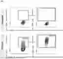


Sources:
- United States Patent and Trademark Office - verify current appl. status at the USPTO↗
Recent applications in this class:
- » 20250346861 2025-11-13
SYSTEMS AND METHODS FOR GENERATING PLATELET LYSATE FROM PLATELET-RICH PLASMA - » 20250327028 2025-10-23
ENGINEERED PLATELETS AS TARGETED PROTEIN DEGRADERS - » 20250320456 2025-10-16
STABLE HUMAN PLATELET LYSATE COMPOSITION, METHODS AND USES THEREOF - » 20250223557 2025-07-10
METHOD TO PRODUCE MAMMAL PLATELET CONCENTRATE, MAMMAL PLATELET CONCENTRATE AND USE THEREOF - » 20250223556 2025-07-10
METHOD FOR PREPARING EXOSOMES FROM HUMAN PLATELET - » 20250027046 2025-01-23
Compositions and Methods for Producing Megakaryocytes - » 20250011719 2025-01-09
NEW METHOD FOR IMPROVING QUALITY OF PLATELETS - » 20240417686 2024-12-19
CD34+CD41DIM MEGAKARYOCYTES PROGENITORS AND USES THEREOF FOR PRODUCING PROPLATELET-BEARING MKS AND/OR PLATELETS - » 20240352418 2024-10-24
METHOD FOR PRODUCING MULTINUCLEATED MEGAKARYOCYTE WITH ENHANCED PLATELET PRODUCTION CAPABILITY, METHOD FOR PRODUCING PLATELETS, METHOD FOR PRODUCING PLATELET PREPARATION, AND METHOD FOR PRODUCING BLOOD PREPARATION - » 20240271094 2024-08-15
COMPOSITIONS AND METHODS FOR PATHOGEN INACTIVATION OF PLATELETS