Method for detecting the myocardial state of the heart and a measuring apparatus for performing this method
US20080119746A1
2008-05-22
11/933,795
2007-11-01
✅ Patent granted
US 7,890,161 B2
2011-02-15
-
-
Mark W Bockelman | Erica Lee
2029-10-02
Abstract:
A method for detecting the myocardial state of the heart has the following method steps, which may be implemented by a corresponding measuring apparatus:
-
- inserting a bipolar cardiological measuring electrode (4) in a heart division (8) at an acute attachment angle (9) of the measuring electrode (4) on the myocardium (6) of less than 90°,
- measuring a cardiological stimulation signal (IEGM) in sequential cardiac cycles,
- determining the positive and negative maximum amplitudes Vp and Vn of the stimulation signal (IEGM) of sequential cardiac cycles,
- ascertaining an asymmetry factor η of the stimulation signal (IEGM) of sequential cardiac cycles according to the equation η=(Vp−|Vn|)/(Vp+|Vn|) and
- storing the asymmetry factor η of sequential cardiac cycles for its analysis.
Assignee:
- BIOTRONIK CRM Patent AG 138 🇨🇭 Baar, Switzerland
Interested in similar patents?
Get notified when new applications in this technology area are published.
Classification:
A61B5/316 » CPC main
Measuring for diagnostic purposes ; Identification of persons; Detecting, measuring or recording bioelectric or biomagnetic signals of the body or parts thereof Modalities, i.e. specific diagnostic methods
A61B5/283 » CPC further
Measuring for diagnostic purposes ; Identification of persons; Detecting, measuring or recording bioelectric or biomagnetic signals of the body or parts thereof; Bioelectric electrodes therefor specially adapted for particular uses for electrocardiography [ECG] Invasive
A61N1/08 IPC
Electrotherapy; Circuits therefor; Details Arrangements or circuits for monitoring, protecting, controlling or indicating
Description
This application takes priority from German Patent Application DE 10 2006 054 474.9 filed 18 Nov. 2006, the specification of which is hereby incorporated herein by reference
BACKGROUND OF THE INVENTION
1. Field of the Invention
The present invention relates to a method for detecting the myocardial state of the heart and a measuring apparatus for performing this method.
2. Description of the Related Art
Cardiological experiments and studies have resulted in the finding that the change of the state of the myocardium frequently results in a change of the direction of the excitation propagation of the cardiological stimulation signals in the heart. This is based, for example, on the different velocities of the excitation propagation in healthy myocardial areas on one hand and, for example, in myocardial areas subject to ischemia on the other hand. A further reason may be the change of the geometry of the heart divisions in the event of cardiac insufficiency or cardiomyopathy, in the event of operational wounds, or in the event of a myocardial infarction. Furthermore, direction changes of the excitation propagation may occur due to a change of the myocardial state before a cardiac flutter or also due to psychological stress situations and strains.
Disturbances of this type may be recognized in principle by complex analyses of cardiological stimulation signals, however, complex multi-electrode measurements are known to be necessary for this purpose, as are performed in an ECG, for example. The corresponding measurement methods and apparatuses may not be implemented in practice in an implant.
BRIEF SUMMARY OF THE INVENTION
Proceeding therefrom, the present invention is based on the object of specifying a method for detecting the myocardial state of the heart and a corresponding measuring apparatus, which may be implemented using simple measuring technology and design and may therefore be implemented in an implant.
This object is achieved in regard to the method by the method steps specified in claim 1 as follows:
-
- inserting a bipolar cardiological measuring electrode in a heart division at an acute attachment angle of the measuring electrode on the myocardium of less than 90°,
- measuring a cardiological stimulation signal in sequential cardiac cycles,
- determining the positive and negative maximum amplitudes Vp and Vn of the stimulation signal of sequential cardiac cycles,
- ascertaining an asymmetry factor η of the stimulation signal of sequential cardiac cycles according to the equation
η=(Vp−|Vn|)/(Vp+|Vn|) and
-
- storing the asymmetry factor η of sequential cardiac cycles for its analysis.
As will be explained in greater detail on the basis of the exemplary embodiment, the present invention proceeds from the finding that in the event of a measuring electrode which is atypical per se and is not placed perpendicularly to the myocardial plane, the change of the direction of the excitation propagation results in a change of the asymmetry of intracardial electrical signals (“IEGM” in short in the following). The present invention makes use of this in that the specified acute attachment angle of the measuring angle is selected as significantly below 90° and preferably approximately 45°. The specified asymmetry factor may be determined from the corresponding stimulation signals and stored for the subsequent diagnosis of its myocardial state, which is not part of the method according to the present invention, in particular for diagnosis of myocardial ischemia and cardiac insufficiency.
A specific IEGM is used depending on the placement of the measuring electrode in a heart division, thus, for a measuring device placed in the ventricle, the R wave is used, and for a measuring electrode placed in the atrium, the P wave of the cardiological stimulation signal is used.
A corresponding measuring apparatus for performing the method described above is the subject matter of claim 5. This measuring apparatus thus has:
-
- a measuring electrode (4), which may be positioned in a heart division (8) at an acute attachment angle (9) to the myocardium (6), and
- an implantable processing apparatus (2), which picks up the measured signals (IEGM) of the measuring electrode (4), for recording, detecting, relaying, and/or analyzing the measured signals to ascertain the asymmetry factor η.
Devices for diagnosing the myocardial state may thus be conceived on the basis of the analysis of the asymmetry factor of the IEGM signals. The simplicity of the method and in particular the slight output signals necessary for this purpose, in the form of the positive and negative maximum amplitudes of a stimulation signal, allow the corresponding method to be applied in modern implantable devices. Preferably, a cardiac pacemaker or defibrillator is thus provided with an integrated analysis apparatus to ascertain the asymmetry factor η.
Alternatively or additionally thereto, the processing apparatus of the measuring apparatus may have a transmitter for relaying the ascertained measured signals to an external analysis station, in which the asymmetry factor η may then be ascertained.
BRIEF DESCRIPTION OF THE FIGURES
Further features, details, and advantages of the present invention may be inferred from the following description, in which an exemplary embodiment of the method according to the present invention and a corresponding measuring apparatus are explained in greater detail.
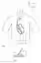
FIG. 1 shows a schematic view of a patient thorax having an implanted cardiac pacemaker and measuring electrode in the right ventricle,
FIG. 2 shows a schematic enlarged view of the measuring electrode anchored in the myocardium,
FIG. 3 shows a diagram of analytically calculated IEGM signals for various directions of the excitation propagation,
FIGS. 4 and 5 show IEGM signals having different asymmetries, and
FIG. 6 shows a time diagram of the blood-pressure (a) and the corresponding IEGM asymmetry factor η (b).
DETAILED DESCRIPTION OF THE INVENTION
FIG. 1 shows a cardiac pacemaker 2 implanted in the thorax 1 of a cardiac patient, in which an analysis apparatus 3 for the above-mentioned asymmetry factor η, which is to be explained in greater detail below, is integrated. This analysis apparatus 3 processes the measured signals of the bipolar measuring electrode 4, whose electrode tip 5 is anchored in the myocardium 6 of the heart 7 in the area of the right ventricle 8. As FIG. 2 shows, the measuring electrode 4 is inclined at an acute angle 9 of approximately 45° to the plane of the myocardium 6.
The measured signals of the measuring electrode 4 or data generated by the analysis apparatus 3 may be transmitted via the transmitter 10 to an external analysis station 11, where it may be processed further and used in the course of so-called “home monitoring”, for example.
Analytical evaluations of medical experiments have shown that in the event of non-perpendicular positioning of the measuring electrode 4 to the plane of the myocardium 6, a change of the direction of the excitation propagation results in a change of the asymmetry of intercardial electrical signals. FIG. 3 shows IEGM signals calculated for several angles α between the direction of the excitation propagation and the projection of the measuring electrode 4 on the myocardial surface.
FIGS. 4 and 5 show IEGM signals in the form of R waves of the measuring electrode 4 inserted into the right ventricle as shown in FIG. 1. The amplitude Vp of the positive wave and the amplitude Vn of the negative wave of the particular signal are plotted. According to the equation
η=(Vp−|Vn|)/(Vp+|Vn|)
the asymmetry factor η is calculated in FIG. 4 as η=0.4 and in FIG. 5 as η=0.97.
The significance of the asymmetry factor η for a pathological situation of the heart may be shown on the basis of FIG. 6. The diagram of (a) shows the blood pressure in the coronary artery during an ischemia, which occurs in a period of time between approximately t=1 minute and t=4 minutes.
With a brief delay, the asymmetry factor η plotted in partial figure (b) passes through a maximum at t=2 minutes, to then sink significantly to a value of approximately a third of the original asymmetry factor.
The significance of the change of the asymmetry factor for the presence of an ischemia is clear.
Claims
What is claimed is:1. A method for detecting the myocardial state of the heart comprising:
inserting a measuring electrode (4) comprising a bipolar cardiological measuring electrode in a heart division (8) at attachment angle (9) comprising an acute attachment angle of said measuring electrode (4) on a myocardium (6) of less than 90°;
measuring a stimulation signal (IEGM) comprising a cardiological stimulation signal in sequential cardiac cycles;
determining positive and negative maximum amplitudes Vp and Vn of said stimulation signal (IEGM) of sequential cardiac cycles;
ascertaining an asymmetry factor η of said stimulation signal (IEGM) of sequential cardiac cycles according to equation η=(Vp−|Vn|)/(Vp+|Vn|); and,
storing said asymmetry factor η of sequential cardiac cycles for analysis.
2. The method according to claim 1 wherein said attachment angle (9) is approximately 45°.
3. The method according to claim 1 wherein an R wave of said cardiological stimulation signal is measured for said measuring electrode (4) inserted into a ventricle of said heart.
4. The method according to claim 1 wherein a P wave of said cardiological stimulation signal is measured for said measuring electrode (4) inserted into an atrium of said heart.
5. A measuring apparatus for detecting the myocardial state of the heart comprising:
a measuring electrode (4), which may be positioned in a heart division (8) at attachment angle (9) comprising an acute attachment angle to a myocardium (6); and,
an implantable processing apparatus (2), configured to pick up measured stimulation signals (IEGM) of said measuring electrode (4) wherein said measured stimulation signals (IEGM) are utilized to ascertain the asymmetry factor η.
6. The measuring apparatus according to claim 5, wherein said measured stimulation signals (IEGM) are recorded, detected, transmitted and/or analyzed.
7. The measuring apparatus according to claim 5, wherein said implantable processing apparatus (2) includes a transmitter (10) configured to relay said measured stimulation signals to an external analysis station (11) in which said asymmetry factor η may be ascertained.
8. The measuring apparatus according to claim 5, wherein said implantable processing apparatus (2) is formed by a cardiac pacemaker or defibrillator.
9. The measuring apparatus according to claim 8, wherein said cardiac pacemaker or defibrillator is provided with an integrated analysis apparatus (3) configured to ascertain said asymmetry factor η.
Images & Drawings included:


Sources:
- United States Patent and Trademark Office - verify current appl. status at the USPTO↗
Recent applications in this class:
- » 20250120642 2025-04-17
NON-INVASIVE METHOD AND SYSTEM FOR MEASURING MYOCARDIAL ISCHEMIA, STENOSIS IDENTIFICATION, LOCALIZATION AND FRACTIONAL FLOW RESERVE ESTIMATION - » 20250082245 2025-03-13
METHOD AND APPARATUS FOR WIDE-BAND PHASE GRADIENT SIGNAL ACQUISITION - » 20250064372 2025-02-27
FAST AUTOBLOCKING FOR PHYSIOLOGICAL WAVEFORM DISPLAY - » 20240407700 2024-12-12
Handheld or Wearable Device for Recording or Sonifying Brain Signals - » 20240389920 2024-11-28
VISION TRANSFORMER SYSTEM AND METHOD CONFIGURED TO GENERATE A PATIENT DIAGNOSIS FROM AN ELECTROCARDIOGRAM - » 20240341656 2024-10-17
SYSTEM AND METHOD FOR ELECTROCARDIOGRAM ANALYSIS AND OPTIMIZATION OF CARDIOPULMONARY RESUSCITATION AND THERAPY DELIVERY - » 20240335151 2024-10-10
SYSTEM AND METHOD TO ANALYZE WAVEFORMS FOR ELECTROMYOGRAPHY TESTING - » 20240293068 2024-09-05
Seamlessly Embedded Heart Rate Monitor - » 20240260880 2024-08-08
Machine Learning (ML)-based Disease-Detection System Using Detection Animals - » 20240245337 2024-07-25
Devices and Methods for Analyzing Electrocardiogram (ECG) signals for Artifact and Notification of Culprit Electrode
Recent applications for this Assignee:
- » 20120277840 2012-11-01
Electrode catheter for interventional use - » 20110263987 2011-10-27
Respiration measurement by means of morphological operators - » 20110263966 2011-10-27
Dissipation device and MRI-safe catheter system - » 20110196463 2011-08-11
Electrode device for active medical implants - » 20110153011 2011-06-23
MRT-adjusted IEGM-scanning - » 20110152973 2011-06-23
Extended noise mode - » 20110152972 2011-06-23
Switched protective device against electromagnetic interference - » 20110152733 2011-06-23
MRT Lorentz vibrator - » 20110152674 2011-06-23
Implantable medical device having magnetic resonance tomography antenna - » 20110152673 2011-06-23
Apparatus and method for communication between MRI system and implantable medical devices