Artificial synthetic cDNA and method for detecting secondary glioblastoma
US20160017436A1
2016-01-21
14/800,563
2015-07-15
✅ Patent granted
US 10,151,000 B2
2018-12-11
-
-
Kenneth R Horlick
McCoy Russell LLP
2036-12-31
Abstract:
The present invention provides an artificial synthetic cDNA (complementary deoxyribonucleic acid). The said artificial synthetic cDNA encodes a fused protein which is specifically presented in secondary glioblastoma, and the said artificial synthetic cDNA can be used as a biomarker for detecting the secondary glioblastoma. The present invention further provides a method for detecting secondary glioblastoma. According to the above technical solutions, the accuracy in distinguishing the secondary glioblastoma from primary glioblastoma is effectively improved in the present invention.
Assignee:
- BEIJING NEUROSURGICAL INSTITUTE 10 🇨🇳 Beijing, China
- Beijing Institute for Brain Disorders 1 🇨🇳 Beijing, China
Applicant:
Interested in similar patents?
Get notified when new applications in this technology area are published.
Classification:
C12Q1/68 IPC
Measuring or testing processes involving enzymes, nucleic acids or microorganisms ; Compositions therefor; Processes of preparing such compositions involving nucleic acids
G01N33/57407 » CPC further
Investigating or analysing materials by specific methods not covered by groups -; Biological material, e.g. blood, urine ; Haemocytometers; Chemical analysis of biological material, e.g. blood, urine; Testing involving biospecific ligand binding methods; Immunological testing; Immunoassay; Biospecific binding assay; Materials therefor for cancer Specifically defined cancers
C12Q2600/158 » CPC further
Oligonucleotides characterized by their use Expression markers
C12Y207/10001 » CPC further
Transferases transferring phosphorus-containing groups (2.7); Protein-tyrosine kinases (2.7.10) Receptor protein-tyrosine kinase (2.7.10.1)
C12Y301/03048 » CPC further
Hydrolases acting on ester bonds (3.1); Phosphoric monoester hydrolases (3.1.3) Protein-tyrosine-phosphatase (3.1.3.48)
G01N2333/912 » CPC further
Assays involving biological materials from specific organisms or of a specific nature; Enzymes; Proenzymes; Transferases (2.) transferring phosphorus containing groups, e.g. kinases (2.7)
G01N2333/916 » CPC further
Assays involving biological materials from specific organisms or of a specific nature; Enzymes; Proenzymes; Hydrolases (3) acting on ester bonds (3.1), e.g. phosphatases (3.1.3), phospholipases C or phospholipases D (3.1.4)
C12P19/34 IPC
Preparation of compounds containing saccharide radicals; Preparation of nitrogen-containing carbohydrates; N-glycosides; Nucleotides Polynucleotides, e.g. nucleic acids, oligoribonucleotides
C12Q1/6886 » CPC main
Measuring or testing processes involving enzymes, nucleic acids or microorganisms ; Compositions therefor; Processes of preparing such compositions involving nucleic acids; Nucleic acid products used in the analysis of nucleic acids, e.g. primers or probes for diseases caused by alterations of genetic material for cancer
C12N9/16 » CPC further
Enzymes; Proenzymes; Compositions thereof ; Processes for preparing, activating, inhibiting, separating or purifying enzymes; Hydrolases (3) acting on ester bonds (3.1)
C12N9/12 » CPC further
Enzymes; Proenzymes; Compositions thereof ; Processes for preparing, activating, inhibiting, separating or purifying enzymes; Transferases (2.) transferring phosphorus containing groups, e.g. kinases (2.7)
G01N33/574 IPC
Investigating or analysing materials by specific methods not covered by groups -; Biological material, e.g. blood, urine ; Haemocytometers; Chemical analysis of biological material, e.g. blood, urine; Testing involving biospecific ligand binding methods; Immunological testing; Immunoassay; Biospecific binding assay; Materials therefor for cancer
Description
CROSS-REFERENCE TO RELATED APPLICATIONS
The present application claims priority to Chinese Patent Application No. 201410342399.1, filed on Jul. 18, 2014, the entire contents of which are hereby incorporated by reference for all purposes.
INCORPORATION BY REFERENCE TO ELECTRONICALLY SUBMITTED MATERIAL
Incorporated by reference in its entirety herein is a computer-readable nucleotide/amino acid sequence listing submitted herewith and identified as follows: 47,116 bytes ASCII (Text) file named “Sequence Listing INN14301,” created Jun. 15, 2015.
FIELD OF THE INVENTION
The present invention relates to the technical field of biotechnology and specifically relates to an artificial synthetic cDNA, a fragment of the artificial synthetic cDNA and a method for detecting secondary glioblastoma.
BACKGROUND OF THE INVENTION
Glioblastoma is glioma with the highest malignant degree in astrocytomas. This tumor locates below the cortex and grows throughout supratentorial cerebral hemisphere in most cases. This tumor grows in an infiltrative manner, often invades several cerebral lobes, and further invades the deep structure and can also affect the contralateral cerebral hemisphere via the callus. This tumor mostly grows in the frontal lobe, followed by the temporal lobe and the parietal lobe, and the tumor can also occur in the occipital lobe/the thalamus, the basal ganglia and the like in a few cases.
The glioblastoma has a high growth rate and short disease course, and for 70-80% of patients, the disease course is 3-6 months, and only 10% of the patients have a disease course of more than 1 year. In the individual cases, the stroke-like episodes may occur due to tumor bleeding. Due to rapid growth of the tumor, the hydrocephalus occurs frequently, and the symptom of increased intracranial pressure is obvious, and almost all the patients suffer from headache, vomiting, papilledema/headache, mental changes, limb weakness, unconsciousness, and speech disorders. The glioblastoma damages brain tissues in an infiltrative manner and causes a series of focal lesion symptoms, and the patients have aphasia, hemiplegia, hemianesthesia, hemianopsia and the like to different extents. Hemiplegia, brain neural damages, hemianesthesia, and hemianopsia can be found by neurological examination. About 33% of the patients have seizures and about 20% of the patients have dementia, hypophrenia, and other mental symptoms.
The glioblastoma can be divided into secondary glioblastoma developed from low-grade astrocytomas and primary glioblastoma which does not show early stage low-grade lesions. But the secondary glioblastoma and the primary glioblastoma are very difficult to be distinguished in histology. At present, although the mutation of isocitrate dehydrogenase (IDH) is only found in the secondary glioblastoma, the mutation of IDH does not occur in part of the secondary glioblastomas.
Thus, the detection of the secondary glioblastoma cannot solely rely on the detection of the mutation of IDH, and a new method for detecting secondary glioblastoma needs to be developed to improve the detection of the secondary glioblastoma.
SUMMARY OF THE INVENTION
In order to further improve the detection accuracy of secondary glioblastoma, the present invention provides an artificial synthetic cDNA, a fragment of the artificial synthetic cDNA, and a method for detecting the secondary glioblastoma.
A fused protein provided by the present invention has relatively high specificity in the appearance of the secondary glioblastoma. In one aspect, the present invention provides an artificial synthetic cDNA, and the artificial synthetic cDNA does not exist in the nature, wherein the artificial synthetic cDNA encodes a certain fused protein; and in a direction from an N terminal to a C terminal, the fused protein is formed of a first protein fragment connected to a second protein fragment, wherein the first protein fragment is as shown in SEQ ID NO: 1, 2, 3 or 4 and the second protein fragment is as shown in SEQ ID NO: 5 or 6.
In another aspect, the present invention further provides an artificial synthetic nucleic acid fragment, and the artificial synthetic nucleic acid fragment does not exist in the nature, wherein the sequence of the artificial synthetic nucleic acid fragment contains the sequence as shown in SEQ ID NO: 14, 15 or 16, and the artificial synthetic nucleic acid fragment is the fragment of the above-mentioned artificial synthetic cDNA.
In still another aspect, the present invention further provides a primer pair, wherein the primer pair contains a first primer as shown in SEQ ID NO: 17 and a second primer as shown in SEQ ID NO: 18, and 5′ ends of the first primer and the second primer are chemically modified.
The present invention can also provide a method for detecting secondary glioblastoma, and the method comprises the following steps: detecting a certain fused protein in a glioblastoma sample to be detected by using an artificially prepared antibody, wherein the fused protein in the direction from an N terminal to a C terminal is formed by connecting a first protein fragment and a second protein fragment, wherein the first protein fragment is as shown in SEQ ID NO: 1, 2, 3 or 4 and the second protein fragment is as shown in SEQ ID NO: 5 or 6; and indicating that the glioblastoma sample to be detected is the secondary glioblastoma if the fused protein is presented in the glioblastoma sample to be detected.
The method may additionally or alternatively comprise the following steps:
detecting the content of a certain fused nucleic acid in a glioblastoma sample to be detected by using a chemically modified nucleic acid probe and/or primers which are prepared artificially, wherein the fused nucleic acid is the nucleic acid coding the fused protein; and indicating that the glioblastoma sample to be detected is the secondary glioblastoma if the fused nucleic acid is presented in the glioblastoma sample to be detected.
Through the above technical solution, the artificial synthetic cDNA provided by the present invention effectively improves the accuracy in distinguishing the secondary glioblastoma from primary glioblastoma.
Other features and advantages of the present invention will be described in detail in the following detailed description of the embodiments.
BRIEF DESCRIPTION OF THE DRAWING
The accompanying drawing is used for providing a further understanding of the present invention and constitutes one part of the description. The accompanying drawing and the following detailed description of the embodiments are used for explaining the present invention rather than limiting the present invention. Wherein,
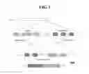
FIG. 1 is a structural schematic diagram of a fused protein formed by connecting a PTPRZ1 protein fragment and an MET protein fragment, which shows the three zoomed-in views of the region of the translocation and subsequent generated cDNA. The said fused protein named D64 is formed of a first protein fragment connected to a second protein fragment, the said first protein fragment is as shown in SEQ ID NO: 4 and the said second protein fragment is as shown in SEQ ID NO: 5 or 6.
DETAILED DESCRIPTION OF THE EMBODIMENTS
In conjunction with the accompanying drawing, the specific embodiments of the present invention will be described below in detail. It should be understood that the specific embodiments described herein are only intended to illustrate and explain the present invention and are not intended to limit the present invention.
In the present invention, unless contrarily indicated, the term “nucleic acid” used herein can be deoxyribonucleic acid or ribonucleic acid; the “nucleic acid” can be single-stranded nucleic acid or double-stranded nucleic acid; the sequence of the “nucleic acid” refers to the sequence of bases; and the “nucleic acid” can have the known modification in the art as long as the modification does not change the base pairing of the “nucleic acid”.
According the present invention, a fused protein is provided, wherein the fused protein in the direction from an N terminal to a C terminal is formed of a first protein fragment connected to a second protein fragment, wherein the first protein fragment is as shown in SEQ ID NO: 1, 2, 3, or 4 and the second protein fragment is as shown in SEQ ID NO: 5 or 6.
SEQ ID NO: 1 is a protein sequence encoded by the coding sequence of the first exon of the human PTPRZ1 gene (whose NCBI Gene ID is 5803).
SEQ ID NO: 2 is the protein sequence encoded by the coding sequence from the first exon to the second exon of the human PTPRZ1 gene.
SEQ ID NO: 3 is the protein sequence encoded by the coding sequence from the first exon to the third exon of the human PTPRZ1 gene.
SEQ ID NO: 4 is the protein sequence encoded by the coding sequence from the first exon to the eighth exon of the human PTPRZ1 gene.
SEQ ID NO: 5 is a fragment of the protein sequence encoded by the sequence from the start of exon 2 to the end of the translated region of the homology isoform 1 of the human MET gene (whose NCBI Gene ID is 4233).
SEQ ID NO: 6 is the fragment of the encoded protein sequence encoded by the sequence from the start of exon 2 to the end of the translated region of the homology isoform 2 of the human MET gene.
In one aspect, the present invention provides an artificial synthetic cDNA, and the artificial synthetic cDNA does not exist in the nature, wherein the artificial synthetic cDNA encodes a certain fused protein; and the fused protein in the direction from an N terminal to a C terminal is formed of a first protein fragment connected to a second protein fragment, wherein the first protein fragment is as shown in SEQ ID NO: 1, 2, 3 or 4 and the second protein fragment is as shown in SEQ ID NO: 5 or 6.
The artificial synthetic cDNA does not exist in nature, including the situation that the artificial synthetic cDNA does not exist in a genomic DNA sequence. The production of the sequence of the artificial synthetic cDNA may include the following events: (1) translocation of genomic DNA causes fusion of the human PTPRZ1 gene and the human MET gene so as to produce fused genomic DNA; (2) the fused genomic DNA is transcribed to obtain fused hnRNA; (3) the fused hnRNA is subjected to splicing to remove the introns and obtain mature fused mRNA; and (4) the mature fused mRNA is subjected to artificial reverse transcription to obtain the artificial synthetic cDNA. For example, as shown in FIG. 1, the human PTPRZ1 gene and the human MET gene are located on human chromosome 7. The translocation of genomic DNA causes the genomic DNA from exon 1 to exon 8 of the human PTPRZ1 gene to fuse with the genomic DNA from exon 2 to the end of the human MET gene so as to produce fused genomic DNA. The fused hnRNA is transcribed and then subjected to splicing to remove the introns and obtain mature fused mRNA; and the mature fused mRNA is subjected to artificial reverse transcription to obtain the artificial synthetic cDNA, which in the direction from 5′ to 3′ is formed of a first nucleic acid fragment connected to a second nucleic acid fragment, wherein the first nucleic acid fragment is as shown in SEQ ID NO: 10, and the second nucleic acid fragment is as shown in SEQ ID NO: 11 or 12.
The sequence of fused nucleic acid coding the above-mentioned fused protein can be obtained by decoding according to an amino acid codon comparison table. Due to the existence of degeneracy of codons, the sequences of a plurality of fused nucleic acids coding the fused protein with the same amino acid sequence can be different from each other.
The artificial synthetic cDNA in the direction from 5′ to 3′ is formed of a first nucleic acid fragment connected to a second nucleic acid fragment, wherein the first nucleic acid fragment is as shown in SEQ ID NO: 7, 8, 9 or 10, and the second nucleic acid fragment is as shown in SEQ ID NO: 11 or 12.
SEQ ID NO: 7 corresponds to a fragment of the translated region of the first exon of the human PTPRZ1 gene, coding the protein fragment as shown in SEQ ID NO: 1.
SEQ ID NO: 8 corresponds to the translated region of the sequence from the first exon to the second exon of the human PTPRZ1 gene, coding the protein fragment as shown in SEQ ID NO: 2.
SEQ ID NO: 9 corresponds to the translated region of the sequence from the first exon to the third exon of the human PTPRZ1 gene, coding the protein fragment as shown in SEQ ID NO: 3.
SEQ ID NO: 10 corresponds to the translated region of the sequence from the first exon to the eighth exon of the human PTPRZ1 gene, coding the protein fragment as shown in SEQ ID NO: 4.
The sequence of SEQ ID NO: 11 corresponds to the fragment starting from exon 2 of the homology isoform 1 of the human MET gene, coding the protein fragment as shown in SEQ ID NO: 5.
The sequence of SEQ ID NO: 12 corresponds to the fragment starting from exon 2 of the homology isoform 2 of the human MET gene, coding the protein fragment as shown in SEQ ID NO: 6.
In another aspect, the present invention further provides an artificial synthetic nucleic acid fragment, and the artificial synthetic nucleic acid fragment does not exist in the nature, wherein the sequence of the artificial synthetic nucleic acid fragment contains the sequence as shown in SEQ ID NO: 14, 15 or 16, and the artificial synthetic nucleic acid fragment is the fragment of the above-mentioned artificial synthetic cDNA.
The sequence of SEQ ID NO: 13 is the fragment of the fused nucleic acid formed of part of the 5′UTR, SEQ ID NO: 7 connected to a respective 5′ part of SEQ ID NO: 11 or 12; and is obtained by performing PCR amplification with a first primer as shown in SEQ ID NO: 17 and a second primer as shown in SEQ ID NO: 18 by taking the cDNA as a template.
The sequence of SEQ ID NO: 14 is the fragment of the fused nucleic acid formed of part of the 5′UTR, SEQ ID NO: 8 connected to a respective 5′ part of SEQ ID NO: 11 or 12; and is obtained by performing PCR amplification with the first primer as shown in SEQ ID NO: 17 and the second primer as shown in SEQ ID NO: 18 by taking the cDNA as the template.
The sequence of SEQ ID NO: 15 is the fragment of the fused nucleic acid formed of part of the 5′UTR, SEQ ID NO: 9 connected to a respective 5′ part of SEQ ID NO: 11 or 12; and is obtained by performing PCR amplification with the first primer as shown in SEQ ID NO: 17 and the second primer as shown in SEQ ID NO: 18 by taking the cDNA as the template.
The sequence of SEQ ID NO: 16 is the fragment of the fused nucleic acid formed of part of the 5′UTR, of SEQ ID NO: 10 connected to a respective 5′ part of SEQ ID NO: 11 or 12; and is obtained by performing PCR amplification with the first primer as shown in SEQ ID NO: 17 and the second primer as shown in SEQ ID NO: 18 by taking the cDNA as the template.
The sequence of the artificial synthetic nucleic acid fragment is the sequence as shown in SEQ ID NO: 14, 15 or 16.
In still another aspect, the present invention further provides a primer pair, wherein the primer pair contains a first primer as shown in SEQ ID NO: 17 and a second primer as shown in SEQ ID NO: 18, and 5′ ends of the first primer and the second primer are chemically modified.
The present invention further provides a method for detecting secondary glioblastoma,
the method comprising the following steps:
detecting a certain fused protein in a glioblastoma sample to be detected by using an artificially prepared antibody, wherein the fused protein in the direction from an N terminal to a C terminal is formed by connecting a first protein fragment and a second protein fragment, wherein the first protein fragment is as shown in SEQ ID NO: 1, 2, 3, or 4 and the second protein fragment is as shown in SEQ ID NO: 5 or 6; and indicating that the glioblastoma sample to be detected is the secondary glioblastoma if the fused protein is presented in the glioblastoma sample to be detected;
Additionally or alternatively, the method comprises the following steps:
detecting a certain fused nucleic acid in a glioblastoma sample to be detected by using a chemically modified nucleic acid probe and/or primers which are prepared artificially, wherein the fused nucleic acid is the nucleic acid coding the fused protein; and indicating that the glioblastoma sample to be detected is the secondary glioblastoma if the fused nucleic acid is presented in the glioblastoma sample to be detected.
The artificially prepared antibody can be a commercially available antibody and can also be the antibody prepared through a conventional monoclonal antibody and/or polyclonal antibody preparation technology. The chemical modification of the nucleic acid probe and/or the chemical modification in the primers can adopt various chemical modifications which are conventionally used in the probe and the primers, for example, the chemical modifications can comprise at least one of phosphorylation modification, biotin modification, digoxin modification, amino-modification and mercapto-modification.
According to the present invention, the average median survival time in the cases with the secondary glioblastoma, in which the fused protein of the invention appeared, was shorter than the average median survival time in the reported cases with the secondary glioblastoma, indicating that in the secondary glioblastoma, the cases in which the fused protein of the invention appeared had poorer prognosis. The fused protein provided by the present invention can also be used as a molecular marker for judging the prognosis of the secondary glioblastoma.
The present invention will be described below in detail through the Examples.
PREPARATION EXAMPLE 1
In this preparation example, secondary glioblastoma samples and primary glioblastoma samples were obtained, and RNA and cDNA of the samples were further obtained.
| TABLE 1 | |||
| No. | Type | ||
| of case | Gender | Age | of disease |
| 1 | M | 44 | pGBM |
| 2 | F | 59 | pGBM |
| 3 | F | 56 | pGBM |
| 4 | F | 48 | pGBM |
| 5 | M | 64 | pGBM |
| 6 | M | 66 | pGBM |
| 7 | M | 59 | pGBM |
| 8 | F | 62 | pGBM |
| 9 | M | 42 | pGBM |
| 10 | M | 81 | pGBM |
| 11 | M | 60 | pGBM |
| 12 | M | 29 | sGBM |
| 13 | M | 26 | pGBM |
| 14 | M | 47 | pGBM |
| 15 | M | 42 | pGBM |
| 16 | F | 43 | pGBM |
| 17 | F | 40 | sGBM |
| 18 | M | 27 | sGBM |
| 19 | M | 42 | pGBM |
| 20 | F | 37 | sGBM |
| 21 | M | 45 | sGBM |
| 22 | M | 54 | sGBM |
| 23 | F | 47 | sGBM |
| 24 | M | 33 | pGBM |
| 25 | M | 63 | pGBM |
| 26 | M | 34 | pGBM |
| 27 | M | 18 | sGBM |
| 28 | M | 33 | sGBM |
| 29 | M | 30 | pGBM |
| 30 | M | 49 | pGBM |
| 31 | M | 43 | pGBM |
| 32 | F | 28 | pGBM |
| 33 | M | 42 | sGBM |
| 34 | F | 62 | pGBM |
| 35 | M | 48 | pGBM |
| 36 | F | 51 | sGBM |
| 37 | F | 40 | pGBM |
| 38 | F | 24 | pGBM |
| 39 | F | 49 | sGBM |
| 40 | M | 51 | sGBM |
| 41 | F | 55 | pGBM |
| 42 | M | 38 | sGBM |
| 43 | M | 54 | pGBM |
| 44 | F | 60 | pGBM |
| 45 | F | 37 | pGBM |
| 46 | F | 59 | pGBM |
| 47 | M | 54 | pGBM |
| 48 | M | 52 | pGBM |
| 49 | M | 46 | pGBM |
| 50 | M | 56 | pGBM |
| 51 | M | 60 | pGBM |
| 52 | F | 63 | pGBM |
| 53 | M | 44 | pGBM |
| 54 | F | 25 | pGBM |
| 55 | M | 42 | pGBM |
| 56 | M | 51 | pGBM |
| 57 | M | 45 | pGBM |
| 58 | F | 50 | sGBM |
| 59 | M | 61 | pGBM |
| 60 | M | 43 | pGBM |
| 61 | F | 54 | pGBM |
| 62 | M | 39 | pGBM |
| 63 | M | 57 | pGBM |
| 64 | F | 64 | pGBM |
| 65 | F | 55 | pGBM |
| 66 | F | 52 | pGBM |
| 67 | M | 57 | pGBM |
| 68 | F | 60 | pGBM |
| 69 | M | 42 | pGBM |
| 70 | M | 46 | sGBM |
| 71 | M | 55 | pGBM |
| 72 | M | 44 | pGBM |
| 73 | M | 57 | pGBM |
| 74 | M | 45 | sGBM |
| 75 | M | 55 | pGBM |
| 76 | M | 40 | pGBM |
| 77 | F | 30 | sGBM |
| 78 | M | 25 | sGBM |
| 79 | M | 51 | sGBM |
By using the operation which is in line with the standard of Medical Ethics Committee, 59 cases of primary glioblastoma samples and 20 cases of secondary glioblastoma samples were collected. For each patient from whom the sample was collected, the consents of the patient and a therapist thereof were obtained, and written proofing materials were also possessed. The diagnosis, the identification, and the differentiation of the primary glioblastoma from the secondary glioblastoma were performed according to a histological method in the literature (Louis D N, et al, 2007. The 2007 WHO classification of tumors of the central nervous system. Acta Neuropathol 114 (2): 97-109). The information of the gender, the age, and the type of the disease of each pathological sample is as shown in Table 1, wherein pGBM represents the primary glioblastoma and sGBM represents the secondary glioblastoma.
A DNA extraction kit (purchased from Qiagen) was used to extract total RNA in the primary glioblastoma samples and the secondary glioblastoma samples according to an operation instruction. By detecting the total RNA by an integrity analyzer, it was confirmed that the RNA integrity number (RIN) was greater than 7.0. A reverse transcription kit (purchased from Invitrogen) was used for synthesizing double-stranded cDNA by using the total RNA as the template according to the operation instruction.
EXAMPLE 1
In this example, RNA sequencing was performed on 59 cases of the primary glioblastoma samples and 20 cases of the secondary glioblastoma samples collected in the preparation example 1.
An RNA library construction kit (purchased from Illumina) was used for constructing an RNA library for the RNA of each sample, and then a sequencing platform (Illumina HiSeq 2000) was used for performing RNA sequencing on each RNA library. The sequences obtained by sequencing were aligned with a reference RNA sequence database (Hg19 Refseq, GRCh37), and the RNA of a fused gene was sought by referring to a method in the literature (McPherson A, et al. 2011. deFuse: an algorithm for gene fused discovery in tumor RNA-Seq data. PLoS Comput Biol 7(5): e1001138).
The results indicate that in the samples as shown in Table 1, the RNA of the fused gene of the present invention is present in a plurality of secondary glioblastoma (sGBM) samples, but the RNA of the fused gene of the present invention is not present in the primary glioblastoma (pGBM) samples. The specific appearance situations are as shown in Table 2.
| TABLE 2 | ||
| Fused protein | No. of cases with appearance |
| Fused gene | First | Second | Primary | Secondary |
| First nucleic | Second nucleic | protein | protein | glioblastoma | glioblastoma | |
| No. | acid fragment | acid fragment | fragment | fragment | (pGBM) | (sGBM) |
| 1 | SEQ ID NO: 7 | SEQ ID NO: 11 | SEQ ID NO: 1 | SEQ ID NO: 5 | 0 | 2 |
| 2 | SEQ ID NO: 8 | SEQ ID NO: 11 | SEQ ID NO: 2 | SEQ ID NO: 5 | 0 | 1 |
| 3 | SEQ ID NO: 9 | SEQ ID NO: 11 | SEQ ID NO: 3 | SEQ ID NO: 5 | 0 | 4 |
| 4 | SEQ ID NO: 10 | SEQ ID NO: 11 | SEQ ID NO: 4 | SEQ ID NO: 5 | 0 | 2 |
| 5 | SEQ ID NO: 7 | SEQ ID NO: 12 | SEQ ID NO: 1 | SEQ ID NO: 6 | 0 | 5 |
| 6 | SEQ ID NO: 8 | SEQ ID NO: 12 | SEQ ID NO: 2 | SEQ ID NO: 6 | 0 | 3 |
| 7 | SEQ ID NO: 9 | SEQ ID NO: 12 | SEQ ID NO: 3 | SEQ ID NO: 6 | 0 | 1 |
| 8 | SEQ ID NO: 10 | SEQ ID NO: 12 | SEQ ID NO: 4 | SEQ ID NO: 6 | 0 | 2 |
| Total | 0 | 20 | ||||
It could be seen from the data in Table 2 that the fused protein described in the present invention specifically appeared in the secondary glioblastoma rather than the primary glioblastoma, so that the fused protein could be used for distinguishing the secondary glioblastoma from the primary glioblastoma. FIG. 1 illustrates the formation process of the fused gene of the present invention as an example, namely, it shows a structural schematic diagram formed by connecting a PTPRZ 1 protein fragment and an MET protein fragment.
In addition, according to the calculation, the average median survival time in the cases with the secondary glioblastoma, in which the fused protein of the present invention appeared was 127 days, which was shorter than the average median survival time (248 days) in the reported cases with the secondary glioblastoma, indicating that in the secondary glioblastoma, the cases in which the fused protein of the present invention appeared had poorer prognosis.
EXAMPLE 2
In this example, PCR verification of the fused protein was performed on the RNA obtained from the 59 cases of the primary glioblastoma samples and the 20 cases of the secondary glioblastoma samples collected in the preparation example 1.
Primers used for PCR verification comprises a first primer as shown in SEQ ID NO: 17 and a second primer as shown in SEQ ID NO: 18. The operation of the PCR was performed according to the synthetic primers and an instruction of a PCR kit. The presence of an amplification band in each PCR product was displayed by agarose gel nucleic acid electrophoresis, and each presented amplification band was recovered by using a DNA gel recovery kit (QlAquick PCR purification kit, purchased from Qiagen), then cloned to a T vector (pGEM-T easy vector, purchased from Promega) and then sequenced by using a DNA sequencer (ABI Prism 3730x1 DNA Sequencer, purchased from Applied Biosystems). The results are as shown in Table 3.
| TABLE 3 | |
| No. of cases |
| Fused gene | Fused protein | Sequence of | Primary | Secondary |
| First nucleic | Second nucleic | First protein | Second protein | amplification | glioblastoma | glioblastoma | |
| No. | acid fragment | acid fragment | fragment | fragment | product | (pGBM) | (sGBM) |
| 1 | SEQ ID NO: 7 | SEQ ID NO: 11 | SEQ ID NO: 1 | SEQ ID NO: 5 | SEQ ID NO: 13 | 0 | 2 |
| 2 | SEQ ID NO: 8 | SEQ ID NO: 11 | SEQ ID NO: 2 | SEQ ID NO: 5 | SEQ ID NO: 14 | 0 | 1 |
| 3 | SEQ ID NO: 9 | SEQ ID NO: 11 | SEQ ID NO: 3 | SEQ ID NO: 5 | SEQ ID NO: 15 | 0 | 4 |
| 4 | SEQ ID NO: 10 | SEQ ID NO: 11 | SEQ ID NO: 4 | SEQ ID NO: 5 | SEQ ID NO: 16 | 0 | 2 |
| 5 | SEQ ID NO: 7 | SEQ ID NO: 12 | SEQ ID NO: 1 | SEQ ID NO: 6 | SEQ ID NO: 13 | 0 | 5 |
| 6 | SEQ ID NO: 8 | SEQ ID NO: 12 | SEQ ID NO: 2 | SEQ ID NO: 6 | SEQ ID NO: 14 | 0 | 3 |
| 7 | SEQ ID NO: 9 | SEQ ID NO: 12 | SEQ ID NO: 3 | SEQ ID NO: 6 | SEQ ID NO: 15 | 0 | 1 |
| 8 | SEQ ID NO: 10 | SEQ ID NO: 12 | SEQ ID NO: 4 | SEQ ID NO: 6 | SEQ ID NO: 16 | 0 | 2 |
| Total | 0 | 20 |
It can be seen from the data in Table 3 that the fragment of the fused gene described in the present invention is specifically presented in the secondary glioblastoma rather than the primary glioblastoma, so that the fragment of the fused gene can be used for distinguishing the secondary glioblastoma from the primary glioblastoma; and specifically, the secondary glioblastoma and the primary glioblastoma can be distinguished by using the PCR method.
In addition, according to the calculation, the average median survival time in the cases with the secondary glioblastoma, in which the fused protein of the invention was presented was 127 days, which was shorter than the average median survival time (248 days) in the reported cases with the secondary glioblastoma, indicating that in the secondary glioblastoma, the cases in which the fused protein of the present invention was presented had poorer prognosis.
In addition, for those samples having cDNA in which the products as shown in SEQ ID: NO: 13-16 have been amplified, the genomic DNA were used as templates to conduct PCR amplifications, by using the first primer as shown in SEQ ID NO: 17 and the second primer as shown in SEQ ID NO: 18, and it was found that in the amplification products of genomic DNA, the product as shown in SEQ ID NO: 13 was present, but the products as shown in SEQ ID NO: 14-16 were not present. The possible reason was that in the genomic DNA of the samples in which the fused gene was present, the nucleic acid fragment as shown in SEQ ID NO: 13 was present between a site as shown in SEQ ID NO: 17 and the site as shown in SEQ ID NO: 18 in the genomic DNA, while the nucleic acid fragments as shown in SEQ ID NO: 14-16 were not present between those sites.
EXAMPLE 3
In this example, immuno-hybridization verification of the fused protein was performed on the total protein samples of the 59 cases of the primary glioblastoma samples and the 20 cases of the secondary glioblastoma samples collected in the preparation example 1.
An antibody used for immuno-hybridization verification was an anti-human MET protein antibody (the antibody was derived from a rabbit and purchased from Abcam, and the product number was ab51067). The size of the non-fused human MET protein was 145kDa, while the molecular weight of the fused protein was increased. The operation of immuno-hybridization was performed by referring to the instruction of the antibody and the instruction of an immuno-hybridization kit. The presence and the positions of immuno-hybridization bands are as shown in Table 4.
| TABLE 4 | |
| Cases in which fused | |
| hybridization band presents |
| Fused gene | Fused protein | Size of fused | Primary | Secondary |
| First nucleic | Second nucleic | First protein | Second protein | hybridization | glioblastoma | glioblastoma | |
| No. | acid fragment | acid fragment | fragment | fragment | band (kDa) | (pGBM) | (sGBM) |
| 1 | SEQ ID NO: 7 | SEQ ID NO: 11 | SEQ ID NO: 13 | SEQ ID NO: 5 | 158 | 0 | 2 |
| 2 | SEQ ID NO: 8 | SEQ ID NO: 11 | SEQ ID NO: 14 | SEQ ID NO: 5 | 160 | 0 | 1 |
| 3 | SEQ ID NO: 9 | SEQ ID NO: 11 | SEQ ID NO: 15 | SEQ ID NO: 5 | 167 | 0 | 4 |
| 4 | SEQ ID NO: 10 | SEQ ID NO: 11 | SEQ ID NO: 16 | SEQ ID NO: 5 | 191 | 0 | 2 |
| 5 | SEQ ID NO: 7 | SEQ ID NO: 12 | SEQ ID NO: 13 | SEQ ID NO: 6 | 159 | 0 | 5 |
| 6 | SEQ ID NO: 8 | SEQ ID NO: 12 | SEQ ID NO: 14 | SEQ ID NO: 6 | 161 | 0 | 3 |
| 7 | SEQ ID NO: 9 | SEQ ID NO: 12 | SEQ ID NO: 15 | SEQ ID NO: 6 | 168 | 0 | 1 |
| 8 | SEQ ID NO: 10 | SEQ ID NO: 12 | SEQ ID NO: 16 | SEQ ID NO: 6 | 192 | 0 | 2 |
| Total | 0 | 20 |
It can be seen from the data in Table 4 that the protein expression product of the fused gene described in the present invention is specifically present in the secondary glioblastoma rather than in the primary glioblastoma, so that the protein expression product of the fused gene can be used for distinguishing the secondary glioblastoma from the primary glioblastoma; and specifically, the secondary glioblastoma and the primary glioblastoma can be distinguished by using the immuno-hybridization method.
In addition, according to the calculation, the average median survival time in the cases with the secondary glioblastoma, in which the fused protein of the present invention appeared was 127 days, which was shorter than the average median survival time (248 days) in the reported cases with the secondary glioblastoma, indicating that in the secondary glioblastoma, the cases in which the fused protein of the invention was presented had poorer prognosis.
COMPARATIVE EXAMPLE 1
According to a method in the literature (Yan H, et al. 2009. IDH1 and IDH2 mutations in gliomas. N Engl J Med 360(8): 765-773.), presence of the mutation of isocitrate dehydrogenase (IDH) was tested in the 20 cases of secondary glioblastoma (sGBM) in the preparation example 1, and it was found from the results that the mutation of IDH only occurred in 12 cases. Thus, the distinguishing of the secondary glioblastoma from the primary glioblastoma by means of the mutation of IDH had relatively low accuracy.
The preferred embodiments of the present invention are described in detail in conjunction with the accompanying drawing. However, the present invention is not limited to the specific details in the embodiments, and in the scope of technical concept, the technical scheme of the present invention can be subjected to a variety of simple modifications, and these simple modifications still belong to the scope of protection of the present invention.
In addition, it needs to be noted that the various specific technical features described in the above embodiments can be combined in any suitable way under the situation that no contradictions exist. In order to avoid the unnecessary repetition, the various possible combination ways will not be described any more herein.
In addition, the various different embodiments of the present invention can also be combined arbitrarily, and the combinations should also be considered as the contents disclosed in the invention as long as the combinations do not depart from the idea of the present invention.
Claims
1. An artificial synthetic cDNA, said artificial synthetic cDNA encoding a certain fused protein; and in a direction from an N terminal to a C terminal, said fused protein is formed of a first protein fragment connected to a second protein fragment, wherein the said first protein fragment is as shown in SEQ ID NO: 1, 2, 3, or 4 and the said second protein fragment is as shown in SEQ ID NO: 5 or 6.
2. The artificial synthetic cDNA according to claim 1, wherein in a direction from 5′ to 3′, said artificial synthetic cDNA is formed of a first nucleic acid fragment connected to a second nucleic acid fragment, wherein the said first nucleic acid fragment is as shown in SEQ ID NO: 7, 8, 9 or 10, and the said second nucleic acid fragment is as shown in SEQ ID NO: 11 or 12.
3. An artificial synthetic nucleic acid fragment, comprising a sequence as shown in SEQ ID NO: 14, 15 or 16, and said artificial synthetic nucleic acid fragment is a fragment of said artificial synthetic cDNA of claim 1.
4. The artificial synthetic nucleic acid fragment according to claim 3, wherein the sequence of the said artificial synthetic nucleic acid fragment is the sequence as shown in SEQ ID NO: 14, 15 or 16.
5. A primer pair, comprising a first primer as shown in SEQ ID NO: 17 and a second primer as shown in SEQ ID NO: 18, and 5′ ends of said first primer and said second primer are chemically modified.
6. A method for detecting secondary glioblastoma, the method comprising the following steps:
detecting a certain fused nucleic acid in a glioblastoma sample by using a chemically modified nucleic acid probe and/or primers which are prepared artificially, wherein said fused nucleic acid is a nucleic acid encoding a fused protein, wherein in a direction from an N terminal to a C terminal, said fused protein is formed of a first protein fragment connected to a second protein fragment, wherein said first protein fragment is as shown in SEQ ID NO: 1, 2, 3, or 4 and said second protein fragment is as shown in SEQ ID NO: 5 or 6; and indicating that said glioblastoma sample is a secondary glioblastoma if the fused nucleic acid is detected in the glioblastoma sample;
additionally or alternatively, the method comprises the following steps:
detecting a certain fused protein in a glioblastoma sample by using an artificially prepared antibody, wherein in a direction from an N terminal to a C terminal, said fused protein is formed of a first protein fragment connected to a second protein fragment, wherein said first protein fragment is as shown in SEQ ID NO: 1, 2, 3, or 4 and said second protein fragment is as shown in SEQ ID NO: 5 or 6; and indicating that said glioblastoma sample is a secondary glioblastoma if the fused protein is detected in the glioblastoma sample.
Images & Drawings included:
Sources:
- United States Patent and Trademark Office - verify current appl. status at the USPTO↗
Recent applications in this class:
- » 20250290156 2025-09-18
EVALUATING OVARIAN CANCER CHEMOTHERAPY RESPONSE USING GENE EXPRESSION DATA AND MACHINE LEARNING - » 20250290155 2025-09-18
CLINICAL WORKFLOW FOR TREATMENT OF BREAST CANCER - » 20250290154 2025-09-18
PREDICTIVE MARKERS FOR IMMUNOTHERAPY - » 20250290153 2025-09-18
METHODS OF DIAGNOSING, DETERMINING THE PROGRESSION OF, AND TREATING A PROSTATE CANCER - » 20250290152 2025-09-18
GENETIC TRIO OF BRAF AND TERT MUTATIONS AND RS2853669TT IN PAPILLARY THYROID CANCER AGGRESSIVENESS - » 20250290151 2025-09-18
Differentially-Methylated Regions of the Genome Useful as Markers of Embryo-Adult Transitions - » 20250290150 2025-09-18
METHODS AND SYSTEMS FOR CHARACTERIZATION, DIAGNOSIS, AND TREATMENT OF CANCER - » 20250290149 2025-09-18
SYSTEMS AND METHODS FOR ENRICHING CELL-FREE MICROBIAL NUCLEIC ACID MOLECULES - » 20250290148 2025-09-18
DIFFERENTIAL ALTERNATIVE SPLICING IN RELAPSED AND REFRACTORY DIFFUSE LARGE-B CELL LYMPHOMA PATIENTS RECEIVING CAR-T THERAPY - » 20250290147 2025-09-18
PROSTATE CANCER MARKERS
Recent applications for this Assignee:
- » 20240052014 2024-02-15
TRUNCATED BODY OF IL7Ra AND USE THEREOF IN PREPARATION OF MEDICATION FOR TREATING TUMOR - » 20230322940 2023-10-12
ANTI-CD44 SINGLE-CHAIN ANTIBODY AND USE THEREOF IN PREPARING DRUG FOR TREATING TUMOR - » 20230310603 2023-10-05
ANTI-CD133 SINGLE-CHAIN ANTIBODY AND USE THEREOF IN PREPARATION OF DRUG FOR TREATING TUMOR - » 20230295294 2023-09-21
ANTI-TIM3 SINGLE-CHAIN ANTIBODY AND USE THEREOF IN PREPARING MEDICINE FOR TREATING TUMOR - » 20230193397 2023-06-22
cDNA, mRNA, PROTEIN, AND KIT AND SYSTEM FOR EVALUATING GLIOMA PROGNOSIS - » 20230160007 2023-05-25
KIT FOR EVALUATING SENSITIVITY OF PATIENT TO MET INHIBITOR - » 20230143605 2023-05-11
KIT AND SYSTEM FOR EVALUATING GLIOMA AND/OR GASTRIC ADENOCARCINOMA PROGNOSIS - » 20210369711 2021-12-02
Uses of compound in preparation of drugs for treating brain glioma - » 20190329033 2019-10-31
DEVICE AND METHOD FOR ELECTROTHROMBOSIS