Antibody
US20170129948A1
2017-05-11
15/317,340
2015-06-12
✅ Patent granted
US 10,590,188 B2
2020-03-17
WO; PCT/GB2015/051737; 20150612
WO; WO2015/189638; 20151217
Julie Wu | Carmencita M Belei
Grimes & Yvon LLP
2035-06-12
Abstract:
The present invention provides an antibody which comprises a variable heavy (VH) chain comprising CDR1, CDR2 and CDR3, and/or a variable light (VL) chain comprising CDR1, CDR2 and CDR3, wherein the CDRs have the same amino acid sequence as those from a complete antibody isolated from a synovial tissue sample of rheumatoid arthritis patients, as listed in Tables 1 and 2 or Tables 1 A and 2 A.
Inventors:
- Constantino Pitzalis 4 🇬🇧 London, United Kingdom
- Michele Bombardieri 1 🇬🇧 London, United Kingdom
- Elisa Corsiero 1 🇬🇧 London, United Kingdom
Assignee:
- QUEEN MARY UNIVERSITY OF LONDON 53 🇬🇧 London, United Kingdom
Applicant:
Interested in similar patents?
Get notified when new applications in this technology area are published.
Classification:
C07K2317/565 » CPC further
Immunoglobulins specific features characterized by immunoglobulin fragments variable (Fv) region, i.e. VH and/or VL Complementarity determining region [CDR]
G01N33/564 » CPC further
Investigating or analysing materials by specific methods not covered by groups -; Biological material, e.g. blood, urine ; Haemocytometers; Chemical analysis of biological material, e.g. blood, urine; Testing involving biospecific ligand binding methods; Immunological testing; Immunoassay; Biospecific binding assay; Materials therefor for pre-existing immune complex or autoimmune disease, i.e. systemic lupus erythematosus, rheumatoid arthritis, multiple sclerosis, rheumatoid factors or complement components C1-C9
G01N2496/00 » CPC further
Reference solutions for assays of biological material
G01N2800/105 » CPC further
Detection or diagnosis of diseases; Musculoskeletal or connective tissue disorders Osteoarthritis, e.g. cartilage alteration, hypertrophy of bone
C07K16/18 » CPC main
Immunoglobulins [IGs], e.g. monoclonal or polyclonal antibodies against material from animals or humans
G01N33/53 IPC
Investigating or analysing materials by specific methods not covered by groups -; Biological material, e.g. blood, urine ; Haemocytometers; Chemical analysis of biological material, e.g. blood, urine; Testing involving biospecific ligand binding methods; Immunological testing Immunoassay; Biospecific binding assay; Materials therefor
C07K2317/21 » CPC further
Immunoglobulins specific features characterized by taxonomic origin from primates, e.g. man
C07K16/44 » CPC further
Immunoglobulins [IGs], e.g. monoclonal or polyclonal antibodies against material not provided for elsewhere, e.g. haptens, metals, DNA, RNA, amino acids
Description
FIELD OF THE INVENTION
The present invention relates to antibodies relevant to rheumatoid arthritis.
BACKGROUND TO THE INVENTION
Inflammatory arthritis is a prominent clinical manifestation in diverse autoimmune disorders including rheumatoid arthritis (RA), psoriatic arthritis (PsA), systemic lupus erythematosus (SLE), Sjogren's syndrome and polymyositis.
Rheumatoid arthritis (RA) is a chronic inflammatory disease that affects approximately 0.5 to 1% of the adult population in northern Europe and North America. It is a systemic inflammatory disease characterized by chronic inflammation in the synovial membrane of affected joints, which ultimately leads to loss of daily function due to chronic pain and fatigue. The majority of patients also experience progressive deterioration of cartilage and bone in the affected joints, which may eventually lead to permanent disability. The long-term prognosis of RA is poor, with approximately 50% of patients experiencing significant functional disability within 10 years from the time of diagnosis. Life expectancy is reduced by an average of 3-10 years.
Inflammatory bone diseases, such as RA, are accompanied by bone loss around affected joints due to increased osteoclastic resorption. This process is mediated largely by increased local production of pro-inflammatory cytokines, of which tumor necrosis factor-α (TNF-α) is a major effector.
In RA specifically, an immune response is thought to be initiated/perpetuated by one or several antigens presenting in the synovial compartment, producing an influx of acute inflammatory cells and lymphocytes into the joint. Successive waves of inflammation lead to the formation of an invasive and erosive tissue called pannus. This contains proliferating fibroblast-like synoviocytes and macrophages that produce proinflammatory cytokines such as TNF-α and interleukin-1 (IL-1). Local release of proteolytic enzymes, various inflammatory mediators, and osteoclast activation contribute to much of the tissue damage. There is loss of articular cartilage and the formation of bone erosion. Surrounding tendons and bursa may become affected by the inflammatory process. Ultimately, the integrity of the joint structure is compromised, producing disability.
B cells are thought to contribute to the immunopathogenesis of RA, predominantly by serving as the precursors of autoantibody-producing cells but also as antigen presenting cells (APC) and pro-inflammatory cytokine producing cells. Autoantibodies such as rheumatoid factor (RF) are detected in the serum and synovial fluid of RA patients. Although the sensitivity of RF in diagnosing RA is 30%-70% in early cases and 80%-85% in progressive cases, the specificity of RF is only ˜40%. The presence of serum anti-immunoglobulin binding protein (BiP) antibodies has been reported in RA sera, and anti-BiP antibodies showed similar sensitivity and specificity as RF. BiP concentrations are elevated in the synovial fluid of RA patients and BiP-responsive T cells are also detected in RA patients. Anti-citrullinated protein/peptide antibodies (ACPAs) have been reported to be specific in the diagnosis of RA and the sensitivity and specificity of anti-CCP antibodies in the diagnosis of RA are 60%-80% and 95%-98%, respectively. A number of additional autoantibody specificities have also been associated with RA, including antibodies to Type II collagen and proteoglycans. The generation of large quantities of these antibodies may lead to immune complex formation and the activation of the complement cascade. This in turn amplifies the immune response and may culminate in local cell lysis.
Current standard therapies for RA which are used to modify the disease process and to delay joint destruction are known as disease modifying anti-rheumatic drugs (DMARDs). Examples of DMARDs include methotrexate, leflunomide and sulfasalazine.
Biologic agents designed to target specific components of the immune system that play role in RA are also used as therapeutics. There are various groups of biologic treatments for RA including: TNF-α inhibitors (etanercept, infliximab and adalimumab), B cell targeted therapy (Rituximab), human IL-1 receptor antagonist (anakinra) and selective co-stimulation modulators (abatacept).
Despite the identification of a number of auto-antibodies associated with RA and improved knowledge of the aetiology of the disease, there remains a subset of patients who do not respond adequately to current therapies.
Further understanding of the molecular mechanisms underlying RA is required. Thus there is a need for the provision of relevant autoantibodies associated with RA.
DESCRIPTION OF THE FIGURES
FIG. 1—A diagram showing the strategy to prepare mononuclear cells from synovial tissue and to generate human monoclonal antibodies from single FACS sorted CD19+ B cell.
FIG. 2—Histological characterization of synovial ELS, single synovial CD19+ cell sorting and VH/VL Ig gene analysis demonstrating intra-synovial antigen-driven B cell affinity maturation and clonal diversification.
(a) Representative immunohistological characterization of synovial tissue samples from RA patients used in this study (RA015/11, RA056/11 and RA057/11). To assess the presence of ELS sequential paraffin tissue sections were stained for CD20 (B cells, left panel), CD3 (T cells, central panel) and CD138 (plasma cells, right panel), respectively. (b) Isolation strategy of single CD19+ RA synovial B cells. Mononuclear cells were surface labelled with fluorochrome-coupled anti-CD19 and anti-CD3 antibodies; the sorting gate strategy for single CD19+CD3-B cell is shown. A total of 50,000 events is shown in the FACS plot. (c) The frequencies of μ, γ, and α heavy chain among all CD19+ B cells for which VH sequences were obtained are shown. (d-e) Ig gene sequences of CD19+ synovial B cells were analysed for the absolute numbers of somatic mutations in VH genes (FRs+CDRs) (shown separately for IgM, IgG and IgA clones in d) as well as VL genes (κ and λ shown separately in e). (f) Frequency of replacement (R) and silent (S) mutation ratio in FR (white) and CDR (black) regions for IgM, IgG and IgA is shown. Significant differences between the R/S ratio in FR vs CDR regions of IgG and IgA clones are shown: (g) IgH CDR3 aa length and (h) number of positively charged aa in the CDR3 is shown for each heavy chain isotype separately. (i) Genealogic trees generated by comparison of Ig VH sequences of synovial B cells. The synovial B cell clones are depicted as white circle, the putative common progenitor as grey circle and the germline sequences as black circle. The number inside the circle corresponds to the name of the clone and the number beside the line represents the additional mutation.
FIG. 3—Characterization of the binding of the RA synovial rmAbs towards citrullinated antigens demonstrates biased immunoreactivity towards citrullinated histones.
(a) Multiplex autoantibody assay (luminex) heatmap. Heatmap tiles reflect the amount of IgG autoantibody binding reactivity based on the fluorescence intensity scale as indicated on the top right. Recombinant rmAb IDs (individual columns, top labelling) and the location of each citrullinated antigen in the assay (individual rows, right side legend) are shown. (b) Column bar graph representation of the mean fluorescence intensity (FI+SEM) of the luminex heatmap for each citrullinated antigen. (c) Pie charts showing the general percentage of reactivity towards citH2A (top) and citH2B (bottom) histones of the RA rmAbs after correction for background signal and the breakdown of the prevalence in each individual synovial tissue. (d-e) Binding of the RA and control rmAbs (30 naïve and memory B cell clones from SS patients) to native and in vitro citrullinated histone H2A and H2B tested by ELISA. Results are grouped according to tissues' donors and shown as increase percentage of binding comparing native vs citrullinated histones. A flu control rmAb is shown in red. The dotted horizontal line represents the cut-off for positivity of the rmAbs which was determined as the mean+2SD of the reactivity of 30 SS control rmAbs (right panel). (f) Binding of the synovial rmAbs (black circles if non-reactive and coloured circles if reactive) and control rmAbs (open circles) to citrullinated histone H2A peptides tested by ELISA (H2A 1-21 Cit; H2A 27-47 Cit; H2A 69-90 Cit; H2A 79-98 Cit; H2A 94-113 Cit). Results are expressed as absorbance at 405 nm. Each coloured circle represents an individual RA rmAb.
FIG. 4—RA synovial rmAbs display selective immunoreactivity towards neutrophil NETs which is dependent on somatic hypermutation.
(a) Representative pictures of PMA-stimulated neutrophils incubated with RA synovial (a.i) vs control SS rmAbs (a.ii) demonstrating selective immunoreactivity of RA rmAbs towards NETs. NETs are clearly evident as web-like structures rich in nuclear material stained by DAPI (blue, left columns) and are strongly bound by RA synovial (but not SS) rmAbs (green, middle columns, with overlap staining in the right columns). Corresponding multiplex tiles reporting the binding of the same rmAb towards citH2A and citH2B histones are reported beside each IF staining (a.iii) demonstrating good accordance with anti-NET staining. (b) Binding of the RA synovial rmAbs to NETs is confirmed also in using PMA-stimulated synovial fluid neutrophils. (c) Pie chart displaying the percentage of synovial rmAbs reacting towards NETs within individual synovial tissue demonstrated that up to 42% of the intrasynovial humoral response is directed towards NETs. (d) Sub-analysis of the ELISA immunoreactivity towards citH2A and citH2B histones demonstrates significantly higher binding in anti-NET+vs anti-NET-clones. (e) Sub-analysis of the anti-citH2A (top) and citH2B (bottom) histone reactivity in ELISA according to the number of somatic mutations in the VH regions of IgM (left), IgG (central) and IgA (right) clones, demonstrates progressive increase immunoreactivity according to the mutational load in all isotypes. (f) Reversal to germline (GL) sequences by overlapping PCR in representative individual anti-NET+RA rmAb invariably abrogated the binding to NETs. The family usage, CDR3 sequence and the total number of somatic mutations in the FR and CDR regions of VH and VL Ig genes prior to reversal to GL sequences is shown beside each IF staining. * p<0.05; ** p<0.01
FIG. 5—ELS+RA synovia are self-maintained and release anti-NET and anti-citrullinated histone antibodies in vivo when engrafted in the Hu-RA/SOD chimeric model.
(a) RA ELS+ synovial tissues RA015/11 and RA056/11 transplanted into SCID mice (arrow depict site of transplant) displayed persistent ELS after 4 weeks post-engraftment as shown in representative pictures of sequential analysis of paraffin embedded sections stained for H&E and for CD20 (B cells), CD3 (T cells) and CD138 (plasma cells) (b). (c) Serum from Hu-RA SCID mice engrafted with RA015/11 and RA056/11 synovia reproduced the reactivity towards NETs in PMA-stimulated neutrophils. Representative pictures with NETs visualised by DAPI (blue) and the binding of human IgG in green are shown. (d) Binding of the human IgG in Hu-RA SCID mice to citrullinated vs unmodified H2A and H2B histones by ELISA confirmed the immunoreactivity observed with the rmAbs from the same patients. Results are shown as increase percentage in immunoreactivity in citrullinated vs native H2A and H2B histones. (e) Stratification of synovial RA grafts based on citH2A and citH2B immunoreactivity vs non-reactive demonstrated increased synovial expression of mRNA transcripts for CXCL13 and LTβ in anti-citH2A and citH2B reactive vs non-reactive samples. * p<0.05
FIG. 6—A Table summarising the reactivity of each antibody to citH2A-H2B in Luminex and to NETs in immunofluorescence. The additional column on the right indicates for which of several citrullinated peptides in H2A and H4 each antibody displayed the strongest binding.
FIG. 7—Micrographs showing reactivity of each antibody against Neutrophil Extracellular Traps (NETs).
SUMMARY OF ASPECTS OF THE INVENTION
The present invention provides antibodies which are relevant to RA. In particular, provided herein are the variable heavy (VH) and variable light (VL) chain sequences which include both the complementarity determining regions (CDRs) and framework regions sequences, derived from full antibody molecules isolated from synovial tissue samples comprising ectopic germinal centres.
Thus in a first aspect the present invention provides an antibody which comprises a variable heavy (VH) chain comprising CDR1, CDR2 and CDR3, and/or a variable light (VL) chain comprising CDR1, CDR2 and CDR3, wherein the CDRs have the same amino acid sequence as those from a complete antibody isolated from a synovial tissue sample, as listed in Tables 1 and 2.
In a related aspect, the present invention provides an antibody which comprises a variable heavy (VH) chain comprising CDR1, CDR2 and CDR3, and/or a variable light (VL) chain comprising CDR1, CDR2 and CDR3, wherein the CDRs have the same amino acid sequence as those from a complete antibody isolated from a synovial tissue sample, as listed in Tables 1 and 2 or Tables 1A and 2A.
The antibody may comprise a VH and VL sequence as shown in Table 3 and Table 4; or a sequence which has at least 90% sequence identity thereto.
The antibody may comprise a VH and VL sequence as shown in Tables 3 and 4 or Tables 3A and 4A; or a sequence which has at least 90% sequence identity thereto.
The antibody may bind Neutrophil extracellular traps (NETs).
The antibody may bind citrullinated histone 2 A (cit-H2A) and/or cit-H2B.
The antibody may be selected from the group consisting of a full length antibody, a single chain antibody, a single-chain variable fragment, a bispecific antibody, a minibody, a domain antibody, a synthetic antibody and an antibody fusion.
In a second aspect, the present invention provides a nucleotide sequence encoding an antibody according to the first aspect of the present invention.
In a third aspect, the present invention provides the use of an antibody according to the first aspect of the invention as a positive control in a diagnostic test for rheumatoid arthritis.
The diagnostic test may be an ELISA assay.
In a fourth aspect, the present invention provides the use of an antibody according to the first aspect of the invention to exacerbate arthritis symptoms in an animal model of rheumatoid arthritis.
DETAILED DESCRIPTION
In a first aspect, the present invention provides antibodies relevant to RA. In particular the present invention relates to VH/VL sequences including CDRs identified from full antibody molecules isolated from synovial tissue samples comprising ectopic germinal centres.
Rheumatoid Arthritis (RA)
RA is a chronic, systemic inflammatory disorder that may affect many tissues and organs, but principally affects synovial joints. It is a disabling and painful condition, which can lead to substantial loss of functioning and mobility if not adequately treated.
The disease process involves an inflammatory response of the synovium, secondary to massive immune cell infiltrate and proliferation of synovial cells, excess synovial fluid, and the development of fibrous tissue (pannus) in the synovium that attacks the cartilage and sub-chondral bone. This often leads to the destruction of articular cartilage and the formation of bone erosions with secondary ankylosis (fusion) of the joints. RA can also produce diffuse inflammation in the lungs, the pericardium, the pleura, the sclera, and also nodular lesions, most commonly in subcutaneous tissue. RA is considered a systemic autoimmune disease as autoimmunity plays a pivotal role in its chronicity and progression.
A number of cell types are involved in the aetiology of RA, including T cells, B cells, monocytes, macrophages, dendritic cells and synovial fibroblasts.
As discussed above, numerous autoantibodies are associated with the RA aetiology and RA is considered to be an autoimmune condition.
Autoantibodies such as rheumatoid factor (RF) are detected in the serum and synovial fluid of RA patients. Although the sensitivity of RF in diagnosing RA is 30%-70% in early cases and 80%-85% in progressive cases, the specificity of RF is only ˜40%. The presence of serum anti-immunoglobulin binding protein (BiP) antibodies has been reported in RA sera, and anti-BiP antibodies showed similar sensitivity and specificity as RF. BiP concentrations are elevated in the synovial fluid of RA patients and BiP-responsive T cells are also detected in RA patients. Anti-citrullinated protein/peptide antibodies (ACPAs) have been reported to be specific in the diagnosis of RA and the sensitivity and specificity of anti-CCP antibodies in the diagnosis of RA are 60%-80% and 95%-98%, respectively. A number of additional autoantibody specificities have also been associated with RA, including antibodies to Type II collagen and proteoglycans. The generation of large quantities of these antibodies may lead to immune complex formation and the activation of the complement cascade. This in turn amplifies the immune response and may culminate in local cell lysis.
The antibodies of the present invention comprise one or more CDR sequences identified from a full antibody molecule isolated from a synovial tissue sample comprising ectopic germinal centres.
Germinal centres are sites where mature B cells rapidly proliferate, differentiate, and undergo somatic hypeiniutation and class switch recombination during an immune response. During this process of rapid division and selection, B cells are known as centroblasts, and once they have stopped proliferating they are known as centrocytes. B cells within germinal centres typically express CD138 and activation-induced-cytidine-deaminase (AID). Germinal centres develop dynamically after the activation of B cells by T-cell dependent antigen.
As used herein, the tem′ germinal centre refers to an ectopic or tertiary lymphatic structure that forms in non-lymphoid tissues and may develop to become a place of autoantibody generation. In the context of RA, germinal centres form in the synovium and are typically characterised by the presence of aggregated T and/or B lymphocytes alongside follicular dendritic cells (FDCs).
FDCs have high expression of complement receptors CR1 and CR2 (CD35 and CD21 respectively) and Fc-receptor FcγRIIb (CD32). Further FDC specific molecular markers include FDC-M1, FDC-M2 and C4.
The identification of germinal centres in a synovial sample of a RA patient may therefore involve determining the presence of cells positive for one or more of the above markers. For example it may involve determining the presence of plasma cells (CD138+) or FDCs (CD35+-CD21+).
Determining the presence of germinal centre in a synovial sample of a RA patient may involve identifying FDCs within B cell aggregates using one or more of the above markers. Determining the presence of germinal centres may involve the identification of CD21+ cells within B cell aggregates in a synovial sample from a RA patient.
Identification of germinal centres may be performed using standard methods which are known in the art. Such methods include, but are not limited to, immunohistochemistry and fluorescence microscopy.
In the context of the present invention, the germinal centres are present in the synovial tissue of a patient suffering from RA. The synovial tissue sample may be isolated from any joint. In particular the synovial tissue sample may be isolated from the hip or knee joint of a patient suffering from RA.
Antibody
The term “antibody” is used herein to relate to an antibody or a functional fragment thereof. By functional fragment, it is meant any portion of an antibody which retains the ability to bind to the same antigen target as the parental antibody.
Binding of the antibody to the antigen is facilitated by the Fab (fragment, antigen binding) region at the N-terminal domain of the antibody. The Fab is composed of one constant and one variable domain from each heavy and light chain of the antibody. The diversity of the antibody repertoire is based on the somatic recombination of variable (V), diversity (D) and joining (J) gene segments. In humans, Ig genes are randomly assembled from about 50 V, 25 D and 6 J gene segments for heavy chains and over 30 potentially functional Vκ and Vλ, light chain genes and 5 Jκ and 4 Jλ genes, respectively.
Variable loops, three each on the VL and VH chains are responsible for binding to the antigen. These loops are referred to as the complementarity determining regions (CDRs). The CDRs (CDR1, CDR2 and CDR3) of each of the VH and VL are arranged non-consecutively. Within the variable domain, CDR1 and CDR2 are found in the V region of the polypeptide chain, and CDR3 includes some of V, all of D (heavy chains only) and J regions. Since most sequence variation associated with immunoglobulins is found in the CDRs, these regions may be referred to as hypervariable regions. Among these, CDR3 shows the greatest variability as it is encoded by a recombination of the VJ in the case of a light chain region and VDJ in the case of heavy chain regions. Regions between CDRs in the variable domain of an immunoglobulin are known as framework regions. These are important for establishing the structure of the VH and VL domains. The variable domains of the VH and VL chains constitute an Fv unit and can interact closely to form a single chain Fv (ScFv) unit.
References to “VH” refer to the variable region of an immunoglobulin heavy chain. References to “VL” refer to the variable region of an immunoglobulin light chain.
The C-terminal domain of an antibody is called the constant region. In most H chains, a hinge region is found. This hinge region is flexible and allows the Fab binding regions to move freely relative to the rest of the molecule. The hinge region is also the place on the molecule most susceptible to the action of proteases which can split the antibody into the antigen binding site (Fab) and the effector (Fc) region.
The domain structure of the antibody molecule is favourable to protein engineering, facilitating the exchange between molecules of functional domains carrying antigen-binding activities (Fabs and Fvs) or effector functions (Fcs). The structure of the antibody also makes it easy to produce antibodies with an antigen recognition capacity joined to molecules such as toxins, lymphocytes, growth factors and detectable or therapeutic agents.
As used herein, “antibody” means a polypeptide having an antigen binding site which comprises at least one complementarity determining region (CDR). The antibody may comprise 3 CDRs and have an antigen binding site which is equivalent to that of a domain antibody (dAb). The antibody may comprise 6 CDRs and have an antigen binding site which is equivalent to that of a classical antibody molecule. The remainder of the polypeptide may be any sequence which provides a suitable scaffold for the antigen binding site and displays it in an appropriate manner for it to bind the antigen. The antibody may be a whole immunoglobulin molecule or a part thereof such as a Fab, F(ab)′2, Fv, single chain Fv (ScFv) fragment or Nanobody. The antibody may be a conjugate of the antibody and another agent or antibody, for example the antibody may be conjugated to a polymer (eg PEG), toxin or label. The antibody may be a bifunctional antibody. The antibody may be non-human, chimeric, humanised or fully human.
Fab, Fv and ScFv fragments with VH and VL joined by a polypeptide linker exhibit specificities and affinities for antigen similar to the original monoclonal antibodies. The ScFv fusion proteins can be produced with a non-antibody molecule attached to either the amino or the carboxy terminus. In these molecules, the Fv can be used for specific targeting of the attached molecule to a cell expressing the appropriate antigen. Bifunctional antibodies can also be created by engineering two different binding specificities into a single antibody chain. Bifunctional Fab, Fv and ScFv antibodies may comprise engineered domains such as CDR grafted or humanised domains.
The antigen-binding domain may be comprised of the heavy and light chains of an immunoglobulin, expressed from separate genes, or may use the light chain of an immunoglobulin and a truncated heavy chain to form a Fab and a F(ab)′2 fragment. Alternatively, truncated forms of both heavy and light chains may be used which assemble to form a Fv fragment. An engineered svFv fragment may also be used, in which case, only a single gene is required to encode the antigen-binding domain.
The present invention provides antibodies as defined in Tables 1 to 4.
| TABLE 1 |
| VH CDR and FR amino acid sequences |
| Ab | ||||
| identifier | FR1 | CDR1 | FR2 | CDR2 |
| RA015- | EVQLEESGPGLVKPSETL | GGSISSY | WSWIRQPAGKGLE | IYTSGST |
| 11_88.1 | SLTCTVS | Y | WIGR | |
| RA015- | QVQLVESGAEVKKPGAS | GYSFTSY | MHWVRQAPGQRL | INDGNG |
| 11_94.1 | VKVSCKAS | A | EWMGW | NT |
| RA015- | EVQLVESGGGLVKPGGS | GFTFSNA | MSWVRQAPGKGL | EKSKAN |
| 11_12.2 | LRLSCAAS | W | EWVGR | GETI |
| RA015- | QVQLVQSGAEVKKPGAS | GYTFTG | MITWVRQAPGQGL | INPNSG |
| 11_19.2 | VKVSCKAS | YY | EWMGW | DT |
| RA015- | EVQLVESGGGLVQPGGS | GFTFSSY | MNWVRQAPGKGL | ISSSGTT |
| 11_83.2 | LRLSCTAS | E | EWVSY | I |
| RA015- | QVQLVESGGGLVQSGGS | GFRFSGH | MHWVRQPAGKGL | ISGNGE |
| 11_58.1 | LRLSCSAS | A | EYISA | AT |
| RA015- | EVQLEESGPGLVKPSQTL | GGSISSG | WSWIRQHPGKGLE | IYYSGS |
| 11_68.1 | SLTCTVS | DYY | WIGY | T |
| RA015- | EVQLVESGAEVKKPGAS | GYTFSD | IHWVRQAPGQGLE | INPHSD |
| 11_81.1 | VKVSCKAS | YF | WMGW | DT |
| RA015- | EVQLVESGGGLVKPGGS | GFTFSTY | MIWVRQAPGKGL | ISGSGSY |
| 11_91.1 | LRLSCAAS | T | EWVSS | I |
| RA015- | EVQLVQSGPEVKKPGTS | GFTSSRS | VQWLRQTRGQRL | IVVGSG |
| 11_95.1 | VKVSCKAS | A | EWIGG | NT |
| RA015- | EVQLVESGGGFVQPGGS | GFSIGNY | LTWVRQAPGKRL | ITGSGG |
| 11_17.2 | LRLSCAAS | A | EWVSS | DT |
| RA015- | EVQLVESGGDLVQPGRS | GFTFDD | MHWVRQAPGKGL | IRWNSD |
| 11_64.2 | LRLSCAAS | YD | EWVSG | TI |
| RA015- | EVQLQESGPGLVKPSGTL | GGSISIT | WTWVRQPPGKGL | IYHSGY |
| 11_66.2 | SLTCAVS | NW | EWIGE | T |
| RA056- | EVQLLESGGGLVQPGGS | GFTFSSY | MSWVRQAPGKGL | ISGSGGS |
| 11_9.2 | LRLSCAAS | A | EWVSA | T |
| RA056- | EVQLVESGGGLVKPGGS | GFTFSSY | MNWVRQAPGKGL | ISSSSSYI |
| 11_34.2 | LRLSCAAS | S | EWVSS | |
| RA056- | EVQLEESGGGVVQPGRS | GFTFSRN | MHWVRQAPGKGL | IWYDGS |
| 11_38.2 | LRLSCAAS | G | EWVAV | NR |
| RA056- | EVQLLESGGGLVQPGGS | GFTFSSY | MSWVRQAPGKGL | ISGSGGS |
| 11_41.2 | LRLSCAAS | A | EWVSA | T |
| RA056- | QVQLQESGPGLVKPSQT | GGSISSG | WSWIRQIIPGKGLE | IYYSGS |
| 11_48.2 | LSLTCTVS | GYY | WIGY | T |
| RA056- | EVQLVESGGGLVQPGGS | GFTFSSY | MHWVRQAPGKGL | ISSNGGS |
| 11_81.2 | LRLSCSAS | A | EYVSA | T |
| RA056- | EVQLVESGAEVKKPGAS | GYTFNT | INWVRQATGQGLE | MNPNSG |
| 11_29.1 | VKVSCKAS | YE | WMGW | DT |
| RA056- | EVQLVESGGGLVKPGGS | GFTFSNA | MSWVRQAPGKGL | IKSKAN |
| 11_33.1 | LRLSCAAS | W | EWVGR | GETI |
| RA056- | QVQLVESGGGLVQPGGS | GFTFSSY | MNWVRQAPGKGL | ICSSGST |
| 11_35.1 | LRLSCAAS | E | EWVSY | I |
| RA056- | EVQLVESGGGVVQPGRS | GFTFSSH | MHWVRQVAGKG | ISDDSSE |
| 11_45.1 | LRLSCGAT | A | LEWVAV | K |
| RA056- | QVQLVESGGGLVQPGES | GFTFGN | MSWVRQAPGKGL | TSGSGG |
| 11_56.1 | LRLSCAAS | YA | AWVAA | ST |
| RA056- | EVQLQESGPRLVKPSETL | GGSISSS | WAWIRQPPGKGL | IYYTGS |
| 11_66.1 | SLTCTVS | DRY | AYIGI | T |
| RA056- | EVQLVESGPGLVRPSQTL | GGSVSS | WSWIRQPPGKGLE | LFYSGTT |
| 11_68.1 | SLTCTVA | GSYH | WIGY | |
| RA056- | QVQLVESGAEVKKPGSS | GGTFSSY | ISWVRQAPGQGLE | IIPIFGTA |
| 11_76.1 | VKVSCKAS | A | WMGG | |
| RA056- | EVQLVESGGGLVQPGGS | GFTFSSY | MNWVRQAPGKGL | IICSDGV |
| 11_80.1 | LRLSCAAS | E | EWVSY | I |
| RA056- | EVQLVESGPGLVKPSETL | GGSISPY | WNWIRQPPGKRLE | VYYNG |
| 11_12.2 | SLTCTVS | Y | WIGY | NT |
| RA056- | QVQLVQSGAEVKKSGES | GYSFTR | IGWVRQMPGKGL | ISPGDSN |
| 11_20.2 | LWISCKGS | YW | EWMGI | T |
| RA056- | EVQLVESGGGVVKPGRS | GFNLSSY | MHWVRQAPGKGL | VWYDG |
| 11_23.2 | LRLSCAAS | G | EWVAV | RNK |
| RA056- | EVQLQESGPRLVKPSETL | GGSISSS | WAWIRQPPGKGL | IYYTGS |
| 11_36.2 | SLTCTVS | DHY | AYIGI | T |
| RA056- | QVQLVESGGDLVQPGRS | GFTFDD | MHWVRQAPGKGL | IRWNSD |
| 11_39.2 | LRLSCAAS | YD | EWVSG | TI |
| RA056- | QVQLVESGGGVVQPGRS | GFTFSNY | IHWVRQAPGKGLE | ISHDGS |
| 11_45.2 | LRLSCAAS | G | WMAF | KK |
| RA056- | QVQLVESGAEVKTPGAS | GYTFTSY | IHWVRQAPGQGLE | INPSAGS |
| 11_54.2 | VKVSCKTS | Y | WMGI | T |
| RA056- | QVQLQQWGAGLLKPSET | GGSFSG | WSWIRQSPGKGLE | VNHSGS |
| 11_56.2 | LSLTCVVY | YY | WIGE | S |
| RA056- | EVQLQQSGPGLVKPSETL | GGSISSY | WSWIRQPPGKGLE | IHHSGS |
| 11_75.2 | SLTCTVS | Y | WIGY | A |
| RA056- | QVQLVQSGGGVVQPGRS | GFTFSGY | MHWVRQAPGKGL | ISFDGSD |
| 11_94.2 | LRLSCAAS | G | EWVAF | K |
| RA056- | EVQLVESGGGLVQPGGS | GFTFTDN | MTWVRQAPGKGL | IRNNGQ |
| 11_95.2 | LRLSCAAS | A | EWVST | NT |
| RA056- | EVQLVESGGGLVQPGGS | GFTFRN | MSWVRQAPGKGL | ISDTGFS |
| 11_95.1 | LRLSCAVS | YA | EWVSS | T |
| RA056- | VQLVEMGGGRIVQPGRS | GFSFSSH | MHWVRQAPGKGL | ISYDGG |
| 11_96.2 | LSLSCAAS | A | EWVAV | DK |
| RA056- | QVQLVQSGADVKKPGAS | GYTFTA | IHWVRQAPGQRLE | INAGNG |
| 11_58.2 | VKISCKAS | YA | WMGW | NT |
| RA056- | EVQLQESGPGLVEPSGTL | GGSITSS | WSWVRQPPGKGP | IYHIGDS |
| 11_93.2 | SLTCVVS | NW | EWIGE | |
| RA057. | QVQLVESGAEVKKPGAS | GYTFTSY | MHWVRQAPGQGL | INPSGGS |
| 11_2.1 | VKVSCKAS | Y | EWMGI | T |
| RA057. | QVQLVESGPGLVKPSQT | GGSISSG | WSWIRQHPGKGLE | IYYSGS |
| 11_17.1 | LSLTCTVS | GYY | WIGY | T |
| RA057. | QVQLVESGGGLVKPGGS | GFTFSSY | MNWVRQAPGKGL | ISSSSSYI |
| 11_28.1 | LRLSCAAS | S | EWVSS | |
| RA057. | QVQLVEWGAGLLKPSET | GGSFSG | WSWIRQPPGKGLE | INHSGS |
| 11_35.1 | LSLTCAVY | YY | WIGE | T |
| RA057. | QVQLVQSGAEVKKPGAS | GYTFTSY | MHWVRQAPGQGL | INPSGGS |
| 11_44.1 | VKVSCKAS | Y | EWMGI | T |
| RA057. | EVQLEESGPGLVKPSETL | GGSISSY | WSWIRQPPGKGLE | IYYSGS |
| 11_51.1 | SLTCTVS | Y | WIGY | T |
| RA057. | QVQLVESGAEVKKPGES | GYSFTSY | IGWVRQMPGKGL | IYPGDS |
| 11_56.1 | LKISCKGS | W | EWMGI | DT |
| RA057. | QVQLVESGGGLVKPGGS | GFTFSSY | MNWVRQAPGKGL | ISSSSSYI |
| 11_61.1 | LRLSCAAS | S | EWVSS | |
| RA057. | QVQLVESGGGLVQPGGS | GFTFSSY | MSWVRQAPGKGL | IKQDGS |
| 11_62.1 | LRLSCAAS | W | EWVAN | EK |
| RA057. | EVQLQESGPGLVKPSETL | GGSISSY | WSWIRQPPGKGLE | IYYSGS |
| 11_67.1 | SLTCTVS | Y | WIGY | T |
| RA057. | QVQLVQSGAEVKKPGAS | GYTFTSY | ISWVRQAPGQGLE | ISAYNG |
| 11_71.1 | VKVSCKAS | G | WMGW | NT |
| RA057. | QVQLVESGAEVKKPGAS | GYTLTEL | MHWVRQAPGKGL | FDPEDG |
| 11_82.1 | VKVSCKVS | S | EWMGG | ET |
| RA057. | QVQLVQSGAEVKKPGAS | GYTFTSY | ISWVRQAPGQGLE | ISAYNG |
| 11_89.1 | VKVSCKAS | G | WMGW | NT |
| RA057. | QVQLVESGGGLVQPGRS | GFTFEDY | MHWVRQVPGKGL | ISWNSV |
| 11_50.1 | LRLSCAAS | A | EWVSS | TI |
| RA057. | QVQLVESGGGLVQPGGS | GFTFYD | MSWVRQAPGKGL | ITLSGVT |
| 11_72.1 | LRLSCAAS | YD | QWVST | A |
| RA057. | QVQLVESGGGLVICPGGS | GFTFSSY | MNWVRQAPGKGL | ISSSSSY |
| 11_78.1 | LRLSCAAS | S | EWVSF | M |
| RA057. | QVQLVESGGGLVQPGGS | GFTFSSY | MNWVRQAPGKGL | ICSSGST |
| 11_80.1 | LRLSCAAS | E | EWVSY | I |
| RA057. | QVQLVESGGGLVQPGGS | GFTFSSY | MHWVRQAPGKGL | IKTDGSI |
| 11_93.1 | LRLSCAAS | W | VWVAR | T |
| RA057. | EVQLVESGGGLVQPGGS | GFSFSSH | MSWVRQAPGKGL | IKADGS |
| 11_25.1 | LRLSCAAP | W | EWVAN | EK |
| RA057. | QVQLVQSGGGLVQPGGS | GFTFSNY | MTWVRQAPGKGL | IKQDGS |
| 11_47.1 | LRLSCAAS | W | EWVAN | QK |
| Ab | ||
| identifier | FR3 | CDR3 |
| RA015- | NYNPSLKSRVTMSVDTSKNQFSLKLSSVT | EVPTPYFDL |
| 11_88.1 | AADTAVYYC | |
| RA015- | KYSQKFQGRVTITRDTSASTAYMGLSSLR | GGEDGYGDSYNAFDL |
| 11_94.1 | SEDTAVYYC | |
| RA015- | DYAAPVKGRFTISRDDSKNTLYLQMNSL | HFESCGGDCSNW |
| 11_12.2 | KTEDTAVYYC | |
| RA015- | NYAQKFQGRVEVITRDTSISAAYMELSSL | VGGGRQLWLKDNYDYF |
| 11_19.2 | RSDDTAVYYC | YMDV |
| RA015- | YYADSVKGRFTISRDNAKNSLYLQ1VIHSL | DMPHFLYSSRWYPFDY |
| 11_83.2 | RAEDTAVYYC | |
| RA015- | YYAGSVKGRFTISRDNFICNTLYLQMTSL | EIVGANRWVPVGP |
| 11_58.1 | RPEDTAVYYC | |
| RA015- | YYNPSLKSRVTISVDTSKNQFSLKLSSVT | AISWADGYYMDV |
| 11_68.1 | AADTAVYYC | |
| RA015- | NIAQKFQGRVTLPMDTSISTAYMEITRLE | GAYGDPLHI |
| 11_81.1 | SDDTAIYYC | |
| RA015- | FYADSVKGRFTISRDNPKNSLYLQMNSL | WRAGVPSYFDY |
| 11_91.1 | RADDTAVYYC | |
| RA015- | NYAPNFQDRVTITWDMSTRTAYMELSSL | GGSYVDY |
| 11_95.1 | RSEDTAVYYC | |
| RA015- | YNADFMKGRFTMSRDLYICNTLYLffMNS | SPTDFWDDYLYYFDS |
| 11_17.2 | LRAEDTAIYYC | |
| RA015- | GYADSVKGRFTISRDNARNSLYLQMNSL | DISSYDDTSGYYYN |
| 11_64.2 | RAEDTALYYC | |
| RA015- | NYNPSLKTRVTISVDKSKNHLSLKLSFVT | KGTYSTDSYDGFDI |
| 11_66.2 | AADTAVYYC | |
| RA056- | YYADSVKGRFTISRDNSKNTLYLQMNSL | CETGERRWYYYGSGTIRE |
| 11_9.2 | RAEDTAVYYC | AFDI |
| RA056- | YYADSVKGRFTISRDNAKNSLYLQMNSL | PRQLGSVWFDP |
| 11_34.2 | RAEDTAVYYC | |
| RA056- | YYTDSVKGRFTISRDNSRNTLYLQMDSL | DRSSSWYFDH |
| 11_38.2 | KPEDTALYYC | |
| RA056- | YYADSVKGRFTISRDNSICNTLYLQMNSL | GSGTFDY |
| 11_41.2 | RAEDTAVYYC | |
| RA056- | YYNPSLKSRVTISVDTSKNQFSLKLSSVT | VSLNSSSSLIHYYYYMDV |
| 11_48.2 | AADTAVYYC | |
| RA056- | YYADSVKGRFTISRDNSKNTLYLQMSSL | VKEYDFWSGYYYRGATR |
| 11_81.2 | RAEDTAVYYC | TTPNFDY |
| RA056- | VYAQKCQGRVSMTRHTSTSTASMELISLI | AAGVGVALDY |
| 11_29.1 | FEDTAVYYC | |
| RA056- | DYAAPVKGRLTISRDDSKNTLYLQMNSL | FIFESCGGDCSNW |
| 11_33.1 | KTEDTAVYYC | |
| RA056- | YYADSVKGRFTISRDNAKNSLYLQMNSL | VHMYYYDSSGYYYDDY |
| 11_35.1 | RAEDTAVYYC | |
| RA056- | YYADSVRGRFIISRDNAKDTVYLQMNSL | PHRLLDSCSSTSCYVVAF |
| 11_45.1 | RPDDTAVYYC | DL |
| RA056- | YYAGSVK*CFTISRDNSKITLYLQVHSLR | GTLSGFATTFDY |
| 11_56.1 | PEDTAVYYC | |
| RA056- | YYNPSLKSRVSISVDTSKNQFSLNVNSVT | RHIGRHYYFDY |
| 11_66.1 | AADTGVYYC | |
| RA056- | KYNPSLKSRVTISTDVSKNQFSLKLKSVT | DASIAARPPWGMDV |
| 11_68.1 | AADTAVYYC | |
| RA056- | NYAQKFQGRVTITADESTSTAYMELSSLR | VRITIFGVVMVKSDNWFD |
| 11_76.1 | SEDTAVYYC | P |
| RA056- | YYADSVKGRFTISRDNAKNSLYLQMNSL | VHLYYYDSSGYYYDDY |
| 11_80.1 | RAEDTAVYYC | |
| RA056- | NYNPSLKSRVTISVDTPKNQFSLRLSSVT | YGVDYFDY |
| 11_12.2 | AADTAVYYC | |
| RA056- | RYSPSFQGQVTISADKSISTAYLQLSSLKA | QGYYDRSPRPHYMDV |
| 11_20.2 | SDIATYYC | |
| RA056- | FYTDSVKGRFTISRDNSINSVYLQMNSLR | VTSRVVAAAGGYFDH |
| 11_23.2 | AEDTAIYYC | |
| RA056- | YYNPSLKSRVSISVDTSKNQFSLNVNSVT | RITIGRHYYFDY |
| 11_36.2 | AADTGVYYC | |
| RA056- | GYADSVKGRFTISRDNARNSLYLQMNSL | DISSYDDTSGYYYN |
| 11_39.2 | RAEDTALYYC | |
| RA056- | NYADSVKGRFTISRDNSKNTLYLQMNRL | DIVVVPAATSLLGGYYYY |
| 11_45.2 | RVEDTAIYHC | YMDV |
| RA056- | TYPQKFQGRVTMTRDRSTSTVYMELSSL | DGLEARRTTSSHPHYYM |
| 11_54.2 | RSEDTAVYYC | DV |
| RA056- | YYNPSLKSRVTISVDTSKDQFSLKLTSVT | KKGRVGIAYMEV |
| 11_56.2 | AADTAVYYC | |
| RA056- | DYNPSLKGRVTISLDTSKKQFSLKLRFVT | TPYPPLDWYFDL |
| 11_75.2 | TADTALYYC | |
| RA056- | YYAASVKGRFTLSRDNSICNTLYLKINSLR | EVREYTDY |
| 11_94.2 | TEDTAVYYC | |
| RA056- | YYTDSVKGRFTISRDNFNNMVYLQMSSL | LVGITHLSAAPWT |
| 11_95.2 | RAEDTAVYYC | |
| RA056- | YYADSVKGRFAISRDNSKNRLYLEMNSL | VPHQLVPIWFDP |
| 11_95.1 | RADDTAIYYC | |
| RA056- | NYADSVRGRFTISRDNSEDTLYLQMNGL | DARGVRNAFDL |
| 11_96.2 | RTEDTAMYFC | |
| RA056- | KYSQKFQGRVTITRDTSANTSYMDLSSLR | SLYCSTHSCSFLIILY |
| 11_58.2 | SEDTAVYFC | |
| RA056- | NYNPSLKSRVTMSVDKSKNQFSLKLRSV | TFWSGSYSRYFDS |
| 11_93.2 | TAADTAIYYC | |
| RA057. | SYAQKFQGRVTMTRDTSTSTVYMELSSL | FGRHDYGGKDDY |
| 11_2.1 | RSEDTAVYYC | |
| RA057. | YYNPSLKSRVTISVDTSKNQFSLKLSSVT | DQITMVRGGDGQNYYYY |
| 11_17.1 | AADTAVYYC | YMDV |
| RA057. | YYADSVKGRFTISRDNAKNSLYLQMNSL | DVGDIVVVTASLDY |
| 11_28.1 | RAEDTAVYYC | |
| RA057. | NYNPSLKSRVTISVDTSKNQFSLKLSSVT | GWAYSSSWYRRMISFDY |
| 11_35.1 | AADTAVYYC | |
| RA057. | SYAQKFQGRVTMTRDTSTSTVYMELSSL | VGGGYYDSSGGALDY |
| 11_44.1 | RSEDTAVYYC | |
| RA057. | NYNPSLKSRVTISVDTSKNQFSLKLSSVT | RVGSPYCGGDCYPAFDI |
| 11_51.1 | AADTAVYYC | |
| RA057. | RYSPSFQGQVTISADKSISTAYLQWSSLK | ILVDCSSTSCYYYYYYMD |
| 11_56.1 | ASDTAMYYC | V |
| RA057. | YYADSVKGRFTISRDNAKNSLYLQMNSL | GGSSWYYFDY |
| 11_61.1 | RAEDTAVYYC | |
| RA057. | YYVDSVKGRFTISRDNAKNSLYLQMNSL | ELFHILSY |
| 11_62.1 | RAEDTAVYYC | |
| RA057. | NYNPSLKSRVTISVDTSKNQFSLKLSSVT | RESSRLGNAFDI |
| 11_67.1 | AADTAVYYC | |
| RA057. | NYAQKLQGRVTMTTDTSTSTAYMELRSL | DLNSYYFDY |
| 11_71.1 | RSDDTAVYYC | |
| RA057. | IYAQKFQGRVTMTEDTSTDTAYMELSSL | PIVLGAFDI |
| 11_82.1 | RSEDTAVYYC | |
| RA057. | NYAQKLQGRVTMTTDTSTSTAYMELRSL | RYCSSTSCYKGSYYYYY |
| 11_89.1 | RSDDTAVYYC | YYMDV |
| RA057. | DYADSVKGRFTISRDNARNSLYLQMNSL | GSYRYYYYCIDV |
| 11_50.1 | RPEDTALYYC | |
| RA057. | YYADSVKGRFTISRDNSKNMVYLQMNSL | HWDS |
| 11_72.1 | RAEDTAVYYC | |
| RA057. | HYADSVKDRFTISRDNANNSLYLQMNSL | LGYDFWSGFIRH |
| 11_78.1 | TAEDTGVYYC | |
| RA057. | YYADSVKGRFTISRDNAKNSLYLQMNSL | VHLYYYDSSGYYYDDY |
| 11_80.1 | RAEDTAVYYC | |
| RA057. | GHADSVKGRFSVSRDNAKNTLYLQMNS | DGGEAYDFWSDNFIRFYF |
| 11_93.1 | LRAEDTGVYFC | YYYMDV |
| RA057. | YYIDSVKGRFSISRDNAKKSLYLQMNSLR | DQVEQQLVLGYFYYYYM |
| 11_25.1 | AEDTAVYYC | DV |
| RA057. | YYVDSVKGRFTISRDNAENSLYLQMNGL | DPRAYDYWSGYYEGYFD |
| 11_47.1 | RAEDTAVYYC | Y |
| TABLE 1A |
| VH CDR and FR amino acid sequences |
| Ab | ||||
| identifier | FR1 | CDR1 | FR2 | CDR2 |
| RA061.11_ | QVQLQESGSGLVRSSQN | GGSVSR | WGWVRQPPGQG | ITHSGT |
| G29.1 | LSLTCSVS | GGAS | LEWIGY | T |
| RA061.11_ | EVQLVESGGGSVQPGGS | GFTFSSH | IHWVRQAPGKGL | INSDG |
| G35.1 | LRLSCAAS | W | VCVSR | SST |
| RA061.11_ | QVQLVESGGGLVQPGGS | RFTFSNY | MNWVRQAPGKG | ISGSG |
| G40.1 | LRLSCATS | A | LEWVSA | GTT |
| RA061.11_ | EVQLQESGPGLVKPSETL | GGSITSD | WGWVRQPPGKG | ISYSGS |
| M43.1 | SLTCTVS | TFY | LEWIAS | T |
| RA061.11_ | EVQLVQSGAEVKKPGAS | GYTFTSY | ISWVRQAPGQGL | ISAYN |
| M44.1 | VKVSCKAS | G | EWMGW | GNT |
| RA061.11_ | QVQLVQSGAEVKKPGAS | GYTFTSY | MHWVRQAPGQG | INPSG |
| M47.1 | VKVSCKAS | Y | LEWMGI | GST |
| RA061.11_ | QVQLVESGGVVVQPGGS | GFTFDDY | IHWVRQAPGKGL | ISWDG |
| G65.1 | LRLSCAAS | A | EWVSL | GST |
| RA061.11_ | QVQLVESGGGLIQPGGSL | GFTVSGN | MSWVRQAPGRGL | IYSTG |
| G66.1 | RLSCAAS | Y | EWVSV | DT |
| RA061.11_ | QVQLVQSGAEVKKPGES | GYTFSNY | IGWVRQMPGKGL | IYTGD |
| G67.1 | LKISCHGS | W | EWMGI | SYS |
| RA061.11_ | EVQLQESGPGLVKPSETL | GGSISSSS | WGWIRQPPGKGL | IYYSG |
| M71.1 | SLTCTVS | YY | EWIGS | ST |
| RA061.11_ | EVQLVESGGGLVQPGGS | GFTFSSY | MSWVRQAPGKGL | ISGSG |
| M72.1 | LRLSCAAS | A | EWVSA | GST |
| RA061.11_ | EVQLVESGGGLVQPGGS | GFTFSSY | MHWVRQAPGKG | INSDG |
| M80.1 | LRLSCAAS | W | LVWVSR | SST |
| RA061.11_ | QVQLVESGGGLVQPGGS | GFTFSSY | MNWVRQAPGKG | ISSSSS |
| M82.1 | LRLSCAAS | S | LEWVSY | TI |
| RA061.11_ | QVQLVQSGGGLVQPGGS | GFTVRSS | VSWLRQTPGKGL | LFSGGS |
| A89.1 | LTLSCAVS | Y | EWVSV | T |
| RA061.11_ | EVQLVESGGGLVQPGGS | GFNFENY | MDWVRQAPGKG | ITWNS |
| A90.1 | LRLSCEAS | A | LEWVSG | GKI |
| RA061.11_ | QVQLVESGGCVVQPGRS | GFTFSTY | MYWVRQAPGEG | ISYHG |
| A95.1 | LRLSCAAS | A | LEWVAV | SNK |
| Ab | ||
| identifier | FR3 | CDR3 |
| RA061.11_G | FSNPSLKSRVMISKDKSQNHFSLSLTSVTV | ARWSTAFDR |
| 29.1 | ADTAVYFC | |
| RA061.11_G | SYADSVKGRFTISRDNAKNMVYLQMNSLR | TSDRRSQFRRSGRAP |
| 35.1 | AEDTAVDLG | WDAFDI |
| RA061.11_G | YYADSVKGRFTISRDNSRNSLYLQMNSLR | VKESVGALLWEIDDW |
| 40.1 | GEDTAVYYC | QFFDY |
| RA061.11_M | FYNPSLKSRVTMSVDTSKNQFSLHLNSVTA | AKHGGGMATSFDY |
| 43.1 | ADTAVFYC | |
| RA061.11_M | NYAQKLQGRVTMTTDTSTSTAYMELRSLR | ARDTDHYFDY |
| 44.1 | SDDTAVYYC | |
| RA061.11_M | SYAQKFQGRVTMTRDTSTSTVYMELSSLR | AREGAIAAAGFDY |
| 47.1 | SEDTAVYYC | |
| RA061.11_G | YYADSVKGRFTISRDNSKNSLYLQMNSLR | AKDTAILFGGSSFDY |
| 65.1 | TEDTALYYC | |
| RA061.11_G | YYAESVKGRFTVSRDDNSKSSVKVVVEQT | LCERKGQWLVQRYG |
| 66.1 | ESRGHGRVL | R |
| RA061.11_G | RYSPSFQGLGDVAVDESLSTAYLEWSSLK | VRQWENRGWSIAY |
| 67.1 | ASDTAMYYC | |
| RA061.11_M | YYNPSLKSRVTISVDTSKNQFSLKLSSVTA | ARHLRYNWFDP |
| 71.1 | ADTAVYYC | |
| RA061.11_M | YYADSVKGRFTISRDNSKNTLYLQMNSLR | AKMLFTPWEVTWLRP |
| 72.1 | AEDTAVYYC | YFDY |
| RA061.11_M | SYADSVKGRFTISRDNAKNTLYLQMNSLR | ASLVPAAGGDY |
| 80.1 | AEDTAVYYC | |
| RA061.11_M | YYADSVKGRFTISRDNAKNSLYLQMNSLR | ARGSPYSSSSSVRGM |
| 82.1 | AEDTAVYYC | DV |
| RA061.11_A | SYADFVKGRFTMSRDNSKNTLYLQMDSLR | AKGGWELTNWFDP |
| 89.1 | SDDTAVYYC | |
| RA061.11_A | HYADSVKGRFTISRDNAKNSLFLQMNNLR | AKASGEDFPDY |
| 90.1 | HEDTALYYC | |
| RA061.11_A | YYADSVKGRFTISRDNSKNTLYLLMNSLR | ARDPGWSGSLMDYYY |
| 95.1 | AEDTAVYYC | GMDV |
| TABLE 2 |
| VL CDR and FR amino acid sequences |
| Ab | ||||
| identifier | FR1 | CDR1 | FR2 | CDR2 |
| RA015.11_ | FVSQTPATLSASVGDRV | QSISSY | LNWYQQKPGKV | AAS |
| KC88.1 | TITCRAS | PKLLIY | ||
| RA015.11_ | MTPTIPVTLSASVGDRV | QSISNW | LAWYQQKPGKA | KAS |
| KC94.1 | TITCRAS | PKLLIY | ||
| RA015.11_L | QSELTQPPSVSVAPGQT | NIGSKS | VHWYQQKPGQA | DDS |
| C12.2 | ARITCGGN | PVLVVY | ||
| RA015.11_ | YHDPQAPLTLSLSPGER | QSVSSSY | LAWYQQKPGQA | GAS |
| KC19.2 | ATLSCRAS | PRLLIY | ||
| RA015.11_ | HDPQAPATLSASVGDR | QGISSY | LAWYQQKPGKA | AAS |
| KC83.2 | VTITCRAS | PNLLIY | ||
| RA015.11_ | MTLIIPVTLSLSPGERAT | QSIRSN | LAWYQQKPGQA | GAS |
| KC58.1 | LSCRAS | PRLLIH | ||
| RA015.11_L | QFVLTQPPSVSGAPGQR | SSNIGAGY | VHWYQQLPGTA | GNS |
| C68.1 | VTISCTGS | D | PKLLIY | |
| RA015.11_L | QSVLTQTPSVSVAPGQT | SIGNRA | VHWYQQKPGQA | DDS |
| C81.1 | AIITCGGH | PVVVVY | ||
| RA015.11_ | LLSLHIPVTLSASVGDR | QDITKY | LNWYQQKPGKA | DVS |
| KC91.1 | VTITCQAS | PKLLIY | ||
| RA015.11_ | SSHIPVTLAVSLGERATI | QSVLYYSN | LTWYQQKPGQPP | WAS |
| KC95.1 | NCKSS | SKNY | KLLIY | |
| RA015.11_ | YDPTAPATLSLSPGERA | QSVRSSY | LAWYQQKPGQA | GAS |
| KC17.2 | TLSCRAS | PRLLIY | ||
| RA015.11_ | LPQAPATLSLSPGERAT | QSVSSY | LAWYQQKPGQA | DAY |
| KC64.2 | LSCRAS | PRLLIY | ||
| RA015.1l_L | QSVLTQPASVSGSPGQSI | SSDVGNYN | VSWYQQHPGKA | EDS |
| C66.2 | TISCTGT | L | PKLMIY | |
| RA056.11_ | RSPKAPVTLSLSPGERA | QSVSSY | LAWYQQKPGQA | DAS |
| KC9.2 | TLSCRAS | PRLLIY | ||
| RA056.11_ | MTPTAPVTLSASVGDR | QGISSY | LAWYQQKPGKA | AAS |
| KC34.2 | VTITCRAS | PKLLIY | ||
| RA056.11_L | QSVLTQPASVSGPPGQSI | NSDVGAY | VSWYQQHPGKA | EVS |
| C38.2 | AISCTGT | NY | PKLMIY | |
| RA056.11_L | QSVLTQPPSVSVAPGKT | NIGSKS | VHWYQQKPGQA | YDS |
| C41.2 | ARITCGGN | PVLVIY | ||
| RA056.11_ | YDPTAPVTLSASVGDRV | QSISSY | LNWYQQKPGKA | AAS |
| KC48.2 | TITCRAS | PKLLIY | ||
| RA056.11_ | PPAPLTLSVSPGERATLS | QSVSSN | LAWYQQKPGQA | GAS |
| KC81.2 | CRAS | PRLLIY | ||
| RA056.11_ | KIVMAQSPATLSLSPGE | QSVHNIY | LPWYQQKPGQA | GTS |
| KC29.1 | RTTLSGRAS | ARLLIY | ||
| RA056.11_L | QSVLTQSPSASASLGAS | SGHSNYA | IAWHQQQPERGP | VNSD |
| C33.1 | VKLTCTLT | RYLMK | GSH | |
| RA056.11_L | QSVLTQPPSASGSPGQS | SSDVGGYN | VSWYQQHPGKA | EVS |
| C35.1 | VTISCTGT | Y | PKLMIY | |
| RA056.11_L | QSVLTQSPSASASLGAS | SGHSNYA | IAWHQQQPERGP | VNSD |
| C45.1 | VKLTCTLT | RYLMK | GSH | |
| RA056.11_L | QSVLTQPASVSGSPGQSI | SSDVGGYN | VSWYQQHPGKA | DVN |
| C56.1 | TISCTGT | H | PKLMIY | |
| RA056.11_L | QSVLTQPRSVSGSPGQS | SSDVGDYK | VSWYQQYPGKA | DVI |
| C66.1 | VTISCTGT | Y | PRLMIY | |
| RA056.11_L | QSVLTQPASVSGSPGQSI | SSDVGSYS | VSWFQQHPGRAP | EGS |
| C68.1 | TISCTGT | L | KLIIY | |
| RA056.11_ | LMTQAPVTLSVSPGERA | QSVSSN | LAWYQQKPGQA | GAS |
| KC76.1 | TLSCRAS | PRLLIY | ||
| RA056.11_L | QSVLTQPASVSGSPGQSI | SSDVGGYN | VSWYQQHPGKA | DVS |
| C80.1 | TISCTGT | Y | PKLMIY | |
| RA056.11_L | QSVLTQPPSVSAAPGQK | SSNIGNNY | VSWYQQLPGTAP | DNN |
| C12.2 | VTISCSGS | KLLIY | ||
| RA056.11_ | SPQAPVTLSLSPGERAT | QSVSSY | LAWYQQKPGQA | DAS |
| KC20.2 | LSCRAS | PRLLIY | ||
| RA056.11_L | QFVLTQSLSVSVALGQT | NIVAKT | VHWYQQKSGQA | RDT |
| C23.2 | ANITCGGH | PVLVIY | ||
| RA056.11_L | QSVLTQPASVSGSPGQSI | SSDVGGYN | VSWYQQHPGKA | DVS |
| C36.2 | TISCTGT | Y | PKLMIY | |
| RA056.11_L | QSVLTQPPSASGTPGQR | SSNIGNNY | VYWYQQLPGTA | RNN |
| C39.2 | VTISCSGS | PKLLIY | ||
| RA056.11_ | PQAPVTLSASVGDRITIT | QSISRY | LNWYQQKPGRA | AAS |
| KC45.2 | CRAS | PNLLIY | ||
| RA056.11_ | DDPKAPATLSLSPGDRA | QSVSSY | LAWYQQKPGQPP | DAS |
| KC54.2 | TLSCRAS | RLLIF | ||
| RA056.11_ | LDDPQDPVSLSASVGDK | QSISSH | LNWYQQQPGKA | AAS |
| KC56.2 | VTITCRAS | PNLLIY | ||
| RA056.11_ | MIQSPVCLAVSLGERAT | QSVSYSSN | LAWYLQRSGQPP | WAS |
| KC75.2 | INCKSS | NKDH | QLLIY | |
| RA056.11_ | MTPQAPVTLSLSPGERA | QSVNYY | LAWYQQKPGRA | DAS |
| KC94.2 | TLSCRAS | PRLLIY | ||
| RA056.11_L | QSVLTQPASVSGSPGQSI | STDLGTYH | VSWYQQHPGKA | EGS |
| C95.2 | TISCAGT | L | PKLLIY | |
| RA056.11_L | QSQLTQPESASGSRGQ | SSDSGGYS | VSGSQQQPGKAP | EVD |
| C95.1 | WITISITGT | Y | KLIIF | |
| RA056.11_ | PQAPATLSASVGDRVTI | QVIRND | LGWYQQKPGNA | AAS |
| KC96.1 | TCRAS | PKRLIY | ||
| RA056.11_ | YDPKAPLTLSLSPGERA | QTVSSSS | LAWYQQKPGQA | SAS |
| KC58.2 | TLSCRAS | PRLLIY | ||
| RA056.11_ | HDPQAPVTLSVSPGERV | QSVYSN | LAWYQLKPGQG | SAS |
| KC93.2 | TLSCRAS | PRLLIY | ||
| RA057.11_ | LTPQDPVTLSASVGDRV | QDISNY | LNWYQQKPGKA | DAS |
| KC2.1 | TITCQAS | PKLLIY | ||
| RA057.11_ | YDPTAPVTLSASVGDRV | QSISSY | LNWYQQKPGKA | AAS |
| KC17.1 | TITCRAS | PKLLIY | ||
| RA057.11_L | QSVLTQPPSASGTPGQR | SSNIGSNT | VNWYQQLPGTA | SNN |
| C28.1 | VTISCSGS | PKLLIY | ||
| RA057.11_ | PALFFSPATLSLSSGERA | QSVISSY | LAWYQQKPGQA | GAS |
| KC35.1 | TLSCRAS | PRLLIY | ||
| RA057.11_ | PQAPATLSASVGDRVTI | QSISSW | LAWYQQKPGKA | KAS |
| KC44.1 | TCRAS | PKLLIY | ||
| RA057.11_ | CSMTSDSSHPASTGDRV | QGISSY | LAWYQQKPGKA | AAS |
| KC51.1 | TITCRAS | PKLLIY | ||
| RA057.11_L | QSVLTQPPSVSVSPGQT | ALPKQY | AYWYQQKPGQA | KDS |
| C56.1 | ARITCSGD | PVLVIY | ||
| RA057.11_L | QSVLTQPPSASGTPGQR | SSNIGSNT | VNWYQQLPGTA | SNN |
| C61.1 | VTISCSGS | PKLLIY | ||
| RA057.11_ | TPQYPLTLSASVGDRVT | QDISNY | LNWYQQKPGKA | DAS |
| KC62.1 | ITCQAS | PKLLIY | ||
| RA057.11_L | QSVLTQPPSASGTPGQR | SSNIGSNT | VNWYQQLPGTA | SNN |
| C62.1 | VTISCSGS | PKLLIY | ||
| RA057.11_L | QSVLTQPASVSGSPGQSI | SSDVGSYN | VSWYQQHPGKA | EGS |
| C67.1 | TISCTGT | L | PKLMIY | |
| RA057.11_ | YEPPIPVTLAVSLGERAT | QSVLYSSN | LAWYQQKPGQPP | WAS |
| KC71.1 | INCKSS | NKNY | KLLIY | |
| RA057.11_ | YDPPAPVTLSLSPGERA | QSVSSSY | LAWYQQKPGQA | GAS |
| KC82.1 | TLSCRAS | PRLLIY | ||
| RA057.11_L | QSVLTQPASVSGSPGQSI | SSDVGSYN | VSWYQQHPGKA | EGS |
| C82.1 | TISCTGT | L | PKLMIY | |
| RA057.11_ | IEPTAPVTLSLSPGERAT | QSVSSSY | LAWYQQKPGQA | GAS |
| KC89.1 | LSCRAS | PRLLIY | ||
| RA057.11_ | HDPQAPFTLSLSPGERA | LSVSSNY | LAWYQQKPGQA | GAS |
| KC50.1 | TMSCRAS | PRLLIY | ||
| RA057.11_L | QSVLTQPPSASGTPGQR | RSNIGSNT | VNWYRQLPGTAP | SND |
| C72.1 | VTISCSGS | KLLIY | ||
| RA057.11_L | QSVLTQPHSVSGSPGKT | SGSIASSY | VQWYQQRPGSSP | EDN |
| C78.1 | VTISCTRS | TTVIY | ||
| RA057.11_ | SCSIFQTPATLSLSPGER | QSVSSNY | LSWYQQKPGQAP | GAS |
| KC80.1 | DTLSCRAS | RLLIY | ||
| RA057.11_L | QSVLTQPASVSGSPGQSI | SSDVGGYD | VSWYQQHPGKA | EVS |
| C93.1 | TISCTGS | Y | PKLMIF | |
| RA057.11_L | QSVLTQPPSKSGTPGQR | RSNIGSTT | VNWFQQLPESAP | SND |
| C25.1 | VTISCYGS | KLLIH | ||
| RA057.11_ | PASPKSPVTLSLSPGERA | QSVGNSF | LAWYQQKPGQT | GAS |
| KC47.1 | TLSCRAS | PRLLIY | ||
| RA057.11_L | QSVLTQPASVSGSPGQSI | SGDVENYN | VSWYQQHPGKA | EVT |
| C47.1 | TISCTGT | V | PKLIIY | |
| Ab | ||
| identifier | FR3 | CDR3 |
| RA015.11_KC | SLQSGVPSRFSGSGSGTDFTLTISSLQPEDF | QQSYSTPYT |
| 88.1 | ATYYC | |
| RA015.11_KC | TLESGVPSRFSGSGSGTEFTLTISSLQPDDF | QQYNSYSWT |
| 94.1 | ATYYC | |
| RA015.11_LC | ERPSGIPERFSGSNSGNTATLTISRVEAGDE | QVWDSSSDHPGV |
| 12.2 | ADYHC | |
| RA015.11_KC | SRATGLPDRFSGSGSGTDFTLTISRLEPEDC | QQYGSSHT |
| 19.2 | AVYYC | |
| RA015.11_KC | TLQSGVPSRFSGSGSGTEFTLTISSLQPEDF | QQLNSYPLT |
| 83.2 | ATYYC | |
| RA015.11_KC | TRTTGIPARFSGSGSGTEFTLTITSLQSEDF | QQYNNWPQST |
| 58.1 | AVYYC | |
| RA015.11_LC | NRPSGVPDRFSGSKSGTSASLAITGLQAED | QSYDSSLSGSV |
| 68.1 | EADYYC | |
| RA015.11_LC | DRPSGIPERFSGSNSGNTATLTISRVEAGD | QVWDSSFDRPD |
| 81.1 | EADYFC | |
| RA015.11_KC | NLETGVPSRFSGSGSGTDFTFTISSLQPEDT | QQYANVFT |
| 91.1 | ATYYC | |
| RA015.11_KC | TRESGVPDRFSGSGSGTDFTLTISSLQAED | QQYYSNPYT |
| 95.1 | VAVYYC | |
| RA015.11_KC | SRATGIPDRISGSGSGTDFTLTISRLEPEDF | QQYGSSPWT |
| 17.2 | VVYYC | |
| RA015.11_KC | NRATGIPARFSGSGSGTDFTLTISSLEPEDF | QQRSNWPGT |
| 64.2 | AVYYC | |
| RA015.11_LC | KRPSGVSNRFSGSKSGNTASLTISGLQAED | CSYAGSSTLYV |
| 66.2 | EADYYC | |
| RA056.11_KC | NRATGIPARFSGSGSGTDFTLTISSLEPEDF | QQRSNWPPT |
| 9.2 | AVYYC | |
| RA056.11_KC | TLQSGVPSRFSGSGSGTDFTLTISSLQPEDF | QQLNSYPLT |
| 34.2 | ATYYC | |
| RA056.11_LC | NRPSGVSDRFSGSKSGNTASLTISGLQAED | SSYTSSSTWV |
| 38.2 | EANYYC | |
| RA056.11_LC | DRPSGIPERFSGSNSGNTATLTISRVEAGD | QVWDSSSDHYV |
| 41.2 | EADYYC | |
| RA056.11_KC | SLQSGVPSRFSGSGSGTDFTLTISSLQPEDF | QQSYSTPYT |
| 48.2 | ATYYC | |
| RA056.11_KC | TRATGIPARFSGSGSGTEFTLTISSLQSEDF | QQYNNWPLWT |
| 81.2 | AVYYC | |
| RA056.11_KC | SRSTGVTDRFSGSGSGTDFTLTISRLESEDF | QHYESSPPVFT |
| 29.1 | AVYFC | |
| RA056.11_LC | NKGDGIPDRFSGSSSGAERYLTISSLQSDD | QTWDTGIQV |
| 33.1 | EADYYC | |
| RA056.11_LC | KRPSGVPDRFSGSKSGNTASLTVSGLQAE | SSYAGSNNYV |
| 35.1 | DEADYYC | |
| RA056.11_LC | NKGDGIPDRFSGSSSGAERYLTISSLQSDD | QTWDTGIQV |
| 45.1 | EADYYC | |
| RA056.11_LC | NRPSGVSHRFSGSKSGNRASLTISGLQAED | SSYTSSSSLLYV |
| 56.1 | EADYYC | |
| RA056.11_LC | KRPSGVPDRFSGSKSDNTASLTISGLQAED | CSYVGSYTVA |
| 66.1 | EADYYC | |
| RA056.11_LC | QRPSGVSNRFSGSKSGNTASLTISGLQTED | CSYAAGNTRV |
| 68.1 | EAHYYC | |
| RA056.11_KC | TRATGIPARFSGSGSGTEFTLTISSLQSEDF | QQYNNLYT |
| 76.1 | AVYYC | |
| RA056.11_LC | NRPSGVSNRFSGSKSGNTASLTISGLQAED | SSYTSSSTVV |
| 80.1 | EADYYC | |
| RA056.11_LC | QRPSGIPDRFSGSKSGTSATLGITGLQTGD | GTWDSSLSAVV |
| 12.2 | EADYYC | |
| RA056.11_KC | NRATGIPARFSGSGSGTDFTLTITNLEPEDF | QQRSNWPPT |
| 20.2 | AVYYC | |
| RA056.11_LC | NRPSRIPERFSGSTSGNTATLTIRTAQAGD | QVWDISSVV |
| 23.2 | EADYYC | |
| RA056.11_LC | NRPSGVSNRFSGSKSGNTASLTISGLQAED | SSYTSSSTLV |
| 36.2 | EADYYC | |
| RA056.11_LC | QRPSGIPDRFSGSKSGTSASLAISGLRSEDE | AAWDDSLSGWV |
| 39.2 | ADYYC | |
| RA056.11_KC | ALQSGVPSRFSGSGSGTDFTLTISSLQPEDF | QQSSTTPLT |
| 45.2 | ATYYC | |
| RA056.11_KC | TRATGIPARFSGSGSGTDFTLTISSLEPEDF | QLRSNWRT |
| 54.2 | AHYYC | |
| RA056.11_KC | TLQYGVPSRFSGSGSGTDFILTISNLQPEDF | QQSFSMPFT |
| 56.2 | ATYYC | |
| RA056.11_KC | TRKSGVPDRFSGSGSGTDFTLTISSLQAED | QQYYITPPT |
| 75.2 | VAVYYC | |
| RA056.11_KC | NRATGVPARFSGRGSGTDFTLTISSLEPED | QLRSNWLLT |
| 94.2 | FAVYYC | |
| RA056.11_LC | RRPSGISDRFSGSKSGDTAALTISGLQAED | CSYAGTWV |
| 95.2 | EADYYC | |
| RA056.11_LC | IRPSGAWDCFCGSKSDYTASATMSRFQAQ | NSISSTSTNNV |
| 95.1 | DEAEYDC | |
| RA056.11_KC | ILQSGVPSRFSGSGFGTEFTLTISSLQPEDF | LQHNSFPWT |
| 96.1 | ATYYC | |
| RA056.11_KC | SRATGIPDRFSGSGSGTDFTLTISRLEPEDS | QQYGSSPGT |
| 58.2 | AVYHC | |
| RA056.11 KC | TRATGIPVRFSGSGSGTEFTLSISSLQSEDF | QQYYNWPPIT |
| 93.2 | AVYLC | |
| RA057.11_KC | NLETGVPSRFSGSGSGTDFTFTISSLQPEDI | QQYDNLPYT |
| 2.1 | ATYYC | |
| RA057.11_KC | SLQSGVPSRFSGSGSGTDFTLTISSLQPEDF | QQSYSTPPLST |
| 17.1 | ATYYC | |
| RA057.11_LC | QRPSGVPDRFSGSKSGTSASLAISGLQSED | AAWDDSLNGVV |
| 28.1 | EADYYC | |
| RA057.11_KC | SRATGIPDRFSGSGSGTDFTLTISRLEPEDF | QQHGSSPYT |
| 35.1 | AVYYC | |
| RA057.11_KC | SLESGVPSRFSGSGSGTEFTLTISSLQPDDF | QQYNSYPWT |
| 44.1 | ATYYC | |
| RA057.11_KC | TLQSGVPSRFSGSGSGTDFTLTISCLQSEDF | QQYYSYPT |
| 51.1 | ATYYC | |
| RA057.11_LC | ERPSGLPERFSGSSSGTTVTLTISGVQAEDE | QSADSSGLV |
| 56.1 | ADYYC | |
| RA057.11_LC | QRPSGVPDRFSGSKSGTSASLAISGLQSED | AAWDDSLNGWV |
| 61.1 | EADYYC | |
| RA057.11_KC | NLETGVPSRFSGSGSGTDFTFTISSLQPEDI | QQYDNLPLT |
| 62.1 | ATYYC | |
| RA057.11_LC | QRPSGVPDRFSGSKSGTSASLAISGLQSED | AAWDDSLNGPV |
| 62.1 | EADYYC | |
| RA057.11_LC | KRPSGVSNRFSGSKSGNTASLTISGLQAED | CSYAGSSTL |
| 67.1 | EADYYC | |
| RA057.11_KC | TRESGVPDRFSGSGSGTDFTLTISSLQAED | QQYYSTPLT |
| 71.1 | VAVYYC | |
| RA057.11_KC | SRATGIPDRFSGSGSGTDFTLTISRLEPEDF | QQYGSSPPYT |
| 82.1 | AVYYC | |
| RA057.11_LC | KRPSGVSNRFSGSKSGNTASLTISGLQAED | CSYAGSPV |
| 82.1 | EADYYC | |
| RA057.11_KC | SRATGIPDRFSGSGSGTDFTLTISRLEPEDF | QQYGSSPLT |
| 89.1 | AVYYC | |
| RA057.11_KC | SRATGIPDRFSGGGSGTDYTLTISRLEPEDF | QQYGSSPVYS |
| 50.1 | AVYYC | |
| RA057.11_LC | QRPSGVPDRFSASKSGTSASLAISGLQSED | SAWDNSLNGYF |
| 72.1 | EADYYC | |
| RA057.11_LC | QRPSGVPDRFSGSIDSSSNSASLTITGLKTE | WSYDNYQEI |
| 78.1 | DEADYYC | |
| RA057.11_KC | SRATGIPDRFSGSGSGTDFTLTISRLEPEDF | QQYGTSPWT |
| 80.1 | AVYYC | |
| RA057.11_LC | NRPSGVSNRFIGSKSGNTASLTISGLQAED | SSYTTSSDLV |
| 93.1 | EADYYC | |
| RA057.11_LC | QRPSGVPDRFSGSKSDTSASLAISGLQSED | AAWDASLKV |
| 25.1 | EADYYC | |
| RA057.11_KC | SRATGIPDRFSGSGSGTDFTLTISRLEREDF | QQYGSSPGT |
| 47.1 | AVYYC | |
| RA057.11_LC | KRPSGVSNRFSGSKSGNTASLTISGLQAED | CSSASFTISWV |
| 47.1 | EADYYC | |
| TABLE 2A |
| VL CDR and FR amino acid sequences |
| Ab | ||||
| identifier | FR1 | CDR1 | FR2 | CDR2 |
| RA061.11_K | CCSMTQSPATLSASVGDR | QDIKKS | FNWYHQKPGRA | DSV |
| C29.1 | VTISCQAN | PKVLIY | ||
| RA061.11_K | SCSMTQSPVTLSASVGDR | QTIYSW | LAWYQQKPGKA | QAS |
| C35.1 | VTITCRAS | PKLLIY | ||
| RA061.11_L | SYELTQPLSVSVALGQTA | NIGSKN | VHWYQQKPGQA | RDS |
| C40.1 | RITCGGN | PVLVIY | ||
| RA061.11_K | CRAMTQSPVTLSVSPGER | QRVSSN | LAWYQQKPGQA | GAS |
| C43.1 | ATLSCRAS | PRLLIY | ||
| RA061.11_K | CCSMTQTPATLSASVGDR | QSISSW | LAWYQQKPGKA | KAS |
| C44.1 | VTITCRAS | PKLLIY | ||
| RA061.11_K | VWSMTQTPGTLSASVGD | QGISNY | LAWFQQKPGKA | AAS |
| C47.1 | RVTITCRAS | PKSLIY | ||
| RA061.11_K | AMTQSPVTLSASVGDRVT | QFISSA | LAWYQQKPGKA | DAS |
| C65.1 | ITCRAS | PKLLIY | ||
| RA061.11_K | VCSMTQSPATLSLSPGER | QSVSTSY | LAWYQQKPGQA | GAS |
| C66.1 | ATLSCRAS | PRLLMY | ||
| RA061.11_K | SWSMTQSPATLSLSAGER | QSVTTF | LAWYQQKPGQA | DAT |
| C67.1 | ATLSCRAS | PRLLIY | ||
| RA061.11_K | VCSMTQTPGTLSLSPGER | QSVSSSY | LAWYQQKPGQA | GAS |
| C71.1 | ATLSCRAS | PRLLIY | ||
| RA061.11_K | VWFMDQSPGALCLSAGE | QSVSSSY | LAWCQQKPFQAP | WCI |
| C72.1 | RATLSCRAS | RLLME | ||
| RA061.11_K | CCSMTQSPVTLPVTLGQP | QSLVHSD | LNWFQQRPGQSP | KVS |
| C80.1 | ASISCRSS | GNTY | RRLIY | |
| RA061.11_K | CWSMTQTPVTLPVTLGQP | QSLVYSD | LNWFQQRPGQSP | KVS |
| C82.1 | ASISCRSS | GNTY | RRLIY | |
| RA061.11_L | QSVLTQPPSVSGSPGQSV | NSDVGTY | VSWYQQPPGTAP | EVN |
| C89.1 | TISCTGT | DR | KLIIY | |
| RA061.11_K | CCSMTQTPGVLGLSPGER | QRKTSTS | LVRYQQRPGQAP | GTS |
| C90.1 | ATLSCRVS | TLLMY | ||
| RA061.11_K | CCALTQSPATLPVTPGEP | QSLLHSN | LAWYLQKPGQSP | LGS |
| C95.1 | ASISCKSS | GYNY | QLLFY | |
| Ab | ||
| identifier | FR3 | CDR3 |
| RA061.11_KC | ILETGVPSRFSGSGSGTHFTLTISSLQPEDIG | QQYEHLPLT |
| 29.1 | TYYC | |
| RA061.11_KC | NLEIGVPSRFSGSGSGTEFTLTISSLQPDDF | QQYSTDSLYT |
| 35.1 | ATYYC | |
| RA061.11_LC | NRPSGIPERFSGSNSGNTATLTISRAQAGDE | QVWDSSTVV |
| 40.1 | ADYYC | |
| RA061.11_KC | TRATGIPARFSGSGSGTDFTLTISDIQSEDF | QHYNNWPPWT |
| 43.1 | AYYYC | |
| RA061.11_KC | SLESGVPSRFSGSGSGTEFTLTISSLQPDDF | QQYNSYSLA |
| 44.1 | ATYYC | |
| RA061.11_KC | SLQSGVPSKFSGSGSGTDFTLAISSLQPEDF | QQYNSYPLT |
| 47.1 | ATYYC | |
| RA061.11_KC | SLESGVPSRFSGSGSGTDFTLTISSLQPEDF | QQFNSYPST |
| 65.1 | ATYYC | |
| RA061.11_KC | RRAAGISDRFSGSGSGTDFALTISRLEPEDF | QEYGSSPGT |
| 66.1 | AVYYC | |
| RA061.11_KC | NRATGIPARFSGSGSGTDFTLTISSLEPEDF | QHRYGWPPG |
| 67.1 | AVYYC | |
| RA061.11_KC | SRATGIPDRFSGSGSGTDFTLTISRLEPEDF | QQYGSSPNT |
| 71.1 | AVYYC | |
| RA061.11_KC | QQGHWHPRQVQWQWVWDKTSLSPSADW | VSSMVAHLS |
| 72.1 | SLKILHCIT | |
| RA061.11_KC | NRDSGVPDRFSGSGSGTDFTLKISRVEAED | MQGTHWPPWT |
| 80.1 | VGVYYC | |
| RA061.11_KC | NWDSGVPDRFSGSGSGTDFTLKISRVEAED | MQGTLHRF |
| 82.1 | VGVYYC | |
| RA061.11_LC | NRPSGVPDRFSGSKSGNTASLTISGLQAED | CSYRSGRTFV |
| 89.1 | EADYYC | |
| RA061.11_KC | NRATGIPDRFSGSGSGTDFTVTISRLEPEDF | QQFDSSPWT |
| 90.1 | AMYYC | |
| RA061.11_KC | DRASGVPDRFSGSGSGTDFTLKISRVEPED | MQGLHTPLT |
| 95.1 | VGVYYC | |
| TABLE 3 |
| VH amino acid sequences (VDJ) |
| Ab | |
| identifier | V-D-J-REGION |
| RA015- | EVQLEESGPGLVKPSETLSLTCTVSGGSISSYYWSWIRQPAGKGLEWIGRIYTSGS |
| 11_88.1 | TNYNPSLKSRVTMSVDTSKNQFSLKLSSVTAADTAVYYCAREVPTPYFDLWGRG |
| TLVTVSS | |
| RA015- | QVQLVESGAEVKKPGASVKVSCKASGYSFTSYAMHWVRQAPGQRLEWMGWIN |
| 11_94.1 | DGNGNTKYSQKFQGRVTITRDTSASTAYMGLSSLRSEDTAVYYCARGGEDGYG |
| DSYNAFDLWGQGTMVTVSQ | |
| RA015- | EVQLVESGGGLVKPGGSLRLSCAASGFTFSNAWMSWVRQAPGKGLEWVGRIKS |
| 11_12.2 | KANGETIDYAAPVKGRFTISRDDSKNTLYLQMNSLKTEDTAVYYCATHFESCGG |
| DCSNWWGQGTLVTVSS | |
| RA015- | QVQLVQSGAEVKKPGASVKVSCKASGYTFTGYYMHWVRQAPGQGLEWMGWI |
| 11_19.2 | NPNSGDTNYAQKFQGRVIMTRDTSISAAYMELSSLRSDDTAVYYCGRVGGGRQL |
| WLKDNYDYFYMDVWGKGTTVTVSS | |
| RA015- | EVQLVESGGGLVQPGGSLRLSCTASGFTFSSYEMNWVRQAPGKGLEWVSYISSS |
| 11_83.2 | GTTIYYADSVKGRFTISRDNAKNSLYLQMHSLRAEDTAVYYCARDMPHFLYSSR |
| WYPFDYWGQGTPVTVSS | |
| RA015- | QVQLVESGGGLVQSGGSLRLSCSASGFRFSGHAMHWVRQPAGKGLEYISAISGN |
| 11_58.1 | GEATYYAGSVKGRFTISRDNFKNTLYLQMTSLRPEDTAVYYCVTEIVGANRWVP |
| VGPWGQGTLVTVSS | |
| RA015- | EVQLEESGPGLVKPSQTLSLTCTVSGGSISSGDYYWSWIRQHPGKGLEWIGYIYY |
| 11_68.1 | SGSTYYNPSLKSRVTISVDTSKNQFSLKLSSVTAADTAVYYCARAISWADGYYM |
| DVWGKGTTVTVSS | |
| RA015- | EVQLVESGAEVKKPGASVKVSCKASGYTFSDYFIHWVRQAPGQGLEWMGWINP |
| 11_81.1 | HSDDTNIAQKFQGRVTLPMDTSISTAYMEITRLESDDTAIYYCARGAYGDPLHIW |
| GQGTVVTVSS | |
| RA015- | EVQLVESGGGLVKPGGSLRLSCAASGFTFSTYTMIWVRQAPGKGLEWVSSISGSG |
| 11_91.1 | SYIFYADSVKGRFTISRDNPKNSLYLQMNSLRADDTAVYYCARWRAGVPSYFDY |
| WGQGTLVTVSS | |
| RA015- | EVQLVQSGPEVKKPGTSVKVSCKASGFTSSRSAVQWLRQTRGQRLEWIGGIVVG |
| 11_95.1 | SGNTNYAPNFQDRVTITWDMSTRTAYMELSSLRSEDTAVYYCARGGSYVDYWG |
| QGTLVTISS | |
| RA015- | EVQLVESGGGFVQPGGSLRLSCAASGFSIGNYALTWVRQAPGKRLEWVSSITGS |
| 11_17.2 | GGDTYNADFMKGRFTMSRDLYKNTLYLHMNSLRAEDTAIYYCAKSPTDFWDD |
| YLYYFDSWGQGTLVTVSS | |
| RA015- | EVQLVESGGDLVQPGRSLRLSCAASGFTFDDYDMHWVRQAPGKGLEWVSGIRW |
| 11_64.2 | NSDTIGYADSVKGRFTISRDNARNSLYLQMNSLRAEDTALYYCAKDISSYDDTSG |
| YYYNWGQGTLVTVSS | |
| RA015- | EVQLQESGPGLVKPSGTLSLTCAVSGGSISITNWWTWVRQPPGKGLEWIGEIYHS |
| 11_66.2 | GYTNYNPSLKTRVTISVDKSKNHLSLKLSFVTAADTAVYYCARKGTYSTDSYDG |
| FDIWGQGTMVTVSS | |
| RA056- | EVQLLESGGGLVQPGGSLRLSCAASGFTFSSYAMSWVRQAPGKGLEWVSAISGS |
| 11_9.2 | GGSTYYADSVKGRFTISRDNSKNTLYLQMNSLRAEDTAVYYCANCETGERRWY |
| YYGSGTIREAFDIWGQGTMVTVSQ | |
| RA056- | EVQLVESGGGLVKPGGSLRLSCAASGFTFSSYSMNWVRQAPGKGLEWVSSISSSS |
| 11_34.2 | SYIYYADSVKGRFTISRDNAKNSLYLQMNSLRAEDTAVYYCARPRQLGSVWFDP |
| WGQGTLVTVSS | |
| RA056- | EVQLEESGGGVVQPGRSLRLSCAASGFTFSRNGMHWVRQAPGKGLEWVAVIWY |
| 11_38.2 | DGSNRYYTDSVKGRFTISRDNSRNTLYLQMDSLKPEDTALYYCAKDRSSSWYFD |
| HWGQGALITISS | |
| RA056- | EVQLLESGGGLVQPGGSLRLSCAASGFTFSSYAMSWVRQAPGKGLEWVSAISGS |
| 11_41.2 | GGSTYYADSVKGRFTISRDNSKNTLYLQMNSLRAEDTAVYYCARGSGTFDYWG |
| QGTLVTVSS | |
| RA056- | QVQLQESGPGLVKPSQTLSLTCTVSGGSISSGGYYWSWIRQHPGKGLEWIGYIYY |
| 11_48.2 | SGSTYYNPSLKSRVTISVDTSKNQFSLKLSSVTAADTAVYYCARVSLNSSSSLIHY |
| YYYMDVWGKGTTVTWRA | |
| RA056- | EVQLVESGGGLVQPGGSLRLSCSASGFTFSSYAMHWVRQAPGKGLEYVSAISSN |
| 11_81.2 | GGSTYYADSVKGRFTISRDNSKNTLYLQMSSLRAEDTAVYYCVKEYDFWSGYY |
| YRGATRTTPNFDYWGQGTLVTVSS | |
| RA056- | EVQLVESGAEVKKPGASVKVSCKASGYTFNTYEINWVRQATGQGLEWMGWMN |
| 11_29.1 | PNSGDTVYAQKCQGRVSMTRHTSTSTASMELISLIFEDTAVYYCARAAGVGVAL |
| DYWGQGTLLTVSS | |
| RA056- | EVQLVESGGGLVKPGGSLRLSCAASGFTFSNAWMSWVRQAPGKGLEWVGRIKS |
| 11_33.1 | KANGETLDYAAPVKGRLTISRDDSKNTLYLQMNSLKTEDTAVYYCATHFESCGG |
| DCSNWWGQGTLVTVSS | |
| RA056- | QVQLVESGGGLVQPGGSLRLSCAASGFTFSSYEMNWVRQAPGKGLEWVSYICSS |
| 11_35.1 | GSTIYYADSVKGRFTISRDNAKNSLYLQMNSLRAEDTAVYYCAGVHMYYYDSS |
| GYYYDDYWGQGTMVTVSS | |
| RA056- | EVQLVESGGGVVQPGRSLRLSCGATGFTFSSHAMHWVRQVAGKGLEWVAVISD |
| 11_45.1 | DSSEKYYADSVRGRFIISRDNAKDTVYLQMNSLRPDDTAVYYCATPHRLLDSCSS |
| TSCYVVAFDLWGHGTMVTVSL | |
| RA056- | QVQLVESGGGLVQPGESLRLSCAASGFTFGNYAMSWVRQAPGKGLAWVAATS |
| 11_56.1 | GSGGSTYYAGSVK*CFTISRDNSKITLYLQVHSLRPEDTAVYYCAKGTLSGFATT |
| FDYWGQGTLVTVSS | |
| RA056- | EVQLQESGPRLVKPSETLSLTCTVSGGSISSSDHYWAWIRQPPGKGLAYIGIIYYT |
| 11_66.1 | GSTYYNPSLKSRVSISVDTSKNQFSLNVNSVTAADTGVYYCARRHIGRHYYFDY |
| WGQGTLVTVSS | |
| RA056- | EVQLVESGPGLVRPSQTLSLTCTVAGGSVSSGSYHWSWIRQPPGKGLEWIGYIFY |
| 11_68.1 | SGTTKYNPSLKSRVTISTDVSKNQFSLKLKSVTAADTAVYYCARDASIAARPPWG |
| MDVWGQGTTVTVSS | |
| RA056- | QVQLVESGAEVKKPGSSVKVSCKASGGTFSSYAISWVRQAPGQGLEWMGGIIPIF |
| 11_76.1 | GTANYAQKFQGRVTITADESTSTAYMELSSLRSEDTAVYYCARVRITIFGVVMV |
| KSDNWFDPWGQGTLVTVSS | |
| RA056- | EVQLVESGGGLVQPGGSLRLSCAASGFTFSSYEMNWVRQAPGKGLEWVSYIICS |
| 11_80.1 | DGVIYYADSVKGRFTISRDNAKNSLYLQMNSLRAEDTAVYYCAGVHLYYYDSS |
| GYYYDDYWGQGTLVTVSS | |
| RA056- | EVQLVESGPGLVKPSETLSLTCTVSGGSISPYYWNWIRQPPGKRLEWIGYVYYNG |
| 11_12.2 | NTNYNPSLKSRVTISVDTPKNQFSLRLSSVTAADTAVYYCSGYGVDYFDYWGQG |
| TLVTVSS | |
| RA056- | QVQLVQSGAEVKKSGESLWISCKGSGYSFTRYWIGWVRQMPGKGLEWMGIISP |
| 11_20.2 | GDSNTRYSPSFQGQVTISADKSISTAYLQLSSLKASDIATYYCARQGYYDRSPRPH |
| YMDVWGKGTTVTVSS | |
| RA056- | EVQLVESGGGVVKPGRSLRLSCAASGFNLSSYGMHWVRQAPGKGLEWVAVVW |
| 11_23.2 | YDGRNKFYTDSVKGRFTISRDNSINSVYLQMNSLRAEDTAIYYCARVTSRVVAA |
| AGGYFDHWGQGTLVTVSS | |
| RA056- | EVQLQESGPRLVKPSETLSLTCTVSGGSISSSDHYWAWIRQPPGKGLAYIGIIYYT |
| 11_36.2 | GSTYYNPSLKSRVSISVDTSKNQFSLNVNSVTAADTGVYYCARRHIGRHYYFDY |
| WGQGTLVTVSS | |
| RA056- | QVQLVESGGDLVQPGRSLRLSCAASGFTFDDYDMHWVRQAPGKGLEWVSGIR |
| 11_39.2 | WNSDTIGYADSVKGRFTISRDNARNSLYLQMNSLRAEDTALYYCAKDISSYDDT |
| SGYYYNWGQGTLVTVSS | |
| RA056- | QVQLVESGGGVVQPGRSLRLSCAASGFTFSNYGIHWVRQAPGKGLEWMAFISHD |
| 11_45.2 | GSKKNYADSVKGRFTISRDNSKNTLYLQMNRLRVEDTAIYHCAKDIVVVPAATS |
| LLGGYYYYYMDVWGKGTTTVTVSS | |
| RA056- | QVQLVESGAEVKTPGASVKVSCKTSGYTFTSYYIHWVRQAPGQGLEWMGIINPS |
| 11_54.2 | AGSTTYPQKFQGRVTMTRDRSTSTVYMELSSLRSEDTAVYYCARDGLEARRTTS |
| SHPHYYMDVWDKGTTVTVSS | |
| RA056- | QVQLQQWGAGLLKPSETLSLTCVVYGGSFSGYYWSWIRQSPGKGLEWIGEVNH |
| 11_56.2 | SGSSYYNPSLKSRVTISVDTSKDQFSLKLTSVTAADTAVYYCAKKKGRVGIAYM |
| EVWDKGTTVTISS | |
| RA056- | EVQLQQSGPGLVKPSETLSLTCTVSGGSISSYYWSWIRQPPGKGLEWIGYIHHSGS |
| 11_75.2 | ADYNPSLKGRVTISLDTSKKQFSLKLRFVTTADTALYYCARTPYPPLDWYFDLW |
| GRGTLVTVSS | |
| RA056- | QVQLVQSGGGVVQPGRSLRLSCAASGFTFSGYGMHWVRQAPGKGLEWVAFISF |
| 11_94.2 | DGSDKYYAASVKGRFTLSRDNSKNTLYLKINSLRTEDTAVYYCAKEVREYTDY |
| WGQGTLVTVSS | |
| RA056- | EVQLVESGGGLVQPGGSLRLSCAASGFTFTDNAMTWVRQAPGKGLEWVSTIRN |
| 11_95.2 | NGQNTYYTDSVKGRFTISRDNFNNMVYLQMSSLRAEDTAVYYCAKLVGITHLS |
| AAPWTWGQGTMVTVSS | |
| RA056- | EVQLVESGGGLVQPGGSLRLSCAVSGFTFRNYAMSWVRQAPGKGLEWVSSISDT |
| 11_95.1 | GFSTYYADSVKGRFAISRDNSKNRLYLEMNSLRADDTAIYYCAKVPHQLVPIWF |
| DPWGQGTQVTVSS | |
| RA056- | VQLVEMGGGRIVQPGRSLSLSCAASGFSFSSHAMHWVRQAPGKGLEWVAVISY |
| 11_96.2 | DGGDKNYADSVRGRFTISRDNSEDTLYLQMNGLRTEDTAMYFCTRDARGVRNA |
| FDLWGQGTMLTVSS | |
| RA056- | QVQLVQSGADVKKPGASVKISCKASGYTFTAYAIHWVRQAPGQRLEWMGWIN |
| 11_58.2 | AGNGNTKYSQKFQGRVTITRDTSANTSYMDLSSLRSEDTAVYFCARSLYCSTHS |
| CSFLHLYWGQGALVTVSS | |
| RA056- | EVQLQESGPGLVEPSGTLSLTCVVSGGSITSSNWWSWVRQPPGKGPEWIGEIYHI |
| 11_93.2 | GDSNYNPSLKSRVTMSVDKSKNQFSLKLRSVTAADTAIYYCARTFWSGSYSRYF |
| DSWGQGTLVTVSS | |
| RA057.1 | QVQLVESGAEVKKPGASVKVSCKASGYTFTSYYMHWVRQAPGQGLEWMGIINP |
| 1_2.1 | SGGSTSYAQKFQGRVTMTRDTSTSTVYMELSSLRSEDTAVYYCARFGRHDYGG |
| KDDYWGQGTLVTVSS | |
| RA057.1 | QVQLVESGPGLVKPSQTLSLTCTVSGGSISSGGYYWSWIRQHPGKGLEWIGYIYY |
| 1_17.1 | SGSTYYNPSLKSRVTISVDTSKNQFSLKLSSVTAADTAVYYCARDQITMVRGGD |
| GQNYYYYYMDVWGKGTTVTVSS | |
| RA057.1 | QVQLVESGGGLVKPGGSLRLSCAASGFTFSSYSMNWVRQAPGKGLEWVSSISSS |
| 1_28.1 | SSYIYYADSVKGRFTISRDNAKNSLYLQMNSLRAEDTAVYYCARDVGDIVVVTA |
| SLDYWGQGTLVTVSS | |
| RA057.1 | QVQLVEWGAGLLKPSETLSLTCAVYGGSFSGYYWSWIRQPPGKGLEWIGEINHS |
| 1_35.1 | GSTNYNPSLKSRVTISVDTSKNQFSLKLSSVTAADTAVYYCARGWAYSSSWYRR |
| MISFDYWGQGTLVTVSS | |
| RA057.1 | QVQLVQSGAEVKKPGASVKVSCKASGYTFTSYYMHWVRQAPGQGLEWMGIINP |
| 1_44.1 | SGGSTSYAQKFQGRVTMTRDTSTSTVYMELSSLRSEDTAVYYCARVGGGYYDSS |
| GGALDYWGQGTLVTVSS | |
| RA057.1 | EVQLEESGPGLVKPSETLSLTCTVSGGSISSYYWSWIRQPPGKGLEWIGYIYYSGS |
| 1_51.1 | TNYNPSLKSRVTISVDTSKNQFSLKLSSVTAADTAVYYCARRVGSPYCGGDCYP |
| AFDIWGQGTMVTVSQ | |
| RA057.1 | QVQLVESGAEVKKPGESLKISCKGSGYSFTSYWIGWVRQMPGKGLEWMGIIYPG |
| 1_56.1 | DSDTRYSPSFQGQVTISADKSISTAYLQWSSLKASDTAMYYCARILVDCSSTSCY |
| YYYYYMDVWGKGTTVTVS | |
| RA057.1 | QVQLVESGGGLVKPGGSLRLSCAASGFTFSSYSMNWVRQAPGKGLEWVSSISSS |
| 1_61.1 | SSYIYYADSVKGRFTISRDNAKNSLYLQMNSLRAEDTAVYYCARGGSSWYYFDY |
| WGQGTLVTVSS | |
| RA057.1 | QVQLVESGGGLVQPGGSLRLSCAASGFTFSSYWMSWVRQAPGKGLEWVANIKQ |
| 1_62.1 | DGSEKYYVDSVKGRFTISRDNAKNSLYLQMNSLRAEDTAVYYCARELFHILSYW |
| GQGTLVTVSS | |
| RA057.1 | EVQLQESGPGLVKPSETLSLTCTVSGGSISSYYWSWIRQPPGKGLEWIGYIYYSGS |
| 1_67.1 | TNYNPSLKSRVTISVDTSKNQFSLKLSSVTAADTAVYYCARRESSRLGNAFDIWG |
| QGTMVTVSQ | |
| RA057.1 | QVQLVQSGAEVKKPGASVKVSCKASGYTFTSYGISWVRQAPGQGLEWMGWISA |
| 1_71.1 | YNGNTNYAQKLQGRVTMTTDTSTSTAYMELRSLRSDDTAVYYCARDLNSYYFD |
| YWGQGTLVTVSS | |
| RA057.1 | QVQLVESGAEVKKPGASVKVSCKVSGYTLTELSMHWVRQAPGKGLEWMGGFD |
| 1_82.1 | PEDGETIYAQKFQGRVTMTEDTSTDTAYMELSSLRSEDTAVYYCATPIVLGAFDI |
| WGQGTMVTVSQ | |
| RA057.1 | QVQLVQSGAEVKKPGASVKVSCKASGYTFTSYGISWVRQAPGQGLEWMGWISA |
| 1_89.1 | YNGNTNYAQKLQGRVTMTTDTSTSTAYMELRSLRSDDTAVYYCARRYCSSTSC |
| YKGSYYYYYYYMDVWGKGTTVTVSS | |
| RA057.1 | QVQLVESGGGLVQPGRSLRLSCAASGFTFEDYAMHWVRQVPGKGLEWVSSISW |
| 1_50.1 | NSVTIDYADSVKGRFTISRDNARNSLYLQMNSLRPEDTALYYCAAGSYRYYYYC |
| IDVWGKGTTVTVSS | |
| RA057.1 | QVQLVESGGGLVQPGGSLRLSCAASGFTFYDYDMSWVRQAPGKGLQWVSTITL |
| 1_72.1 | SGVTAYYADSVKGRFTISRDNSKNMVYLQMNSLRAEDTAVYYCAKHWDSWGQ |
| GTPVTVSS | |
| RA057.1 | QVQLVESGGGLVKPGGSLRLSCAASGFTFSSYSMNWVRQAPGKGLEWVSFISSS |
| 1_78.1 | SSYMHYADSVKDRFIISRDNANNSLYLQMNSLTAEDTGVYYCARLGYDFWSGH |
| RHWGQGTLVTVSS | |
| RA057.1 | QVQLVESGGGLVQPGGSLRLSCAASGFTFSSYEMNWVRQAPGKGLEWVSYICSS |
| 1_80.1 | GSTIYYADSVKGRFTISRDNAKNSLYLQMNSLRAEDTAVYYCVGVHLYYYDSSG |
| YYYDDYWGQGTLVTVSS | |
| RA057.1 | QVQLVESGGGLVQPGGSLRLSCAASGFTFSSYWMHWVRQAPGKGLVWVARIKT |
| 1_93.1 | DGSITGHADSVKGRFSVSRDNAKNTLYLQMNSLRAEDTGVYFCARDGGEAYDF |
| WSDNHRFYFYYYMDVWGKGTTVSVSS | |
| RA057.1 | EVQLVESGGGLVQPGGSLRLSCAAPGFSFSSHWMSWVRQAPGKGLEWVANIKA |
| 1_25.1 | DGSEKYYIDSVKGRFSISRDNAKKSLYLQMNSLRAEDTAVYYCARDQVEQQLVL |
| GYFYYYYMDVWGKGTTVTVSS | |
| RA057.1 | QVQLVQSGGGLVQPGGSLRLSCAASGFTFSNYWMTWVRQAPGKGLEWVANIK |
| 1_47.1 | QDGSQKYYVDSVKGRFTISRDNAENSLYLQMNGLRAEDTAVYYCARDPRAYDY |
| WSGYYEGYFDYWGQGSLVTVSS | |
| TABLE 3A |
| VH amino acid sequences (VDJ) |
| Ab | |
| identifier | V-D-J-REGION |
| RA061.1 | QVQLQESGSGLVRSSQNLSLTCSVSGGSVSRGGASWGWVRQPPGQGLEWIGYIT |
| 1_G29.1 | HSGTTFSNPSLKSRVMISKDKSQNHFSLSLTSVTVADTAVYFCARWSTAFDRWG |
| QGTLVTVSS | |
| RA061.1 | EVQLVESGGGSVQPGGSLRLSCAASGFTFSSHWIHWVRQAPGKGLVCVSRINSD |
| 1_G35.1 | GSSTSYADSVKGRFTISRDNAKNMVYLQMNSLRAEDTAVDLGTSDRRSQFRRSG |
| RAPWDAFDIWGQGTMVTVSS | |
| RA061.1 | QVQLVESGGGLVQPGGSLRLSCATSRFTFSNYAMNWVRQAPGKGLEWVSAISGS |
| 1_G40.1 | GGTTYYADSVKGRFTISRDNSRNSLYLQMNSLRGEDTAVYYCVKESVGALLWEI |
| DDWQFFDYWGQGTLVTVSS | |
| RA061.1 | EVQLQESGPGLVKPSETLSLTCTVSGGSITSDTFYWGWVRQPPGKGLEWIASISYS |
| 1_M43.1 | GSTFYNPSLKSRVTMSVDTSKNQFSLHLNSVTAADTAVFYCAKHGGGMATSFD |
| YWGQGTLVTVSS | |
| RA061.1 | EVQLVQSGAEVKKPGASVKVSCKASGYTFTSYGISWVRQAPGQGLEWMGWISA |
| 1_M44.1 | YNGNTNYAQKLQGRVTMTTDTSTSTAYMELRSLRSDDTAVYYCARDTDHYFD |
| YWGQGTLVTVSS | |
| RA061.1 | QVQLVQSGAEVKKPGASVKVSCKASGYTFTSYYMHWVRQAPGQGLEWMGIINP |
| 1_M47.1 | SGGSTSYAQKFQGRVTMTRDTSTSTVYMELSSLRSEDTAVYYCAREGAIAAAGF |
| DYWGQGTLVTVSS | |
| RA061.1 | QVQLVESGGVVVQPGGSLRLSCAASGFTFDDYAIHWVRQAPGKGLEWVSLISW |
| 1_G65.1 | DGGSTYYADSVKGRFTISRDNSKNSLYLQMNSLRTEDTALYYCAKDTAILFGGSS |
| FDYWGQGTLVTVSS | |
| RA061.1 | QVQLVESGGGLIQPGGSLRLSCAASGFTVSGNYMSWVRQAPGRGLEWVSVIYST |
| 1_G66.1 | GDTYYAESVKGRFTVSRDDNSKSSVKVVVEQTESRGHGRVLLCERKGQWLVQR |
| YGRLGQGTTVTVSS | |
| RA061.1 | QVQLVQSGAEVKKPGESLKISCHGSGYTFSNYWIGWVRQMPGKGLEWMGIIYT |
| 1_G67.1 | GDSYSRYSPSFQGLGDVAVDESLSTAYLEWSSLKASDTAMYYCVRQWENRGWS |
| IAYWGQGTLVTVSS | |
| RA061.1 | EVQLQESGPGLVKPSETLSLTCTVSGGSISSSSYYWGWIRQPPGKGLEWIGSIYYS |
| 1_M71.1 | GSTYYNPSLKSRVTISVDTSKNQFSLKLSSVTAADTAVYYCARHLRYNWFDPWG |
| QGTLVTVSS | |
| RA061.1 | EVQLVESGGGLVQPGGSLRLSCAASGFTFSSYAMSWVRQAPGKGLEWVSAISGS |
| 1_M72.1 | GGSTYYADSVKGRFTISRDNSKNTLYLQMNSLRAEDTAVYYCAKMLFTPWEVT |
| WLRPYFDYWGQGTLVTVSS | |
| RA061.1 | EVQLVESGGGLVQPGGSLRLSCAASGFTFSSYWMHWVRQAPGKGLVWVSRINS |
| 1_M80.1 | DGSSTSYADSVKGRFTISRDNAKNTLYLQMNSLRAEDTAVYYCASLVPAAGGD |
| YWGQGTLVTVSS | |
| RA061.1 | QVQLVESGGGLVQPGGSLRLSCAASGFTFSSYSMNWVRQAPGKGLEWVSYISSS |
| 1_M82.1 | SSTIYYADSVKGRFTISRDNAKNSLYLQMNSLRAEDTAVYYCARGSPYSSSSSVR |
| GMDVWGQGTTVTVSS | |
| RA061.1 | QVQLVQSGGGLVQPGGSLTLSCAVSGFTVRSSYVSWLRQTPGKGLEWVSVIFSG |
| l_A89.1 | GSTSYADFVKGRFTMSRDNSKNTLYLQMDSLRSDDTAVYYCAKGGWELTNWF |
| DPWGQGTLVTVSS | |
| RA061.1 | EVQLVESGGGLVQPGGSLRLSCEASGFNFENYAMDWVRQAPGKGLEWVSGITW |
| 1_A90.1 | NSGKIHYADSVKGRFTISRDNAKNSLFLQMNNLRHEDTALYYCAKASGEDFPDY |
| WGQGTLVTVSS | |
| RA061.1 | QVQLVESGGCVVQPGRSLRLSCAASGFTFSTYAMYWVRQAPGEGLEWVAVISY |
| 1_A95.1 | HGSNKYYADSVKGRFTISRDNSKNTLYLLMNSLRAEDTAVYYCARDPGWSGSI |
| MDYYYGMDVWGQGTTVIVSP | |
| TABLE 4 |
| VL amino acid sequence (VJ) |
| Ab | |
| identifier | V-J-REGION |
| RA015.11_ | FVSQTPATLSASVGDRVTITCRASQSISSYLNWYQQKPGKVPKLLIYAASSLQS |
| KC88.1 | GVPSRFSGSGSGTDFTLTISSLQPEDFATYYCQQSYSTPYTFGQGTKLEIK |
| RA015.11_ | MTPTIPVTLSASVGDRVTITCRASQSISNWLAWYQQKPGKAPKLLIYKASTLES |
| KC94.1 | GVPSRFSGSGSGTEFTLTISSLQPDDFATYYCQQYNSYSWTFGQGTKVEIK |
| RA015.11_ | QSELTQPPSVSVAPGQTARITCGGNNIGSKSVHWYQQKPGQAPVLVVYDDSER |
| LC12.2 | PSGIPERFSGSNSGNTATLTISRVEAGDEADYHCQVWDSSSDHPGVFGGGTKLT |
| V | |
| RA015.11_ | YHDPQAPLTLSLSPGERATLSCRASQSVSSSYLAWYQQKPGQAPRLLIYGASSR |
| KC19.2 | ATGIPDRFSGSGSGTDFTLTISRLEPEDCAVYYCQQYGSSHTFGQGTKLEIK |
| RA015.11_ | HDPQAPATLSASVGDRVTITCRASQGISSYLAWYQQKPGKAPNLLIYAASTLQS |
| KC83.2 | GVPSRFSGSGSGTEFTLTISSLQPEDFATYYCQQLNSYPLTFGGGTKVEIK |
| RA015.11_ | MTLIIPVTLSLSPGERATLSCRASQSIRSNLAWYQQKPGQAPRLLIHGASTRTTG |
| KC58.1 | IPARFSGSGSGTEFTLTITSLQSEDFAVYYCQQYNNWPQSTFGQGTKVEIR |
| RA015.11_ | QFVLTQPPSVSGAPGQRVTISCTGSSSNIGAGYDVHWYQQLPGTAPKLLIYGNS |
| LC68.1 | NRPSGVPDRFSGSKSGTSASLAITGLQAEDEADYYCQSYDSSLSGSVFGGGTTL |
| TVL | |
| RA015.11_ | QSVLTQTPSVSVAPGQTAIITCGGHSIGNRAVHWYQQKPGQAPVVVVYDDSD |
| LC81.1 | RPSGIPERFSGSNSGNTATLTISRVEAGDEADYFCQVWDSSFDRPDFGTGTKVT |
| VL | |
| RA015.11_ | LLSLHIPVTLSASVGDRVTITCQASQDITKYLNWYQQKPGKAPKLLIYDVSNLE |
| KC91.1 | TGVPSRFSGSGSGTDFTFTISSLQPEDTATYYCQQYANVFTFGPGTKVDIK |
| RA015.11_ | SSHIPVTLAVSLGERATINCKSSQSVLYYSNSKNYLTWYQQKPGQPPKLLIYWA |
| KC95.1 | STRESGVPDRFSGSGSGTDFTLTISSLQAEDVAVYYCQQYYSNPYTFGQGTKVE |
| IK | |
| RA015.11_ | YDPTAPATLSLSPGERATLSCRASQSVRSSYLAWYQQKPGQAPRLLIYGASSRA |
| KC17.2 | TGIPDRISGSGSGTDFTLTISRLEPEDFVVYYCQQYGSSPWTFGQGTKVEIK |
| RA015.11_ | LPQAPATLSLSPGERATLSCRASQSVSSYLAWYQQKPGQAPRLLIYDAYNRAT |
| KC64.2 | GIPARFSGSGSGTDFTLTISSLEPEDFAVYYCQQRSNWPGTFGQGTKVEIK |
| RA015.11_ | QSVLTQPASVSGSPGQSITISCTGTSSDVGNYNLVSWYQQHPGKAPKLMIYEDS |
| LC66.2 | KRPSGVSNRFSGSKSGNTASLTISGLQAEDEADYYCCSYAGSSTLYVFGAGTK |
| VTVL | |
| RA056.11_ | RSPKAPVTLSLSPGERATLSCRASQSVSSYLAWYQQKPGQAPRLLIYDASNRAT |
| KC9.2 | GIPARFSGSGSGTDFTLTISSLEPEDFAVYYCQQRSNWPPTFGGGTKVEIK |
| RA056.11_ | MTPTAPVTLSASVGDRVTITCRASQGISSYLAWYQQKPGKAPKLLIYAASTLQS |
| KC34.2 | GVPSRFSGSGSGTDFTLTISSLQPEDFATYYCQQLNSYPLTFGGGTKVEIK |
| RA056.11_ | QSVLTQPASVSGPPGQSIAISCTGTNSDVGAYNYVSWYQQHPGKAPKLMIYEV |
| LC38.2 | SNRPSGVSDRFSGSKSGNTASLTISGLQAEDEANYYCSSYTSSSTWVFGGGTKL |
| TVL | |
| RA056.11_ | QSVLTQPPSVSVAPGKTARITCGGNNIGSKSVHWYQQKPGQAPVLVIYYDSDR |
| LC41.2 | PSGLPERFSGSNSGNTATLTISRVEAGDEADYYCQVWDSSSDHYVFGTGTKVT |
| VL | |
| RA056.11_ | YDPTAPVTLSASVGDRVTITCRASQSISSYLNWYQQKPGKAPKLLIYAASSLQS |
| KC48.2 | GVPSRFSGSGSGTDFTLTISSLQPEDFATYYCQQSYSTPYTFGQGTKLEIK |
| RA056.11_ | PPAPLTLSVSPGERATLSCRASQSVSSNLAWYQQKPGQAPRLLIYGASTRATGI |
| KC81.2 | PARFSGSGSGTEFTLTISSLQSEDFAVYYCQQYNNWPLWTFGQGTKLEIK |
| RA056.11_ | KIVMAQSPATLSLSPGERTTLSGRASQSVHNIYLPWYQQKPGQAARLLIYGTSS |
| KC29.1 | RSTGVTDRFSGSGSGTDFTLTISRLESEDFAVYFCQHYESSPPVFTFGPGTKVDI |
| K | |
| RA056.11_ | QSVLTQSPSASASLGASVKLTCTLTSGHSNYAIAWHQQQPERGPRYLMKVNSD |
| LC33.1 | GSHNKGDGIPDRFSGSSSGAERYLTISSLQSDDEADYYCQTWDTGIQVFGPGTK |
| VTVL | |
| RA056.11_ | QSVLTQPPSASGSPGQSVTISCTGTSSDVGGYNYVSWYQQHPGKAPKLMIYEV |
| LC35.1 | SKRPSGVPDRFSGSKSGNTASLTVSGLQAEDEADYYCSSYAGSNNYVFGTGTK |
| VTVL | |
| RA056.11_ | QSVLTQSPSASASLGASVKLTCTLTSGHSNYAIAWHQQQPERGPRYLMKVNSD |
| LC45.1 | GSHNKGDGIPDRFSGSSSGAERYLTISSLQSDDEADYYCQTWDTGIQVFGPGTK |
| VTVL | |
| RA056.11_ | QSVLTQPASVSGSPGQSITISCTGTSSDVGGYNHVSWYQQHPGKAPKLMIYDV |
| LC56.1 | NNRPSGVSHRFSGSKSGNRASLTISGLQAEDEADYYCSSYTSSSSLLYVFGSGT |
| KVTVL | |
| RA056.11_ | QSVLTQPRSVSGSPGQSVTISCTGTSSDVGDYKYVSWYQQYPGKAPRLMIYDV |
| LC66.1 | IKRPSGVPDRFSGSKSDNTASLTISGLQAEDEADYYCCSYVGSYTVAFGGGTKL |
| TVL | |
| RA056.11_ | QSVLTQPASVSGSPGQSITISCTGTSSDVGSYSLVSWFQQHPGRAPKLIIYEGSQ |
| LC68.1 | RPSGVSNRFSGSKSGNTASLTISGLQTEDEAHYYCCSYAAGNTRVFGGGTKLT |
| VL | |
| RA056.11_ | LMTQAPVTLSVSPGERATLSCRASQSVSSNLAWYQQKPGQAPRLLIYGASTRA |
| KC76.1 | TGIPARFSGSGSGTEFTLTISSLQSEDFAVYYCQQYNNLYTFGQGTKLEIK |
| RA056.11_ | QSVLTQPASVSGSPGQSITISCTGTSSDVGGYNYVSWYQQHPGKAPKLMIYDV |
| LC80.1 | SNRPSGVSNRFSGSKSGNTASLTISGLQAEDEADYYCSSYTSSSTVVFGGGTKL |
| TVL | |
| RA056.11_ | QSVLTQPPSVSAAPGQKVTISCSGSSSNIGNNYVSWYQQLPGTAPKLLIYDNNQ |
| LC12.2 | RPSGIPDRFSGSKSGTSATLGITGLQTGDEADYYCGTWDSSLSAVVFGGGTKLT |
| VL | |
| RA056.11_ | SPQAPVTLSLSPGERATLSCRASQSVSSYLAWYQQKPGQAPRLLIYDASNRAT |
| KC20.2 | GIPARFSGSGSGTDFTLTITNLEPEDFAVYYCQQRSNWPPTFGQGTKVEIK |
| RA056.11_ | QFVLTQSLSVSVALGQTANITCGGHNIVAKTVHWYQQKSGQAPVLVIYRDTN |
| LC23.2 | RPSRLPERFSGSTSGNTATLTIRTAQAGDEADYYCQVWDISSVVFGGGTKLTVL |
| RA056.11_ | QSVLTQPASVSGSPGQSITISCTGTSSDVGGYNYVSWYQQHPGKAPKLMIYDV |
| LC36.2 | SNRPSGVSNRFSGSKSGNTASLTISGLQAEDEADYYCSSYTSSSTLVFGGGTKL |
| TVL | |
| RA056.11_ | QSVLTQPPSASGTPGQRVTISCSGSSSNIGNNYVYWYQQLPGTAPKLLIYRNNQ |
| LC39.2 | RPSGIPDRFSGSKSGTSASLAISGLRSEDEADYYCAAWDDSLSGWVFGGGTKL |
| TVL | |
| RA056.11_ | PQAPVTLSASVGDRITITCRASQSISRYLNWYQQKPGRAPNLLIYAASALQSGV |
| KC45.2 | PSRFSGSGSGTDFTLTISSLQPEDFATYYCQQSSTTPLTFGGGTKVEIN |
| RA056.11_ | DDPKAPATLSLSPGDRATLSCRASQSVSSYLAWYQQKPGQPPRLLIFDASTRAT |
| KC54.2 | GIPARFSGSGSGTDFTLTISSLEPEDFAHYYCQLRSNWRTFGGGTKVEIK |
| RA056.11_ | LDDPQDPVSLSASVGDKVTITCRASQSISSHLNWYQQQPGKAPNLLIYAASTLQ |
| KC56.2 | YGVPSRFSGSGSGTDFILTISNLQPEDFATYYCQQSFSMPFTFGPGTKVDVK |
| RA056.11_ | MIQSPVCLAVSLGERATINCKSSQSVSYSSNNKDHLAWYLQRSGQPPQLLIYW |
| KC75.2 | ASTRKSGVPDRFSGSGSGTDFTLTISSLQAEDVAVYYCQQYYITPPTFGQGTKV |
| EIK | |
| RA056.11_ | MTPQAPVTLSLSPGERATLSCRASQSVNYYLAWYQQKPGRAPRLLIYDASNRA |
| KC94.2 | TGVPARFSGRGSGTDFTLTISSLEPEDFAVYYCQLRSNWLLTFGGGTNVEIK |
| RA056.11_ | QSVLTQPASVSGSPGQSITISCAGTSTDLGTYHLVSWYQQHPGKAPKLLIYEGS |
| LC95.2 | RRPSGISDRFSGSKSGDTAALTISGLQAEDEADYYCCSYAGTWVFGGGTKVTV |
| L | |
| RA056.11_ | QSQLTQPESASGSRGQWITISITGTSSDSGGYSYVSGSQQQPGKAPKLIIFEVDIR |
| LC95.1 | PSGAWDCFCGSKSDYTASATMSRFQAQDEAEYDCNSISSTSTNNVFGRRTTGR |
| PSIRQLRRLGD | |
| RA056.11_ | PQAPATLSASVGDRVTITCRASQVIRNDLGWYQQKPGNAPKRLIYAASILQSG |
| KC96.1 | VPSRFSGSGFGTEFTLTISSLQPEDFATYYCLQHNSFPWTFGQGTKVEIK |
| RA056.11_ | YDPKAPLTLSLSPGERATLSCRASQTVSSSSLAWYQQKPGQAPRLLIYSASSRA |
| KC58.2 | TGIPDRFSGSGSGTDFTLTISRLEPEDSAVYHCQQYGSSPGTFGQGTKLEIK |
| RA056.11_ | HDPQAPVTLSVSPGERVTLSCRASQSVYSNLAWYQLKPGQGPRLLIYSASTRA |
| KC93.2 | TGIPVRFSGSGSGTEFTLSISSLQSEDFAVYLCQQYYNWPPITFGQGTRLESK |
| RA057.11_ | LTPQDPVTLSASVGDRVTITCQASQDISNYLNWYQQKPGKAPKLLIYDASNLE |
| KC2.1 | TGVPSRFSGSGSGTDFTFTISSLQPEDIATYYCQQYDNLPYTFGQGTKLEIK |
| RA057.11_ | YDPTAPVTLSASVGDRVTITCRASQSISSYLNWYQQKPGKAPKLLIYAASSLQS |
| KC17.1 | GVPSRFSGSGSGTDFTLTISSLQPEDFATYYCQQSYSTPPLSTFGPGTKVDIK |
| RA057.11_ | QSVLTQPPSASGTPGQRVTISCSGSSSNIGSNTVNWYQQLPGTAPKLLIYSNNQ |
| LC28.1 | RPSGVPDRFSGSKSGTSASLAISGLQSEDEADYYCAAWDDSLNGVVFGGGTKL |
| TVL | |
| RA057.11_ | PALFFSPATLSLSSGERATLSCRASQSVISSYLAWYQQKPGQAPRLLIYGASSRA |
| KC35.1 | TGIPDRFSGSGSGTDFTLTISRLEPEDFAVYYCQQHGSSPYTFGQGTKLEIK |
| RA057.11_ | PQAPATLSASVGDRVTITCRASQSISSWLAWYQQKPGKAPKLLIYKASSLESGV |
| KC44.1 | PSRFSGSGSGTEFTLTISSLQPDDFATYYCQQYNSYPWTFGRRDQRW |
| RA057.11_ | CSMTSDSSHPASTGDRVTITCRASQGISSYLAWYQQKPGKAPKLLIYAASTLQS |
| KC51.1 | GVPSRFSGSGSGTDFTLTISCLQSEDFATYYCQQYYSYPTFGPGTKVDIK |
| RA057.11_ | QSVLTQPPSVSVSPGQTARITCSGDALPKQYAYWYQQKPGQAPVLVIYKDSER |
| LC56.1 | PSGIPERFSGSSSGTTVTLTISGVQAEDEADYYCQSADSSGLVFGGGTKLTVL |
| RA057.11_ | QSVLTQPPSASGTPGQRVTISCSGSSSNIGSNTVNWYQQLPGTAPKLLIYSNNQ |
| LC61.1 | RPSGVPDRFSGSKSGTSASLAISGLQSEDEADYYCAAWDDSLNGWVFGGRTKL |
| TVL | |
| RA057.11_ | TPQYPLTLSASVGDRVTITCQASQDISNYLNWYQQKPGKAPKLLIYDASNLET |
| KC62.1 | GVPSRFSGSGSGTDFTFTISSLQPEDIATYYCQQYDNLPLTFGQGTKLEIK |
| RA057.11_ | QSVLTQPPSASGTPGQRVTISCSGSSSNIGSNTVNWYQQLPGTAPKLLIYSNNQ |
| LC62.1 | RPSGVPDRFSGSKSGTSASLAISGLQSEDEADYYCAAWDDSLNGPVFGGGTKL |
| TS | |
| RA057.11_ | QSVLTQPASVSGSPGQSITISCIGTSSDVGSYNLVSWYQQHPGKAPKLMIYEGS |
| LC67.1 | KRPSGVSNRFSGSKSGNTASLTISGLQAEDEADYYCCSYAGSSTLFGGGTKLTV |
| L | |
| RA057.11_ | YEPPIPVTLAVSLGERATINCKSSQSVLYSSNNKNYLAWYQQKPGQPPKLLIYW |
| KC71.1 | ASTRESGVPDRFSGSGSGTDFTLTISSLQAEDVAVYYCQQYYSTPLTFGGGTKV |
| EIK | |
| RA057.11_ | YDPPAPVTLSLSPGERATLSCRASQSVSSSYLAWYQQKPGQAPRLLIYGASSRA |
| KC82.1 | TGIPDRFSGSGSGTDFTLTISRLEPEDFAVYYCQQYGSSPPYTFGQGTKLEIK |
| RA057.11_ | QSVLTQPASVSGSPGQSITISCTGTSSDVGSYNLVSWYQQHPGKAPKLMIYEGS |
| LC82.1 | KRPSGVSNRFSGSKSGNTASLTISGLQAEDEADYYCCSYAGSPVFGGGTKLTV |
| L | |
| RA057.11_ | IEPTAPVTLSLSPGERATLSCRASQSVSSSYLAWYQQKPGQAPRLLIYGASSRAT |
| KC89.1 | GIPDRFSGSGSGTDFTLTISRLEPEDFAVYYCQQYGSSPLTFGGGTKVEIK |
| RA057.11_ | HDPQAPFTLSLSPGERATMSCRASLSVSSNYLAWYQQKPGQAPRLLIYGASSR |
| KC50.1 | ATGIPDRFSGGGSGTDYTLTISRLEPEDFAVYYCQQYGSSPVYSFGQGTKLEIK |
| RA057.11_ | QSVLTQPPSASGTPGQRVTISCSGSRSNIGSNTVNWYRQLPGTAPKLLIYSNDQ |
| LC72.1 | RPSGVPDRFSASKSGTSASLAISGLQSEDEADYYCSAWDNSLNGYFFGPGTKV |
| TVL | |
| RA057.11_ | QSVLTQPHSVSGSPGKTVTISCTRSSGSIASSYVQWYQQRPGSSPTTVIYEDNQR |
| LC78.1 | PSGVPDRFSGSIDSSSNSASLTITGLKTEDEADYYCWSYDNYQEIFGSGTTVTVL |
| RA057.11_ | SCSIFQTPATLSLSPGERDTLSCRASQSVSSNYLSWYQQKPGQAPRLLIYGASSR |
| KC80.1 | ATGIPDRFSGSGSGTDFTLTISRLEPEDFAVYYCQQYGTSPWTFGQGTKVEIK |
| RA057.11_ | QSVLTQPASVSGSPGQSITISCIGSSSDVGGYDYVSWYQQHPGKAPKLMIFEVS |
| LC93.1 | NRPSGVSNRFIGSKSGNTASLTISGLQAEDEADYYCSSYTTSSDLVFGGGTKLT |
| VL | |
| RA057.11_ | QSVLTQPPSKSGTPGQRVTISCYGSRSNIGSTTVNWFQQLPESAFKLLIHSNDQR |
| LC25.1 | PSGVPDRFSGSKSDTSASLAISGLQSEDEADYYCAAWDASLKVFLLGTGTKVT |
| VL | |
| RA057.11_ | PASPKSPVTLSLSPGERATLSCRASQSVGNSFLAWYQQKPGQTPRLLIYGASSR |
| KC47.1 | ATGIPDRFSGSGSGTDFTLTISRLEREDFAVYYCQQYGSSPGTFGQGTKVEVK |
| RA057.11_ | QSVLTQPASVSGSPGQSITISCTGTSGDVENYNVVSWYQQHPGKAPKLIIYEVT |
| LC47.1 | KRPSGVSNRFSGSKSGNTASLTISGLQAEDEADYYCCSSASFTISWVFGGGTKL |
| TVL | |
| TABLE 4A |
| VL amino acid sequence (VJ) |
| Ab | |
| identifier | V-J-REGION |
| RA061.11_ | CCSMTQSPATLSASVGDRVTISCQANQDIKKSFNWYHQKPGRAPKVLIYDSVIL |
| KC29.1 | ETGVPSRFSGSGSGTHFTLTISSLQPEDIGTYYCQQYEHLPLTFGGGTKVELK |
| RA061.11_ | SCSMTQSPVTLSASVGDRVTITCRASQTIYSWLAWYQQKPGKAPKLLIYQASN |
| KC35.1 | LEIGVPSRFSGSGSGTEFTLTISSLQPDDFATYYCQQYSTDSLYTFGQGTKLEIK |
| RA061.11_ | SYELTQPLSVSVALGQTARITCGGNNIGSKNVHWYQQKPGQAPVLVIYRDSNR |
| LC40.1 | PSGIPERFSGSNSGNTATLTISRAQAGDEADYYCQVWDSSTVVFGGGTKLTVL |
| RA061.11_ | CRAMTQSPVTLSVSPGERATLSCRASQRVSSNLAWYQQKPGQAPRLLIYGAST |
| KC43.1 | RATGIPARFSGSGSGTDFTLTISDIQSEDFAYYYCQHYNNWPPWTFGQGTKVEI |
| K | |
| RA061.11_ | CCSMTQTPATLSASVGDRVTITCRASQSISSWLAWYQQKPGKAPKLLIYKASSL |
| KC44.1 | ESGVPSRFSGSGSGTEFTLTISSLQPDDFATYYCQQYNSYSLAFGQGTKVEIK |
| RA061.11_ | VWSMTQTPGTLSASVGDRVTITCRASQGISNYLAWFQQKPGKAPKSLIYAASS |
| KC47.1 | LQSGVPSKFSGSGSGTDFTLAISSLQPEDFATYYCQQYNSYPLTFGGGTKVEIK |
| RA061.11_ | AMTQSPVTLSASVGDRVTITCRASQFISSALAWYQQKPGKAPKLLIYDASSLES |
| KC65.1 | GVPSRFSGSGSGTDFTLTISSLQPEDFATYYCQQFNSYPSTFGPGTKVDIK |
| RA061.11_ | VCSMTQSPATLSLSPGERATLSCRASQSVSTSYLAWYQQKPGQAPRLLMYGAS |
| KC66.1 | RRAAGISDRFSGSGSGTDFALTISRLEPEDFAVYYCQEYGSSPGTFGQGTKLEIK |
| RA061.11_ | SWSMTQSPATLSLSAGERATLSCRASQSVTTFLAWYQQKPGQAPRLLIYDATN |
| KC67.1 | RATGIPARFSGSGSGTDFTLTISSLEPEDFAVYYCQHRYGWPPGFGGGTKVEIK |
| RA061.11_ | VCSMTQTPGTLSLSPGERATLSCRASQSVSSSYLAWYQQKPGQAPRLLIYGASS |
| KC71.1 | RATGIPDRFSGSGSGTDFTLTISRLEPEDFAVYYCQQYGSSPNTFGGGTKVEIK |
| RA061.11_ | VWFMDQSPGALCLSAGERATLSCRASQSVSSSYLAWCQQKPFQAPRLLMEWC |
| KC72.1 | IQQGHWHPRQVQWQWVWDKTSLSPSADWSLKILHCITVSSMVAHLSLSAEGP |
| RWRSN | |
| RA061.11_ | CCSMTQSPVTLPVTLGQPASISCRSSQSLVHSDGNTYLNWFQQRPGQSPRRLIY |
| KC80.1 | KVSNRDSGVPDRFSGSGSGTDFTLKISRVEAEDVGVYYCMQGTHWPPWTFGQ |
| GTKVEIK | |
| RA061.11_ | CWSMTQTPVTLPVTLGQPASISCRSSQSLVYSDGNTYLNWFQQRPGQSPRRLIY |
| KC82.1 | KVSNWDSGVPDRFSGSGSGTDFTLKISRVEAEDVGVYYCMQGTLHRFFGGGT |
| KVEIK | |
| RA061.11_ | QSVLTQPPSVSGSPGQSVTISCTGTNSDVGTYDRVSWYQQPPGTAPKLIIYEVN |
| LC89.1 | NRPSGVPDRFSGSKSGNTASLTISGLQAEDEADYYCCSYRSGRTFVFGTGTKVT |
| VL | |
| RA061.11_ | CCSMTQTPGVLGLSPGERATLSCRVSQRKTSTSLVRYQQRPGQAPTLLMYGTS |
| KC90.1 | NRATGIPDRFSGSGSGTDFTVTISRLEPEDFAMYYCQQFDSSPWTFGQGTKVEF |
| T | |
| RA061.11_ | CCALTQSPATLPVTPGEPASISCKSSQSLLHSNGYNYLAWYLQKPGQSPQLLFY |
| KC95.1 | LGSDRASGVPDRFSGSGSGTDFTLKISRVEPEDVGVYYCMQGLHTPLTFGGGT |
| KVEIK | |
The antibody according to the present invention comprises 3 or 6 of the CDR sequences as provided in Tables 1 and 2 or Tables 1A and 2A. For example the antibody may comprise three CDR sequences as provided in Tables 1 or 2, or Tables 1A or 2A, if it is a domain antibody (either all three VH or all three VL).
In particular, the antibody may comprise a CDR3 sequence as provided in Table 1 or Table 1A.
The antibody may comprise a VH and VL CDR3 pair as provided in Tables 1 and 2 or Tables 1A and 2A. The antibody may comprise the corresponding CDR1, CDR2 and CDR3 sequences from a VH/VL pair as provided in Tables 1 and 2 or Tables 1A and 2A. The term “corresponding” means that the CDR sequences are associated with the same antibody identifier. The term “pair” means that the VH and VL are associated with the same antibody identifier.
The CDR sequence may be identical to a sequence provided in Table 1 or 2, or Table 1A or 2A. The CDR sequence may comprise one, two, three, four or five amino acid substitutions compared to a CDR sequence provided in Table 1 or 2, or Table 1A or 2A. The CDR sequence may have 80, 90, 95, 97, 98 or 99% identity with a CDR sequence provided in Table 1 or 2, or Table 1A or 2A.
The antibody may comprise a VH and/or VL sequence as provided in Table 3 or 4, or Table 3A or 4A, respectively. Table 3 and Table 3A provide the VDJ sequence of VH chain and Table 4 and Table 4A provide the VJ sequence of VL chain. The antibody may comprise a VH/VL pair of sequences as provided in Tables 3 and 4 or Tables 3A and 4A.
The VH or VL sequence may have 70, 80, 90, 95, 97, 98 or 99% identity with a VH or VL sequence provided in Table 3 or 4, or Table 3A or 4A.
Identity comparisons can be conducted by eye, or more usually, with the aid of readily available sequence comparison programs. These commercially available computer programs can calculate % identity between two or more sequences. A suitable computer program for carrying out such an alignment is the GCG Wisconsin Bestfit package (University of Wisconsin, U.S.A.; Devereux et al., 1984, Nucleotide sequences Research 12:387). Examples of other software than can perfoim sequence comparisons include, but are not limited to, the BLAST package, in particular IgBlast (see Ausubel et al., 1999 ibid—Chapter 18), FASTA, in particular IMGT, (Atschul et al., 1990, J. Mol. Biol., 403-410) and the GENEWORKS suite of comparison tools. Both BLAST and FASTA are available for offline and online searching.
Once the software has produced an optimal alignment, it is possible to calculate % identity. The software typically does this as part of the sequence comparison and generates a numerical result.
The antibody of the present invention may be a chimeric antibody. Chimeric antibodies may be produced by transplanting antibody variable domains from one species (for example, a mouse) onto antibody constant domains from another species (for example a human).
The antibody of the present invention may be a full-length, classical antibody. For example the antibody may be an IgG, IgM or IgA molecule.
The antibody may be a functional antibody fragment. Specific antibody fragments include, but are not limited to, (i) the Fab fragment consisting of VL, VH, CL and CH1 domains, (ii) the Fd fragment consisting of the VH and CH1 domains, (iii) the Fv fragment consisting of the VL and VH domains of a single antibody, (iv) the dAb fragment, which consists of a single variable domain, (v) isolated CDR regions, (vi) F(ab′)2 fragments, a bivalent fragment comprising two linked Fab fragments (vii) single chain Fv molecules (scFv), wherein a VH domain and a VL domain are linked by a peptide linker which allows the two domains to associate to form an antigen binding site, (viii) bispecific single chain Fv dimers, and (ix) “diabodies” or “triabodies”, multivalent or multispecific fragments constructed by gene fusion. The antibody fragments may be modified. For example, the molecules may be stabilized by the incorporation of disulphide bridges linking the VH and VL domains.
The present invention also provides heavy and light chain dimers of the antibodies of the invention, or any minimal fragment thereof such as Fvs or single chain antibodies. Single chain antibodies are formed by linking the heavy and light chain fragments of the Fv region via an amino acid bridge, resulting in a single chain polypeptide.
The antibody described herein may be a multispecific antibody, and notably a bispecific antibody, also sometimes referred to as “diabodies”. These are antibodies that bind to two (or more) different antigens. Diabodies can be manufactured in a variety of ways known in the art, e.g., prepared chemically or from hybrid hybridomas. The antibody may be a minibody. Minibodies are minimized antibody-like proteins comprising a scFv joined to a CH3 domain. In some cases, the scFv can be joined to the Fc region, and may include some or all of the hinge region.
The antibody may be a domain antibody (also referred to as a single-domain antibody or nanobody). This is an antibody fragment containing a single monomeric single variable antibody domain. Examples of single-domain antibodies include, but are not limited to, VHH fragments originally found in camelids and VNAR fragments originally found in cartilaginous fishes. Single-domain antibodies may also be generated by splitting the dimeric variable domains from common IgG molecules into monomers.
The antibody may be a synthetic antibody (also referred to as an antibody mimetic). Antibody mimetics include, but are not limited to, Affibodies, DARPins, Anticalins, Avimers, Versabodies and Duocalins.
Antibodies of the present invention may be produced by suitable methods which are well known in the art. Nucleotide sequences encoding immunoglobulins or immunoglobulin-like molecules can be expressed in a variety of heterologous expression systems. Large glycosylated proteins including immunoglobulins are efficiently secreted and assembled from eukaryotic cells, particularly mammalian cells. Small, non-glycosylated fragments such as Fab, Fv or scFv fragments can be produced in functional form in mammalian cells or bacterial cells.
Thus, one method of making an antibody of the present invention involves introducing a nucleotide sequence of the present invention into a replicable vector, introducing the vector into a compatible host cell, and growing the host cell under conditions which bring about replication of the vector. The vector may be recovered from the host cell.
Preferably, a nucleotide sequence of the present invention which is inserted into a vector is operably linked to a control sequence that is capable of providing for the expression of the coding sequence by the host cell, i.e. the vector is an expression vector. The polypeptide produced by a host recombinant cell may be secreted or may be contained intracellularly depending on the sequence and/or vector used. As will be understood by those of skill in the art, expression vectors containing the polypeptide coding sequences can be designed with signal sequences which direct secretion of the polypeptide coding sequences through a particular prokaryotic or eukaryotic cell membrane.
The vectors described herein may be transformed or transfected into a suitable host cell to provide for expression of a polypeptide comprising a peptide sequence as provided by the present invention. This process may comprise culturing a host cell transformed with an expression vector under conditions to provide for expression by the vector a coding sequence comprising a polypeptide sequence of the present invention and optionally recovery of the expressed polypeptide. The vectors, for example, may be a plasmid or virus vector providing an origin of replication, a promoter to the expression of the said polypeptide and optionally a regulator of the promoter. The vectors may contain one or more selectable marker genes, for example an ampicillin resistance gene in the case of a bacterial plasmid or a neomycin resistance gene for a mammalian vector. The expression of a polypeptide sequence of the invention may be constitutive such that it is continually produced, or inducible, such that a stimulus is required to initiate expression. In the case of an inducible promoter, polypeptide production can be initiated when require by, for example, addition of an inducer substance to the culture medium, for example dexamethasone or IPTG.
Purification of expressed antibodies may be performed using techniques which are well known in the art. For example antibodies may be purified by ion exchange chromatography or affinity chromatography using Protein A, Protein G or Protein L.
Also provided herein is an entity (i.e. a peptide) which binds to the antigen binding site of an antibody according to the present invention. Such entities may be identified using competitive binding assays which are well-known in the art, for example using radiolabeled competitive binding assays.
These competitive binding entities may be termed ‘mimotopes’. A mimotope is a macromolecule, often a peptide, which mimics the structure of an epitope. Because of this property it causes an antibody response similar to the one elicited by the epitope. An antibody for a given epitope antigen will recognize a mimotope which mimics that epitope. Mimotopes to antibodies of the present invention may be identified using phage display techniques and are useful a therapeutic entities and vaccines.
Neutrophil Extracellular Traps
The antibodies of the present invention may bind Neutrophil extracellular traps (NETs).
NETs are networks of extracellular fibres, primarily composed of DNA, released from neutrophils, which bind pathogens.
Upon in vitro activation with the pharmacological agent Phorbol 12-myristate 13-acetate (PMA), interleukin 8 (IL-8) or lipopolysaccharide (LPS), neutrophils release granule proteins and chromatin to form an extracellular fibril matrix known as NETs through an active process. Analysis by immunofluorescence corroborated that NETs contained proteins from azurophilic granules (neutrophil elastase, cathepsin G and myeloperoxidase) as well as proteins from specific granules (lactoferrin) and tertiary granules (gelatinase), yet CD63, actin, tubulin and various other cytoplasmatic proteins were not present. NETs provide for a high local concentration of antimicrobial components and bind microbes extracellularly independent of phagocytic uptake. In addition to their antimicrobial properties, NETs may serve as a physical barrier that prevents further spread of the pathogens. Furthermore, delivering the granule proteins into NETs may keep potentially injurious proteins like proteases from diffusing away and inducing damage in tissue adjacent to the site of inflammation.
High-resolution scanning electron microscopy has shown that NETs consist of stretches of DNA and globular protein domains with diameters of 15-17 nm and 25 nm, respectively. These aggregate into larger threads with a diameter of 50 nm. However, under flow conditions, NETs can form much larger structures, hundreds of nanometers in length and width.
The formation of NETs is regulated by the lipoxygenase pathway—during certain forms of activation (including contact with bacteria), oxidized products of 5-lipoxygenase in the neutrophils form 5-HETE-phospholipids that inhibit NET formation. Evidence from laboratory experiments suggests that NETs are removed by macrophages.
The contribution of NETs to autoimmunity in RA has been highlighted by evidence that RA unstimulated synovial neutrophils display enhanced NETosis. Is has also been shown that NETosis is correlated with autoantibodies to citrullinated antigens (ACPA) and with systemic inflammatory markers.
Citrullinated Histone 2A, Histone 2B and Histone H4
An antibody of the invention may specifically bind (i.e. target) a citrullinated protein derived from a neutrophil extracellular traps (NETs).
For example, the antibody may bind citrullinated histone 2 A (cit-H2A) and/or cit-H2B and/or cit-H4.
NETs are known to comprise a chromatin meshwork and citrullinated histones. Citrullination is a post-translational modification catalysed by the peptidyl arginine deiminases (PAD). In particular, type IV (PAD4), has been suggested to play an important role in the histones citrullination during NETosis.
Citrullination or deimination is the conversion of the amino acid arginine in a protein into the amino acid citrulline in which peptidylarginine deiminases (PADs) replace the primary ketimine group (═NH) by a ketone group (═O). Arginine is positively charged at a neutral pH, whereas citrulline is uncharged. This increases the hydrophobicity of the protein, leading to changes in protein folding. Therefore, citrullination can change the structure and function of proteins.
Histones proteins, H2A, H2B, H3 and H4 have been identified in NETs and it has been reported that an increase in histone citrullination is associated with chromatin &condensation during NETs formation.
Nucleotide Sequences
The present invention also provides nucleotide sequences encoding the VH and/or VL peptide sequences or the CDR peptide sequences described herein. The present invention also provides nucleotide sequences encoding antibodies comprising the VH and/or VL peptide sequences or the CDR peptide sequences described herein.
It will be understood that numerous different nucleotide sequences can encode the same VH and/or VL peptide sequences, or CDR peptide sequences as a result of the degeneracy of the genetic code. In addition, it is to be understood that the skilled persons may, using routine techniques, make nucleotide substitutions that do not affect the polypeptide encoded by the nucleotide sequence. This may be performed to reflect the codon usage of any particular host organism in which the polypeptide of the present invention is to be expressed.
Variants and homologues of the nucleotide sequences described herein may be obtained using degenerative PCR, which will use primers designed to target sequences within the variants and homologues encoding conserved amino acids sequences within the sequences of the present invention. Alternatively, such nucleotide sequences may be obtained by site directed mutagenesis of characterised sequences. This may be useful for example where silent nucleotide sequences are required to sequences to optimise codon preferences for a particular host cell in which the nucleotide sequences are being expressed. Other sequence changes may be desired in order to introduce restriction enzyme recognition sites, or alter the binding specificity of the polypeptide encoded by the nucleotide sequences.
Due to the inherent degeneracy of the genetic code, other DNA sequences which encode substantially the same or a functionally equivalent amino acid sequence, may be used to clone and express the polypeptide. As will be understood by those skilled in the art, it may be advantageous to produce the polypeptides of the present invention possessing non-naturally occurring codons. Codons preferred by a particular prokaryotic or eukaryotic host can be selected, for example, to increase the rate of the polypeptide expression or to produce recombinant RNA transcripts having desired properties, such as a longer half-life, than transcripts produced from naturally occurring sequence.
Uses
In one aspect, the present invention provides the use of an antibody according to the first aspect of the invention in a diagnostic test for RA. Specifically provided is the use of an antibody as a positive control in a diagnostic test for RA.
The term “positive control” is used herein according to its normal meaning to refer to an agent used to assess the validity of a test result (i.e. herein the test result is the result of a sample derived from a subject).
The diagnostic test involves determining the presence of antibodies against a specific antigen in a sample derived from a RA patient by contacting the sample to be tested with a candidate antigen. Binding of antibodies within the sample to the candidate antigen is determined using detection methods known in the art (i.e. radiolabelling, fluorescence etc.). The antibody according to the present invention which is used as positive control is therefore an antibody which has been determined to bind the specific candidate antigen and thus provides a reliable positive signal to demonstrate the validity of the assay.
The sample may be a synovial tissue or a synovial fluid sample. The sample may be derived from any synovial joint, for example the knee or hip joint of a subject. The sample may be a peripheral blood sample or a serum sample.
The diagnostic test may be an ELISA, immunofluorescence, western blot or a chip-based high throughput assay.
The present invention also provides the use of an antibody according to the first aspect of the invention for exacerbating or increasing the symptoms in an animal model of RA. Such a use comprises administering an antibody according to the present invention to the animal to either induce or exacerbate arthritis symptoms. Alternatively, the antibody could prevent/improve experimental arthritis in animal models of RA.
Animal models for RA are well known in the art. For example the present use may be performed with the collagen-induced arthritis (CIA) model, the collagen-antibody-induced arthritis model, Zymosan-induced arthritis model, Antigen-induced arthritis model, TNF-α transgenic mouse model of inflammatory arthritis, K/B×N model, SKG model, Human/SCID chimeric mice or Human DR4-CD4 mice.
In another aspect, an antibody according to the present invention may be used to provide a therapeutic agent for use in treating RA. For example, the present invention provides the use of an antibody according to the first aspect of the invention to identify mimotopes to the antibody.
Mimotopes of the epitopes bound by antibodies of the present invention may be for use in treating RA. For example the mimotopes may be provided in as a vaccine for use in the treatment of RA.
Method
The present invention also provides a method for determining the antibody repertoire of B cells obtained from a synovial tissue sample, said method comprising the steps of: i) disrupting the tissue sample and generating a single cell suspension, ii) isolating individual B cells; and iii) amplifying and determining the VH and VL sequences of the individual B cells.
The sample may be from a subject with RA. The synovial joint may display active inflammation at the time the sample is taken. The tissue sample may comprise germinal centres.
The term “isolating individual B cells” refers to the act of separating a B cell from the remainder of the cells within the population. Method for isolating individual B cells include fluorescence-activated cell sorting (FACS) using a B cell specific cell marker such as CD19 and/or CD20. The B cells should be isolated such that an individual B cell is separated from the remaining B cells present within the sample. This may involve, for example, separating individual B cells into individual tubes or individual wells of a PCR plate.
The nucleotide sequence encoding the VH and VL regions expressed by individual B cells isolated may be determined using a number of techniques which are well known in the art. Such techniques typically involve the isolation of mRNA from the cell, followed by generation of cDNA and determining the nucleotide sequence of regions of interest using specific primers and first-generation sequencing techniques. Amplification of the VH and/or VL sequences may be performed by nested PCR. Alternatively the sequence of the VH and VL regions of an individual B cell may be determined using second-generation sequencing techniques.
Determining the nucleotide sequence encoding the VH and VL domains expressed by an individual B cell may comprise identifying the nucleotide sequence encoding for the whole of the VH or VL domain. Alternatively, determining the nucleotide sequence encoding the VH and VL domains may comprise determining the sequence of the CDRs. Determining the sequence encoding the VH and VL domains may comprise determining the nucleotide sequence encoding the CDR3.
The method of may further comprise the step of determining the level of sequence identity shared between the VH and VL regions of different individual B cells isolated from the tissue sample. The level of sequence identity shared by VH and/or VL sequences isolated from B cells may further be used in order to identify sequences which have arisen through affinity maturation.
The sequences derived from antibodies identified according to the method of the present invention are thus sequences which are directly associated with the in vivo site of disease (i.e. a synovial joint in RA). In particular pairs of VH/VL sequences identified using the method of the present invention are VH/VL pairs which are shown to occur in the disease setting. Further, antibody sequences which have arisen via affinity maturation within local germinal centres are highly relevant to disease processes.
The “antibody repertoire” of a tissue sample therefore refers to the VH and/or VL encoding nucleotide sequences or the CDR encoding nucleotide sequences present within a population of B cells isolated from the tissue sample. As the VH and VL domains are the primary determinants antigen-recognition by an antibody, identifying the antibody repertoire of a tissue sample allows the range of antigens recognised within said tissue sample to be determined.
The present invention also provides an antibody which targets a citrullinated protein derived from a neutrophil extracellular traps (NETs).
The citrullinated protein may be citrullinated histone 2 A (cit-H2A) and/or cit-H2B.
The antibody may be identified using the method outlined above.
The invention will now be further described by way of Examples, which are meant to serve to assist one of ordinary skill in the art in carrying out the invention and are not intended in any way to limit the scope of the invention.
EXAMPLES
Example 1—Generation of Synovial B Cell Monoclonal Antibodies
Preparation of Mononuclear Cells from Synovial Tissue and Phenotypic Characterisation by Fluorescence Activated Cell Sorting (FACS)
Mononuclear cells were isolated from synovial tissue specimens obtained from hip or knee joint replacement surgery.
Single CD19+ lesional B cells from synovial cell suspension obtained from 3 joint replacements of ELS+/ACPA+RA patients were sorted (FIG. 2a-b). Sequence analysis on a total of 139 different VH/JH regions and 175 VL regions (Vκ=94; Vλ=81) demonstrated that the VH/VL gene repertoire of the synovial B cells was not significantly different compared to peripheral blood (PB) CD5-IgM+ B cells of healthy. IgG and IgA synovial B cell clones showed significantly higher number of SHM in their VH region compared to IgM, ˜50% of which displayed germline sequences (FIG. 2d); additionally the number of SHM in VL was higher in κ compared to λ chains (FIG. 1e). Switched B cell clones showed i) high ratios of replacement (R) to silent (S) mutations in CDR1-2 compared to the FR1-3 regions (FIG. 2f), ii) a shorter CDR3 length compared to unswitched un-mutated IgM+ clones (FIG. 2g) and iii) a higher frequency of positively charged aa frequently used by autoreactive B cells
Example 2—Determination of the Immunoreactivity of the Isolated Synovial Antibodies
Matching VH and VL Ig genes from 66 individual B cells were cloned into specific expression vectors and produced in vitro full recombinant monoclonal antibodies (rmAbs) as complete IgG1. Sufficient yield (>5 μg/ml) was obtained from 59 rmAbs (RA015/11=12; RA056/11=26; RA057/11=21) which were used for downstream analysis.
The rmAbs were screened in an autoantigen microarray platform which contains >300 peptides and proteins in their native and post-translationally modified form (Robinson et al.) and a multiplex antigen assay containing 20 RA-associated citrullinated antigens. Several RA rmAbs showed strong immunoreactivity towards citrullinated histones H2A (citH2A) and citH2B by multiplex assay (FIG. 3a) with reactivity to histories H2A and H2B also frequently observed in the protein array heatmap. Quantitative analysis confirmed that the strongest reactivity was directed against citH2A and citH2B followed by citrullinated vimentin and fibrinogen (FIG. 3b).
Additionally, 5 rmAbs displayed binding to different citrullinated antigens, highlighting the existence of clones with multiple citrullinated reactivity. Overall, 41% (24 out of 59) and 34% (20 out of 59) of the clones were considered as reactive against citH2A and citH2B, respectively (FIG. 3c). Such reactivity was confirmed to be disease-specific as it was not detectable in 30 control rmAbs from circulating naïve and memory B cells of 5 Sjögren's syndrome (SS) patients.
The immunoreactivity of the RA rmAbs towards the native vs citrullinated form of H2A and H2B histories was determined by ELISA. As shown in FIG. 3d-e, a significant increase was detected in the binding to citH2A/H2B compared to native H2A/H2B histories in a large proportion of rmAbs from ELS+ACPA+RA synovial B cells but not in either naïve or memory SS B cells or flu control rmAb. While no immunodominant reactivity was identified to synthetic citrullinated H2A peptides spanning the whole histone H2A length, different antibodies recognised different citrullinated H2A epitopes, suggesting the occurrence of in situ “epitope spreading” (FIG. 3f). It was also demonstrated that the immunoreactivity observed against citrullinated histones or multiple citrullinated antigens was not due to polyreactivity, a phenomenon frequently observed in rmAbs generated from naïve B cells. Accordingly, only 1/59 clones (RA057/11.35.1) displayed polyreactivity against multiple structurally unrelated antigens such as ss/dsDNA, LPS and insulin.
Synovial rmAbs displayed strong binding to NETs generated from either PB neutrophils of healthy donors (FIG. 4a.i) or from RA SF neutrophils (FIG. 4b) in large proportion: 33%, 42% and 19% of the total synovial antibody response of patients RA015/11, RA056/11 and RA057/11, respectively (FIG. 3c). Immunoreactivity of the RA rmAbs was restricted to NETs with negligible binding to the nucleus of neutrophils not undergoing NETosis. Conversely, none of the rmAbs generated from SS patients displayed NET reactivity (FIG. 4a.ii-3b). Reactivity towards NETs in the cell-based assay was strongly associated with higher immunobinding to citrullinated histones in the multiplex assay (see multiplex tiles in FIG. 4a.iii) and in ELISA (FIG. 4d).
Immunolabelling of NETs using RA rmAbs demonstrated exact colocalization with an anti-citH4 polyclonal antibody, confirming that anti-NET synovial mAbs specifically bind citrullinated histones externalized during NETosis. Several RA rmAbs also reacted with a band corresponding to citH4 in immunoblot using acid-extracted NET proteins from PB PMA-stimulated neutrophils as substrate. Another important question was whether affinity maturation via SHM was required for the binding of the RA rmAbs to NET antigens.
A progressive increase in the mutational load within the VH Ig genes was associated with higher reactivity to citrullinated histones in all isotypes tested, with the strongest difference observed in IgG-switched clones (FIG. 4e). Selected highly mutated rmAbs with strong NETs reactivity in immunofluorescence to the corresponding VH and VL Ig germline sequences by overlapping PCR were reverted. rmAbs reverted into their germline sequence invariably lost all the reactivity towards NETs at the identical concentration (FIG. 4f). Overall, these data strongly suggest that antigen-driven SHM is required for the immunoreactivity of RA synovial B cell clones to NET-associated autoantigens.
Example 3—rmAbs in In Vivo Disease
An in vivo chimeric human RA/SCID mouse transplantation model was utilised (FIG. 5a), whereby a total of 31 SCID mice were transplanted with synovial tissues from either patient. RA synovial ELS were self-maintained for several weeks in the absence of recirculating immune cells (FIG. 5b) and released IgG ACPA autoantibodies (measured as total anti-CCP IgG, not shown). Strikingly, mouse sera from mice transplanted with RA015/11 or RA056/11 grafts contained autoreactive human anti-NET IgG (FIG. 5c) and/or anti-citH2A/citH2B histones antibodies (FIG. 5d). Additionally, mouse sera reactive against citrullinated histones/NETs displayed higher tissue levels of CXCL13, CXCR5 and LTβ mRNA, which are master regulators of ectopic lymphoid neogenesis and are selectively unpregulated in ELS+RA synovium (FIG. 5e). These data provides direct demonstration that the presence of ELS is associated with functional activation of autoreactive B cells and the production of anti-NET autoantibodies.
Materials and Methods
Patients
Three synovial tissues from total joint replacement (2 knees and 1 hip) were obtained after informed consent (LREC 05/Q0703/198) from ACPA+RA patients (all females, main age 70.5 year, range 66-75, all on combination DMARD therapy including methotrexate) diagnosed according to the revised ACR criteria. Synovial tissue was dissected and processed as previously described (Humby, F., et al. PLoS medicine 6, e1 (2009)).
Histological Characterization of Lymphocytic Aggregates within RA Synovial Tissue
Sequential paraffin-embedded 3 μm sections of synovial tissue were stained for the markers CD3, CD20 and CD138 following routine H&E staining to classify the lymphocytic infiltration as aggregate or diffuse, as previously reported (Humby, F., et al).
Synovial Mononuclear Cell Isolation, FACS Labelling and CD19+ Cell Sorting
Mononuclear cells were isolated from fresh synovial tissue specimens obtained as above. Briefly, the synovial tissue was cut into small pieces and enzymatically digested in 1.5 ml RPMI (supplemented with 2% FBS) with 37 μl collagenase D (100 mg/ml, Roche) and 2 DNase I (10 mg/ml) at 37° C. for 1 hour under shaking in a water-bath with tiny magnetic stirrers. After the first digestion, the sample was incubated in 1.5 ml RPMI (supplemented with 2% FBS) with 37 μl collagenase/dispase mix solution (100 mg/ml, Roche) and 2 μl DNase I (10 mg/ml) at 37° C. for 30 min under shaking in the water-bath. After the second incubation, 15 μl of 0.5 M EDTA were added to stop the reaction. The samples were then filtered through 40 μm cell strainer (Sigma) to remove undigested tissue and centrifuged at 1200 rpm for 10 min. The cells were resuspended in complete tissue culture media. Cells viability was determined by Trypan blue exclusion test. Immunofluorescence labeling for flow cytometry was performed by staining the purified mononuclear cells on ice with PerCPCy5.5 anti-human CD19 (clone SJ25C1; BD Biosciences) and FITC anti-human CD3 (clone HIT3a, eBioscience) in order to differentiate CD3-CD19+ B cells from CD3+CD19− T cells. Incubation with antibodies was performed in the dark at 4° C. for 30 min in PBS+2% fetal calf serum (FCS). Flow cytometric analysis and sorting was performed with a FACSAria flow cytometer (Becton Dickinson). Single CD19+ cells were sorted directly into 96-well plates (Eppendorf) containing 4 μl/well of ice-cold 0.5×PBS, 100 mM DTT (Invitrogen), 40 U/μl RNasin Ribonuclease Inhibitor (Promega) as previously described 12. Plates were sealed with adhesive PCR foil (4titude) and immediately frozen on dry ice before storage at −80° C.
Single Cell RT-PCR and Immunoglobulin VH and VL Gene Amplification
cDNA was synthesized in a total volume of 14.5 μl per well in the original 96-well sorting plate. In brief, total RNA from single cells was reverse transcribed in nuclease-free water (Qiagen) using 300 ng/μl random hexamer primers (Roche), 25 mM each nucleotide dNTP-mix (Invitrogen), 100 mM DTT (Invitrogen), 10% NP-40 (Sigma), 40 U/μl RNasin (Promega), and 50 U Superscript III reverse transcriptase (Invitrogen). Reverse transcription, single-cell RT-PCR reactions, and immunoglobulin V gene amplification were performed. Briefly, for each cell IgH and corresponding IgL chain (Igκ and Igλ) gene transcripts were amplified independently by nested PCR starting from 3 μl of cDNA as template. cDNA from CD3-CD19+ B cells isolated from synovial tissue was amplified using reverse primers that bind the Cμ, Cγ or Cα constant region in three independent nested PCR. All PCR reactions were performed in 96-well plates in a total volume of 40 μl per well containing 50 mM each primer 12, 25 mM each nucleotide dNTPmix (Invitrogen) and 1.2 U HotStar Taq DNA polymerase (Qiagen). All nested PCR reactions with family-specific primers were performed with 3 μl of unpurified first PCR product.
Ig Gene Sequence Analysis
Aliquots of VH, Vκ and Vλ. chains second PCR products were sequenced with the respective reverse primer (Beckman Coulter Genomics) and the sequences were analyzed by IgBlast (http://www.ncbi.nlm.nih.gov/igblast/) to identify germline V(D)J gene segments with highest homology. IgH complementary determining region CDR3 length and the number of positively (Histidine (H), Arginine (R), Lysine (K)) and negatively charged (Aspartate (D), Glutamate (E)) amino acids were determined. CDR3 length was determined as indicated in IgBlast by counting the amino acid residues following framework region FR3 up to the conserved tryptophan-glycine motif in all JH segments or up to the conserved phenylalanine-glycine motif in JL segments. The V gene somatic mutations was performed using IMGTN-QUEST search page (http://imgt.org/INIGT_vquest) in order to characterize the silent versus non-silent mutation in each FR region and CDR region to determine the R/S ratio.
Expression Vector Cloning and Monoclonal Antibody Production
The expression vector cloning strategy and antibody production were performed. Briefly, before cloning all PCR products were digested with the respective restriction enzymes AgeI, Sall, BsiWI and XhoI (all from NEB). Digested PCR products were ligated using the T4 DNA Ligase (NEB) into human IgG1, Igκ or Igλ expression vector. Competent E. coli DH10β bacteria (New England Biolabs) were transformed at 42° C. with 3 μl of the ligation product. Colonies were screened by PCR and PCR products of the expected size (650 bp for Igγ1, 700 bp for Igκ and 590 bp for Igλ) were sequenced to confirm identity with the original PCR products. To express the antibodies in vitro, cells of the Human Embryonic Kidney (HEK) 293T cell line were cultured in 6 well plates (Falcon, BD) and co-transfected with plasmids encoding the IgH and IgL chain originally amplified from the same B cell. Transient transfection of exponentially growing 293T cells was performed by Polyethylenimine (Sigma) at 60-70% cell confluency. Tissue culture supernatants with the secreted antibodies were stored at 4° C. with 0.05% sodium azide. Recombinant antibody concentrations were determined by IgG ELISA before and after purification with Protein G beads (GE Healthcare).
Synovial Antigen Microarray Profiling
The synovial antigen microarray production, probing and scanning protocol has been previously described (Hueber, W., et al. Arthritis and rheumatism 52, 2645-2655 (2005)). Briefly, each antigen was robotically spotted in ordered arrays onto poly-L-lysine microscope slides at 0.2 mg/ml concentration. Each array was blocked with PBS 1×, 3% FCS and 0.05% Tween 20 overnight on a rocking platform at 4° C. Arrays were probed with the rmAbs at a working concentration of 10 μg/ml for 1 hour on a rocking platform at 4° C. followed by washing and incubation with Cy3-conjugated goat anti-human secondary antibody. The arrays were scanned using a GenePix 4400A scanner and the net mean pixel intensities of each feature were determined using GenePix Pro 7.0 software. The net median pixel intensity of each feature above the background was used.
Multiplex Autoantibody Assay
The multiplex autoantibodies assay containing 20 citrullinated RA-associated antigens was performed as previously published (Robinson, W. H, Nature medicine 8, 295-301 (2002)). Briefly, the rmAbs were added at a final concentration of 10 μg/ml to custom Bio-Plex™ beads associated with RA putative autoantigens and incubated at room temperature for lhour. After washing, PE anti-human IgG antibody was added to the beads and incubated at room temperature. After another wash, the beads mix was passed through a laser detector using a Luminex 200 running Bio-Plex Software V.5.0 (Bio-Rad, Hercules, Calif., USA). The fluorescence of PE detected reflects the amount of antibodies that bind to the beads.
Arginine Deimination of Histone H2A and H2B
Histones H2A and H2B purified from bovine thymus tissue (ImmunoVision) were incubated at 1 mg/ml with rabbit skeletal muscle PAD (7 U/mg fibrinogen; Sigma) in 0.1 M Tris-HCl (pH 7.4), 10 mM CaCl2, and 5 mM DTT for 2 h at 50° C. After incubation each histone was stored at −80° C. in aliquots of 100 μl each.
ELISA Assay for Anti-Citrullinated H2A and H2B
ELISA plates (Thermo Scientific) were coated with 50 μl/well citrullinated or unmodified histones H2A or H2B at a final concentration of 10 μg/ml in 1×PBS. Plates were washed with 1×PBS and 0.1% Tween 20 before incubation for 1 hour with 200 μl/well 1% BSA in 1×PBS and washed again. Samples were transferred into the ELISA plate at a concentration of 10 μg/ml and incubated for 2 hours (SCID serum was diluted 1:10). Unbound antibodies were removed by washing before incubation for 1 hour with 50 μl/well of horseradish peroxidase (HRP) coupled goat anti-human IgG (Becton Dickinson). Assays were developed using TMB Substrate Reagent Set (BD OptEIA). Optical densities (OD) were measured at 450 nm. All steps were performed at room temperature.
ELISA for Anti-citH2A Peptides
ELISA plates (Costar™ 96-well half area plates) were coated with 25 μl/well citrullinated or peptides derived from H2A at a final concentration of 10 μg/ml in 1×PBS and incubated overnight at 4° C. Plates were then washed with 1×PBS before saturation for 1 hour with 75 μl/well 1% Porcine Gelatin in 1×PBS and washed again. Samples diluted in 1×PBS, 0.5% Porcine gelatin, 0.05% Tween-20 at a concentration of 10 μg/ml, were transferred into the ELISA plate and incubated for 2 hours at RT. Unbound antibodies were removed by 3 washings in 1×PBX, 0.05% Tween-20, before incubation for 2 hour RT with 25 μl/well of horseradish peroxidase (HRP) coupled goat anti-human IgG (Becton Dickinson) 1/3000 in dilution buffer. Assays were developed using Alkaline Phosphatase (Sigma). Optical densities (OD) were measured at 405 nm.
Characterization of Polyreactivity by ELISA
To test the reactivity against different allo- and auto-antigens, supernatants were tested for polyreactivity against double and single-stranded DNA (dsDNA and ssDNA), lipopolysaccharide (LPS) and insulin by ELISA Antibodies that reacted against at least two structurally diverse self- and non-self-antigens were defined as polyreactive. Internal controls for polyreactivity were added on each plate consisting of the recombinant monoclonal antibodies mGO53 (negative), JB40 (low polyreactive), and ED38 (highly polyreactive).
Neutrophils Isolation, Stimulation of NETosis and Immunofluorescence Microscopy on NETs
Neutrophils were isolated from peripheral blood of healthy blood donors or the synovial fluid of 2 RA patients using discontinuous gradient centrifugation. For immunofluorescence microscopy, purified neutrophils were seeded onto cell culture cover slides at 2×10⁵ cells/well and activated with 100 nM PMA for 4 h at 37° C. After fixation in 4% (final concentration) paraformaldehyde and incubation with protein block solution (DAKO), NETs were stained with RA synovial rmAbs or SS control rmAbs diluted in PBS 1× for 1 hour at room temperature. As positive control, a polyclonal rabbit anti-histone H4 (citrulline 3; Millipore) was diluted in DAKO antibody diluent (DAKO) for 2 h at RT. After 3 washes with TBS 1×, Alexa 488 goat anti-human IgG (Invitrogen, 1:200) or Alexa 555 goat anti-rabbit IgG (Invitrogen, 1:200) diluted in antibody diluent (DAKO) was added for 30 min at room temperature. After further washes, DAPI (Invitrogen) was added to visualize the NETs. All sections were visualised using an Olympus BX60 microscope. All monoclonal antibodies have been tested at a final concentration of 10 μg/ml.
Neutrophils Preparation and NETs Protein Acid Extraction for Immunoblotting
In preparation for immunoblotting, neutrophils obtained from buffy coats of healthy donors were seeded in Petri dishes in RPMI at 2×10⁶ cells/well and activated with 100 nM phorbol myristate acetate (PMA) for 4 h at 37° C. After removing the medium, the wells were washed 2×10 min with Dulbecco-modified phosphate buffer saline (D-PBS) and incubated for 20 min at 37° C. with 10 U/ml DNase I (Sigma) in RPMI. DNase activity was stopped by adding EDTA 5 mM (final concentration). The samples were then centrifuged at 3000 g to remove intact cells and intact nuclei; the supernatants containing NETs proteins were processed as described below. NETs were incubated overnight in H2SO4 0.2 M at 4° C. with agitation. Acid extracted proteins were then precipitated with 33% trichloroacetic acid (TCA) for 2 h at 4° C., washed twice with acetone and suspended in ddH2O27. Protein concentrations were determined using bicinchoninic acid (BCA) Protein Assay (Pierce, Rockford, Ill., USA).
SDS-PAGE and Immunoblotting
Acid extracted proteins from NETs were resolved in a 16.5% Tris-Tricine-sodium dodecyl sulfate polyacrylamide gel electrophoresis (SDS PAGE) (Bio-Rad, Hercules, Calif., USA) under non-reducing conditions and blotted onto polyvinylidenfluoride (PVDF) (Millipore, Billerica, Mass., USA). The membrane strips were saturated for 30 min at room temperature in tris buffer saline (TBS) containing 5% bovine serum albumine (BSA) and 0.05% Tween-20, and incubated overnight at 4° C. with the synovial rmAbs at 10 μg/ml, control rmAbs at 10 μg/ml, anti-Histone H4 (Upstate, Millipore), and anti-histone H4 (citrulline 3) (Upstate, Millipore) rabbit antisera diluted 1:500. HRP coupled goat antihuman IgG (1:30000) and goat anti-rabbit (1:5000) diluted in TBS containing 0.1% Tween-20 were used as detection antibodies for the rmAbs and histones, respectively, and incubated 30 min at room temperature. Peroxidase activity was visualised by means of enhanced chemiluminescence using Luminata Western HRP Substrate (Millipore). Control rmAbs derive from single sorted naïve and memory B cells of Sjögren's syndrome patients. Images were acquired and analysed using the VersaDoc Imaging System and QuantityOne analysis software (Bio-Rad).
Overlap PCR to Revert Mutated IgH and IgL Chain Genes to Germline Sequence
Mutated VH and VL regions were reverted into their germline (GL) counterpart sequence. This consisted of two (if J gene germline) or three (if J gene mutated) independent first PCR reactions followed by a nested overlapping PCR to join the amplicons generated with the first PCRs. As templates for the first reactions we used plasmids containing the rmAbs clone specific CDR3 regions and plasmids derived from naïve B cells, containing the corresponding unmutated VH and VL genes. All reverted IgH and IgL chain PCR products were sequenced before and after cloning to confirm the absence of mutations. GL antibodies were expressed and tested in fluorescence microscopy on NETs as described above.
RA Synovial Tissue Transplantation into SCID Mice
Human synovium from the same 2 RA patients (RA015/11 and RA056/11) undergoing arthroplasty from which the monoclonal antibodies were generated were transplanted subcutaneously into Beige SCID-17 mice. Four weeks posttransplantation animals were sacrificed and underwent terminal bleed. Serum was collected and stored at −20° C. for subsequent analysis of human APCA, anti-NETs and anticitrullinated histone antibodies. Furthermore, at culling each synovial graft was harvested and divided into two parts; one part was paraffin embedded for later histological characterization and one part was stored in RNA-later at −80° C. for quantitative real-time RT PCR.
All publications mentioned in the above specification are herein incorporated by reference. Various modifications and variations of the described methods and system of the invention will be apparent to those skilled in the art without departing from the scope and spirit of the invention. Although the invention has been described in connection with specific preferred embodiments, it should be understood that the invention as claimed should not be unduly limited to such specific embodiments. Indeed, various modifications of the described modes for carrying out the invention which are obvious to those skilled in molecular biology or related fields are intended to be within the scope of the following claims.
Claims
1. An antibody which comprises a variable heavy (VH) chain comprising CDR1, CDR2 and CDR3, and/or a variable light (VL) chain comprising CDR1, CDR2 and CDR3, wherein the CDRs have the same amino acid sequence as those from a complete antibody isolated from a synovial tissue sample, as listed in Tables 1 and 2 or Tables 1A and 2A.
2. An antibody according to claim 1 which comprises a VH and VL sequence as shown in Tables 3 and 4 or Tables 3A and 4A; or a sequence which has at least 90% sequence identity thereto.
3. An antibody according to claim 1 or 2 which binds Neutrophil extracellular traps (NETS).
4. An antibody according to any preceding claim, which binds citrullinated histone 2 A (cit-H2A) and/or cit-H2B.
5. An antibody according to any preceding claim, wherein the antibody is selected from the group consisting of a full length antibody, a single chain antibody, a single-chain variable fragment, a bispecific antibody, a minibody, a domain antibody, a synthetic antibody and an antibody fusion.
6. A nucleotide sequence encoding an antibody according to any of claims 1 to 5.
7. The use of an antibody according to any of claims 1 to 5 as a positive control in a diagnostic test for rheumatoid arthritis.
8. The use according to claim 7 wherein the diagnostic test is an ELISA assay.
9. The use of an antibody according to any of claims 1 to 5 to exacerbate arthritis symptoms in an animal model of rheumatoid arthritis.
Images & Drawings included:

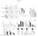
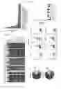
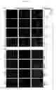
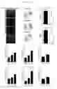
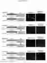
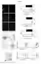
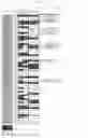
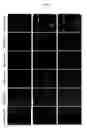

Sources:
- United States Patent and Trademark Office - verify current appl. status at the USPTO↗
Similar patent applications:
- » 20230348570
EPITOPE OF ANTIBODY AGAINST STRUCTURAL PROTEIN OF SARS-COV-2, ANTIBODY REACTING WITH EPITOPE, METHOD FOR DETECTING SARS-COV-2 USING ANTIBODY, DETECTION KIT FOR SARS-COV-2 CONTAINING ANTIBODY, METHOD FOR DETECTING ANTI-SARS-COV-2 ANTIBODY CONTAINING POLYPEPTIDE OF EPITOPE, DETECTION KIT FOR ANTI-SARS-COV-2 ANTIBODY CONTAINING POLYPEPTIDE OF EPITOPE, VACCINE FOR SARS-COV-2 CONTAINING POLYPEPTIDE OF EPITOPE, AND THERAPEUTIC AGENT FOR SARS-COV-2 INFECTION CONTAINING ANTIBODY - » 20090203538
Method of classifying antibody, method of identifying antigen, method of obtaining antibody or antibody set, method of constructing antibody panel and antibody or antibody set and use of the same - » 20140235833
Method of classifying antibody, method of identifying antigen, method of obtaining antibody or antibody set, method of constructing antibody panel and antibody or antibody set and use of the same - » 20250122306
ANTI-TISSUE FACTOR ANTIBODIES AND ANTIBODY CONJUGATES, COMPOSITIONS COMPRISING ANTI-TISSUE FACTOR ANTIBODIES OR ANTIBODY CONJUGATES, AND METHODS OF MAKING AND USING ANTI-TISSUE FACTOR ANTIBODIES AND ANTIBODY CONJUGATES - » 20060247424
Anti-idiotype antibody, method of constructing the anti-idiotype antibody, and method of preparing idiotype antibody using the anti-idiotype antibody - » 20210324088
ANTIBODIES, ACTIVATABLE ANTIBODIES, BISPECIFIC ANTIBODIES, AND BISPECIFIC ACTIVATABLE ANTIBODIES AND METHODS OF USE THEREOF - » 20220251176
FUSION PROTEIN COMPRISING ANTI-MESOTHELIN ANTIBODY, ANTI-CD3 ANTIBODY OR ANTI-EGFR ANTIBODY, BISPECIFIC OR TRISPECIFIC ANTIBODY COMPRISING SAME, AND USES THEREOF - » 20160237113
Antibody purification method, antibody obtained therefrom, novel antibody purification method using cation exchanger, and antibody obtained therefrom - » 20190135943
Antibodies, activatable antibodies, bispecific antibodies, and bispecific activatable antibodies and methods of use thereof - » 20060078951
Monoclonal antibody selectively recognizing listeria monocytogenes, hybridoma producing the antibody, test kit comprising the antibody and detection method of listeria monocytogenes using the antibody
Recent applications in this class:
- » 20250289877 2025-09-18
Antibodies to Amyloid Beta - » 20250289876 2025-09-18
CONNEXIN 43 ANTIBODIES AND USE THEREOF - » 20250289875 2025-09-18
GRP78 NANOBODIES - » 20250289874 2025-09-18
ANTIBODY AGAINST P-TAU 217 AND USE THEREOF - » 20250289873 2025-09-18
COMPLEMENT FACTOR H RELATED 4-SPECIFIC ANTIBODIES AND USES THEREOF - » 20250282856 2025-09-11
INHIBITORS OF COMPLEMENT FACTOR H - » 20250282855 2025-09-11
COMPOSITIONS AND METHODS FOR TREATING MULTIPLE SYSTEM ATROPHY (MSA) - » 20250282854 2025-09-11
TAU BINDING COMPOUNDS - » 20250282853 2025-09-11
UTILIZATION OF IMMORTALIZED B CELLS TO IDENTIFY SDCBP AS A NOVEL THERAPEUTIC TARGET IN OVARIAN CARCINOMA - » 20250282852 2025-09-11
METHODS OF TREATING NEUROLOGICAL DISEASES
Recent applications for this Assignee:
- » 20250251399 2025-08-07
BIOMARKERS FOR PANCREATIC CANCER - » 20250145667 2025-05-08
PEPTIDE - » 20250044280 2025-02-06
METHODS FOR DIAGNOSING OR PREDICTING A CARDIAC INFLAMMATORY CONDITION - » 20240368241 2024-11-07
SYNDECAN PEPTIDES - » 20240058232 2024-02-22
Composition Comprising Calcium Orthophosphate and a Bioactive Glass Comprising Fluorine - » 20240036048 2024-02-01
MEANS AND METHODS FOR DIAGNOSING PANCREATIC CANCER - » 20240025955 2024-01-25
AGRIN POLYPEPTIDE AND USES THEREOF - » 20220325317 2022-10-13
METHODS FOR GENERATING A POPULATION OF POLYNUCLEOTIDE MOLECULES - » 20220233462 2022-07-28
Biodegradable multilayer nanocapsules for the delivery of biologically active agents in target cells - » 20220042998 2022-02-10
Biomarkers for pancreatic cancer