Method of synthesizing biogenic elemental selenium nanostructure using enterobacter cloacae and application thereof
US20170191083A1
2017-07-06
15/397,711
2017-01-03
✅ Patent granted
US 10,087,466 B2
2018-10-02
-
-
Vera Afremova
Shuang Chang | PSK Intellectual Property Group, LLC
2037-01-03
Abstract:
A method of synthesizing biogenic elemental selenium nanostructure using Enterobacter cloacae and its application. The method uses E. cloacae Z0206 to reduce selenite to zero valence selenium and forms nano-sized elemental selenium particles, including steps of inoculating activated E. cloacae Z0206 to fermentation broth, adding sodium selenite solution, shaking and incubating, collecting the fermentation broth and separating the elemental selenium nanoparticles.
Inventors:
- Yizhen Wang 3 🇨🇳 Hangzhou, China
- Deguang Song 1 🇨🇳 Hangzhou, China
- Zeqing Lu 1 🇨🇳 Hangzhou, China
- Fengqin Wang 1 🇨🇭 Hangzhou, Switzerland
- Yuanzhi Cheng 1 🇨🇭 Hangzhou, Switzerland
- Xiaoxiao Li 1 🇨🇳 Hangzhou, China
- Fengqin Wang 2 🇨🇳 Hangzhou, China
- Yuanzhi Cheng 1 🇨🇳 Hangzhou, China
Assignee:
- ZHEJIANG UNIVERSITY 49 🇨🇳 HANGZHOU, Zhejiang Province, China
Applicant:
Interested in similar patents?
Get notified when new applications in this technology area are published.
Classification:
C12P3/00 » CPC main
Preparation of elements or inorganic compounds except carbon dioxide
C01B19/02 » CPC further
Selenium; Tellurium; Compounds thereof Elemental selenium or tellurium
A23L33/16 » CPC further
Modifying nutritive qualities of foods; Dietetic products; Preparation or treatment thereof using additives Inorganic salts, minerals or trace elements
Description
CROSS-REFERENCE TO RELATED APPLICATIONS
This non-provisional application claims priority to and benefit of, under 35 U.S.C. §119(a), Patent Application No. 201610000452.9 filed in P. R. China on Jan. 4, 2016, the entire content of which is hereby incorporated by reference.
FIELD OF THE INVENTION
The present invention relates to the field of microbial application, nanostructure elemental selenium synthesis and biological feed additives, more particularly to a method for synthesizing biogenic elemental Selenium nanostructures using bacteria and its application in pig production.
BACKGROUND OF THE INVENTION
Selenium is one of the essential trace elements for human and animals, which is fundamental to health. Selenium-deficiency in human affects nervous system, reproductive system, immune system and cardiovascular system. Selenium-deficiency in animals often results in dysfunction in reproductive system, decrease of reproductive performance, growth inhibition, and muscle lesions (such as white muscle disease, cardiomyopathy and skeletal muscle myopathy), etc. Human and animals maintain selenium requirements by obtaining selenium from soil through food chain. People with selenium intake deficiency are facing health risks. Currently recognized as the safest and most efficient selenium supplement form is Selenomethionine (SeMet). Comparing with SeMet, elemental selenium is regarded as biological inert with neither activity nor toxicity. However, recent years, research has indicated that comparing with SeMet, nano-sized elemental selenium particles possess similar biological activity and even lower toxicity.
Selenium nanoparticle biogenesis mostly uses ascorbic acid, sodium thiosulfate, sodium sulphite or hydrazine to reduce selenium dioxide, selenite or selenate to synthesize selenium nanostructure. Chemically synthesized selenium nanostructures require surfactant or stabilizer (proteins or polysaccharides, etc.) to maintain stability, otherwise, they transform easily into black, toxic elemental selenium. In addition, the process of selenium nanostructure synthesis introduces toxic materials, which pollutes the environment. It has been proven that some bacteria could reduce selenium oxyanions to zero-valence selenium and form nanostructure particles, which are sphere shaped, uniform sized, and more stable, comparing with chemically synthesized selenium nanostructures. Moreover, bacterial fermentation isn't usually influenced by temperature and season. Bacterial fermentation for biogenic selenium nanostructure synthesis is also characterized by strong production capacity and short production cycle. Therefore, using bacteria to synthesize biogenic elemental selenium nanostructure may be a safe and efficient way for the future.
The present invention relates to a method of using E. cloacae to synthesize biogenic elemental selenium nanostructures.
SUMMARY OF THE INVENTION
Therefore, the objective of the present invention is to provide a method of using E. cloacae to synthesize biogenic elemental selenium nanostructures and to overcome drawbacks of the existing technologies.
In one aspect, the present invention provides a method of using E. cloacae to synthesize biogenic elemental selenium nanostructures using selenite as the starting material and E. cloacae as the fermentative bacteria.
Preferably, the method includes the following steps:
-
- inoculate activated E. cloacae Z0206 cells to fermentation broth, add sodium selenite solution into the fermentation broth;
- shake and incubate;
- collect the fermentation broth and centrifuge, collect the supernatant after the centrifugation, centrifuge the supernatant once again, re-suspend the sediment with double distilled water, ultrasonicate the suspension, add hexane, mix and stratify by standing. The biogenic elemental selenium nanostructures should present in the lower red aqueous phase. The parameters of the first centrifugation are 4° C., 5,000×g, 15 min. The parameters of the second centrifugation are 4° C., 25,000×g, 15 min. The parameters of the ultrasonication are 25 W, 5 s on/5 s off, 15 min. The volume of hexane is half of the volume of the suspension;
Preferably, in the first step, the broth composition contains sucrose (25 g·L−1), yeast extraction (5 g·L−1), tryptone (5 g·L−1), K2HPO4·3H2O (2.62 g·L−1), KH2PO4 (1 g·L−1) and MgSO4 (0.5 g·L−1); the initial pH value is 7.5.
Preferably, in the first step, the final concentration of sodium selenite in the broth is 10 mM.
Preferably, in the first step, the temperature, rotation speed and incubation time are 32° C., 250 rpm and 144 hours, respectively.
An application of the biogenic elemental selenium nanostructures synthesized using the method of the present invention, where the biogenic elemental selenium nanostructures replace the use of sodium selenite in pig feed.
The present invention further optimizes all the parameters of the synthesis and separation process, overcomes the potential defect of biogenic elemental selenium nanostructures and the uncertainty of production. The present invention is easy to produce and to be applied at industrial scale.
The biogenic elemental selenium nanostructures and biogenic elemental selenium nanostructure-polysaccharides complex could replace sodium selenite in pig feed, which could improve antioxidative capacity and immunity, and promote the growth of pig.
BRIEF DESCRIPTION OF THE DRAWINGS
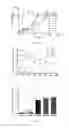
FIG. 1: Effect of different concentrations of sodium selenite on the growth of E. cloacae Z0206;
FIG. 2: Changes of sodium selenite residue in E. cloacae Z0206 fermentation broth at different concentrations of sodium selenite;
FIG. 3: Average consumption rate of sodium selenite by E. cloacae Z0206 at different concentrations of sodium selenite;
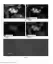
FIG. 4: Environmental scanning electronic microscope (ESEM) analysis and energy dispersive x-ray spectrum (EDX) analysis of E. cloacae Z0206 cells and biogenic elemental selenium nanostructures. (A) ESEM images; (B) Merge of the element distribution maps of carbon and selenium; (C) Element distribution map of carbon; (D) Element distribution map of selenium; (E) EDX analysis.
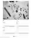
FIG. 5: Transmission electronic microscope (TEM) analysis and EDX analysis of E. cloacae Z0206 cell and biogenic elemental selenium nanostructures. (A) TEM images; (B) EDX analysis of particle 1; (C) EDX analysis of particle 2; (D) EDX analysis of particle 3; (E) EDX analysis of particle 4.
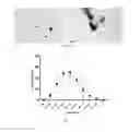
FIG. 6: TEM images of separated biogenic elemental selenium nanostructures.
FIG. 7: Size analysis of separated biogenic elemental selenium nanostructures.
DETAILED DESCRIPTION OF THE INVENTION
Embodiment 1: Effect of Different Concentrations of Sodium Selenite on E. Cloacae Z0206 Growth and Selenite Reduction Rate
-
- 1. Broth preparation
- LB broth: 10 g NaCl, 10 g tryptone and 5 g yeast extraction were dissolved in 1 L double distilled water, sterilized at 100 kPa, 121° C. for 20 min.
- LB agar plate: 10 g NaCl, 10 g tryptone, 5 g yeast extraction and 15 g agar were dissolved in 1 L double distilled water, sterilized at 100 kPa, 121° C. for 20 min.
- Fermentation broth: 25 g sucrose, 5 g yeast extraction, 5 g tryptone, 2.62 g K2HPO4·3H2O, 1 g KH2PO4 and 0.5 g MgSO4 were dissolved in 1 L double distilled water, adjusted the initial pH value to 7.5, sterilized at 67 kPa, 115° C. for 30 min.
- 2. Bacteria activation
- Bacterial stock from −80° C. was thawed, a loop of bacteria was taken and streaked on LB agar plate, cultivated at 32° C. for 24 h.
- A single colony was picked and inoculated into LB broth, cultivated at 32° C., 250 rpm for 18 h.
- 3. Inoculation and fermentation
- Cell density was adjusted to OD600=0.5 with PBS, 1% of the bacteria cells were inoculated to fermentation broth containing 0 mM, 0.5 mM, 1 mM, 5 mM, 10 mM and 15 mM sodium selenite, respectively. Each concentration gradient was repeated for three times. Cells were fermented at 32° C., 250 rpm, and the fermentation broth was collected at 4 h, 8 h, 12 h, 16 h, 20 h, 24 h, 36 h, 48 h, 72 h and 96 h after inoculation for detecting cell protein content in order to characterize bacterial cell density; fermentation broth was also collected at 0 h, 12 h, 24 h, 36 h, 48 h, 72 h, 96 h, 120 h, 144 h and 168 h after inoculation for detecting sodium selenite residue.
- 4. Results
- 1. Broth preparation
As shown in FIG. 1, growth of control group E. cloacae Z0206 reached stationary phase at 12 h, while addition of sodium selenite significantly decreased the growth rate of E. cloacae Z0206, and the inhibition increased with the increase of sodium selenite concentration.
FIGS. 2-3 show the sodium selenite reduction rates of E. cloacae Z0206 at different concentrations of sodium selenite. E. cloacae Z0206 started to consume sodium selenite at the beginning of cultivation. Sodium selenite of groups of 0.5 mM, 1 mM, 5 mM and 10 mM was completely consumed at 48 h, 48 h, 72 h and 144 h after inoculation, respectively. Sodium selenite reduction rate of the 15 mM group was only 91.35%±1.40% at the end of point of monitoring (168 h).
Embodiment 2: Synthesis of Biogenic Elemental Selenium Nanostructure Using E. Cloacae Z0206 and Sodium Selenite
Activated E. cloacae Z0206 cell density was adjusted to OD600=0.5 with PBS, 1% of the bacteria cells were inoculated to the fermentation broth, sodium selenite solution was added to a final concentration of 10 mM, cultivated at 32° C., 250 rpm for 144 h.
Embodiment 3: Electronic Microscope Analysis and Energy-Dispersive X-Ray Spectroscopy Analysis of Bacteria Cells and Biogenic Elemental Selenium Nanostructures
1 mL fermentation broth was collected according to the steps of Embodiment 2. After centrifugation, the sediment was washed three times with PBS and fixed with 2.5% glutaraldehyde solution for 12 h. The sediment was then washed again three times with PBS, followed by 1% osmic acid fixation, ethanol gradient dehydration, isoamyl acetate treatment, critical point drying and gold plating. Samples were analysed with environmental scanning electron microscope (ESEM) and energy-dispersive X-ray spectroscopy (EDX) analysis.
As shown in FIG. 4, E. cloacae Z0206 reduced sodium selenite to regular spherical nanoparticles (FIG. 4A, indicated by arrow). These particles were monodisperse with sizes range between 100 nm to 200 nm. The extracellular nanoparticles were identified as elemental nano-selenium by EDX analysis, and these particles may be capped with a layer of carbon-containing organic matter.
1 mL fermentation broth was collected according to the steps of Embodiment 2. After centrifugation, the sediment was washed three times with PBS and fixed with 2.5% glutaraldehyde solution for 12 h. The sediment was then washed again three times with PBS, followed by agarose pre-embedding. The samples were treated with 1% osmic acid, ethanol gradient and embedding agent, followed by heating at 70° C., slicing and dying. The samples were analyzed with transmission electron microscopy (TEM) and EDX.
As shown in FIG. 5, TEM analysis confirmed that E. cloacae Z0206 reduced sodium selenite to extracellularly located spherical nanoparticles, with sizes ranging between 100 nm to 200 nm. EDX analysis confirmed that these particles were elemental nano-selenium.
Embodiment 4: Biogenic Elemental Selenium Nanostructure Separation, Purification and Characterization
-
- 1. Fermentation broth according to Embodiment 2 was collected, centrifuged at 5,000×g for 15 min, the sediment was discarded.
- 2. The supernatant was centrifuged at 4° C., 25,000×g for 15 min, the supernatant was discarded and the sediment was collected.
- 3. The sediment was re-suspended with double distilled water, ultrasonicated at 25 W, 5 s on/5 s off for 15 min.
- 4. Hexane (half volume of above mentioned suspension) was added, vortexed and mixed, and let stand to stratification. The biogenic elemental selenium nanoparticles were present in the lower aqueous phase and the lower aqueous phase was collected, which was a biogenic elemental selenium nanoparticle suspension.
- 5. A drop of the biogenic elemental selenium nanoparticle suspension was added to a copper net, dried with paper filter, and analysed with TEM;
- 6. A drop of the biogenic elemental selenium nanoparticle suspension was analysed with nano-sizer to measure particle size.
As shown in FIG. 6, TEM analysis showed that the biogenic elemental selenium nanoparticles were monodisperse, regular spheres, with sizes range between 100 nm to 200 nm. Particle size analysis (FIG. 7) showed that the biogenic elemental selenium nanoparticles ranged between 80 nm to 250 nm, with the average size of 144.10±1.14 nm.
Embodiment 5: Biogenic Elemental Selenium Nanostructure-Polysaccharides Complex Separation
-
- 1. The fermentation broth according to Embodiment 2 was collected by centrifuging at 5,000×g for 15 min. The supernatant was collected.
- 2. Pre-cooled ethanol (3-fold volumes of the supernatant) was added to the supernatant, centrifuged at 5,000×g for 15 min. The obtained sediment was the biogenic elemental selenium nanostructure-polysaccharides complex.
Embodiment 6: Application of Biogenic Elemental Selenium Nanostructure in Pig
-
- 1. Experiment design
Ninety 28-day-old weaned pigs were randomly divided into three groups, with three replicates per group, and ten pigs per replicate. The control group was fed with basic diet, the experimental group 1 was fed with basic diet supplied with 0.3 mg/kg Na2SeO3, and the experimental group 2 was fed with basic diet supplied with 0.14 mg/kg biogenic elemental selenium nanostructure. Diet composition and nutrient levels were shown in Table 1.
| TABLE 1 |
| Basic diet and nutrient levels |
| Composition (%) | Nutrient levels |
| Corn | 59.3 | Digestible energy (MJ/kg) | 14.43 |
| Dehulled soybean meal | 8.0 | Crude protein (%) | 19.50 |
| Extruded soybean | 6.0 | Ca (%) | 0.79 |
| Fermented soybean meal | 7.0 | Total P (%) | 0.60 |
| Whey powder | 6.0 | Lys (%) | 1.50 |
| Plasma protein | 2.5 | Thr (%) | 0.98 |
| Fish meal | 3.0 | Met (%) | 0.52 |
| Soybean oil | 2.5 | Trp (%) | 0.18 |
| monocalcium phosphate | 0.9 | Se (%) | 0.10 |
| Stone powder | 0.8 | ||
| Premix | 4.0 | ||
-
- 2. Feeding and management
During the experimental process, pigs were kept in pigpen with slatted floor, automatic feeder and duckbill type drinker. Anthelmintic work and vaccination were performed according to the farm management.
Growth Measurement
Pig body weight was measured at 28-day-old and 67-day-old, respectively. Food consumption data was collected. Average daily feed intake, average daily weight gain and feed/gain ratio was calculated.
Sample Collection
After 12 h fasting, pig blood were collected and allowed to coagulation. The blood was centrifuged at 4° C., 3,000×g for 15 min. Serum was collected and stored at −80° C.
Serum Antioxidative and Immune Function Measurement
Serum total antioxidative ability, glutathione peroxidase (GPx) activity, superoxide dismutase (SOD) activity, and malondialdehyde (MDA) concentration were detected using relevant kits according to the manufacturer's instruction.
Serum IgG and IgM were measured using turbidimetric inhibition immuno assay.
Serum inflammatory cytokine tumor necrosis factor alpha (TNF-α), interleukin-2 (IL-2) and interleukin-6 (IL-6) were determined using enzme linked immunosorbent assay (ELISA) kit according to manufacturer's instruction.
Statistics
One-way analysis of variance (ANOVA) followed by a lease significant difference (LSD) multiple comparison test was used to determine the statistical significance for multiple comparisons, P<0.05 was considered statistically significant. All statistical tests were carried out using SPSS 22 software. All data are presented as the mean±SD.
Results
-
- (1) Effect of biogenic elemental selenium nanostructure on pig growth
| TABLE 2 |
| Effect of biogenic elemental selenium nanostructure on pig growth |
| Experimental | Experimental | ||
| Item | Control | group 1 | group 2 |
| Initial | 8.08 ± 0.06 | 8.12 ± 0.10 | 8.13 ± 0.10 |
| weight (kg) | |||
| Final | 23.75 ± 0.49a | 24.17 ± 0.76a | 25.72 ± 0.83b |
| weight (kg) | |||
| Average daily | 668.36 ± 18.37 | 654.17 ± 37.64 | 679.23 ± 49.77 |
| feed intake (g) | |||
| Average daily | 401.71 ± 11.56a | 411.54 ± 20.63a | 451.03 ± 23.97b |
| gain (g) | |||
| Feed/gain ratio | 1.67 ± 0.03a | 1.59 ± 0.01b | 1.50 ± 0.03c |
As shown in Table 2, even though there was no significant difference in average daily feed intake, pigs fed with biogenic elemental selenium nanostructure (experimental group 2) had significant increase of the average daily gain, and decreased feed/gain ratio. The effect of promoting growth by biogenic elemental selenium nanostructure was better than by sodium selenite (experimental group 1).
-
- (2) Effect of BNS on pig antioxidative function
| TABLE 3 |
| Effect of biogenic elemental selenium nanostructure |
| on pig antioxidative function |
| Experiment | Experiment | ||
| Item | Control | group 1 | group 2 |
| T-AOC (U/mL) | 2.26 ± 0.11a | 2.51 ± 0.16a | 2.93 ± 0.14b |
| GPx (U/mL) | 219.00 ± 11.72a | 238.56 ± 13.07a | 275.05 ± 15.54b |
| SOD (U/mL) | 138.68 ± 4.54a | 147.94 ± 5.89a | 164.48 ± 6.00b |
| MDA | 3.53 ± 0.14a | 3.31 ± 0.10a | 2.73 ± 0.10b |
| (nmol/mL) | |||
As shown in Table 3, compared with control group and experimental group 1, biogenic elemental selenium nanostructure significantly increased the activity of T-AOC, GPx and SOD, and decreased MDA concentration in experimental group 2. No significant difference of the parameters was shown comparing experimental group 1 with the control group.
-
- (3) Effect of biogenic elemental selenium nanostructure on pig immune cytokines expression
| TABLE 4 |
| Effect of BNS on pig immune cytokines expression |
| Experimental | Experimental | ||
| Item | Control | group 1 | group 2 |
| IgG (g/L) | 2.39 ± 0.09a | 2.66 ± 0.23ab | 3.68 ± 0.35c |
| IgM (g/L) | 0.66 ± 0.07a | 0.71 ± 0.07a | 1.03 ± 0.12b |
| TNF-α (ng/L) | 33.66 ± 5.17a | 41.86 ± 4.50ab | 56.01 ± 4.20c |
| IL-2 (ng/L) | 88.24 ± 7.98a | 108.19 ± 10.26ab | 131.38 ± 11.80b |
| IL-6 (ng/L) | 479.44 ± 26.84a | 571.10 ± 38.62ab | 707.25 ± 52.95c |
As shown in Table 4, comparing with the control group, Na2SeO3 and biogenic elemental selenium nanostructure significantly elevated serum levels of IgG, IgM, TNF-α, IL-2 and IL-6. The elevated effects on all the serum levels of the biogenic elemental selenium nanostructure group reached to significant levels.
Conclusions
Dietary supplementation with biogenic elemental selenium nanostructure could significantly promote pig growth, decrease feed/gain ratio; and increase the levels of antioxidative activity and immune cytokines. All of the effects were more effective than those of sodium selenite.
Claims
What is claimed is:1. A method of synthesizing biogenic elemental selenium nanoparticles using selenite as starting materials and E. cloacae Z0206 as fermentative bacteria, comprising the steps of:
inoculating activated E. cloacae Z0206 cells to a fermentation broth, and adding sodium selenite solution to obtain an inoculated broth;
shaking and incubating the inoculated broth to obtain a fermented broth; and
collecting the fermented broth, performing a first centrifugation on the fermented broth to obtain a supernatant, performing a second centrifugation on the supernatant to obtain a sediment, re-suspending the sediment with double distilled water to obtain a suspension, ultrasonicating the suspension, adding hexane, and mixing and stratifying by standing, wherein parameters of the first centrifugation are 4° C., 5,000×g, 15 minutes, parameters of the second centrifugation are 4° C., 25,000×g, 15 minutes, parameters of the ultrasonication are 25 W, 5 seconds on and 5 seconds off for 15 minutes, and the volume of the hexane is half of the volume of the suspension.
2. The method of claim 1, wherein the fermentation broth consists of sucrose (25 g·L−1), yeast extraction (5 g·L−1), tryptone (5 g·L−1), K2HPO4·3H2O (2.62 g·L−1), KH2PO4 (1 g·L−1) and MgSO4 (0.5 g·L−1), with an initial pH value of 7.5.
3. The method of claim 1, wherein the final concentration of sodium selenite in the fermentation broth is 10 mM.
4. The method of claim 1, wherein the temperature, rotation speed and incubation time are 32° C., 250 rpm and 144 hours, respectively.
Images & Drawings included:
Sources:
- United States Patent and Trademark Office - verify current appl. status at the USPTO↗
Recent applications in this class:
- » 20250283118 2025-09-11
MODIFIED BACTERIA AND METHODS OF USE FOR BIOGLASS MICROLENSES - » 20250270594 2025-08-28
RARE EARTH ELEMENT PHOSPHATES AND PROCESS FOR MAKING - » 20250257369 2025-08-14
ENGINEERED CLOSTRIDIUM THERMOCELLUM FOR CO-UTILIZATION OF HEMICELLULOSE AND CELLULOSE - » 20250236893 2025-07-24
ENHANCING THE SEQUESTRATION OF CARBON DIOXIDE IN DOWNHOLE CARBONATE-BASED RESERVOIRS VIA INJECTING UREASE ENZYME - » 20250109414 2025-04-03
Continuous Flow Reactor for Conversion of Wastewater to Biohydrogen Using Activated Sludge and Microalgae - » 20250066821 2025-02-27
PROCESSES AND SYSTEMS FOR BIOLOGICAL HYDROGEN PRODUCTION FROM ORGANIC WASTE USING YEAST - » 20250059572 2025-02-20
METHOD FOR EMPLOYING CORN PLANT RESIDUE IN THE PRODUCTION OF DIRECT REDUCED IRON - » 20250051804 2025-02-13
Method to Transform Phosphorus in an Antibiotic into Sulfur - » 20250011818 2025-01-09
Method for Treating Recycled Aggregates via Microbial-Induced Calcite Precipitation - » 20240401086 2024-12-05
INTEGRATED GAS FERMENTATION AND CARBON BLACK PROCESSES
Recent applications for this Assignee:
- » 20220096422 2022-03-31
BLOOD-BRAIN BARRIER PERMEABILITY REGULATOR AND USE THEREOF - » 20220079929 2022-03-17
USE OF AXITINIB AND ANALOGS THEREOF IN PREPARING BLOOD-BRAIN BARRIER PERMEABILITY REGULATOR - » 20190378622 2019-12-12
Method for prediction of cortical spiking trains - » 20190154848 2019-05-23
14C testing bottle, testing device and method, sampling and preparation system and method - » 20190093118 2019-03-28
Gene combination and use thereof - » 20190010102 2019-01-10
Method for adsorption separation of propylene and propyne - » 20180038353 2018-02-08
Method and apparatus for solar power generation through gas volumetric heat absorption based on characteristic absorption spectrum - » 20180038310 2018-02-08
Radiation thermal absorber based on characteristic absorption spectrum, and stirling engine and operation method thereof - » 20180030077 2018-02-01
Process for preparing high-purity L-arabinose by using arabic gum as raw material - » 20180016308 2018-01-18
Human alpha-defensin 5 variant and uses thereof