Method and apparatus for accessing matter disposed within an internal body vessel
US20180360643A1
2018-12-20
16/000,376
2018-06-05
✅ Patent granted
US 11,160,682 B2
2021-11-02
-
-
Andrew J Mensh
Carter, DeLuca & Farrell LLP
2040-01-04
Abstract:
A method for accessing matter within an intestine, includes creating an opening extending through an abdominal wall of an abdominal cavity, accessing an intestinal end segment of an intestine within the abdominal cavity, positioning the intestinal end segment of the intestine in registration with the opening extending through the abdominal wall, attaching the intestinal end segment to cutaneous tissue surrounding the opening to establish fluid communication between a lumen of the intestine and the opening through the abdominal wall, securing an access device adjacent the opening through the abdominal wall and connecting a receptacle to the access device configured to be in fluid communication with the lumen of the intestine to collect matter within the intestine.
Assignee:
- Covidien LP 8,015 🇺🇸 Mansfield, MA, United States
Applicant:
Interested in similar patents?
Get notified when new applications in this technology area are published.
Classification:
A61B17/04 IPC
Surgical instruments, devices or methods, e.g. tourniquets for suturing wounds; Holders or packages for needles or suture materials
A61B17/34 IPC
Surgical instruments, devices or methods, e.g. tourniquets Trocars; Puncturing needles
A61B17/3415 » CPC further
Surgical instruments, devices or methods, e.g. tourniquets; Trocars; Puncturing needles for introducing tubes or catheters, e.g. gastrostomy tubes, drain catheters
A61F2/0063 » CPC further
Filters implantable into blood vessels; Prostheses, i.e. artificial substitutes or replacements for parts of the body; Appliances for connecting them with the body; Devices providing patency to, or preventing collapsing of, tubular structures of the body, e.g. stents Implantable repair or support meshes, e.g. hernia meshes
A61F5/445 » CPC main
Orthopaedic methods or devices for non-surgical treatment of bones or joints ; Nursing devices; Anti-rape devices; Devices worn by the patient for reception of urine, faeces, catamenial or other discharge; Portable urination aids ; Colostomy devices Colostomy, ileostomy or urethrostomy devices
A61B17/0469 » CPC further
Surgical instruments, devices or methods, e.g. tourniquets for suturing wounds; Holders or packages for needles or suture materials Suturing instruments for use in minimally invasive surgery, e.g. endoscopic surgery
A61B17/115 IPC
Surgical instruments, devices or methods, e.g. tourniquets for performing anastomosis; Buttons for anastomosis Staplers for performing anastomosis in a single operation
A61B17/0487 » CPC further
Surgical instruments, devices or methods, e.g. tourniquets for suturing wounds; Holders or packages for needles or suture materials Suture clamps, clips or locks, e.g. for replacing suture knots; Instruments for applying or removing suture clamps, clips or locks
A61F5/448 » CPC further
Orthopaedic methods or devices for non-surgical treatment of bones or joints ; Nursing devices; Anti-rape devices; Devices worn by the patient for reception of urine, faeces, catamenial or other discharge; Portable urination aids ; Colostomy devices; Colostomy, ileostomy or urethrostomy devices Means for attaching bag to seal ring
A61B17/1114 » CPC further
Surgical instruments, devices or methods, e.g. tourniquets for performing anastomosis; Buttons for anastomosis of the digestive tract, e.g. bowels or oesophagus
A61F2/00 IPC
Filters implantable into blood vessels; Prostheses, i.e. artificial substitutes or replacements for parts of the body; Appliances for connecting them with the body; Devices providing patency to, or preventing collapsing of, tubular structures of the body, e.g. stents
A61F2005/4455 » CPC further
Orthopaedic methods or devices for non-surgical treatment of bones or joints ; Nursing devices; Anti-rape devices; Devices worn by the patient for reception of urine, faeces, catamenial or other discharge; Portable urination aids ; Colostomy devices; Colostomy, ileostomy or urethrostomy devices Implantable
A61F5/449 » CPC further
Orthopaedic methods or devices for non-surgical treatment of bones or joints ; Nursing devices; Anti-rape devices; Devices worn by the patient for reception of urine, faeces, catamenial or other discharge; Portable urination aids ; Colostomy devices; Colostomy, ileostomy or urethrostomy devices Body securing means, e.g. belts, garments
A61B17/11 IPC
Surgical instruments, devices or methods, e.g. tourniquets for performing anastomosis; Buttons for anastomosis
A61B17/068 » CPC further
Surgical instruments, devices or methods, e.g. tourniquets Surgical staplers, e.g. containing multiple staples or clamps
A61B17/1155 » CPC further
Surgical instruments, devices or methods, e.g. tourniquets for performing anastomosis; Buttons for anastomosis; Staplers for performing anastomosis in a single operation Circular staplers comprising a plurality of staples
Description
TECHNICAL FIELD
The present disclosure generally relates to a surgical procedure and associated apparatus for accessing internal body vessels. More specifically, the present disclosure relates to a surgical procedure and an apparatus for performing an ostomy for removal of waste material from an intestine.
BACKGROUND
Exteriorization of an internal body vessel such as the intestine is called a stoma. Stomas may be created in conjunction with an ostomy procedure by inverting and suturing a bisected portion of an intestine to the exterior of the abdominal wall to provide internal access into the intestine for collecting fecal matter. Complications associated with stomas can include leaks, bleeding, necrosis, stenosis, dermal infection, prolapse, etc. Accordingly, a need exists to develop improved methods for reducing risks and complications typically associated with ostomy procedures.
SUMMARY
Accordingly, the present disclosure is directed to a method for accessing matter within an intestine, including creating an opening extending through an abdominal wall of an abdominal cavity, accessing an intestinal end segment of an intestine within the abdominal cavity, positioning the intestinal end segment of the intestine in registration with the opening extending through the abdominal wall, attaching the intestinal end segment to cutaneous tissue surrounding the opening to establish fluid communication between a lumen of the intestine and the opening through the abdominal wall, securing an access device adjacent the opening through the abdominal wall and connecting a receptacle to the access device configured to be in fluid communication with the lumen of the intestine to collect matter within the intestine.
In embodiments, the method includes coupling the intestinal end segment of the intestine to an anvil of a stapling instrument, connecting the anvil to a stapling instrument and firing the stapling instrument to deliver fasteners through the cutaneous tissue surrounding the opening and within the intestinal end segment of the intestine.
In some embodiments, securing the access device includes positioning a connector segment of the access device through the opening and within the lumen of the intestinal end segment of the intestine.
In certain embodiments, the method includes establishing a fluid tight seal about the connector segment of the access device within the lumen of the intestinal end segment of the intestine. In embodiments, establishing the fluid tight seal includes inflating an inflatable bladder disposed about the connector segment of the access device to engage an inner wall of the intestinal end segment. In some embodiments, establishing the fluid tight seal includes suturing the intestinal end segment of the intestine to the connector segment of the access device. In certain embodiments, establishing the fluid tight seal includes adhering the connector segment of the access device within the lumen of the intestinal end segment of the intestine.
In embodiments, coupling the intestinal end segment of the intestine includes arranging the intestinal end segment about an anvil head of the anvil. In some embodiments, firing the stapling instrument includes approximating the anvil head relative to a staple cartridge of the stapling instrument. In certain embodiments, firing the stapling instrument includes delivering an annular array of staples where the staples are at least partially deformed by the anvil head of the anvil. In embodiments, attaching the intestinal end segment of the intestine includes applying a purse string about the intestinal end segment and the anvil head.
In certain embodiments, firing the stapling instrument includes delivering staples through facial tissue underlying the cutaneous tissue.
In some embodiments, positioning the intestinal end segment includes exposing end margins of the intestinal end segment external of the opening, and wherein firing the stapling instrument includes delivering the fasteners through the end margins of the intestinal end segment.
In some embodiments, the method includes attaching a mesh to the intestinal end segment and to muscle tissue beneath the cutaneous tissue.
The method and associated apparatuses for forming an ostomy to collect matter from an intestine according to the present disclosure obviates the need to invert the end margins and create a stoma by directly attaching the intestinal segment to the skin or cutaneous layer about an ostomy extending through the abdominal wall. Thus, complications typically associated with stoma formation including, e.g., leakage of intestinal fluids, infections and other dermal complications, are avoided. The access device utilized in the methodologies supports the intestinal segment during treatment and provides a mechanism to attach to a collection or ostomy bag without producing undue strain on the abdominal wall thereby preserving the integrity of the ostomy.
Other advantages will be appreciated by the following description.
BRIEF DESCRIPTION OF THE DRAWINGS
The accompanying drawings, which are incorporated in and constitute a part of this specification, illustrate embodiments of the disclosure and, together with a general description of the disclosure given above, and the detailed description given below, serve to explain the principles of the disclosure, wherein:
FIG. 1 is a schematic illustration, in partial cross-section, of an abdominal region of a subject;
FIG. 2 is a schematic illustration, in partial cross-section, of the abdominal region illustrating an intestine resected and an opening created in the abdominal wall in connection with performing an ostomy procedure in accordance with an illustrative embodiment of the present disclosure;
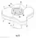
FIG. 3 is a perspective view of an circular anastomosis instrument for use in performing the ostomy procedure;
FIG. 4 is a schematic illustration of the intestinal end segment positioned about the anvil head of the circular anastomosis instrument and the fastener cartridge positioned adjacent the opening in the abdominal cavity prior to actuation of the instrument;
FIG. 5 is a schematic illustration of the intestinal end segment attached to cutaneous tissue subsequent to firing the circular anastomosis instrument to form the ostomy;
FIG. 6 is a perspective view illustrating the staple line created in the cutaneous tissue for attaching the intestinal end segment and forming the ostomy;
FIG. 7 is a perspective view of an illustrative embodiment of an access device for insertion within the ostomy;
FIGS. 8-9 are perspective views illustrating a sequence of insertion of the access device of FIG. 7 within the ostomy;
FIG. 10 is a cross-sectional view taken along the lines 10-10 of FIG. 9;
FIG. 11 is a perspective view illustrating securing of the access device to the abdominal wall in registration with the ostomy;
FIG. 12 is a perspective view of one illustrative embodiment of the access device incorporating an external inflatable bladder;
FIG. 13 is a cross-sectional view illustrating the access device of FIG. 12 positioned within an ostomy;
FIG. 14 is a perspective view illustrating an alternative methodology and associated access device for forming an ostomy;
FIG. 15 is a perspective view illustrating insertion of the access device of FIG. 14 within the ostomy; and
FIG. 16 is a cross-sectional view taken along the line 16-16 of FIG. 15.
DETAILED DESCRIPTION
Aspects of the present disclosure are described in detail with reference to the drawings, in which like reference numerals designate identical or corresponding elements in each of the several views. As used herein, the term “distal” or “leading” refers to that portion of the device that is farther from the user, while the term “proximal” or “trailing” refers to that portion of the device that is closer to the user. As used herein, the term “clinician” refers to a doctor, nurse, or other care provider and may include support personnel. In the following description, well-known functions or constructions are not described in detail to avoid obscuring the present disclosure in unnecessary detail.
The following discussion will focus on methodologies and associated apparatus(es) in performing an ostomy procedure, particularly, in securing the end margins of an intestine to abdominal tissue in connection with a colostomy or ileostomy procedure. However, the guide apparatus has application in other ostomy procedures including urostomy, gastrostomy and jejunostomy procedures.
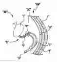
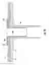
Referring to FIG. 1, an abdominal region “AR” of a subject's body generally includes an abdominal wall “AW” having an outer cutaneous or skin layer “s” (e.g., epidermis, dermis, and hypodermis), a fascia layer “f” including connective tissue and an inner muscle layer “m” (e.g., anterior rectus sheath) that enshrouds organs, vessels, and/or other tissue for performing various bodily functions such as digestion. For instance, as part of a digestive system “D” of a subject's body, the stomach “S” and the intestines “I” are supported in the abdominal cavity “AC.” In the course of a natural digestion process, the stomach “S” and the intestines “I” collaborate with the rest of the digestive system “D” to process food and excrete fecal matter “P” through the anus “A.” Unfortunately, as a result of disease or injury to the intestines “I,” for example, it may become necessary to bypass natural digestion through the anus “A” by performing an ostomy through the abdominal region “AB” in order to safely excrete the fecal matter “P” from the intestine “I” of the subject's body.
Referring now to FIG. 2, in accordance with one exemplative methodology of the present disclosure, an internal body organ or vessel, such as the intestine “I”, can be accessed through an incision or opening “o” formed, e.g., with a scalpel through the skin layer “s”, the fascia layer “f” and the inner muscle layer “m” of the abdominal wall “AW.” The clinician introduces one or more instruments through the opening “o” and bisects the intestine “I” into a first intestinal end segment “I1” and a second intestinal end segment “I2”. The second intestinal end segment “I2” may be sealed closed with any suitable instrument such as an ultrasonic and/or electrosurgical forceps (not shown). For a detailed description of the construction and operation of one example of such a forceps, reference is made to U.S. Pat. No. 8,444,664, the entire contents of which is incorporated by reference herein.
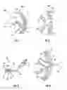
With the intestine “I” bisected into first and second intestinal end segments “I1”, “I2”, attention is directed to connecting the first intestinal segment “I1” about the opening “o” within the abdominal wall “AW”. In embodiments, the first intestinal end segment “I1” may be secured to the abdominal wall “AW” with a stapling apparatus such as the circular fastener apparatus 10 depicted in FIG. 3. In general, the circular fastener apparatus 10 includes a handle assembly 12 that supports an approximation knob 14 and a firing trigger 16. The circular fastener apparatus 10 further includes an elongated shaft 18 that extends distally from the handle assembly 12 to an end effector assembly 20 supported on a distal end portion of the elongated shaft 18. The end effector assembly 20 includes a shell assembly 22 and an anvil 24 that are movable between an unapproximated position (FIG. 3) and an approximated position in response to rotation of the approximation knob 14 of the handle assembly 12. The shell assembly 22 includes a cartridge 26 that supports an annular array of fasteners (e.g., rectangular and/or round wire staples—not shown) in fastener-retaining slots 28 defined in the cartridge 26. The anvil 24 includes an anvil head 30 defining fastener-forming pockets 32 that are configured to form the fasteners upon firing of the fasteners from the cartridge 26. For a detailed description of the construction and operation of one example of such a circular fastener apparatus, reference is made to U.S. Pat. No. 8,011,554, the entire contents of which is incorporated by reference herein.
With reference to FIG. 4, in one exemplary procedure, the skin layer “s” is separated from the fascia “f” at least along locations adjacent the opening “o”. Thereafter, the anvil head 30 of the stapling apparatus 10 is introduced through the opening “o” within the abdominal wall “AW” and positioned within the first intestinal end segment “I1”. The first intestinal end segment “I1” is secured about the anvil head 30 with the use of purse string sutures or the like. The cartridge 26 is positioned external of the skin layer “s”. The approximation knob 14 is then rotated to move the end effector assembly 20 to the approximated position (FIG. 4) so that an end portion of the first intestinal segment “I1” and the skin layer “s” are clamped between the anvil head 30 and the cartridge 26. The firing trigger 16 of the handle assembly 12 is actuated to fire the fasteners from the fastener-retaining slots 28 of the cartridge 26 for formation against the fastener-forming pockets 32 of the anvil 30 to fasten the first intestinal end segment “I1” to the skin layer “s”. The circular fastener apparatus 10 can then be removed.
FIGS. 5-6 illustrates the skin layer “s” attached to the first intestinal end segment “I1”. The fasteners 34 are depicted schematically and penetrate only the skin layer “s” not the underlying fascia layer “f” or muscle layer “m” to connect to the first intestinal segment “I1” within the abdominal cavity “AC”. Thus, the first intestinal segment “I1” does not extend through the opening “o” in the abdominal wall “AW”, but, rather is secured to the inner surface of the skin layer “s”. Thus, formation of a stoma with the first intestinal segment “I1” on the exterior of the skin layer “s” or outside the abdominal cavity “AC” is avoided along with the accompanying limitations associated therewith, including, e.g., leakage of intestinal fluids, infections and other dermal complications. FIG. 6 illustrates the annular array of fasteners 34 penetrating the skin layer “s” to connect with the first intestinal segment “I1” to form the ostomy. In some embodiments it is contemplated that some fascia tissue of the fascia layer “f” may be captured by the fasteners 34.
In addition, a surgical mesh 50 (e.g., a hernia mesh) (FIG. 5) may be secured to the muscle layer “m” and the first intestinal end segment “I1” to stabilize the first intestinal end segment “I1” within the abdominal cavity “AC” to minimize the potential of herniation. For a detailed description of one example of a hernia mesh, reference is made to U.S. Pat. No. 9,005,308, the entire contents of which is incorporated by reference herein.
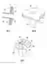
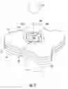
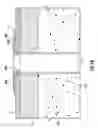
Referring now to FIG. 7, an access device 100 may be mounted to the abdominal wall “AW” in registration with the opening “o” and the first intestinal end segment “I1” to provide a conduit for passage of waste material (e.g., fecal matter “P”) to a collection device such as a colostomy or ileostomy bag (not shown). The access device 100 includes a flange segment 102 and a connector segment 104 extending from the flange segment 102 and defining a longitudinal axis “x”. The flange segment 102 includes a plurality of spaced openings 106, which, in embodiments may be disposed in equidistant radial spaced relation. The openings 106 are configured to receive fasteners to couple the access device 100 to the skin layer “s”. The connector segment 104 may be cylindrical and extends for a distance sufficient to be received through the opening “o” and within the first intestinal end segment “I1”. The flange segment 102 and the connector segment 104 define a longitudinal passage 108 therethrough to permit passage of the fecal matter “P” to the collection device.
The access device 100 may be formed from any biocompatible material including stainless steel, titanium or one or more polymers. The biocompatible polymer may be biodegradable, non-biodegradable or a combination of biodegradable and non-biodegradable. The term “biodegradable” as used herein is defined to include both bioabsorbable and bioresorbable materials.
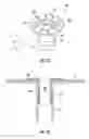
In use, with reference to FIGS. 8-9, the connector segment 104 of the access device 100 is introduced through the opening “o” in the abdominal wall “AW” and the flange segment 102 is positioned against the outer surface of the skin layer “s”. In FIGS. 8-9, the fascia layer “f” and inner muscle layer “m” removed for clarity purposes. In embodiments, the connector segment 104 may be secured within the first intestinal segment “I1” with cements, adhesives, etc. which may be applied prior to, or subsequent to, insertion of the connector segment 104 into the first intestinal segment “I1”. The connector segment 104 may establish a seal with the internal surface of the first intestinal segment “I1”. With the access device 100 appropriately positioned in registration with the first intestinal end segment “I1”, the flange segment 102 is secured to the skin layer “s” through application of at least one fastener 110 as depicted in FIGS. 10-11. In embodiments, the circular fastener apparatus 10 described hereinabove in connection with FIG. 3 may be utilized to deliver an annular array of fasteners 110 (e.g., staples) within the flange segment 102. Other instruments for application of the fasteners 110 are disclosed in commonly assigned U.S. Patent Publication Nos.: 2014/0121684 to Criscuolo and 2014/0276972 to Abuzaina et al., the entire contents of each disclosure being incorporated by reference herein. In the alternative, the flange segment 102 may be secured to the skin layer “s” via suturing.
The fasteners 110 may be delivered through the skin layer “s” without penetrating through the underlying fascia layer “f” or muscle layer “m”. Thus, the fasteners 110 connect the skin layer “s” directly to the intestinal end segment “I1”. It is contemplated, however, that at least some fascia tissue may be captured by the fasteners 110.
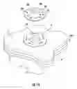
With the access device secured relative to the opening “o”, a collection device, depicted schematically as reference numeral 150, (FIG. 11) may be mounted to the flange segment 102 of the access device 100. The collection device 150 may be a colostomy bag or the like. Any methodology for mounting the collection device 150 to the flange segment 102 is envisioned. The collection device 150 receives intestinal fluids and/or fecal matter passing through the longitudinal passage 108 of the access device 100.
Thus, the aforedescribed methodology performs an ostomy procedure without requiring inversion and exposure of the intestine as in conventional stoma formation. The access device 100 supports the first intestinal end segment “I1” during treatment and provides a mechanism to attach a collection device 150 without producing undue strain within the opening “o” of the abdominal wall and on the first intestinal segment “I” thereby preserving the integrity of the ostomy. The ostomy is effected by connecting the skin layer “s” directly to the first intestinal end segment “I1” which may be advantages in certain conditions, e.g., in dealing with obese subjects, where access to the muscle layer “m” is limited.
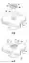
FIGS. 12-13 illustrate an embodiment of an access device for use in the surgical procedure, which may optionally include an inflatable balloon or cuff 112 disposed on the exterior surface of the connector segment 104. The inflatable balloon 112 is in fluid communication with a source of fluids 114 through conduit 116 (shown schematically in FIG. 12). The conduit 116 may be a separate tube extending within the connector segment 102 or, may be a channel extending through the wall of the connector segment 104. The inflatable balloon 112 is configured to transition from the deflated condition depicted in FIG. 11 to the inflated condition depicted in FIG. 12 to engage the internal surface of the first intestinal segment “I1” to secure the connector segment 104 and within the first intestinal end segment “I1”. The inflatable balloon 112 also may establish a seal about the connector segment 104 within the first intestinal end segment “I1” to prevent passage of fluids or fecal matter.
FIGS. 14-16 illustrate another methodology and access device for connecting the first intestinal end segment “I1” about the opening “o” within the abdominal wall “AW”. In accordance with this methodology, the first intestinal end segment “I1” is pulled through the opening “o” and at least partially inverted as in a conventional stoma formation procedure to establish a stoma “t”. An annular array of fasteners 120 may be applied to the tissue of the stoma “t” to connect the first intestinal end segment “I1” and the skin layer “s” in a similar manner discussed hereinafter. In embodiments, the fasteners 120 do not engage or penetrate the underlying fascia layer “f” and muscle layer “m”. An access device 200 including a flange segment 202 and a connector segment 204 is positioned relative to the stoma “t”. The flange segment 202 is substantially similar to the flange segment 102 of the access device of FIGS. 7-12. The connector segment 204 may be shorter in length than the corresponding connector segment 104 of the access device 100 of FIGS. 7-11. In use, the connector segment 204 is positioned within the opening “o” in the abdominal wall “AW” with the flange segment 202 engaging the outer surface of the skin layer “s”. Staples or fasteners 206 are applied through or about the openings 208 in the flange segment 202 to the end margins of the first intestinal end segment “I1” and secure the flange segment 202 to the skin layer “s”. The fasteners 206 may also penetrate underlying fascia tissue of the fascia layer “f”. A collection device may be coupled to the flange segment 202 as discussed hereinabove.
Persons skilled in the art will understand that the structures and methods specifically described herein and shown in the accompanying figures are non-limiting exemplary embodiments, and that the description, disclosure, and figures should be construed merely as exemplary of particular embodiments. It is to be understood, therefore, that the present disclosure is not limited to the precise embodiments described, and that various other changes and modifications may be effected by one skilled in the art without departing from the scope or spirit of the disclosure. Additionally, the elements and features shown or described in connection with certain embodiments may be combined with the elements and features of certain other embodiments without departing from the scope of the present disclosure, and that such modifications and variations are also included within the scope of the present disclosure. Accordingly, the subject matter of the present disclosure is not limited by what has been particularly shown and described.
Claims
What is claimed is:1. A method for accessing matter within an intestine, comprising:
creating an opening extending through an abdominal wall of an abdominal cavity;
accessing an intestinal end segment of an intestine within the abdominal cavity;
positioning the intestinal end segment of the intestine in registration with the opening extending through the abdominal wall;
attaching the intestinal end segment to cutaneous tissue surrounding the opening to establish fluid communication between a lumen of the intestine and the opening through the abdominal wall;
securing an access device adjacent the opening through the abdominal wall; and
connecting a receptacle to the access device configured to be in fluid communication with the lumen of the intestine to collect matter within the intestine.
2. The method of claim 1 including:
coupling the intestinal end segment of the intestine to an anvil of a stapling instrument;
connecting the anvil to a stapling instrument;
firing the stapling instrument to deliver fasteners through the cutaneous tissue surrounding the opening and within the intestinal end segment of the intestine.
3. The method of claim 2 wherein securing the access device includes positioning a connector segment of the access device through the opening and within the lumen of the intestinal end segment of the intestine.
4. The method of claim 3 including establishing a fluid tight seal about the connector segment of the access device within the lumen of the intestinal end segment of the intestine.
5. The method of claim 4 wherein establishing the fluid tight seal includes inflating an inflatable bladder disposed about the connector segment of the access device to engage an inner wall of the intestinal end segment.
6. The method of claim 4 wherein establishing the fluid tight seal includes suturing the intestinal end segment of the intestine to the connector segment of the access device.
7. The method of claim 4 wherein establishing the fluid tight seal includes adhering the connector segment of the access device within the lumen of the intestinal end segment of the intestine.
8. The method of claim 2 wherein attaching the intestinal end segment of the intestine includes arranging the intestinal end segment about an anvil head of the anvil.
9. The method of claim 8 wherein firing the stapling instrument includes approximating the anvil head relative to a staple cartridge of the stapling instrument.
10. The method of claim 9 wherein firing the stapling instrument includes delivering an annular array of staples, the staples being at least partially deformed by the anvil head of the anvil.
11. The method of claim 10 wherein attaching the intestinal end segment of the intestine includes applying a purse string about the intestinal end segment and the anvil head.
12. The method of claim 2 wherein firing the stapling instrument includes delivering staples through facial tissue underlying the cutaneous tissue.
13. The method of claim 2 wherein positioning the intestinal end segment includes exposing end margins of the intestinal end segment external of the opening, and wherein firing the stapling instrument includes delivering the fasteners through the end margins of the intestinal end segment.
14. The method of claim 2 including attaching a mesh to the intestinal end segment and to muscle tissue beneath the cutaneous tissue.
Images & Drawings included:
Sources:
- United States Patent and Trademark Office - verify current appl. status at the USPTO↗
Similar patent applications:
Recent applications in this class:
- » 20250275865 2025-09-04
AN UROSTOMY BAG WITH A SENSOR TO DETERMINE AND COMMUNICATE A URINE LEVEL TO USER - » 20250262085 2025-08-21
A BASEPLATE FOR AN OSTOMY APPLIANCE - » 20250228695 2025-07-17
OSTOMY BARRIER APPLIANCE WITH SELF-ACTIVATING ADJUSTABLE CONVEXITY - » 20250195258 2025-06-19
OSTOMY POUCH - » 20250169981 2025-05-29
LID - » 20250152403 2025-05-15
OSTOMY IMPLANT DEVICE AND METHOD - » 20250152402 2025-05-15
CHARGING DOCK FOR OSTOMY LEAKAGE DETECTION SYSTEM - » 20250143912 2025-05-08
OSTOMY POUCH WITH VIEWING OPTION WINDOW - » 20250134695 2025-05-01
OSTOMY BAG AND METHOD FOR OSTOMY MANAGEMENT - » 20250134694 2025-05-01
MEDICAL COMPONENT AND EXCREMENT STORAGE DEVICE
Recent applications for this Assignee:
- » 20250292086 2025-09-18
SYSTEMS AND METHODS FOR ESTIMATING TISSUE PARAMETERS USING SURGICAL DEVICES - » 20250288348 2025-09-18
ANTI-BACKDRIVE MECHANISM FOR VESSEL SEALING INSTRUMENT - » 20250288312 2025-09-18
APPARATUS FOR ENDOSCOPIC PROCEDURES - » 20250281198 2025-09-11
ARTICULATING ULTRASONIC SURGICAL INSTRUMENTS AND SYSTEMS - » 20250281176 2025-09-11
HAND-HELD SURGICAL INSTRUMENTS - » 20250275766 2025-09-04
HANDHELD ELECTROMECHANICAL SURGICAL SYSTEM - » 20250268625 2025-08-28
SURGICAL ACCESS DEVICE WITH ACTIVE SMOKE FILTRATION - » 20250268624 2025-08-28
SEAL ASSEMBLIES FOR SURGICAL ACCESS ASSEMBLIES - » 20250268595 2025-08-28
HAND-HELD SURGICAL INSTRUMENTS - » 20250261945 2025-08-21
ADAPTER, EXTENSION, AND CONNECTOR ASSEMBLIES FOR SURGICAL DEVICES