Method for preparing multimeric forms of the hepatitis c virus (HCV) envelope glycoprotein 2 (HCV E2)
US20190284230A1
2019-09-19
16/337,900
2017-09-22
✅ Patent granted
US 12,162,906 B2
2024-12-10
WO; PCT/AU2017/051037; 20170922
WO; WO2018/058177; 20180405
Jeffrey S Parkin
Knobbe, Martens, Olson & Bear, LLP
2038-06-12
Abstract:
A method of preparing extracellularly assembled higher order antigen from a native lower order antigen the method comprising the following steps: (i) contacting lower order antigen with a solution comprising a reducing agent for a time and under 5 conditions sufficient to reduce one or more native cysteines; and (ii) removing or diluting the reducing agent or contacting the reduced lower order antigen with an oxidising agent, to elicit assembly of lower order antigen from (i) into an assembled higher order antigen; wherein at least 10% of the lower order antigen is converted to higher order antigen in step (ii) and whereby the assembled higher order antigen 10 displays at least reduced binding to non-neutralizing antibodies compared to the lower order antigen and retains binding to at least one neutralizing antibody. A method of producing a vaccine composition comprising following the steps of the method and then mixing the assembled higher order antigen with a pharmaceutically or physiologically acceptable diluent, carrier or adjuvant. A composition comprising a 15 higher order extracellularly assembled antigen, wherein the assembled antigen displays at least reduced binding to a non-neutralizing antibody compared to a native control higher order antigen. Use of the assembled higher order antigen to stimulate an immune response or for the detection and/or isolation of an immune cell such as a B-cell specific for the antigen.
Inventors:
- Heidi DRUMMER 5 🇦🇺 Melbourne, Australia
- Pantelis POUMBOURIOS 4 🇦🇺 Melbourne, Australia
- Heidi Drummer 2 🇦🇺 Melbourne, Victoria, Australia
- Pantelis Poumbourios 1 🇦🇺 Melbourne, Victoria, Australia
- Robert Center 1 🇦🇺 Melbourne, Victoria, Australia
- Robert Center 2 🇦🇺 Melbourne, Australia
Assignee:
- Macfarlane Burnet Institute for Medical Research and Public Health Limited 1 🇦🇺 Melbourne, Australia
Applicant:
Interested in similar patents?
Get notified when new applications in this technology area are published.
Classification:
C07K16/109 » CPC further
Immunoglobulins [IGs], e.g. monoclonal or polyclonal antibodies against material from viruses from RNA viruses, e.g. hepatitis E virus; Togaviridae, e.g. flavivirus, rubella virus, hog cholera virus Hepatitis C virus; Hepatitis G virus
C07K2317/33 » CPC further
Immunoglobulins specific features characterized by aspects of specificity or valency Crossreactivity, e.g. for species or epitope, or lack of said crossreactivity
C07K2317/76 » CPC further
Immunoglobulins specific features characterized by effect upon binding to a cell or to an antigen Antagonist effect on antigen, e.g. neutralization or inhibition of binding
C12N2770/24222 » CPC further
ssRNA viruses positive-sense; Details; Flaviviridae; Hepacivirus, e.g. hepatitis C virus, hepatitis G virus New viral proteins or individual genes, new structural or functional aspects of known viral proteins or genes
A61K39/29 » CPC further
Medicinal preparations containing antigens or antibodies; Viral antigens Hepatitis virus
G01N33/569 IPC
Investigating or analysing materials by specific methods not covered by groups -; Biological material, e.g. blood, urine ; Haemocytometers; Chemical analysis of biological material, e.g. blood, urine; Testing involving biospecific ligand binding methods; Immunological testing; Immunoassay; Biospecific binding assay; Materials therefor for microorganisms, e.g. protozoa, bacteria, viruses
C07K16/10 IPC
Immunoglobulins [IGs], e.g. monoclonal or polyclonal antibodies against material from viruses from RNA viruses, e.g. hepatitis E virus
C07K1/1136 » CPC further
General methods for the preparation of peptides, i.e. processes for the organic chemical preparation of peptides or proteins of any length by chemical modification of precursor peptides without change of the primary structure by reversible modification of the secondary, tertiary or quarternary structure, e.g. using denaturating or stabilising agents
A61K39/12 » CPC further
Medicinal preparations containing antigens or antibodies Viral antigens
C07K14/1833 » CPC further
Peptides having more than 20 amino acids; Gastrins; Somatostatins; Melanotropins; Derivatives thereof from viruses; RNA viruses; Togaviridae; Flaviviridae; Flaviviridae, e.g. pestivirus, mucosal disease virus, bovine viral diarrhoea virus, classical swine fever virus (hog cholera virus), border disease virus Hepatitis C; Hepatitis NANB
G01N33/5047 » CPC further
Investigating or analysing materials by specific methods not covered by groups -; Biological material, e.g. blood, urine ; Haemocytometers; Chemical analysis of biological material, e.g. blood, urine; Testing involving biospecific ligand binding methods; Immunological testing involving human or animal cells for testing or evaluating the effect of chemical or biological compounds, e.g. drugs, cosmetics involving specific cell types Cells of the immune system
G01N33/56966 » CPC further
Investigating or analysing materials by specific methods not covered by groups -; Biological material, e.g. blood, urine ; Haemocytometers; Chemical analysis of biological material, e.g. blood, urine; Testing involving biospecific ligand binding methods; Immunological testing; Immunoassay; Biospecific binding assay; Materials therefor for microorganisms, e.g. protozoa, bacteria, viruses Animal cells
G01N33/5767 » CPC further
Investigating or analysing materials by specific methods not covered by groups -; Biological material, e.g. blood, urine ; Haemocytometers; Chemical analysis of biological material, e.g. blood, urine; Testing involving biospecific ligand binding methods; Immunological testing; Immunoassay; Biospecific binding assay; Materials therefor for hepatitis non-A, non-B hepatitis
A61K2039/645 » CPC further
Medicinal preparations containing antigens or antibodies characterised by the architecture of the carrier-antigen complex, e.g. repetition of carrier-antigen units Dendrimers; Multiple antigen peptides
C12N2770/24234 » CPC further
ssRNA viruses positive-sense; Details; Flaviviridae; Hepacivirus, e.g. hepatitis C virus, hepatitis G virus Use of virus or viral component as vaccine, e.g. live-attenuated or inactivated virus, VLP, viral protein
C07K1/113 IPC
General methods for the preparation of peptides, i.e. processes for the organic chemical preparation of peptides or proteins of any length by chemical modification of precursor peptides without change of the primary structure
C07K1/13 » CPC further
General methods for the preparation of peptides, i.e. processes for the organic chemical preparation of peptides or proteins of any length Labelling of peptides
C07K1/22 » CPC further
General methods for the preparation of peptides, i.e. processes for the organic chemical preparation of peptides or proteins of any length; Extraction; Separation; Purification by chromatography Affinity chromatography or related techniques based upon selective absorption processes
C07K14/005 » CPC further
Peptides having more than 20 amino acids; Gastrins; Somatostatins; Melanotropins; Derivatives thereof from viruses
C07K14/18 IPC
Peptides having more than 20 amino acids; Gastrins; Somatostatins; Melanotropins; Derivatives thereof from viruses; RNA viruses Togaviridae; Flaviviridae
G01N33/50 IPC
Investigating or analysing materials by specific methods not covered by groups -; Biological material, e.g. blood, urine ; Haemocytometers Chemical analysis of biological material, e.g. blood, urine; Testing involving biospecific ligand binding methods; Immunological testing
A61K39/00 IPC
Medicinal preparations containing antigens or antibodies
C07K1/1133 » CPC main
General methods for the preparation of peptides, i.e. processes for the organic chemical preparation of peptides or proteins of any length by chemical modification of precursor peptides without change of the primary structure by redox-reactions involving cystein/cystin side chains
A61P31/14 » CPC further
Antiinfectives, i.e. antibiotics, antiseptics, chemotherapeutics; Antivirals for RNA viruses
G01N33/576 IPC
Investigating or analysing materials by specific methods not covered by groups -; Biological material, e.g. blood, urine ; Haemocytometers; Chemical analysis of biological material, e.g. blood, urine; Testing involving biospecific ligand binding methods; Immunological testing; Immunoassay; Biospecific binding assay; Materials therefor for hepatitis
Description
FIELD
The present specification relates generally to vaccine and diagnostic compositions. In particular, the specification facilitates the production of higher order forms of antigens of interest, such as HCV envelope 2 (E2) glycoprotein and HIV envelope protein. Higher order antigens are suitable for vaccine production and ex vivo binding applications.
BACKGROUND
Bibliographic details of references in the subject specification are also listed at the end of the specification.
Reference to any prior art in this specification is not, and should not be taken as, acknowledgement or any form of suggestion that this prior art forms part of the common general knowledge in any country.
Hepatitis C virus is a highly variable pathogen that chronically infects 3% of the world's population and is a major cause of chronic liver disease. HCV circulates as 7 highly divergent genotypes (G1-G7) and over 67 subtypes (a, b, c etc.), for which no preventative vaccine is available. Recently, direct acting antivirals (DAAs) have reached the clinic enabling viral clearance to be achieved in >95% of treated individuals. However, DAAs cannot prevent reinfection and their high cost places a major economic burden on health care systems. Furthermore, an estimated 50 million people have undiagnosed infections providing a means for continued viral spread. A prophylactic vaccine would prevent new infections, and possibly reinfections, and significantly augment elimination programs involving the use of DAAs. A prophylactic vaccine has therefore been recognized by the World Health Organization as a priority for development.
The major surface glycoprotein, E2 attaches virions to the host cell receptor CD81 and generates genotype specific and broadly (cross-genotype protective) neutralizing antibodies (bNAbs). In natural infections, 30% of individuals clear their infection spontaneously and this has been correlated with rapid induction of bNAb and broadly reactive T cell responses. In addition, cocktails of broadly neutralizing monoclonal antibodies (NMAb) can prevent and clear established HCV infection in small animal models of HCV. However, previous vaccination studies conducted in animals using vaccines containing full-length or truncated forms of E2 and a phase I clinical trial of recombinant HCV glycoproteins did not elicit high titre NAbs, and very limited cross-genotype neutralization was observed. HCV E2 is highly glycosylated, undergoes rapid sequence change and possesses multiple variable regions, which are all implicated in immune evasion. Non-neutralizing antibodies have been proposed to interfere with the binding of neutralizing antibodies.
Prior art approaches to developing improved HCV vaccines have focused on modifying E2 to remove variable regions while retaining CD81 binding. Recently, the inventors determined that oligomeric and particularly, high molecular weight oligomers (HMW forms) are better than monomers at generating the desired broadly neutralizing antibody responses. This approach has been promising but high molecular weight (HMW) forms of wild type HCV E2 and modified E2 lacking hypervariable regions (e.g. Delta123 HCV E2) are generally expressed in low yield and are structurally heterogeneous which creates challenges for vaccine production. Currently oligomers are produced by transfection of plasmids expressing E2 into host cells and allowing the cell to fold E2 into native oligomers. Oligomers are then separated from other components as required.
It is against this background that the present inventor/s have developed a strategy for producing oligomeric forms of E2 with several potential advantages for commercial vaccine manufacture purposes, such as, in some embodiments, reduced cost and complexity of purification. The method is broadly applicable to any antigen of interest and for the production of assembled higher order forms of an antigen.
SUMMARY
In one aspect, the disclosure enables a method of preparing an extracellularly assembled higher order antigen from a native lower order antigen, the method comprising the following steps: (i) contacting a lower order antigen with a solution comprising a reducing agent for a time and under conditions sufficient to reduce one or more native cysteines; and (ii) removing or diluting the reducing agent or contacting the reduced lower order antigen with an oxidising agent, to elicit assembly of lower order antigen from (i) into an assembled higher order antigen. In one embodiment of the method, at least 10% of the lower order antigen is converted to higher order antigen in step (ii). In one embodiment of the method, the assembled higher order antigen displays at least reduced binding to non-neutralizing antibodies compared to the lower order antigen. In one embodiment of the method, the assembled higher order antigen retains binding to at least one neutralizing antibody.
In one embodiment, the antigen is a viral envelope antigen. In one embodiment the viral envelope antigen is HCV or HIV.
In another embodiment, the antigen is a cancer antigen.
In one embodiment, the higher order cancer antigen is a tetramer, pentamer, hexamer, decamer etc up to a 23 mer.
In one embodiment, there is provided a method of preparing an extracellularly assembled higher order antigen from a native lower order antigen, the method comprising the following steps: (i) contacting a lower order antigen with a solution comprising a reducing agent for a time and under conditions sufficient to reduce one or more native cysteines; and (ii) removing or diluting the reducing agent or contacting the reduced lower order antigen with an oxidising agent, to elicit assembly of lower order antigen from (i) into an assembled higher order antigen; wherein at least 10% of the lower order antigen is converted to higher order antigen in step (ii) and whereby the assembled higher order antigen displays at least reduced binding to non-neutralizing antibodies compared to the lower order antigen and retains binding to at least one neutralizing antibody.
In one embodiment, higher order antigen is purified and mixed with a pharmaceutically acceptable diluent, carrier or adjuvant prior to delivery to a subject as a preventative or therapeutic vaccine.
In one embodiment, the disclosure enables a method of preparing extracellularly assembled higher order hepatitis C virus (HCV) envelope glycoprotein 2 (E2) antigen from native lower order HCV E2 the method comprising the following steps: (i) contacting lower order E2 with a solution comprising a reducing agent for a time and under conditions sufficient to reduce one or more native cysteines; and (ii) removing or diluting the reducing agent or contacting the reduced E2 with an oxidising agent, to elicit reassembly of lower order E2 from (i) into higher order HCV E2. In one embodiment of the method at least 20% of the lower order E2 antigen is assembled in to higher order antigen in step (ii). In one embodiment, the assembled higher order HCV E2 displays at least reduced binding to non-neutralizing antibodies compared to the lower order E2 and retains binding to at least one neutralizing antibody.
Accordingly, in one embodiment, the disclosure enables a method of preparing extracellularly assembled higher order hepatitis C virus (HCV) envelope glycoprotein 2 (E2) antigen from native lower order HCV E2 the method comprising the following steps: (i) contacting lower order E2 with a solution comprising a reducing agent for a time and under conditions sufficient to reduce one or more native cysteines; and (ii) removing or diluting the reducing agent or contacting the reduced E2 with an oxidising agent, to elicit reassembly of lower order E2 from (i) into higher order HCV E2, wherein at least 20% of the lower order antigen is assembled in to higher order antigen in step (ii) and whereby the assembled higher order HCV E2 displays at least reduced binding to non-neutralizing antibodies compared to the lower order E2 and retains binding to at least one neutralizing antibody.
In another one embodiment, the disclosure enables a method of preparing extracellularly assembled higher order HIV envelope glycoprotein antigen from native lower order HIV env the method comprising the following steps: (i) contacting lower order HIV env with a solution comprising a reducing agent for a time and under conditions sufficient to reduce one or more native cysteines; and (ii) removing or diluting the reducing agent or contacting the reduced Env with an oxidising agent, to elicit reassembly of lower order Env from (i) into higher order HIV Env, wherein at least 10% of the lower order antigen is assembled into higher order antigen in step (ii).
In one embodiment, first steps (i) and (ii) are repeated with a solution comprising residual lower order antigen from step (ii) in order to improve the efficiency of the method of assembly of lower order antigen into higher order antigen.
Surprisingly, even cysteine modified versions of HCV E2 monomers, such as the Ala7 construct also assemble into higher order forms using the presently described methods. As cysteine modified forms such as Ala7 are expressed recombinantly in predominantly monomeric form it would be useful to produce higher order forms from this material rather than purifying monomeric forms from a mixture of different forms as are usually produced with, for example, HCV E2 RBD forms or Delta123 forms. Thus, the finding that cysteine modified forms also assemble in the present methods facilitate vaccine manufacture by providing a useful source of lower order or monomer. Furthermore, as determined herein lower order antigen that did not initially assemble into higher order forms was able to form higher order forms when the method was repeated, enabling even higher yields of higher order forms.
In one embodiment of the method, in step (i) or prior to step (i) the solution comprising lower order antigen is substantially depleted of native oligomer or higher order antigen.
In one embodiment, at least 25%, at least 30%, at least 40%, at least 50%, at least 60% or at least 70%, or at least 80%, or at least 90% or at least 95% or more of the lower order antigen is converted into higher order antigen.
In another embodiment, the inventors found genotype specific differences in the yield of higher order antigen. Accordingly, in one embodiment, the method further comprises selecting a viral or antigen genotype which generates the greatest yield of assembled oligomeric antigen.
In one embodiment, the assembled higher order antigen retains or exceeds the ability of a native control higher order antigen to bind or elicit one or more neutralizing antibodies.
In one embodiment, the assembled higher order antigen is the receptor-binding domain (RBD) of HCV E2.
In one embodiment, the assembled higher order HCV E2 antigen lacks all or part of a hypervariable region such as one or more of hypervariable region 1 (HVR1) or a part thereof, the hypervariable region 2 (HVR2) or a part thereof and/or the intergenotypic variable region (igVR/VR3) or a part thereof.
In one embodiment, the assembled oligomeric antigen comprises a non-cysteine substitution or mutation in one or more of amino acid residues selected from the group comprising: C581, C585, C652, C677, C494, C486, C459, C452, C564, C597, and C569. As discussed herein use of cysteine modified forms surprisingly provides a source of lower order antigen able to assemble into higher order forms.
In another aspect, the disclosure provides a method of producing a vaccine composition comprising the herein disclosed assembly method comprising the following steps: (i) contacting a lower order antigen with a solution comprising a reducing agent for a time and under conditions sufficient to reduce one or more native cysteines; and (ii) removing or diluting the reducing agent or contacting the reduced lower order antigen with an oxidising agent, to elicit assembly of lower order antigen from (i) into an assembled higher order antigen, after which the assembled higher order antigen is admixed with a pharmaceutically or physiologically acceptable diluent, carrier or an adjuvant.
In some embodiments, the antigen is a viral envelope antigen or cancer antigen.
In some embodiments, the viral envelope antigen is a hepatitis virus antigen or an HIV envelope antigen.
In another aspect, the disclosure provides an assembled higher order antigen, or composition comprising same, produced from a lower order antigen by the herein disclosed method comprising the following steps: (i) contacting a lower order antigen with a solution comprising a reducing agent for a time and under conditions sufficient to reduce one or more native cysteines; and (ii) removing or diluting the reducing agent or contacting the reduced lower order antigen with an oxidising agent, to elicit assembly of lower order antigen from (i) into an assembled higher order antigen.
In one embodiment there is a enabled a composition comprising a higher order extracellularly assembled antigen. In one embodiment, the higher order assembled antigen displays a more favourable immunogenic profile compared to the counterpart control antigen produced in a host cell. In one embodiment, a favourable immunogenic profile includes greater occlusion of one or more non-neutralizing epitopes and/or greater exposure of one or more neutralizing epitopes of viral envelope antigens. In one embodiment, a favourable immunogenic profile includes an assembled antigen determining a more effective immune response compared to the counterpart control antigen assembled into higher order antigen in the environment of the host cell. In one embodiment a favourable immunogenic profile includes a stronger immune response able to reduce or prevent tumour development in a subject. In one embodiment, the assembled antigen is distinguishable from a native control higher order antigen by displaying reduced binding to the non-neutralizing antibody compared to a native control higher order (oligomeric) antigen. Reduced binding includes no detectable binding (the epitope is occluded in the assembled antigen and fails to bind to a non-neutralizing antibody while the epitope is not occluded in the native control antigen which binds the non-neutralizing antibody). In particular, the epitope recognised by antibody CBH4G is occluded in assembled E2 as described herein.
In another embodiment the present disclosure enables a composition comprising a higher order extracellularly assembled HCV E2 glycoprotein antigen, wherein the assembled E2 displays reduced binding to the non-neutralizing antibody. In particular, non-neutralizing antibody CBH4G displayed reduced binding to assembled E2 compared to a native control oligomeric HCV E2. This antibody is described in the literature, for example, in Keck et al. PLos Pathogens: 8(4) e1002653, April 2012. Also, antibody 2A12 displayed reduced binding to assembled E2 relative to a native control oligomeric HCV E2. Antibody panels and how to generate them are described in Vietheer et al. Hepatology: 65(4), 1117-1131, 2017 incorporated herein by reference, and references referred to therein such as references 5, 33-36, 17 and 37 and supplemental materials available from the publisher.
Thus, in one embodiment the assembled antigen has reduced exposed epitope and/or epitope residues bound by antibody 2A12 or antibody CBH4G or is not recognised by non-neutralizing antibodies, such as antibody 2A12 or antibody CBH4G known in the art.
In another embodiment the present disclosure enables a composition comprising a higher order extracellularly assembled higher order viral envelope antigen, wherein the assembled viral env antigen displays reduced binding to the non-neutralizing antibody compared to a native control higher order viral envelope antigen.
In an embodiment, the refolded oligomeric antigen displays at least one characteristic selected from the group consisting of:
-
- (i) reduced binding to non-neutralizing antibodies relative to a control native antigen form or monomeric form:
- (ii) at least substantially the same binding to neutralizing antibodies relative to a control native antigen form;
- (iii) elicits the production of lower titres of non-neutralizing antibodies relative to a control native antigen form or monomeric antigen;
- (iv) elicits the production of neutralizing antibodies;
- (v) elicits the production of broadly neutralizing antibodies;
- (vi) optionally elicits the production of higher titres of neutralizing antibodies; and
- (vii) optionally elicits the production of higher titres of broadly neutralizing antibodies.
In another embodiment the present disclosure enables a composition comprising a higher order extracellularly assembled higher order HIV envelope antigen, wherein the assembled HIV env antigen displays reduced binding to the non-neutralizing antibody compared to a native control higher order HIV envelope antigen.
Suitable controls will be well understood by the skilled addressee in the context of the present disclosure. In one embodiment, the control antigen is the same genotype as the test antigen of interest, produced recombinantly in a host cell, such as a bacterial or a mammalian, yeast, plant or insect cell.
In one embodiment, the assembled higher order antigen is the receptor-binding domain (RBD) of HCV E2.
In one embodiment, the HCV E2 antigen lacks all or part of a hypervariable region such as one or more of hypervariable region 1 (HVR1) or a part thereof, the hypervariable region 2 (HVR2) or a part thereof and/or the intergenotypic variable region (igVR/VR3) or a part thereof. Examples of HVR1, HVR2 and igVR/VR3 sequences are shown in FIG. 12.
In some embodiments, the E2 antigen comprises a non-cysteine substitution or mutation in one or more of amino acid residues selected from the group comprising: C581, C585, C652, C677, C494, C486, C459, C452, C564, C597, and C569. In one embodiment, reference to one or more amino acids in this context means 2, 3, 4, 5, 6, or 7 amino acids are non-cysteine substitutions or are deleted or otherwise mutated.
In one embodiment the present disclosure provides a composition comprising assembled higher order HCV E2 antigen (assembled from monomeric E2) and a pharmaceutically or physiologically acceptable carrier and/or diluent.
In one embodiment, the composition comprises a pharmaceutically or physiologically acceptable diluent, carrier or adjuvant. In one embodiment, antigens may be presented in any form of carrier suitable for a vaccine composition.
In one embodiment, the disclosure provides for the use of the composition as described herein in the preparation of a medicament for the treatment or prevention of a viral infection such HCV or HIV infection, or cancer, or a condition associated with a viral envelope or cancer antigen.
In one embodiment, the disclosure provides for the use of the assembled higher order antigen, or composition in the preparation of a diagnostic agent for the diagnosis or monitoring of a condition associated with the antigen such as for E2, HCV infection or monitoring of an anti-HCV treatment protocol.
In one embodiment, the disclosure provides for a method for eliciting an immune response in a subject or patient, the method comprising administering to the subject or patient an effective amount of the herein described assembled higher order antigen, or the composition as herein described for a time and under conditions sufficient to elicit an immune response.
In one embodiment, the disclosure provides for a method for immunizing a subject against infection from HCV, comprising administering to the subject the assembled oligomeric antigen, or the composition as described herein.
In one embodiment, the disclosure provides for a method for treating or preventing HCV infection in a subject, comprising administering to the subject the assembled oligomeric antigen, or the composition as described for a time and under conditions sufficient to treat or prevent HCV infection.
In one embodiment, the composition further comprises a second higher order antigen from a different pathogen.
In one embodiment, the assembled higher order antigen comprises a detectable or purification tag.
In one embodiment, the disclosure provides for producing a purified antibody against the higher order assembled antigen as described herein, comprising administering an effective amount of antigen to a subject and purifying the antibody produced.
In one embodiment there is provided an antibody that specifically recognises the assembled higher order antigen/E2 antigen as described herein. Alternatively or in addition, antibodies can be identified that recognise an epitope exposed on native not assembled antigen, or assembled and not native antigen.
In one embodiment there is provided a kit, or a solid or semi-solid substrate, comprising the assembled higher order antigen as described herein, or the composition as described herein.
In one embodiment, the disclosure provides for the use of the assembled higher order antigen produced from a lower order antigen by the herein disclosed method comprising the following steps: (i) contacting a lower order antigen with a solution comprising a reducing agent for a time and under conditions sufficient to reduce one or more native cysteines; and (ii) removing or diluting the reducing agent or contacting the reduced lower order antigen with an oxidising agent, to elicit assembly of lower order antigen from (i) into an assembled higher order antigen, or the composition comprising a higher order extracellularly assembled higher order cancer antigen or viral envelope antigen, wherein the assembled viral env antigen displays reduced binding to the non-neutralizing antibody compared to a native control higher order viral envelope antigen, to bind to/detect an antigen specific immune cell.
In one embodiment, the subject higher order assembled antigens are suitable for or when used for the detection and/or isolation of an immune cell such as a B-cell specific for the antigen. In an embodiment, the immune cell is a T-cell specific for an antigen.
In another embodiment, the disclosure enables a method for detection and/or isolation of an immune cell/B-cell specific for an antigen comprising: i) labelling an immune cell/B-cell specific for an antigen with the assembled higher order antigen as described herein and ii) detecting and/or isolating the labelled immune cell/B-cell cytometrically.
In another embodiment, the disclosure enables a method for detection and/or isolation of an immune cell/B-cell specific for antigen such as HCV or HIV comprising: i) labelling an immune cell/B-cell specific for antigen such as HCV or HIV with the assembled higher order for of the antigen; and ii) detecting and/or isolating the labelled immune cell/B-cell cytometrically.
This summary is not an exhaustive recitation of all embodiments described in the specification.
BRIEF DESCRIPTION OF THE FIGURES
Some figures contain colour representations or entities. Coloured versions of the figures are available from the patentee upon request or from an appropriate Patent Office. A fee may be imposed if obtained from a Patent Office.
FIG. 1 shows agarose gel electrophoresis of Δ123-containing plasmids. Top row of bands represent the vector backbones and bottom row of bands represent the Δ123 inserts, which were isolated from the vector by digesting with restriction enzymes. pcDNA-H77cΔ123-HIS and pcDNA-S52Δ123-HIS were both digested with NheI and XbaI, while pcDNA-Con1Δ123-HIS was digested with NheI and XhoI. Expected lengths of the inserts released from H77c, Con1 and S52 Δ123-containing plasmids are 762 bp, 759 and 710 bp, respectively. GeneRuler 1 kb DNA ladder was used as a size marker (M).
FIG. 2 shows transfection and purification of Δ123. (A-B) Protein expression from transient transfections of pcDNA-Con1Δ123-HIS (A) and pcDNA-S52Δ123-HIS (B) into FS293F cells analysed via sandwich ELISA. Cell culture supernatants harvested on days 3, 5, 7 and 9 post-transfection were diluted by a factor of 2 before half-logarithmic serial dilutions of these were applied to dimeric MBP-CD81-LEL113-201 coated enzyme immunoassay plates. Half-logarithmic serial dilutions of monomeric H77c Δ123 (1 μg/mL) and cell culture supernatant containing HIV gp140, which lacks a 6×HIS tag, were also included as positive and negative controls, respectively. This was followed by the addition of rabbit anti-HIS antibodies diluted to 1/1000 and detection with a single dilution of HRP-conjugated goat anti-rabbit immunoglobulins diluted to 1/1000. (C-D) Con1 Δ123 (C) and S52 Δ123 (D) after affinity chromatography analysed via sandwich ELISA conducted in the same way. Proteins contained in cell culture supernatants were applied to cobalt-charged TALON beads to allow binding of the 6×HIS tagged Δ123, and the supernatant after TALON interaction was collected as the flow through. Proteins were then washed and eluted into three separate fractions. All samples were diluted as appropriate to equalise their amounts for ELISA relative to the original preparation.
FIG. 3 shows gel filtration profiles of Δ123. (A) Affinity-purified H77c Δ123 glycoproteins produced from stable transfection in FS293F cells and analysed by gel filtration chromatography using Superdex 200. Dotted lines and peak heights indicate the fractions that correspond to monomers (78), dimers (70), HMW2 (59) and HMW1 (46) Δ123. (B-C) shows gel filtration chromatography using Superdex200 of affinity-purified Con1 Δ123 (B) and S52 Δ123 (C) produced from round 1 of transient transfections into FS293F cells. Dotted lines and peak heights indicate the fractions that correspond to monomers, dimers, HMW2 and HMW1, respectively, labelled by the numbers 77, 68, 66, 46 and 78, 69, 65, 46 for Con1 and S52, respectively. (D-E) show transient transfections of Con1 Δ123 (D) and S52 Δ123 (E) into FS293F cells were repeated (i.e. round 2) and Δ123 glycoproteins produced were affinity-purified before subjected to analysis by gel filtration chromatography in the same way as round 1.
FIG. 4 shows gel filtration of fractionated Δ123 analysed via SDS-PAGE. Non-reducing SDS-PAGE and coomassie staining of gel filtration fractionated (A) Con1 Δ123 (B) and S52 Δ123, with 20 uL from each of two consecutive fractions pooled and loaded onto a 5-12% polyacrylamide gradient gel. Reducing SDS-PAGE and Coomassie staining of gel filtration fractionated (C) Con1 Δ123 and (D) S52 Δ123, with the same volumes as non-reducing SDS-PAGE loaded onto a 12% acrylamide gel. Bio-Rad broad-range SDS-PAGE standards were loaded as markers (M).
FIG. 5 (A) shows an example of the ability of a MAb to bind monomeric and HMW1 Δ123. Monomeric Δ123 (red) (2 μg/mL) and HMW1 Δ123 (blue) (5 μg/mL) and BSA (green) as a negative control were coated onto enzyme immunoassay plates. Half-logarithmic serial dilutions of the primary antibody was then added and binding was detected with the appropriate HRP-conjugated secondary antibody. Using this technique, the relative binding of a panel MAb towards HMW1 Δ123 were then calculated by comparing the mid-point of each binding curve, which was taken as half of the highest optical density value. The fold difference in binding was calculated relative to monomer and H77c HMW1 Δ123 for construction of Table 7. (B) shows the reactivity of different strains of Δ123 to a panel of MAbs. Single dilution point assessment was performed, whereby H77c, Con1 and S52 Δ123 monomers (5 μg/mL) were coated onto enzyme immunoassay plates as well as BSA (5 μg/mL) as a negative control. A single dilution of the primary antibodies was added and binding detected with the appropriate species specific HRP-conjugated secondary antibodies.
FIG. 6 (A) shows small scale Tris(2-carboxyethyl)phosphine hydrochloride (TCEP) reduction. H77 Δ123 monomers coated on an enzyme immunoassay plate were reduced with different concentrations of TCEP (0-500 mM) prepared in PBS pH 9.6 for 30 min at 37° C. A single dilution of the primary antibodies was added, including conformation-dependent mouse H53 (1 μg/mL) as well as rabbit anti-HIS (1/1000) and human anti-CMV R04 (1/1000), which represent positive and negative controls, respectively. Binding was detected with the appropriate HRP-conjugated secondary antibodies. Reactivity towards each of the primary antibodies was tested in triplicate, and the mean optical density values plotted as a line graph with error bars showing the standard deviations. (B) shows monomeric H77c Δ123 treated with TCEP and BMOE. Non-reducing SDS-PAGE and Coomassie staining of monomeric H77c Δ123 (10 μg) after TCEP reduction and refolding with BMOE crosslinkers under different conditions labelled 1-7, which are described in Table 8. Δ123 samples were loaded onto a 5-12% polyacrylamide gradient gel, along with monomeric and HMW1 H77c Δ123 (5 μs each) as controls as well as broad-range SDS-PAGE markers (M).
FIG. 7 (A) shows monomeric H77c Δ123 treated with glutathione. Non-reducing SDS-PAGE and coomassie staining of monomeric H77c Δ123 (10 μg) after refolding with GSH and GSSG using the redox-shuffling system under different conditions labelled 1-5, which are described in Table 9. H77c Δ123 samples were loaded onto a 5-12% polyacrylamide gradient gel, along with monomeric and HMW1 H77c Δ123 (5 μg each) as controls as well as broad-range SDS-PAGE markers (M). (B) shows gel filtration profiles of monomeric H77c Δ123 treated with glutathione. Gel filtration chromatography of untreated monomeric H77c Δ123 using Superdex 200. (C) shows gel filtration chromatography of H77c Δ123 after 10 μg/μL of the glycoprotein was treated with 2 mM GSH and 0.4 mM GSSG using Superdex 200. Dotted lines and peak heights indicate the fractions that correspond to monomers (78) and dimers (69).
FIG. 8 (A) shows small scale DTT reduction. H77c Δ123 monomers, coated on an enzyme immunoassay plate, were reduced with different concentrations of DTT (0-10 mM) prepared in carbonate-bicarbonate buffer pH 9.6 for 30 min at 37° C. A single dilution of the primary antibodies was added, including conformation-dependent mouse H53 (1 μg/mL) as well as rabbit anti-HIS (1/1000) and human anti-CMV R04, which represent positive and negative controls, respectively. Binding was detected with the appropriate HRP-conjugated secondary antibodies. Reactivity towards each of the primary antibodies was tested in triplicate, and the mean optical density values plotted as a line graph with error bars showing the standard deviations. (B) shows monomeric H77c Δ123 treated with DTT. Non-reducing SDS-PAGE and Coomassie staining of monomeric H77c Δ123 (10 μg) after DTT reduction under different conditions (labelled 1-12 and described in table 3.6) and refolding using the slow dilution method. H77c Δ123 samples were loaded onto a 5-12% polyacrylamide gradient gel, along with monomeric and HMW1 H77c Δ123 (5 μg each) as controls as well as broad-range SDS-PAGE markers (M). (C) shows gel filtration chromatography of untreated monomeric H77c Δ123 using Superdex 200. (D) shows gel filtration chromatography of H77c Δ123 after 1 μg/μL of the glycoprotein was treated with 1 mM DTT using Superdex 200. Dotted lines and peak heights indicate the fractions that correspond to monomers (77-78), dimers (69) and HMW Δ123 (59).
FIG. 9 shows the antigenic characterization of DTT-treated Δ123 and ALA7Δ 123. DTT-treated Δ123 and ALA7Δ 123 has been characterized by assessment of binding with the antibodies (A) AR3C (B) CBH4G (C) HC84.27 and (D) HCV1 as described in Example 15.
FIG. 10 shows the analysis of products generated from refolding DTT-treated Δ123 using gel filtration chromatography. The profile of untreated monomers is shown, followed by increasing numbers of treatments (1, 2, or 3 hits) with DTT as described in Example 14.
FIG. 11 shows the analysis of products generated from refolding non-refolded Δ123 monomers and refolding of Δ123 in the presence of protease inhibitors using gel filtration chromatography as described in Example 14.
FIG. 12 provides a ClustalW alignment of the corresponding E2661 region of HCV isolates used. H77c (AF009606, Gla, SEQ ID NO: 11), J6 (AF177036, G2a, SEQ ID NO: 16), s52 (GU814263, G3a, SEQ ID NO: 13), ED43 (GU814265, G4a, SEQ ID NO: 10), SA13 (AF064490, GSa, SEQ ID NO: 12), EUHK2 (Y12083, G6a, SEQ ID NO: 14), QC69 (EF108306, Gla, SEQ ID NO: 15). HVR1, HVR2 and igVR/VR3 are shown in red/orange. Residues corresponding to amino acid residues GFLASLFY, YTWGENETD and YRLWHF of ED43 are CD81 binding regions.
FIG. 13 shows the nucleotide construct for: Δ123Ala7 codon optimized; Underlined regions correspond to restriction enzyme sites GGTACC=Kpnl, GGATCC=BamHI, CTCGAG=XhoI and the protein sequence for Δ123Ala7.
FIG. 14 to FIG. 14c display a ClustalOmega amino acid alignment of the protein sequences: AF009606 coding sequence (SEQ ID NO:6), AF009606 Full length E2 (SEQ ID NO:7), AF009606 E2661 (SEQ ID NO:8), WT_E2661 (SEQ ID NO: 4) and Δ123 (SEQ ID NO: 3). The underlined region corresponds to residues 630-635.
FIG. 15 (A) shows non-reducing SDS-PAGE of samples reduced with different concentrations of β-mercaptoethanol and assembled. Precision size markers are shown on the left with molecular weights indicated in kDa. Indicative size of monomer, dimer and high molecular weight forms shown on right. (B) shows gel filtration chromatography of monomeric Δ123 after treatment with 100 mM BME (dashed line) or untreated (solid line) using Superdex 200.
FIG. 16 shows antibody titres in animals vaccinated with assembled HCV proteins. Final bleeds from animals vaccinated with antigens listed in Table 15 were analysed for their ability to bind to monomeric Δ123 protein. Reciprocal antibody titres for each group are shown. Horizontal line is the mean, with upper and lower bars representing the standard deviation. The difference in antibody titre between 2-F and 4-F+5-F was statistically significant (p=0.0281) using a Mann-Whitney unpaired t-test. Prism v7.0.
FIG. 17 shows antibody titres to epitope I in animals vaccinated with assembled HCV proteins. Final bleeds from animals vaccinated with antigens listed in Table 15 were analysed for their ability to bind to the synthetic peptide encompassing residues 409-428 of the Genotype 1a H77c sequence. Reciprocal antibody titres for each group are shown. Horizontal line is the mean, with upper and lower bars representing the standard deviation. Differences in antibody titres between groups was determined using a Mann-Whitney unpaired t-test. Prism v7.0.
FIG. 18 shows antibody titres to epitope III in animals vaccinated with assembled HCV proteins. Final bleeds from animals vaccinated with antigens listed in Table 15 were analysed for their ability to bind to the synthetic peptide encompassing residues 523-549 of the Genotype 1a H77c sequence. Reciprocal antibody titres for each group are shown. Horizontal line is the mean, with upper and lower bars representing the standard deviation. Differences in antibody titres between groups was determined using a Mann-Whitney unpaired t-test. Prism v7.0.
FIG. 19 shows antibody titres to genotype 2a epitope I in animals vaccinated with assembled HCV proteins. Final bleeds from animals vaccinated with antigens listed in Table 15 were analysed for their ability to bind to the synthetic peptide encompassing residues 409-428 of the Genotype 2a J6 sequence. Reciprocal antibody titres for each group are shown. Horizontal line is the mean, with upper and lower bars representing the standard deviation. Differences in antibody titres between groups was determined using a Mann-Whitney unpaired t-test. Prism v7.0.
FIG. 20 shows the ability of immune serum to inhibit interaction between HCV E2 and its cellular receptor CD81. Final bleeds from animals vaccinated with antigens listed in Table 15 were analysed for their ability to inhibit the binding between (A) H77c Gla E2 and CD81, and (B) J6 G2a E2 and CD81. Reciprocal antibody titres for each group are shown. Horizontal line is the mean, with upper and lower bars representing the standard deviation. Differences in antibody titres between groups was determined using a Mann-Whitney unpaired t-test. Prism v7.0.
FIG. 21 shows the ability of immune serum to prevent infection of liver cells with genotype 1a viruses. Final bleeds from animals vaccinated with antigens listed in Table 15 were analysed for their ability to prevent infection with Gla HCVpp. Reciprocal antibody titres for each group are shown. Horizontal line is the mean.
FIG. 22 shows the specificity of HMW1 and monomer immune sera. Serial dilutions of guinea pig sera were added to a constant amount of HCV1 (A), HC84-27 (B), AR3C (C) and 2A12 (D). Antibodies were added to monomeric Δ123 and bound MAb was detected with anti-Human Fab2. Groups were compared using Mann-Whitney t test (Prism v 7).
FIG. 23 shows a FACS plot of sorted B cell population using assembled Δ123. CD19 positive and anti-E2 positive B cells were detected with anti-CD19 Cy7 antibody and assembled Δ123.
FIG. 24 shows size-exclusion chromatography-Multi angle light scattering to determine size of assembled proteins. Overlay of UV (A280 nm) signal and molar mass of assembled Δ123A7 (blue) and assembled Δ123 (red) samples.
FIG. 25 shows size-exclusion chromatography of Δ123A7 monomers that were not assembled into HMW forms following treatment with DTT that were subjected to a second round of denaturation with DTT followed by assembly. The monomer and HMW species are indicated by arrows and the % of each shown in the table.
FIG. 26 shows size-exclusion chromatography of RBD monomers subjected to denaturation with DTT followed by assembly. The monomer and HMW species are indicated by arrows and the % of each shown in the table.
FIG. 27 shows size-exclusion chromatography of RBDA7 monomers subjected to denaturation with DTT followed by assembly. The monomer and HMW species are indicated by arrows and the % of each shown in the table.
FIG. 28 shows size-exclusion chromatography of env monomers subjected to denaturation with DTT followed by assembly. The HMW species is indicated by an arrow.
BRIEF DESCRIPTION OF THE TABLES
Table 1 shows the transient transfection conditions.
Table 2 shows a list of MAbs.
Table 3 shows the conditions for DTT reduction.
Table 4 shows the conditions for protein refolding with glutathione.
Table 5 shows the conditions for TCEP reduction.
Table 6 shows the expression of the different oligomeric forms of Δ123. Percentage of monomers, dimers, HMW2 and HMW1 were calculated by dividing the area under their corresponding peak on the gel filtration curves (FIG. 3B-E) by the total area under the curve. Area under the curve was quantified using the UNICORN control software by GE Healthcare Life Sciences.
Table 7 shows the antigenic characterisation of HIMW1 Δ123.
Table 8 shows the conditions for TCEP reduction and BMOE-mediated refolding of Δ123.
Table 9 shows the conditions for H77c Δ123 refolding with glutathione. The ratio of monomers vs. dimers was calculated by dividing the densitometry of monomers by the densitometry of dimers from non-reducing SDS-PAGE analysis of glutathione treated H77c Δ123 (FIG. 7A). Densitometry was quantified using the LI-COR Odyssey system.
Table 10 shows the quantification of multimer formation from glutathione-treated monomeric H77c Δ123. Area under the monomer and dimer peaks on the gel filtration curves (FIG. 7B) calculated as a percentage of the total area under the curve, and ratio of monomers vs. multimers calculated by dividing the area under the monomer peak by that of the dimer peak.
Table 11 shows the quantification of multimer formation from DTT-treated monomeric H77c Δ123. Area under the monomer and BMW peaks on the gel filtration curves (FIG. 7C-D) calculated as a percentage of the total area under the curve, and ratio of monomers (76 min) vs. multimers (58 min) calculated by dividing the area under the monomer peak by that of multimer peak.
Table 12 shows the quantification of multimer formation from DTT-treated monomeric H77c Δ123. Area under the monomer and multimer peaks on the gel filtration curves (FIG. 7B) calculated as a percentage of the total area under the curve, and ratio of monomers vs. multimers calculated by dividing the area under the monomer peak by that of the multimer peak.
Table 13 shows the methods which generated refolding protein using the antigen Δ123.
Table 14 shows the % refolding protein generated by differing refolding methods.
Table 15 shows the immunization groups.
Table 16 shows the statistical analysis of immune sera reactivity to monomeric Δ123.
Table 17 shows the statistical analysis of the ability of immune serum to bind to H77c epitope I.
Table 18 shows the statistical analysis of the ability of immune serum to bind to H77c epitope III.
Table 19 shows the statistical analysis of the ability of immune serum to bind to J6 epitope I.
Table 20 shows the statistical analysis of the ability of immune serum to block H77c Gla E2 binding to CD81.
Table 21 shows the statistical analysis of the ability of immune serum to block JFH-1 G2a E2 binding to CD81.
Table 22 shows the statistical analysis of the ability of immune serum to prevent H77c HCV viruses infecting liver cells.
Table 23 shows the statistical analysis of the ability of immune serum to prevent binding of HCV1.
Table 24 shows the statistical analysis of the ability of immune serum to prevent binding of HC84-27.
Table 25 shows the statistical analysis of the ability of immune serum to prevent binding of AR3C.
Table 26 shows the statistical analysis of the ability of immune serum to prevent binding of 2A12.
Table 27 shows the SEC-MALS of reassembled proteins.
KEY TO SEQUENCE LISTING
SEQ ID NO: 1: DNA construct for codon optimised Δ123Ala7.
SEQ ID NO: 2: DNA sequence encoding codon optimised Δ123Ala7.
SEQ ID NO: 3: Amino acid sequence Δ123Ala7.
SEQ ID NO: 4: Amino acid sequence encoding WT E2661 (RBD).
SEQ ID NO: 5: Δ123 E2661.
SEQ ID NO: 6: Amino acid residues corresponding to the AF009606 coding sequence.
SEQ ID NO: 7: Amino acid residues corresponding to AF009606 full length E2 protein sequence.
SEQ ID NO: 8: Amino acid residues corresponding to AF009606 E2661.
SEQ ID NO: 9: N-Terminal signal sequence.
SEQ ID NO: 10: Amino acid sequence ED43.
SEQ ID NO: 11: Amino acid sequence H77c.
SEQ ID NO: 12: Amino acid sequence SA13.
SEQ ID NO: 13: Amino acid sequence s52.
SEQ ID NO: 14: Amino acid sequence EUHK2.
SEQ ID NO: 15: Amino acid sequence QC69.
SEQ ID NO: 16: Amino acid sequence J6.
SEQ ID NO: 17: Amino acid residues corresponding to HIV env lacking the C-terminal transmembrane domain and cytoplasmic tail.
SEQ ID NO: 18: Amino acid residues corresponding to HIV env with an N-terminal leader sequence.
SEQ ID NO: 19: DNA sequence encoding codon H77cΔ123.
SEQ ID NO: 20: DNA sequence encoding Con1 Δ123.
SEQ ID NO: 21: DNA sequence encoding s52Δ123.
SEQ ID NO: 22: Human trypsinogen signal peptide.
SEQ ID NO: 23: Human tissue plasminogen activator signal peptide (tPA).
SEQ ID NO: 24: Six His tag.
DETAILED DISCUSSION OF EMBODIMENTS
The subject disclosure is not limited to particular screening procedures for agents, specific formulations of agents and various medical methodologies, as such may vary.
Each embodiment in this specification is to be applied mutatis mutandis to every other embodiment unless expressly stated otherwise.
Unless defined otherwise, all technical and scientific terms used herein have the same meanings as commonly understood by one of ordinary skill in the art to which this invention belongs. Any materials and methods similar or equivalent to those described herein can be used to practice or test the present invention. Practitioners are particularly directed to Sambrook et al., 1989 (supra), Coligan et al. Current Protocols In Protein Science, John Wiley & Sons, Inc., 1995-1997, in particular Chapters 1, 5 and 6. and Ausubel et al., Current Protocols in Molecular Biology, Supplement 47, John Wiley & Sons, New York, 1999; Colowick and Kaplan, eds., Methods In Enzymology, Academic Press, Inc.; Weir and Blackwell, eds., Handbook of Experimental Immunology, Vols. I-IV, Blackwell Scientific Publications, 1986; Joklik ed., Virology, 3rd Edition, 1988; Fields and Knipe, eds, Fundamental Virology, 2nd Edition, 1991; Fields et al., eds, Virology, 3rd Edition, Lippincott-Raven, Philadelphia, Pa., 1996, for definitions and terms of the art and other methods known to the person skilled in the art. Reference may also be made to Staby, Rathore and Ahuga, eds Preparative Chromatography for Separation of Proteins; Whiley, 2017, in particular Chapters 3 and 7. Also, Wen, Ellis, Pujar, eds, Vaccine Development and Manufacturing, Wiley, 2014, in particular Chapters 4, 6, 8, 11. Reference may also be made to WO2008022401, WO2012016290 and WO2012068637 for methods and materials.
Definitions
Throughout this specification, unless the context requires otherwise, the word “comprise”, or variations such as “comprises” or “comprising”, will be understood to imply the inclusion of a stated element or integer or group of elements or integers but not the exclusion of any other element or integer or group of elements or integers. By “consisting of” is meant including, and limited to, whatever follows the phrase “consisting of”. Thus, the phrase “consisting of” indicates that the listed elements are required or mandatory, and that no other elements may be present. By “consisting essentially of” is meant including any elements listed after the phrase, and limited to other elements that do not interfere with or contribute to the activity or action specified in the disclosure for the listed elements.
As used herein the singular forms “a”, “an” and “the” include plural aspects unless the context clearly dictates otherwise. Thus, for example, reference to “a composition” includes a single composition, as well as two or more compositions; reference to “an agent” includes one agent, as well as two or more agents; reference to “the disclosure” includes single and multiple aspects of the disclosure and so forth.
While the examples illustrate the assembly of oligomeric HCV E2 from monomeric HCV E2 and the assembly of oligomeric HIV env from monomeric HIV env, the present disclosure is not limited to these particular examples and extends to the assembly of viral envelope proteins and cancer antigens for vaccine production or in ex vivo binding applications. In this respect, the method provides for the production of higher order antigens from lower order antigens. Reference to “higher order” antigens means trimers or larger multiples, while “lower order forms” means monomers or dimers. The terms “antigen” “species” and “form” are used interchangeably.
Reference to “native” means the antigen has been assembled using cellular machinery within a cell. The terms “assembled” and “folded” or “refolded” are used interchangeably although “assembled” is used to convey the partial de novo nature of the assembled antigen. Reference to assembled means “cell-free” assembled. In one embodiment, assembled antigens are as effective or more effective immuonogens than their native counterparts.
As used herein “non-neutralizing antibodies” refers to antibodies that bind to a viral antigen but do not decrease or disrupt viral entry. In one embodiment, in it refers to antibodies that bind to E2 but do not decrease or disrupt viral entry. In relation to cancer antigens, reference to non-neutralizing antibodies means antibodies that do not mediate tumor cell killing.
As used herein “neutralizing antibodies” refers to antibodies that bind to the viral antigen and when bound inhibits viral entry. In one embodiment, it refers to antibodies that bind to E2 and when bound to E2 inhibits viral entry. In relation to cancer antigens, reference to neutralizing antibodies means antibodies that directly or indirectly mediate tumor cell killing.
As used herein “broadly neutralizing antibodies” refers to antibodies that provide cross protection against multiple genotypes or subtypes of an antigen/HCV antigen.
Higher order or oligomeric forms are structural forms of the antigen including trimers and greater fold forms. Higher order antigens or forms or species are assembled from lower order forms, antigens or species. Lower order forms include monomeric or dimeric forms of the antigen.
Reference to a “control” will be understood by the skilled person and means employing a comparator or comparators that will generate or is likely to generate meaningful results in the context of the invention. Typically, a control is a counterpart produced in a host cell or cell free expression system.
Any viral envelope antigen may be engineered using the methods described in this specification. Non-limiting examples of viral families include Adenoviridae, African swine fever-like viruses, Arenaviridae, Arterivirus, Astroviridae, Baculoviridae, Birnaviridae, Bunyaviridae, Caliciviridae, Circoviridae, Coronaviridae, Deltavirus, Filoviridae, Flaviviridae, Hepadnaviridae, Hepeviridae, Herpesviridae, Orthomyxoviridae, Paramyxoviridae, Picornaviridae, Poxyviridae, Reoviridae, Retroviridae and Rhabdoviridae. Particular virus envelope antigens are from Paramyxoviridae, Retroviridae and Filoviridae.
Non-limiting examples of viral envelope antigens are derived from pathogenic viruses such as influenza haemagglutinin (HA); a lentivirus, such as HIV-1 glycoprotein (gp) 120 including the R2 subtype or HIV-2 gp125; a coronavirus, such as SARS Si glycoprotein; a paramyxovirus, such as respiratory syncytial virus (RSV) F2; or a flavivirus, such as Dengue virus E protein.
One important group of antigens are derived from pathogens such as the primary systemic fungal pathogens of man such Coccidioides immitis, Histoplasma capsulatum, Blastomyces dermatitidis, and Paracoccidioides brasiliensis. Important opportunistic fungal pathogens which tend to rely upon an immunocompromised host include Cryptococcus neoformans, Pneumocystis jiroveci, Candida spp., Aspergillus spp., Penicillium marneffei, and Zygomycetes, Trichosporon beigelii, and Fusarium spp. A range of pathogenic fungi are associated with immunocompromised subjects including those with AIDS, with chemotherapy induced neutropenia or patients undergoing haematopoietic stem cell transplantation, among others.
In some embodiments, the antigen is derived from a microbe including a bacterium, fungus, virus, algae, parasite, (including ecto- or endo-parasites) prion, oomycetes, slime, moulds, nematode, mycoplasma and the like. By way of non-limiting example, the microbe is selected from one or more of the following orders, genera or species: Acinetobacter, Actinobacillus, Actinomycetes, Actinomyces, Aeromonas, Bacillus, Bacteroides, Bordetella, Borrelia, Brucella, Burkholderia, Campylobacter, Citrobacter, Clostridium, Corynebacterium, Enterobacter, Enterococcus, Erysipelothrix, Escherichia, Francisella, Haemophilus, Helicobacter, Klebsiella, Legionella, Leptospira, Listeria, Micrococcus, Moraxella, Morganella, Mycobacterium (tuberculosis), Nocardia, Neisseria, Pasteurella, Plesiomonas, Propionibacterium, Proteus, Providencia, Pseudomonas, Rhodococcus, Salmonella, Serratia, Shigella, Staphylococcus, Stenotrophomonas, Streptococcus, Treponema, Vibrio (cholera) and Yersinia (plague), Adenoviridae, African swine fever-like viruses, Arenaviridae (such as viral haemorrhagic fevers, Lassa fever), Astroviridae (astroviruses) Bunyaviridae (La Crosse), Calicivirid (Norovirus), Coronaviridae (Corona virus), Filoviridae (such as Ebola virus, Marburg virus), Parvoviridae (B19 virus), Flaviviridae (such as hepatitis C virus, Dengue viruses), Hepadnaviridae (such as hepatitis B virus, Deltavirus), Herpesviridae (herpes simplex virus, varicella zoster virus), Orthomyxoviridae (influenza virus) Papovaviridae (papilloma virus) Paramyxoviridae (such as human parainfluenza viruses, mumps virus, measles virus, human respiratory syncytial virus, Picornaviridae (common cold virus), Poxyiridae (small pox virus, orf virus, monkey poxvirus), Reoviridae (rotavirus), Retroviridae (human immunodeficiency virus), Paroviridae (paroviruses), Papillomaviridae, (papillomaviruses), alphaviruses, Rhabdoviridae (rabies virus), Trypanosoma, Leishmania, Giardia, Trichomonas, Entamoeba, Naegleria, Acanthamoeba, Plasmodium, Toxoplasma, Cryptosporidium, Isospora, Balantidium, Schistosoma, Echinostoma, Fasciolopsis, Clonorchis, Fasciola, Opisthorchis, Paragonimus, Pseudophyllidea (e.g., Diphyllobothrium), Cyclophyllidea (e.g., Taenia). Pathogenic nematodes include species from the orders; Rhabditida (e.g., Strongyloides), Strongylida (e.g., Ancylostoma), Ascarida (e.g., Ascaris, Toxocara), Spirurida (e.g., Dracunculus, Brugia, Onchocerca, Wucheria), and Adenophorea (e.g., Trichuris and Trichinella), Prototheca, Ptiesteria, Absidia, Aspergillus, Blastomyces, Candida (yeast), Cladophialophera, Coccidioides, Cryptococcus, Cunninghamella, Fusarium, Histoplasma, Madurella, Malassezia, Microsporum, Mucor, Paecilomyces, Paracoccidioides, Penicillium, Pneumocystis, Pseudallescheria, Rhizopus, Rhodotorula, Scedosporium, Sporothrix, Trichophyton and Trichosporon. For the avoidance of doubt the pathogen may include an emerging pathogen.
Illustrative cancer antigens include CD antigens, glycoproteins, glycolipids (gangliosides), carbohydrates (Lewis-Y) vascular targets (VEGF/R), growth factors and stromal or extracellular matrix antigens (FAP, Tenascin) etc. For example, the following as listed: KS ¼ pan-carcinoma antigen, ovarian carcinoma antigen (CA125), prostatic acid phosphate, prostate specific antigen, melanoma-associated antigen p97, melanoma antigen gp75, high molecular weight melanoma antigen (HMW-MAA), prostate specific membrane antigen, carcinoembryonic antigen (CEA), polymorphic epithelial mucin antigen, human milk fat globule antigen, colorectal tumor-associated antigens, CEA, TAG-72, LEA, Burkitt's lymphoma antigen-38.13, CD19, human B-lymphoma antigen-CD20, CD33, melanoma specific antigens, ganglioside GD2, ganglioside GD3, ganglioside GM2, ganglioside GM3, tumor-specific transplantation type of cell-surface antigen (TSTA), virally-induced tumor antigens, T-antigen DNA tumor viruses, Envelope antigens of RNA tumor viruses, oncofetal antigen-alpha-fetoprotein, CEA of colon, bladder tumor oncofetal antigen, differentiation antigen, human lung carcinoma antigen L6, L20, antigens of fibrosarcoma, human leukemia T cell antigen-Gp37, neoglycoprotein, sphingolipids, breast cancer antigen, EGFR (Epidermal growth factor receptor), HER2 antigen, polymorphic epithelial mucin, malignant human lymphocyte antigen-APO-1, differentiation antigen, including I antigen found in fetal erythrocytes, primary endoderm, I antigen found in adult erythrocytes, preimplantation embryos, I (Ma) found in gastric adenocarcinomas, M18, M39 found in breast epithelium, SSEA-1 found in myeloid cells, VEP8, VEP9, Myl, VIM-D5, Du56-22 found in colorectal cancer, TRA-1-85 (blood group H), C14 found in colonic adenocarcinoma, F3 found in lung adenocarcinoma, AH6 found in gastric cancer, Y hapten, LeY found in embryonal carcinoma cells, TL5 (blood group A), EGF receptor found in A431 cells, E1 series (blood group B) found in pancreatic cancer, FC10. 2 found in embryonal carcinoma cells, gastric adenocarcinoma antigen, CO-514 (blood group Lea) found in Adenocarcinoma, NS-10 found in adenocarcinomas, CO-43 (blood groupLeb), G49 found in EGF receptor of A431 cells, MH2 (blood groupALeb/Ley) found in colonic adenocarcinoma, 19.9 found in colon cancer, gastric cancer mucins, TsA7 found in myeloid cells, R24 found in melanoma, 4.2, GD3, D1.1, OFA-1, GM2, OFA-2, GD2, and M1:22:25:8 found in embryonal carcinoma cells, and SSEA-3 and SSEA-4 found in 4 to 8-cell stage embryos.
As used herein the term “human immunodeficiency virus” or “HIV” refers to an enveloped positive single-stranded RNA member of the genus Lentivirus and part of the family Retroviridae. Over time HIV causes acquired immunodeficiency syndrome (AIDS). As used herein the term refers to any HIV genotype, for example, but not limited to HIV1 or HIV2 or any group or subtype thereof. In an embodiment, HIV-1 is from group M, N, O or P. In an embodiment, HIV-1 is subtype is selected from A, B, C, D, E, F, G, H, J, K or a circulating recombinant form (CRF) thereof. HIV encodes the envelope proteins glycoprotein (gp) 120 and env.
As used herein the term “hepatitis C virus” or “HCV” refers to an enveloped positive sense, single-stranded RNA virus belonging to the genus Hepacivirus of the Flaviviridae Family. As used herein the term refers to HCV of any genotype, for example, but not limited to strains of HCV genotype 1 (G1), HCV genotype 2 (G2), HCV genotype 3 (G3), HCV genotype 4 (G4), HCV genotype 5 (G5), HCV genotype 6 (G6), HCV genotype 7 (G7) and can include any subtype thereof e.g. subtype a, b, c, d, e, etc. HCV encodes two glycoproteins E1 and E2 which are required for viral entry into host cells.
As used herein “HCV E2” also referred to as “E2” includes an E2 polypeptide from any genotype/subtype of HCV. In an embodiment, E2 is derived from HCV genotype G1, G2, G3, G4, G5, G6, G7, or a chimeric version thereof. Derived from means directly or indirectly based on one or more of these genotypes. Genotypes vary naturally and may be further modified by man and such functional variants, comprising typically conservative mutations are encompassed. Functional variants comprising one or more amino acid mutations are known to the skilled addressee, and may include functional variants comprising a recombinant E2 ectodomain. The terms further include variants, including portions of the full length E2 polypeptide that, for example, mediate receptor binding, antibody binding by one or more antibodies that recognise conformation or other epitopes and/or mediate E1E2 dimer formation. The term includes modified forms of E2 such as modifications to increase immunogenicity (Delta 123 forms) or monomer production (eg. “Ala7”).
One illustrative parental HCV E2 polypeptide is a receptor binding portion of E2 polypeptide comprising amino acids 384-661 of genotype H77 1a (E2 661 or E2e) or a corresponding portion from another HCV genotype. Accordingly, E2 polypeptides enabled include all or part of the ectodomain that is required for CD81-binding absent the transmembrane domain. Further variants may include the addition or deletion/disruption of sequences necessary for cleavage or secretion. For example, E384TH may be included, deleted or modified to modify signal peptide cleavage and glycoprotein secretion (McCaffrey et al., 2007). In an embodiment, the E2 polypeptide lacks one or more hypervariable regions or a part thereof. In an embodiment, E2 lacks hypervariable regions, such as one or more of: the hypervariable region 1 (HVR1) or a part thereof, the hypervariable region 2 (HVR2) or a part thereof, and intergenotypic variable region (igVR/VR3) or a part thereof.
In an embodiment, the E2 lacks, HVR1, HVR2 and igVR/VR3. In an embodiment, E2 is Δ123. In an embodiment, E2 comprises the sequence as set forth in SEQ ID NO: 3, 4, 5, 6, 7, or 8; or a fragment thereof that retains CD81 binding activity; or a sequence at least 50%, or at least 55%, or at least 60%, or at least 65%, or at least 70%, or at least 75%, or at least 80%, or at least 85%, or at least 90%, or at least 95%, or at least 97%, or at least 98%, or at least 99% identical thereto.
In an embodiment, E2 comprises zero or one or more mutated or disrupted cysteine/s. Thus, in one embodiment, 1, 2, 3, 4, 5, 6, 7, 8, 9, 10, 11, 12, 13, 14, or 15 cysteines are deleted or disrupted. In one embodiment, these are selected from: C581, C585, C652, C677, C494, C486, C459, C452, C564, C597, C569 and/or C620. In an embodiment, the mutated or disrupted cysteines are C486, C581 and C652. In an embodiment, the mutated or disrupted cysteines are C581, C585 and C652. In an embodiment, the mutated or disrupted cysteines are C452, C486, C581 and C652. In an embodiment, the mutated or disrupted cysteines are C569, C581, C585 and C652. In an embodiment, the mutated or disrupted cysteines are C486, C581 and C652. In an embodiment, the mutated or disrupted cysteines are C452, C486, C569, C581, C585 and C652. In an embodiment, the mutated or disrupted cysteines are C452, C486, C569, C581, C585, C597 and C652. Throughout this specification, including the claims all numbering of polypeptide residues of the HCV glycoprotein E2 is based on the prototype HCV-H77 polypeptide sequence, Genbank Accession No AF009606 (SEQ ID: NO: 6) shown in FIG. 14. The mature form of E2 is encompassed by amino acid residues 384 to 746 of SEQ ID NO: 6, presented separately in SEQ ID NO: 7. Modifications referred to herein are made with reference to the amino acid numbering shown in SEQ ID NO:6 and as shown in FIG. 14.
One illustrative cysteine mutated version of E2661 comprises mutation or disruption of the following cysteines: C581, C585, C652, C486, C452, C597, and C569. This mutant is referred to as “Ala7.” Further cysteine modified versions of E2 are described in International publication no. WO 2012/016290 incorporated herein in its entirety.
As used herein “CD81” refers to cluster of differentiation 81, which is a transmembrane protein of the tetraspanin superfamily and is a HCV host receptor.
The receptor binding domain (RBD) of HCV E2 comprises CD81-binding motifs and folds and oligomerises into a spectrum of different species each containing different disulfide and glycan arrangements. The inventor/s have done considerable work to identify the disulfide bonding arrangement on monomeric and dimeric WT E2661 and Δ123 E2661 which suggests that both of these proteins, even as monomeric proteins are actually heterogeneous and present in multiple alternately intramolecular disulfide bonded forms.
When HCV E2 is produced recombinantly, generally 20-30% is oligomeric and approximately 70% is monomeric, depending upon the genotype employed (as determined herein). More monomer is produced with the cysteine modified forms such as Ala7.
As an example, only approximately 20% of a form of the E2 receptor binding domain (residues 384-661) in which hypervariable region 1 (HVR1), hypervariable region 2 (HVR2) and the intergenotypic variable region (igVR or VR3) have been removed referred to as “delta123” or “Δ123” generated from stable transfection of the Genotype 1a H77c sequence of delta123 into FS293F cells is of the HMW forms, compared to 64.9% for monomers (FIG. 3A). As shown herein this percentage can be increased by selecting a genotype that produces increased amounts of oligomeric forms.
Conventionally, reduction and refolding methods are employed to regenerate lower order species from undesirable aggregates. In accordance with the present invention reduction and re-folding is employed to generate oligomers from monomers or oligomers from monomers and oligomers.
In one embodiment, the present invention provides a method of preparing a refolded recombinant oligomeric hepatitis C virus (HCV) envelope glycoprotein 2 (E2) from native HCV E2, said method comprising the following steps:
-
- (i) contacting native E2 with a solution comprising a reducing agent for a time and under conditions sufficient to reduce one or more native cysteines (or disulfide bonds); and
- (ii) removing the reducing agent or contacting the reduced native E2 with an oxidising agent to elicit refolding of reduced monomeric E2 into refolded oligomeric HCV E2;
wherein at least 20% of the monomers are converted to oligomers in step (ii) and the refolded oligomeric HCV E2 displays at least reduced binding to non-neutralizing antibodies compared to the monomeric E2.
In an embodiment, step (i) is performed twice before step (ii). In an embodiment, step (ii) is performed three or more times before step (ii). In some embodiment, steps (i) and (ii) are reparteed two or more times.
In an embodiment, the refolded oligomeric HCV E2 displays at least one characteristic selected from the group consisting of:
-
- (i) reduced binding to non-neutralizing antibodies relative to a control native HCV E2 form or monomeric E2:
- (ii) at least substantially the same binding to neutralizing antibodies relative to a control native HCV E2 form;
- (iii) elicits the production of lower titres of non-neutralizing antibodies relative to a control native HCV E2 form or monomeric E2;
- (iv) elicits the production of neutralizing antibodies;
- (v) elicits the production of broadly neutralizing antibodies;
- (vi) optionally elicits the production of higher titres of neutralizing antibodies; and
- (vii) optionally elicits the production of higher titres of broadly neutralizing antibodies.
Native HCV E2 monomers can be efficiently produced and effectively purified from a mixture of different HCV E2 species. Native monomeric HCV E2 production as described herein or as known in the art. Native monomeric and oligomeric HCV E2 production as described herein or as known in the art. Typically, protein is produced recombinantly in host cells transformed with a suitable expression vector encoding HCV E2.
Suitable mammalian cell lines include, but are not limited to, BHK, VERO, HT1080, 293, 293T, FS293F, Expi293, RD, COS-7, CHO, Jurkat, HUT, SUPT, C8166, MOLT4/clone8, MT-2, MT-4, H9, PML, CEM, myeloma cells (e.g., SB20 cells) and CEMX174 are available, for example, from the ATCC. Other host cells include without limitation yeast, e.g. Pichia pastoris, or insect cells such as Sf9 cells.
The cells may be cultured in a 500 mL, a 1 L, a 1.5 L, a 2 L, a 2.5 L or a 3 L volume. In one example, the cells are cultured using a batch cell culture process. In one example, the cells are cultured using a perfusion cell culture process. In one example, the cells are cultured in a seed medium and a production medium. In one example, the cells are cultured in a stirred-tank reactor. In one example, the volume of the reactor is from about 1 L to about 2500 L. In one example, the reactor is a 1 L reactor, a 1.5 L reactor, a 2 L reactor, a 2.5 L reactor or a 3 L reactor. In one example, the cells are cultured in a wave bioreactor. In one example, the cells are cultured in a cell factory system e.g. a Nunc cell factory system.
Synthetic DNA may be recombinantly expressed by molecular cloning into an expression vector containing a suitable promoter and other appropriate transcription regulatory elements, and transferred into prokaryotic or eukaryotic host cells to produce recombinant protein. Techniques for such manipulations are described by Sambrook et al., Molecular Cloning: A Laboratory Manual (2nd ed.). Cold Spring Harbour Laboratory, Cold Spring Harbour, N.Y., 1989; Ausubel et al., Current Protocols in Molecular Biology, Green Pub. Associates and Wiley-Interscience, New York, 1988.
For example, a construct for expression in yeast preferably contains a synthetic gene, with related transcriptional and translational control sequences operatively linked to it, such as a promoter (such as GAL 10, GALT, ADH1, TDH3 or PGK), and termination sequences (such as the S. cerevisiae ADH1 terminator). The yeast may be selected from the group consisting of: Saccharomyces cerevisiae, Hansenula polymorpha, Pichia pastoris, Kluyveromyces fragilis, Kluyveromyces lactis, and Schizosaccharomyces pombe. See also Yeast Genetics: Rose et al., A Laboratory Course Manual, Cold Spring Harbor Laboratory, Cold Spring Harbor, N.Y., 1990. Nucleic acid molecules can be codon optimized for expression in yeast as known in the art (see Sharp and Cowe, Yeast, 7: 657-678, 1991). Appropriate vectors and control elements for any given cell type can be selected by one having ordinary skill in the art in view of the teachings of the present specification and information known in the art about expression vectors.
Vectors available for cloning and expression in host cell lines are well known in the art, and include but are not limited to vectors for cloning and expression in mammalian cell lines or yeast (fungal) cells, vectors for cloning and expression in bacterial cell lines, vectors for cloning and expression in phage and vectors for cloning and expression in insect cell lines. The expressed proteins can be recovered using standard protein purification methods. Translational control elements have been reviewed by M. Kozak (e.g., Kozak, Mamm Genome, 7(8): 563-74, 1996; Kozak, Biochimie., 76(9): 815-21, 1994; Kozak, J Cell Biol, 108(2): 229-241, 1989; Kozak and Shatkin, Methods Enzymol, 60: 360-375, 1979).
Illustrative polynucleotides encoding HCV E2 are provided in the sequence listing and include the polynucleotide sequences set out in SEQ ID NOs: 1 or 2.
Native HCV E2 may be monomeric, dimeric, trimeric, tetrameric, pentameric up to say 23-mers, including forms having a molecular mass of more than 100 kDa or more than 200 kDa (such as HMW1, or HMW2 forms). Monomeric and oligomeric forms may be selected based on size, gel filtration characteristics, antibody reactivity etc. Expressed protein may be purified from cellular components by affinity chromatography, such as by antibody affinity chromatography.
In one embodiment, the method increases the purity of refolded oligomeric HCV E2 compared to native HCV E2 before treatment with the method as described herein. In one embodiment, the method increases the purity of refolded oligomeric HCV E2 compared to native oligomeric HCV E2 isolated from cell culture. As used herein, the term “purified” or “purity” refers to separation of oligomeric HCV E2 from contaminants i.e. cellular or viral contaminants, such as but not limited, proteins, lipids, nucleic acids and carbohydrates.
Furthermore, in one embodiment the method increases the concentration of oligomeric HCV E2 in a sample i.e. from a sample containing monomeric HCV E2 treatment of the sample with the method as described herein increases the concentration of oligomeric HCV E2 by at least 10%, or by at least 20%, or by at least 30%, or by at least 40%, or by at least 50%, or by at least 60%, or by at least 70%, or by at least 80%, or by at least 90%, or by at least 95%. In an embodiment, the method increases the concentration of dimers by at least 10%, or by at least 20%, or by at least 30%, or by at least 40%, or by at least 50%, or by at least 60%, or by at least 70%, or by at least 80%, or by at least 90%, or by at least 95%. In an embodiment, the method increases the concentration of trimeric and/or higher order forms by at least 10%, or by at least 20%, or by at least 30%, or by at least 40%, or by at least 50%, or by at least 60%, or by at least 70%, or by at least 80%, or by at least 90%, or by at least 95%.
In one embodiment, refolded oligomeric HCV E2 shows modified stability compared to a native control oligomeric HCV E2 i.e. modified thermodynamic stability and/or kinetic stability. In one embodiment, refolded oligomeric HCV E2 shows increased stability compared to a native control oligomeric HCV E2.
In one embodiment, refolded oligomeric HCV E2 shows equivalent binding to non-neutralizing antibodies as native oligomeric HCV E2. In an embodiment, the oligomer is a dimer, or a trimer or a tetramer or a pentamer up to a 23 mer.
Partial reduction of HCV E2 monomer is preferred. Partial reduction is accomplished when some antibody reactivity with antibodies that recognize conformational epitopes is retained. For example, 10%, 20% or 30% of the protein conformation may be retained in partial reduction protocols used herein.
In one embodiment, reducing conditions include a pH of more than about 7 depending upon the reducing agent used.
The term “oligomer” or “multimer” includes versions of the antigen or E2 that are folded as trimers, tetramers, etc., or high molecular weight (HMW) forms. Different forms are identified, for example, by their migration pattern in non-reducing gel electrophoresis, or by gel filtration chromatography or by their antibody reactivity. In one embodiment, monomers, dimers, HMW2 and HMW delta123 forms have molecular masses of about 46, 97, 239 and 2400, respectively.
In one specific embodiment, the lower order form comprises a dimeric form or the lower order form comprises a monomeric form.
In one embodiment, the oligomeric form is a trimeric form.
In one embodiment, the oligomeric form is trimeric and/or higher order forms.
In one embodiment, the oligomeric form a higher order form such as HMW2 or HMW described herein, or modified forms thereof as described herein.
In one embodiment, the native E2 is a modified form of E2 comprising the receptor binding domain, and lacking the stem region and transmembrane domain.
As known in the art, the transmembrane domain is residues 715-746 and the stem region is residues 662-714. In one embodiment, this form is WT E2661.
As described herein, all native forms of HCV E2 comprise different intramolecular and intermolecular (for dimers and higher) disulfide bonded forms. In accordance with one embodiment of the present disclosure, monomers are reduced and refolded into an oligomer, oligomers are reduced and refolded into oligomers, or monomers and oligomers are reduced and refolded into dimers or oligomers.
In one embodiment of the method, at least 25%, at least 30%, at least 40%, at least 50%, at least 60% or at least 70% of the monomers are converted to oligomers.
In another embodiment, at least 70%, 71%, 72%, 73%, 74%, 75%, 76%, 77%, 78%, 79%, 80%, 81%, 82%, 83%, 84%, 85%, or at least 86%, 87%, 88%, 89%, 90%, 91%, 92%, 93%, 94%, 95%, 97%, 98%, 99% of monomers are converted to oligomers.
In one embodiment, the oligomer has an apparent molecular mass of more than 100 kDa. In one embodiment, the oligomer has an apparent molecular mass of more than 200 kDa. In one embodiment, the oligomer has an apparent molecular mass of more than 2000 kDa.
In one embodiment, the method further comprises selecting the HCV genotype which generates the greatest yield of refolded oligomeric HCV E2. In an embodiment, the HCV genotype is selected from G1, G2, G3, G4, G5, G6, G7 and/or other HCV genotype. In an illustrative non-limiting embodiment the HCV genotype is Con1 or S52.
In one embodiment, refolded oligomeric HCV E2 displays reduced binding to antibody CBH4G compared to a control oligomeric HCV E2 not prepared by monomer re-folding. Native oligomers are an example of a control oligomeric HCV E2 not prepared by extracellular monomer re-folding.
In one embodiment of the method, the refolded oligomeric HCV E2 substantially retains the ability of a control oligomeric HCV E2 to bind neutralizing antibodies. In one embodiment of the method, the oligomeric HCV E2 substantially retains the ability of a control oligomeric HCV E2 to bind broadly neutralizing antibodies.
In one embodiment, reducing agents are reversible reducing agents and are for example selected from DTT or 2-mercaptoethanol, Tris(2-carboxyethyl)phosphine, reduced glutathione, thioglycolic acid or other thiol containing agent.
In one embodiment of the method, the reducing agent is dithiothreitol employed at about 0.1-1.0 mM to cause partial reduction of native E2.
In one embodiment of the method, the solution is a buffer at pH>7-9.
In one embodiment, antigen concentration is 1 mg/ml in carbonate buffer pH 9.6. 0.6 mM DTT (final concentration) is used and incubation is for 1 hour at 37° C. The solution is slowly diluted with PBS pH6.8 and the antigen concentration and buffer exchanged with PBS pH 6.8.
In one embodiment, the native E2 (and consequently the refolded oligomer) lacks all or part of a hypervariable region such as one or more of hypervariable region 1 (HVR1) or a part thereof, the hypervariable region 2 (HVR2) or a part thereof and/or the intergenotypic variable region (igVR/VR3) or a part thereof.
The receptor binding domain comprises variable regions HVR1, HVR2 and igVR that play a role in immune evasion by focusing the immune response on regions that are non-essential for viral invasion and shielding the epitopes of broadly neutralising antibodies. Deletion of at least part of HVR2 and/or igVR and/or HVR1 is effected to focus the immune response on conserved epitopes that elicit broadly neutralising antibodies.
A construct comprising the RBD and lacking all three variable regions is referred to as E2661 delta123 or Δ123 as described in WO 2008/022401. The deleted regions are optionally replaced with a flexible linker sequence described in WO08/022401 (incorporated herein in its entirety) such as a peptide sequence of up to 20 amino acids comprising residues selected form the group comprising Gly, Ser, Ala and Arg (for example GSSG or ETHGSSG).
In an embodiment, the native E2 comprises a signal sequence or protein tag to aid purification. In an embodiment, the signal sequence results in secretion of the protein from a cell. In an embodiment, the signal sequence is an N-terminal signal sequence comprising the amino acid sequence “MNPLLILTFVAAALA” provided in SEQ ID NO: 9. In an embodiment, the protein tag is a C-terminal His-tag. In an embodiment, the C-terminal His-tag comprises the amino acid sequence “HHHHHH”.
The inventors have discovered that even monomeric E2 is heterogenous and displays different intramolecular disulfide arrangements. They reasoned that reduction of monomers would provide a good starting point for refolding a more homogeneous oligomer.
In one embodiment, the native E2 comprises a non-cysteine substitution or mutation in one or more of amino acid residues selected from: C581, C585, C652, C677, C494, C486, C459, C452, C564, C597, C569 and/or C620. Surprisingly, even HCV E2 monomers with seven cysteine residues substituted for non-cysteine residues, C452, C486, C569, C597, C581, C585, C652 non-cysteine substitutions (referred to as “Ala7” refolded into higher levels of oligomers.
E2 comprises 18 highly conserved cysteine residues that form 9 intramolecular disulfide bonds in the ER that scaffold the tertiary structure of the glycoprotein. As described in WO 12/016290 (incorporated herein by reference in its entirety), some of these disulfides are dispensable for CD81 binding and MAb H53 binding. Also, cysteine modified forms, including removal of seven cysteine residues, significantly increases monomer production and reduces the production of oligomers. Normally, when E2 is produced recombinantly in host cells it produces a mixture of different monomers and oligomeric forms. WO 12/016290 described the production of various cysteine modified HCV E2. Other constructs disclosed in WO 12/016290 are contemplated herein such as Ala substituted forms having 2, 3, or 4 cysteines mutated or disrupted selected from C452, C486, C569, and C597. E2661 lacks the cysteine at C677 and has 17 cysteines
In some embodiments the native HCV E2 is cysteine modified form such as Ala4, Ala5, Ala6 or Ala7 modified form of HCV E2 which when expressed recombinantly produces at least 40% monomers and less than 70% oligomers, or at least 50% monomers and less than 50% oligomers in native form.
As determined herein, mutagenesis of C620-Ala blocks high molecular weight oligomer formation. Accordingly native HCV E2 forms are generally C620 for the production of higher order forms.
In another broad aspect, the present description provides a method of preparing a recombinant oligomeric viral envelope glycoprotein from a monomeric envelope glycoprotein, said process comprising the following steps:
(i) contacting native E2 with a solution comprising a reducing agent for a time and under conditions sufficient to partially reduce one or more disulfide bonds; and
(ii) removing the reducing agent or contacting E2 from (i) with an oxidising agent to elicit refolding of monomeric E2 into oligomeric envelope glycoprotein;
and wherein at least 20% of the monomers are converted to a oligomer in step (ii), and the oligomers display at least reduced binding to non-neutralizing antibodies compared to the monomeric glycoprotein.
In an embodiment, step (i) is performed twice before step (ii). In an embodiment, step (ii) is performed three times before step (ii).
In another embodiment, the present specification enables a composition comprising a recombinant refolded oligomeric hepatitis C virus (HCV) E2 glycoprotein produced by the reduction and re-folding methods herein before described.
In one embodiment, the refolded oligomeric proteins comprise an amino acid set out in one of SEQ ID NO: 3, 4, 5, 6, 7, 8 or a truncated or modified version, or functional variant thereof. Functional variants and modified forms may display enhanced immunogenicity compared to the pre-modified form or any other suitable control.
In one embodiment, the composition comprises a recombinant refolded oligomeric hepatitis C virus (HCV) E2 glycoprotein produced by the herein described reduction and re-folding method and wherein the oligomeric HCV E2 displays reduced binding to a non-neutralizing antibody compared to a control HMW E2 not prepared by monomer reduction and re-folding.
In one embodiment, the composition comprises a refolded recombinant oligomeric hepatitis C virus (HCV) E2 glycoprotein produced by the herein described reduction and re-folding method and wherein the refolded oligomeric HCV E2 displays reduced binding to antibody CBH4G or AR3C compared to a control HMW E2 not prepared by monomer reduction and re-folding.
In one embodiment, the composition comprises a refolded oligomeric HCV E2 wherein the refolded oligomeric HCV E2 displays reduced binding to antibody CBH4G compared to a control HMW E2 not prepared by monomer reduction and re-folding. In one embodiment, the composition comprises a refolded oligomeric HCV E2 wherein the refolded oligomeric HCV E2 displays reduced binding to antibody AR3C compared to a control HMW E2 not prepared by monomer reduction and re-folding.
In one embodiment, the composition comprises a monomer refolded oligomeric HCV E2 and a pharmaceutically or physiologically acceptable carrier and/or diluent.
In one embodiment, the composition further comprises an adjuvant.
A person skilled in the art will appreciate that the adjuvant can be any agent that enhances the ability of the composition to induce an immune response. In an embodiment, the adjuvant may act by increasing the immune response to the antigen. In an embodiment, the adjuvant may increase the Th1 and/or Th2 immune response. In an embodiment, the adjuvant may be, an alum salt or other mineral adjuvant; a tensoactive agent; a bacterial derivative; a vehicle or slow release material or a cytokine such as those described in Petrovsky et al (2004) and Wilson-Welder et al. (2009). In one embodiment, the adjuvant may be selected from aluminium phosphate, aluminum hydroxide, potassium aluminum sulfate (alum), calcium phosphate, Freund's complete adjuvant, Freund's incomplete adjuvant, MF-59, a saponin, QS-21, lipopolysaccharide (LPS), monophosphoyl lipid A (MPLA), a Th1 activating peptide (e.g. IMP321), a TLR-2 Ligand (e.g. OspA, muramyl dipeptide (MDP), macrophage activating lipopeptide-2 (MALP-2)), a CpG adjuvant, pertussis toxin, heat liable toxin (LTK63 and LT-R192G), diphtheria toxin, Imiquimod, Addavax, ISCOMATRIX, granulocyte macrophage-colony stimulating factor (GM-CSF), IL-12, IL-6, IL-4, IL-2, IL-1, IFN-g, ASO4 (a liposome formulation containing MPLA and QS-21), glycerine, and oil emulsions such as paraffin, mineral oil, lanolin, squalene, ISA-70, and montanide. In one illustrative embodiment, the adjuvant is a saponin based adjuvant. In a related aspect, the adjuvant is a saponin based adjuvant further comprises cholesterol and sterol, an illustrative example of which is ISCOMATRIX adjuvant. In one illustrative embodiment, the adjuvant is MF59. In one illustrative embodiment, the adjuvant is Addavax. In one, embodiment the adjuvant is a carbohydrate adjuvant, for example, a glucan, dextran, lentinan, glucomannan or galactomannan.
In one embodiment, the present specification provides for the use of the composition comprising a refolded oligomeric HCV E2 in, or in the preparation of a medicament for, the treatment or prevention of HCV infection.
In one embodiment, the present specification provides for the use of the composition comprising a refolded oligomeric HCV E2 in, or in the preparation of a diagnostic agent for the diagnosis or monitoring of HCV infection or monitoring of an anti-HCV treatment protocol.
In one embodiment, the composition comprises more than 70% or more than 80% by weight, trimeric or trimeric and higher order forms of HCV E2.
In one embodiment, the composition comprises more than 70% or more than 80%, by weight, dimeric HCV E2.
In one embodiment, the composition comprises more than 70% or more than 80%, by weight, higher order forms of HCV E2 glycoprotein.
In one embodiment, the present specification provides a method for eliciting an immune response in a subject or patient, the method comprising administering to the subject or patient an effective amount of the composition comprising a refolded oligomeric HCV E2 as described herein for a time and under conditions sufficient to elicit an immune response.
In accordance with these embodiments, the composition is generally administered for a time and under conditions sufficient to elicit an immune response comprising the generation broadly neutralizing antibodies. The compositions of the present invention may be administered as a single dose or application. Alternatively, the compositions may involve repeat doses or applications, for example the compositions may be administered 2, 3, 4, 5, 6, 7, 8, 9, 10 or more times.
The present invention further provides a method for eliciting an immune response in a subject or patient, the method comprising administering to the subject or patient an effective amount of a composition comprising refolded oligomeric HCV E2 as described herein. A composition, particularly a vaccine composition for immunizing a subject against infection from hepatitis C virus comprising refolded oligomeric HCV E2 as described herein is also contemplated by the present invention.
The term “vaccine” as used herein refers to a pharmaceutical composition comprising at least one immunologically active component that induces an immunological response in a subject and possibly but not necessarily one or more additional components that enhance the immunological activity of said active component (for example an adjuvant). A vaccine may additionally comprise further components typical to pharmaceutical compositions. The immunologically active component of a vaccine comprises a higher order assembled antigen such as a refolded oligomeric HCV E2 composition. The terms “vaccine” and “vaccine composition” are used interchangeably in the present invention.
“Subjects” contemplated in the present invention are humans or animals including laboratory or art accepted test or vehicle animals. “Patients” include human subjects in need of treatment or prophylaxis.
In one embodiment, the specification also enables a method for immunizing a subject against a condition associated with an antigen, comprising administering to the subject the composition comprising assembled or refolded oligomeric antigen as described herein.
In one embodiment, the specification also enables a method for immunizing a subject against infection from HCV, comprising administering to the subject the composition comprising extracellularly refolded oligomeric HCV E2 as described herein.
In one embodiment, the specification also enables method for treating or preventing HCV infection in a subject, comprising administering to the subject the composition comprising extracellularly refolded oligomeric HCV E2 as described herein for a time and under conditions sufficient to treat or prevent HCV infection.
The terms “effective amount” including “therapeutically effective amount” and “prophylactically effective amount” as used herein mean a sufficient amount of a composition of the present invention either in a single dose or as part of a series or slow release system which provides the desired therapeutic, preventative, or physiological effect in some subjects. Undesirable effects, e.g. side effects, may sometimes manifest along with the desired therapeutic effect; hence, a practitioner balances the potential benefits against the potential risks in determining an appropriate “effective amount”. The exact amount of composition required will vary from subject to subject, depending on the species, age and general condition of the subject, mode of administration and the like. Thus, it may not be possible to specify an exact ‘effective amount’. However, an appropriate ‘effective amount’ in any individual case may be determined by one of ordinary skill in the art using routine skills or experimentation. One of ordinary skill in the art would be able to determine the required amounts based on such factors as prior administration of the compositions or other agents, the subject's size, the severity of a subject's symptoms or the severity of symptoms in an infected population, viral load, and the particular composition or route of administration selected.
The term “treatment” refers to any measurable or statistically significant amelioration in at least some subjects in one or more symptoms of a condition associated with the antigen such as the E2 antigen and HCV infection, or in the risk of developing advanced symptoms of HCV or the risk of transmitting HCV.
The terms “prevention” and “prophylaxis” and the like are used interchangeably and include administration of a composition of the present invention to a subject not known to be infected with HCV for the purpose of prevention or attenuating a subsequent infection or reducing the risk of becoming infected or reducing the severity or onset of a condition or signs of a condition associated with HCV infection.
The administration of the vaccine composition is generally for prophylactic purposes. The prophylactic administration of the composition serves to prevent or attenuate any subsequent infection. In an embodiment, the vaccine composition is to prevent re-infection of a subject be a pathogen. A “pharmacologically acceptable” composition is one tolerated by a recipient patient. It is contemplated that an effective amount of the vaccine is administered. An “effective amount” is an amount sufficient to achieve a desired biological effect such as to induce enough humoral or cellular immunity. This may be dependent upon the type of vaccine, the age, sex, health, and weight of the recipient. Examples of desired biological effects include, but are not limited to, production of no symptoms, reduction in symptoms, reduction in virus titre in tissues or nasal secretions, complete protection against infection by hepatitis C virus, and partial protection against infection by hepatitis C virus.
In some embodiments, a vaccine or composition of the present invention is physiologically significant if its presence results in a detectable change in the physiology of a recipient patient that enhances or indicates an enhancement in at least one primary or secondary humoral or cellular immune response against at least one strain of an infectious hepatitis C virus. The vaccine composition is administered to protect against viral infection. The “protection” need not be absolute, i.e., the hepatitis C infection need not be totally prevented or eradicated, if there is a statistically significant improvement compared with a control population or set of patients. Protection may be limited to reducing the severity or rapidity of onset of symptoms of the hepatitis C virus infection.
In one embodiment, a vaccine composition of the present invention is provided to a subject either before the onset of infection (so as to prevent or attenuate an anticipated infection) or after the initiation of an infection, and thereby protects against viral infection. In some embodiments, a vaccine composition of the present invention is provided to a subject before or after onset of infection, to reduce viral transmission between subjects.
It will be further appreciated that compositions of the present invention can be administered as the sole active pharmaceutical agent, or used in combination with one or more agents to treat or prevent viral or cancer conditions, such as hepatitis C infections or symptoms associated with HCV infection. Other agents to be administered in combination with a composition or a combination of compositions of the present invention include therapies for disease caused by HCV infection or that suppress HCV viral replication by direct or indirect mechanisms. These agents include, but are not limited to, host immune modulators (for example, interferon-alpha, pegylated interferon-alpha, consensus interferon, interferon-beta, interferon-gamma, CpG oligonucleotides and the like); antiviral compounds that inhibit host cell functions such as inosine monophosphate dehydrogenase (for example, ribavirin and the like); cytokines that modulate immune function (for example, interleukin 2, interleukin 6, and interleukin 12); a compound that enhances the development of type 1 helper T cell response; interfering RNA; anti-sense RNA; vaccines comprising HCV antigens or antigen adjuvant combinations directed against HCV; agents that interact with host cellular components to block viral protein synthesis by inhibiting the internal ribosome entry site (IRES) initiated translation step of HCV viral replication or to block viral particle maturation and release with agents targeted toward the viroporin family of membrane proteins such as, for example, HCV P7 and the like; and any agent or combination of agents that inhibit the replication of HCV by targeting other proteins of the viral genome involved in the viral replication and/or interfere with the function of other viral targets, such as inhibitors of NS3/NS4A protease, NS3 helicase, NS5B polymerase, NS4A protein and NS5A protein.
According to yet another embodiment, the pharmaceutical compositions of the present invention may further comprise other inhibitor(s) of targets in the HCV life cycle, including, but not limited to, helicase, polymerase, metalloprotease, NS4A protein, NS5A protein, and internal ribosome entry site (IRES).
Administration is generally for a time and under conditions sufficient to elicit an immune response comprising the generation of E2-specific antibodies or cellular immune response. The immunogenic compositions may be administered in a convenient manner such as by the pulmonary, oral, intravenous (where water soluble), intraperitoneal, intramuscular, subcutaneous, intradermal, intrathecal or suppository routes or implanting (e.g. using slow release, formulations). Administration may be systemic or local, although systemic is more convenient. Other contemplated routes of administration are by patch, cellular transfer, implant, sublingually, intraocularly, topically, orally, rectally, vaginally, nasally or transdermally.
As used herein, an “immune response” refers to the reaction of the body as a whole to the presence of a composition of the present invention which includes making antibodies and developing immunity to the composition. Therefore, an immune response to an antigen also includes the development in a subject of a humoral and/or cellular immune response to the antigen of interest. A “humoral immune response” is mediated by antibodies' produced by plasma cells. A “cellular immune response” is one mediated by T lymphocytes and/or other white blood cells. As used herein, “antibody titres” can be defined as the highest dilution in post-immune sera that resulted in a value greater than that of pre-immune samples for each subject.
Embodiments of the present invention also provide assays for assessing an immune response to the refolded oligomeric antigen such as HCV E2. The assays may comprise in vivo assays, such as assays to measure antibody responses and delayed type hypersensitivity responses. In an embodiment, the assay to measure antibody responses primarily may measure B-cell function as well as B-cell/T-cell interactions. For the antibody response assay, antibody titres in the blood may be compared following an antigenic challenge.
The specification provides a method for producing a purified antibody against refolded oligomeric antigen such as HCV E2 as described herein, comprising: administering to a subject an effective amount of refolded oligomeric antigen such as HCV E2; and purifying the antibody produced.
In another embodiment, the present invention provides antibodies raised against refolded oligomeric antigen such as HCV E2. Specific antibodies recognise the assembled antigen while failing to recognise the native antigen and/or vice versa Antibodies may be polyclonal or monoclonal. Further, antibodies may be selected for diagnostic, prognostic, therapeutic, prophylactic, and screening purposes typically using criteria known to those of skill in the relevant art.
The terms “antibody” and “antibodies” include polyclonal and monoclonal antibodies and all the various forms derived from monoclonal antibodies, including but not limited to full-length antibodies (e.g. having an intact Fc region), antigen-binding fragments, including for example, Fv, Fab, Fab′ and F(ab′).sub.2 fragments; and antibody-derived polypeptides produced using recombinant methods such as single chain antibodies. The terms “antibody” and “antibodies” as used herein also refer to human antibodies produced for example in transgenic animals or through phage display, as well as antibodies, human or humanized antibodies, primatized antibodies or deimmunized antibodies. It also includes other forms of antibodies that may be therapeutically acceptable and antigen-binding fragments thereof, for example single domain antibodies derived from cartilagenous marine animals or Camelidae, or from libraries based on such antibodies. The selection of fragmented or modified forms of the antibodies may also involve consideration of any affect the fragments or modified forms have on the half-lives of the antibody or fragment.
In some embodiments, the antibody is provided with a pharmaceutically or pharmacologically acceptable carrier, diluent or excipient. In other embodiments, the antibody is selected for diagnosis or prognosis. In some embodiments, kits comprising refolded oligomeric forms of HCV E2 glycoprotein antibodies are provided.
A “pharmaceutically acceptable carrier and/or a diluent” is a pharmaceutical vehicle comprised of a material that is not otherwise undesirable i.e., it is unlikely to cause a substantial adverse reaction by itself or with the active composition. Carriers may include all solvents, dispersion media, coatings, antibacterial and antifungal agents, agents for adjusting tonicity, increasing or decreasing absorption or clearance rates, buffers for maintaining pH, chelating agents, membrane or barrier crossing agents. A pharmaceutically acceptable salt is a salt that is not otherwise undesirable. The agent or composition comprising the agent may be administered in the form of pharmaceutically acceptable non-toxic salts, such as acid addition salts or metal complexes.
For oral administration, the compositions can be formulated into solid or liquid preparations such as capsules, pills, tablets, lozenges, powders, suspensions or emulsions. In preparing the compositions in oral dosage form, any of the usual pharmaceutical media may be employed, such as, for example, water, glycols, oils, alcohols, flavoring agents, preservatives, coloring agents, suspending agents, and the like in the case of oral liquid preparations (such as, for example, suspensions, elixirs and solutions); or carriers such as starches, sugars, diluents, granulating agents, lubricants, binders, disintegrating agents and the like in the case of oral solid preparations (such as, for example, powders, capsules and tablets). Because of their ease in administration, tablets and capsules represent the most advantageous oral dosage unit form, in which case solid pharmaceutical carriers are obviously employed. Tablets may contain a binder such as tragacanth, corn starch or gelatin; a disintegrating agent, such as alginic acid; and a lubricant, such as magnesium stearate. If desired, tablets may be sugar-coated or enteric-coated by standard techniques. The active composition can be encapsulated to make it stable to passage through the gastrointestinal tract. See for example, International Patent Publication No. WO 96/11698.
For parenteral administration, the composition may be dissolved in a carrier and administered as a solution or a suspension. For transmucosal or transdermal (including patch) delivery, appropriate penetrants known in the art are used for delivering the composition. For inhalation, delivery uses any convenient system such as dry powder aerosol, liquid delivery systems, air jet nebulizers, propellant systems. For example, the formulation can be administered in the form of an aerosol or mist. The compositions may also be delivered in a sustained delivery or sustained release format. For example, biodegradable microspheres or capsules or other polymer configurations capable of sustained delivery can be included in the formulation. Formulations can be modified to alter pharmacokinetics and biodistribution. For a general discussion of pharmacokinetics, see, e.g., Remington's Pharmaceutical Sciences, 18th Ed., Mack Publishing Company, Easton, Pa., U.S.A. 1990 (supra). In some embodiments the formulations may be incorporated in lipid monolayers or bilayers such as liposomes or micelles. Targeting therapies known in the art may be used to deliver the agents more specifically to certain types of cells or tissues.
The actual amount of active agent administered and the rate and time-course of administration will depend on the nature and severity of the disease. Prescription of treatment, e.g. decisions on dosage, timing, etc. is within the responsibility of general practitioners or specialists and typically takes into account the condition of the individual patient, the site of delivery, the method of administration and other factors known to practitioners. Examples of techniques and protocols can be found in Remington's Pharmaceutical Sciences, 1990 (supra).
Sustained-release preparations that may be prepared are particularly convenient for inducing immune responses. Examples of sustained-release preparations include semipermeable matrices of solid hydrophobic polymers containing the polypeptide, which matrices are in the form of shaped articles, e.g., films, or microcapsules. Examples of sustained-release matrices include polyesters, hydrogels (for example, poly(2-hydroxyethyl-methacrylate), or poly(vinylalcohol)), polylactides, copolymers of L-glutamic acid and ethyl-L-glutamate, non-degradable ethylene-vinyl acetate, degradable lactic acid-glycolic acid copolymers, and poly-D-(−)-3-hydroxybutyric acid. While polymers such as ethylene-vinyl acetate and lactic acid-glycolic acid enable release of molecules for over 100 days, certain hydrogels release proteins for shorter time periods. Liposomes may be used which are of the small (about 200-800 Angstroms) unilamellar type in which the lipid content is greater than about 30% cholesterol, the selected proportion being adjusted for the optimal therapy.
Stabilization of proteins may be achieved by modifying sulfhydryl residues, lyophilizing from acidic solutions, controlling moisture content, using appropriate additives, and developing specific polymer matrix compositions. The in vivo half-life of proteins may be extended using techniques known in the art, including, for example, by the attachment of other elements such as polyethyleneglycol (PEG) groups.
Prime-boost immunization strategies as disclosed in the art are contemplated. See for example International Publication No. WO/2003/047617. Thus, compositions may be in the form of a vaccine, priming or boosting agent.
In one embodiment, the present specification provides a kit, or a solid or semi-solid substrate, comprising the composition comprising a reduced and refolded oligomeric HCV E2 form as described herein.
The term “isolated” means material that is substantially or essentially free from components, that normally accompany it in its native state. For example, an “isolated nucleic acid molecule” refers to a nucleic acid or polynucleotide, isolated from the sequences which flank it in a naturally-occurring state, e.g., a DNA fragment which has been removed from the sequences that are normally adjacent to the fragment. Without limitation, an isolated nucleic acid, polynucleotide, peptide, or polypeptide can refer to a native sequence that is isolated by purification or to a sequence that is produced by recombinant or synthetic means. In some embodiments, purified refolded oligomeric HCV E2 is at least 95 to 99% pure.
A person skilled in the art will appreciate that the refolded oligomer HCV E2 produced by the methods as described herein can be purified by any method known to a person skilled in the art, including, for example one or more of the following steps: centrifugation, microfiltration, antibody purification, depth filtration ultrafiltration, diafiltration, precipitation, bead chromatography (for example size exclusion chromatography, ion exchange chromatography or affinity chromatography), membrane adsorber (for example ion exchange chromatography or affinity chromatography). In one example, the refolded oligomeric HCV E2 produced by the methods as described herein comprises a protein tag, such as a HIS-tag which aids purification of the refolded oligomeric HCV E2.
In one embodiment, the specification enables a method of preparing a refolded recombinant oligomeric hepatitis C virus (HCV) envelope glycoprotein 2 (E2) from recombinant HCV E2, wherein the recombinant E2 is monomeric E2 or comprises a mixture of monomeric and oligomeric recombinant E2 said method comprising the following steps: (i) contacting recombinant E2 with a solution comprising a reducing agent for a time and under conditions sufficient to reduce one or more monomer disulfide bonds; and (ii) removing the reducing agent or contacting the monomer with an oxidising agent to elicit refolding of recombinant E2 into oligomeric HCV E2; wherein the refolded recombinant oligomeric E2 comprises at least 20% more oligomers than recombinant E2 prior to refolding (ii) and the refolded oligomeric HCV E2 displays reduced binding to non-neutralizing antibodies compared to the recombinant monomeric E2.
In some embodiments refolded oligomeric E2 comprises dimers, trimers, and/or higher order forms of E2.
In another aspect the present specification provides a more general method of preparing a refolded recombinant oligomeric viral envelope glycoprotein from a native envelope glycoprotein, said process comprising the following steps: (i) contacting native E2 with a solution comprising a reducing agent for a time and under conditions sufficient to partially reduce one or more monomer disulfide bonds or native cysteines; and
(ii) removing the reducing agent or contacting the reduced native HCV E2 with an oxidising agent to elicit refolding of reduced monomeric envelope glycoprotein into oligomeric envelope glycoprotein;
and wherein at least 20% of the monomers are converted to refolded oligomers in step (ii), and the oligomers display at least reduced binding to non-neutralizing antibodies compared to the monomeric glycoprotein.
A person skilled in the art will appreciate that once produced higher order antigen as described may be purified by any method know to a person skilled in the art. For example “purifying” or “purification” may comprise one or more of the following steps: centrifugation (e.g. ultracentrifugation), chromatography (e.g. size exclusion chromatography, ion exchange chromatography or affinity chromatography), and/or membrane adsorber (for example ion exchange chromatography or affinity chromatography), filtration (e.g. membrane filtration, ultrafiltration or diafiltration).
A person skilled in the art would appreciate that purified higher order antigen can be formulated into pharmaceutical formulations such as vaccine compositions. Such compositions can comprise one or more of: an adjuvant, an excipient, a binder, a preservative, a carrier coupling, a buffering agent, a stabilizing agent, an emulsifying agents, a wetting agent, a non-viral vector and a transfection facilitating compound. A skilled person would further understand that such vaccine compositions can be lyophilized. In one embodiment, the vaccine composition produced is suitable for human use. In another embodiment, the vaccine is suitable for veterinary use.
The higher order antigen or composition as described herein may be used to bind to/detect antigen specific immune cell. In an embodiment, the immune cell is a B-cell. In an embodiment, the immune cell is a T-cell. Detection of antigenic immune cells indicate that a subject has been infected with the pathogen associated with the antigen and/or that the subject has been vaccinated with an antigen. In an embodiment, the higher order antigen as described herein, or the composition as described herein may be used for the detection and/or isolation of B-cells specific for an enveloped virus. In an embodiment, the enveloped virus is HCV. In an embodiment, the enveloped virus is HIV.
A person skilled in the art would appreciate that higher order antigens of the present invention are suitable for use in diagnostic assays and/or isolation of an immune cell. In an embodiment, the higher order antigen may be used as a stain or marker for of an immune cell in flow cytometry. In an embodiment, the higher order antigen is complexed with a marker, dye or fluorophore to allow for detection using flow cytometry. In an embodiment, the marker is d-biotin.
Isolation of immune cell/s may involve a “tetramer assay” or “tetramer stain”, for example as described in Altman et al., (1996), Vollers et al., 2008 and Dolton et al, 2015, wherein a higher order antigen as described herein replaces the role of the major histocompatibility complex (MHC) tetramer in the assay.
In an embodiment, the invention provides a method for detection and/or isolation of immune cells/B-cell specific for HCV comprising: i) labelling an immune cell/B-cell specific for HCV with the higher order antigen as describe herein; and ii) detecting and/or isolating the labelled immune cell/B-cell cytometrically.
Reference to variants includes parts, derivatives, and chemical analogs. Chemical analogs contemplated include modification of side chains, incorporation of unnatural amino acids and/or their derivatives during synthesis and the use of linkers or cross-linkers or other methods to inter alia impose conformational constraints.
The disclosure is completed with the following non-limiting examples of the assembly method and characteristics of the assembled higher order antigens. As noted above, although the disclosure is illustrated using hepatitis and HIV envelope antigens, the invention extends to antigens produced using the subject methods, forming higher order oligomers from lower order antigens in order to enhance the antigenicity and immunogenicity of the antigen. As described herein, the higher order antigens are further characterised using a range of techniques known in the art. However, analysis to date shows the assembled oligomers, produced by the reduction and assembly methods described herein display antibody binding profiles not displayed by control antigens produced intracellularly and therefore the instant compositions can be distinguished from prior art antigens on this basis.
EXAMPLES
Example 1: Expression Vectors and Plasmids
Cloning.
The DNA sequence of HCV E2 Δ123 fused with a C-terminal 6× histidine (HIS) tag (SEQ ID NO: 24) and was cloned into a pcDNA3.1 mammalian expression vector (Invitrogen). Con1 genotype 1b (G1b) Δ123 (SEQ ID NO:20) was cloned by CSL. H77c genotype 1a (G1a) Δ123 (SEQ ID NO:19) was also cloned by CSL, but into a proprietary CSL-modified pcDNA3.1 vector. The resultant plasmids containing H77c Δ123 and Con1 Δ123 were named pcDNA-H77cΔ123-HIS and pcDNA-Con1Δ123-HIS, respectively. The DNA sequence of S52 G3a Δ123 (SEQ ID NO:21) was ligated into the NheI and XbaI sites of pcDNA3.1 using T4 DNA ligase to produce pcDNA-S52Δ123-HIS.
DNA Expression and Purification.
pcDNA-Con1Δ123-HIS and pcDNA-S52Δ123-HIS were transformed into DH5a Escherichia coli using the heat shock method (Froger and Hall, 2007) and then grown on Luria-Bertani (LB) ampicillin (100 μg/mL) agar plates. Single colonies were cultured in ampicillin-containing (100 μg/mL) LB broth and DNA plasmids were extracted using the QIAGEN plasmid maxi prep kit according to the manufacturer's recommendations. To confirm successful ligation and scale up of the cloned plasmids, pcDNA-Con1Δ123-HIS and pcDNA-S52Δ123-HIS from the maxi prep were digested with NheI and XhoI; and NheI and XbaI restriction enzymes, respectively. Resultant digests were then subjected to agarose gel electrophoresis and the gel was viewed using the Gel Doc XR+ system (Bio-Rad Laboratories) and the Quantity One 1-D analysis software (Bio-Rad Laboratories) to confirm the expected restriction pattern. Plasmids were also sequenced (by Micromon) using BigDye Terminator v3.1 chemistry and the sequences were analysed using CLC Sequence Viewer (QIAGEN) and FinchTV (Geospiza).
Example 2: Protein Expression and Purification
Transient Transfection.
FreeStyle 293-F (FS293F) cells (Invitrogen), derived from the human embryonic kidney 293 cell line, were seeded at 1×10⁶ viable cells/mL in 150 mL in 1 L Erlenmeyer cell culture flasks (Corning) and maintained in FreeStyle 293 (FS293) expression media (Invitrogen). Cells were incubated at 37° C. in a humidified atmosphere with 8% CO2 using a Steri-Cycle CO2 incubator (Thermo Electron Corporation), and on an orbital shaker rotating at 4× relative centrifugal force (RCF). Transient transfections of pcDNA-Con1Δ123-HIS and pcDNA-S52Δ123-HIS into FS293F cells were performed using the 293fectin transfection reagent (Invitrogen) according to the manufacturer's instructions, using the volumes shown in Table 1. On day 1 post-transfection, 100 mL FS293 expression medium was added, raising the total transfection volume to 250 mL. Cell cultures were also supplemented with 0.5% lupin peptone (Solabia Biotechnology) and 0.02% Pluronic F-68 (Gibco). A half media change was performed on days 3, 5 and 7 post-transfection, whereby half of the cell culture supernatant (125 mL) was harvested by centrifugation at 300×RCF for 5 min. Pelleted cells were resuspended in 125 mL of fresh FS293 expression medium, also supplemented with 0.5% lupin peptone and 0.02% Pluronic F-68, and then returned to the cell culture flasks. Finally, a full harvest of the cell culture supernatant (250 mL) was performed on day 9 post-transfection. All harvested supernatants were subjected to further centrifugation at 15,344×RCF for 30 min and pellets were discarded to remove residual cells and cellular debris. Cell counting was performed on each day of harvest using the trypan blue dye exclusion method and a hemocytometer to determine the cell density and viability.
Affinity Chromatography.
Cell culture supernatants containing secreted HIS-tagged Δ123 glycoproteins were applied to 10 mL (i.e. 1 column volume) cobalt-charged TALON metal affinity resin (ClonTech) for 2 h on a rocker at room temperature (RT) to allow binding of the HIS-tagged proteins. The beads were washed twice with 20 column volumes of wash buffer (50 mM sodium phosphate, 300 mM sodium chloride, pH 7.0) at a flow rate of 5 mL/min and eluted with 5 column volumes of elution buffer (50 mM sodium phosphate, 300 mM sodium chloride, 200 mM imidazole, pH 7.0) at a flow rate of 1 mL/min. Proteins contained within eluates were concentrated using Amicon Ultra centrifugal filter units (Merck Millipore) with a molecular weight cut-off (MWCO) of 30 kDa, washed in phosphate-buffered saline (PBS, Gibco) which was adjusted to pH 6.8 (PBS pH 6.8) using hydrochloric acid (HCl) and stored at 4° C. until used.
Example 3: Gel Filtration Chromatography
Proteins were subjected to gel filtration chromatography on a Superdex 200 prep grade 16/600 column (GE Healthcare) using the ÄKTA fast protein liquid chromatography (FPLC) system (GE Healthcare). The run was conducted at a flow rate of 0.5 mL/min using filtered and degassed PBS pH 6.8 as the running buffer. Fractions containing the desired oligomeric species were pooled then concentrated using Amicon Ultra centrifugal filter units with a MWCO of 10 kDa and stored in PBS pH 6.8 until used. Protein concentrations were determined by spectrophotometry at an optical density of 280 nm (OD280) using a cuvette with a 1 cm path length and calculated in mg/mL using the following formula:
extinction coefficient×absorbance at OD280
The amino acid sequences of Δ123 were used to determine the extinction coefficients, which were calculated using the following formula:
molecular weight (Da)/((5690×#tryptophans)+(1280×#tyrosines))
Example 4: Polyacrylamide Gel Electrophoresis (PAGE)
Proteins were analysed via sodium dodecyl sulfate (SDS)-PAGE under reducing or non-reducing conditions as appropriate. For reducing SDS-PAGE, protein samples were heated at 100° C. for 5 minutes in 1× sample buffer containing 2% (v/v) β-mercaptoethanol and then loaded onto a 12% acrylamide gel along with broad-range SDS-PAGE standards (Bio-Rad Laboratories). Electrophoresis was conducted using a vertical electrophoresis apparatus (CLP) in 1× running buffer (25 mM Tris base, 192 mM glycine, 0.1% SDS, pH 8.3) at 180V for ˜1.5 h. Non-reducing SDS-PAGE was performed in the same way, but with a 4-12% polyacrylamide gradient gel and without 3-mercaptoethanol in the sample buffer. Gels were stained with 0.25% (v/v) Coomassie brilliant blue R-250 (Bio-Rad Laboratories), 10% (v/v) acetic acid and 50% (v/v) methanol, then destained with 10% (v/v) acetic acid and 50% (v/v) methanol and scanned using an Odyssey infrared imaging system (LI-COR). Band intensities were also quantitated using the Odyssey system.
Example 5: Antibodies
Monoclonal antibodies (MAbs) AR3A, AR3B, AR3C and AR3D kind gifts from Dr Mansun Law, The Scripps Research Institute (Law et al. 2008). CBH-4B, CBH-4D, HC-11 and HCV-1 were kind gifts from Dr. Steven Foung, Standford University. H52 and H53 were kind gifts of Dr. Jean Dubuisson and Dr. Harry Greenberg. MAbs 1, 7, 10, 12, 16, 20, 24 and 60 were produced by our laboratory in collaboration with CSL Ltd (Table 2). Suitable antibody panels are described in the literature, for example, in Keck et al. PLos Pathogens: 8(4) e1002653, April 2012. Also, antibody 2A12 displayed reduced binding to assembled E2 relative to a native control oligomeric HCV E2. Antibody panels and how to generate them are described in Vietheer P. et al. Hepatology: 65(4), 1117-1131, 2017 incorporated herein by reference, and references referred to therein such as references 5, 33-36, 17 and 37 and supplemental materials, available from the publisher.
Example 6: Enzyme Linked Immunosorbent Assays (ELISA)
Sandwich ELISA. To confirm successful protein expression and purification, sandwich ELISA were performed using Maxisorb flat-bottom 96 well plates (Nunc). Wells were coated with 5 μg/mL dimeric maltose binding protein (MBP)-CD81-LEL113-201 in 50 mM carbonate-bicarbonate buffer pH 9.6 and then incubated overnight at 4° C. Plates were washed 4 times in PBS containing 0.05% Tween 20 (PBST) in this and subsequent washing steps and then blocked with 10 mg/mL bovine serum albumin (BSA, Sigma-Aldrich) in PBS (BSA10PBS) for 1 h at RT. After washing, half-logarithmic serial dilutions of tissue culture supernatants were performed in PBST containing 5 mg/mL BSA (BSASPBST), followed by incubation for 1 h at RT. After washing, a single dilution of rabbit anti-6×HIS epitope tag antibodies (Rockland Immunochemicals) in BSASPBST were added and incubated for 1 h at RT. Following washing, a single dilution of goat anti-rabbit immunoglobulins (Dako) conjugated to horse radish peroxidase (HRP) was added. After washing, ELISA development was performed using 3,3′,5,5′-Tetramethylbenzidine (TMB) substrate (Sigma-Aldrich) according to the manufacturer's instructions and the reaction was stopped with 1M HCl. Finally, optical densities were measured at 450 nm using a Multiskan Ascent microplate reader (Thermo Electron Corporation).
Single Dilution Point Assessments and Direct ELISA.
To Assess the Reactivity of Δ123 to a panel of antibodies known in the art such as the panel of antibodies listed in Example 5/Table 2, single dilution point assessments were initially performed whereby Maxisorb flat-bottom 96 well plates were directly coated with 5 μg/mL monomeric Δ123 and then incubated overnight at 4° C. After washing and blocking with BSA10PBS, a single dilution of the primary antibodies was added, followed by the addition of secondary antibody (Dako), with washing and 1 hour incubation at RT in between each addition. The plates were developed and measured as described above for sandwich ELISAs. Direct ELISA was performed by the same method, but wells were coated with either 2 μg/mL monomeric Δ123 or 5 μg/mL HMW1 Δ123 as appropriate and the primary antibodies were subjected to half-logarithmic serial dilutions in BSA5PBST.
Example 7: Protein Reduction and Refolding
Small Scale Reduction.
To determine the optimal DTT (Pierce) concentration for use in protein reduction, a Maxisorb flat-bottom 96 well plate was coated with 5 μg/mL monomeric H77c Δ123 in 50 mM carbonate-bicarbonate buffer of pH 9.6, and incubated at RT for a minimum of 2 h. After washing 4 times with PBST, different concentrations of DTT (0-10 mM) prepared in carbonate-bicarbonate buffer were added and incubated at 37° C. for 30 min. The plate was then immediately washed 6 times in PBST to remove the DTT, followed by blocking with BSA10PBS. The primary antibodies, which include anti-HIS, H53 and R04, followed by the appropriate secondary antibodies were added, as described in Example 6. Finally, bound antibodies were detected and measured as described in Example 6. The same experiment was repeated using TCEP as the reducing agent.
Reduction and Refolding with DTT in Solution.
H77c Δ123 monomers prepared in carbonate-bicarbonate buffer were subjected to DTT reduction under different conditions (Table 3) to determine the requirements for optimal HMW Δ123 formation. Protein refolding was conducted by means of slow dilution, which was achieved through 3 stepwise additions of PBS pH 6.8 at a constant volume that was half of the initial sample volume. For example, 4×200 μL of PBS pH 6.8 would be added to a 400 μL sample. Each addition was followed by 15 min incubation at RT. DTT was removed using Amicon Ultra centrifugal filter units with a MWCO of 10 kDa, followed by two rinses in PBS pH 6.8. The refolded proteins were then analysed by non-reducing SDS-PAGE as described in Example 4. Larger scale refolding was conducted if multimers were successfully formed and analysed by gel filtration chromatography, as described in Example 3.
Reduction and Refolding Using the Redox-Shuffling System.
A ratio of 1:5 oxidised L-glutathione (GSSG, Pierce) to reduced L-glutathione (GSH, Pierce) was added to H77c Δ123 monomers prepared in carbonate-bicarbonate buffer, with different conditions explored (Table 4). The reaction was stopped by removing L-glutathione and the refolded proteins were then analysed, as described in Example 7. Larger scale refolding was conducted if multimers were successfully formed and analysed by gel filtration chromatography, as described in Example 3.
Crosslinking Tris(2-carboxyethyl)phosphine hydrochloride (TCEP)-Reduced Proteins with Bismaleimidoethane (BMOE).
Different concentrations of TCEP were added to H77c Δ123 monomers, prepared in PBS pH 6.8 at different concentrations (Table 5), and incubated for 30 min at 37° C. BMOE (Sigma), dissolved in dimethyl sulphoxide (Sigma) immediately prior to use, was then added to a final concentration of 0.2 mM and incubated for 1 h at RT as instructed by the manufacturer. The reaction was stopped by removing BMOE and TCEP, and the refolded proteins were then analysed, as described in Example 7.
Example 8: Production of Δ123
The DNA sequences of Δ123 from strains H77c, Con1 and S52 were incorporated into pcDNA3.1 based vectors, giving rise to the following plasmids: pcDNA-H77cΔ123-HIS, pcDNA-Con1Δ123-HIS and pcDNA-S52Δ123-HIS. These plasmids displayed the expected restriction patterns on agarose gel electrophoresis, with bands representing H77c, Con1 and S52 Δ123 inserts all corresponding to their expected lengths of 762, 759 and 710 bp, respectively (FIG. 1). H77c Δ123 was cloned into a proprietary CSL-modified pcDNA3.1 vector, accounting for the larger vector size observed compared to the standard Invitrogen vectors containing Con1 and S52, which each migrated to the same horizontal position (FIG. 1).
Transient transfections of Con1 and S52 Δ123 plasmids into mammalian FS293F cells were performed to produce Δ123 glycoproteins. Cells were maintained in suspension in FS293 expression media supplemented with Pluronic F-68 to protect cells against hydrodynamic forces, and lupin peptone to boost growth efficiency. Cells were seeded at 1×10⁶ viable cells/mL, and the cell viability was maintained at approximately 90% from days 0 to 9 post-transfection. To confirm successful expression of Δ123 glycoproteins, cell culture supernatants were analysed via sandwich ELISA (FIG. 2A-B). Half-logarithmic serial dilutions of supernatants harvested on days 3, 5, 7 and 9 post-transfection were applied to dimeric MBP-CD81-LEL113-201-coated enzyme immunoassay plates, and captured Δ123 glycoproteins were detected with rabbit anti-his antibodies and HRP-conjugated goat anti-rabbit immunoglobulins. Monomeric H77c Δ123 at a concentration of 1 μg/mL in the first well served as a positive control, whereas HIV envelope protein gp140-containing supernatant diluted to 1/2 in the first well served as a negative control. All cell culture supernatants exhibited positive binding to CD81, indicating the presence of Δ123 glycoproteins in the supernatants and thus successful protein expression. Furthermore, curves representing cell culture supernatants appeared above the positive control, indicating yields >1 μg/mL of Δ123 glycoproteins—approximately 4 μg/mL and 20 μg/mL for Con1 and S52, respectively. Optical density (OD) values were highest for harvests performed at days 5 and 7, suggesting that protein expression was somewhat more efficient at these intermediate time points. It was also noted that supernatants from the S52 Δ123 transfection demonstrated greater levels of CD81 binding compared to that of Con1 Δ123, suggesting more efficient protein expression for the S52 strain or that S52 Δ123 has a stronger affinity for CD81.
To isolate Δ123 glycoproteins from the cell culture supernatants, affinity chromatography was performed using TALON beads, a sepharose matrix charged with cobalt, which efficiently binds to his-tagged proteins with higher specificity than conventional nickel-affinity supports. The effectiveness of protein purification was also analysed via sandwich ELISA in the same way as conducted for cell culture supernatants (FIG. 2 C-D). Eluates were sequentially collected into three separate fractions, with the proteins eluted in the first fraction showing similar levels of CD81 binding as the supernatant before purification, indicating that fraction 1 contained most of the Δ123 glycoproteins. The proteins eluted in fraction 2 exhibited decreased CD81 binding, indicating that a lesser amount of Δ123 was present, while that of fraction 3 showed negligible CD81 binding and produced a curve that aligned with the HIV gp140 negative control. Supernatants after purification (i.e. flow through) and the wash from S52 Δ123 purification showed virtually similar levels of CD81 binding to the positive control, while low levels were observed for that of Con1 Δ123, indicating that Con1 Δ123 purification was performed more effectively.
Example 9: Δ123 Expression Profiles
The DNA sequences of Δ123 from strains previous analysis of affinity-purified H77c Δ123 expression by gel filtration chromatography revealed that H77c Δ123 exists as a spectrum of differently sized species (FIG. 3A). These consist of monomers, dimers, HMW2 and HMW1 Δ123, which have molecular masses of 46, 97, 239 and 2402 kDa respectively, and vary in their antigenicity as well as immunogenicity (H. Drummer, unpublished data). HMW species are able to induce bNAbs, whereas monomers induce type-specific NAbs (H. Drummer, unpublished data). However, only 23.13% of Δ123 generated from stable transfection of H77c Δ123 into FS293F cells (performed by Dr. Rob J Center prior to the commencement of this project) were of the HMW form, compared to 64.90% for monomers (FIG. 3A and Table 6).
Δ123 from any genotype should be equally able to elicit bNAbs as it comprises the most conserved regions of E2. However, the expression profile of WT E2 and Δ123 will vary between different genotypes. To determine whether Con1 Δ123 and/or S52 Δ123 express higher yields of HMW species, affinity-purified Δ123 glycoproteins from transient transfections into FS293F cells were analysed by gel filtration chromatography on a Superdex 200 prep grade 16/600 column using the ÄKTA FPLC system. Quantification of the area under the peaks using the UNICORN software revealed that both the Con1 (FIG. 3B) and S52 (FIG. 3C) strains indeed generated higher yields of HMW Δ123, with 46.02% (Con1) and 44.88% (S52) of all Δ123 glycoproteins generated of the HMW form (HMW1 and HMW2 combined) compared to 23.13% for H77c (Table 6).
To assess the reproducibility of these results, transient transfections and purification of Δ123 were repeated, and gel filtration chromatography was again performed to analyse Δ123 expression. On the second round, the expression level of Con1 Δ123 was approximately double that of round 1 while that of S52 Δ123 remained relatively stable, as shown by the scale on the y-axes (FIG. 3D-E). Despite the variation in expression levels, it was important to note that the expression profiles of Con1 and S52 Δ123 remained similar for both round 1 and round 2. In comparison to H77c, an improvement in the yield of HMW Δ123 was again observed, with similar proportions of HMW species generated on the second round—46.98% and 41.56% for Con1 and S52, respectively (Table 6).
Gel filtration fractionated Δ123 glycoproteins were analysed via SDS-PAGE. Non-reducing SDS-PAGE on 5-12% polyacrylamide gradient gels corroborated the findings from gel filtration chromatography, again demonstrating that Δ123 exists as a variety of disulfide linked oligomeric forms and displaying an inversely proportional relationship between band intensity and molecular weight (FIG. 4A-B). Reducing SDS-PAGE analyses were also conducted on 12% acrylamide gels using β-mercaptoethanol, a reducing agent that disrupts disulfide bonding, which in turn also disrupts hydrophobic contacts. Results reveal that disulfide bonds are indispensable for the formation and stabilisation of higher order structures as these all migrated as monomers once their disulfides were cleaved by β-mercaptoethanol (FIG. 4C-D). Hydrophobic interactions may contribute to the formation of higher order species.
These data reveal that the Con1 and S52 strains indeed express higher yields of HMW Δ123 compared to H77c and demonstrates one possible method to improve HMW Δ123 production.
Example 10: Antigenic Characterisation of HMW Δ123
To assess whether HMW Δ123 generated by the Con1 and S52 strains recapitulate the antigenic pattern of that of H77c, gel filtration fractionated monomeric and HMW1 Δ123 from all three strains were tested against a panel of conformation sensitive and insensitive MAbs in an ELISA format (FIG. 5A). Single dilution point assessments (FIG. 5B) were initially performed as a rapid test to determine which MAbs exhibit positive reactivity towards Δ123 of H77c and either or both of Con1 and S52, thereby allowing the antigenicity between strains to be compared. The MAbs that fulfilled this criterion were further tested in direct ELISAs, whereby half-logarithmic serial dilutions of the MAbs were applied to enzyme immunoassay plates coated separately with monomeric and HMW1 Δ123, followed by detection with the appropriate HRP-conjugated secondary antibodies. Relative binding of each MAb towards HMW1 Δ123 were then calculated by comparing the mid-point of each binding curve (Table 7), which was taken as half of the highest optical density value.
While single dilution point assessments show that conformation sensitive mouse H53 and MAbs 1, 7, 12, 16 and 20 all exhibited positive reactivity towards H77c Δ123, negligible reactivity towards Con1 and S52 Δ123 was demonstrated, indicating that the epitope integrity of these two strains were altered in a way that rendered the target epitopes of these MAbs unrecognisable, and that these MAbs are type-specific. The contrary was displayed by conformation sensitive human AR3A, where there was positive reactivity towards Con1 and S52 Δ123, but not for H77c (FIG. 5B).
Out of the 11 MAbs that were further tested in direct ELISAs, 8 (AR3B, AR3C, AR3D, CBH-4B, CBH-4D, HC-11, MAbs 10 and 60) showed reduced binding to the HMW form of Δ123 when compared to the monomeric form (Table 7), indicating that their target epitopes were more occluded. Only mouse H52 demonstrated a markedly enhanced ability to bind HMW1 Δ123 for all three strains (Table 7), suggesting improved access to the residue C652.
When comparing the ability of each MAb to bind the HMW forms of Con1 and S52 to that of H77c, 5 (HCV1, AR3B, AR3C, CBH-4D, HC-11) out of the 11 MAbs tested exhibited increased or equivalent levels of binding. Increased binding was also demonstrated by AR3D and H52, but only towards Con1 Δ123, while MAbs 10 and 24 exhibited reduced levels of binding towards both strains (Table 7).
Example 11: Reduction and Refolding of Δ123 Using TCEP and BMOE
The ability to refold Δ123 monomers into monomers by partially reducing monomeric Δ123 to generate free sulfhydryls, followed by re-conjugation of these sulfhydryls into intermolecular disulfide bonds that will mediate multimer formation was assessed.
Small scale reduction of Δ123 using TCEP, a sulfhydryl-free reducing agent, was trialled as a rapid test to determine the optimal concentrations of TCEP required for partial reduction of Δ123 (FIG. 6). This was executed by exposing monomeric H77c Δ123 coated onto an enzyme immunoassay plate, to different concentrations of TCEP (0-500 mM) for 30 min at 37° C., allowing time for the reduction to occur. To determine the extent of structural change resulting from TCEP reduction, a single dilution of the conformation-dependent mouse H53 antibody was applied to the plate, and binding detected with HRP-conjugated anti-mouse antibodies. H77c Δ123 treated with 10-500 mM TCEP exhibited progressively reduced binding to H53, with all optical density values greater than that of the R04 negative control (a human MAb against the human cytomegalovirus). These results show that the conformation of Δ123 was not entirely destroyed as H53 recognition was retained to a degree, indicating that partial reduction was achieved and that reduction was progressively increased with increasing TCEP concentration. Furthermore, positive anti-HIS binding occurred consistently in the presence of different concentrations of TCEP, eliminating the possibility that loss of bound Δ123 could account for reduced H53 binding. Since retention of Δ123 conformation was considered necessary to preserve the antigenic and immunogenic properties of Δ123 and to allow subsequent reformation into HMW Δ123, it was determined that TCEP concentrations between 10-200 mM were suitable for further protein reduction experiments.
Reduction on enzyme immunoassay plate-bound proteins allows rapid testing of numerous reagent concentrations. However, reduction in solution is more appropriate in terms of potential vaccine production. Therefore, H77c Δ123 monomers were prepared in PBS pH 6.8 as TCEP is most effective at a pH range of 6 to 8. Various concentrations of Δ123 were also prepared to investigate whether different amounts of proteins initially present would affect the efficiency of conversion of monomer to HMW forms. TCEP was then added at concentrations guided by the previous small scale reduction experiment. To subsequently transform partially reduced Δ123 monomers into BMW Δ123, reduction was followed by protein refolding, a process that involves disulfide reformation and reshuffling. This was facilitated through the addition of BMOE, a short homobifunctional, maleimide crosslinker that forms thioether bonds with free sulfhydryls at a pH range of 6.5 to 7.5. Sulfhydryl groups are conjugated as a result, thus mimicking disulfide reformation. Furthermore, TCEP is sulfhydryl-free and is thus appropriate for use in conjunction with sulfhydryl-reactive BMOE crosslinkers, justifying the use of TCEP as a reducing agent in this experiment. Refolded Δ123 glycoproteins were analysed via non-reducing SDS-PAGE (FIG. 6B). All samples displayed similar migration patterns to the monomer control and the mock control, which was also subjected to the reduction and refolding procedure but in the absence of TCEP and BMOE (lane 7 on FIG. 6B and Table 8). No bands aligned with that of the BMW Δ123 control, revealing that TCEP reduction and refolding using BMOE were unsuccessful at converting monomeric Δ123 to HMW Δ123.
Example 12: Reduction and Refolding Using the Redox-Shuffling System
The redox-shuffling system was utilised to promote oxidation between reduced sulfhydryls, whereby reduction and refolding was conducted simultaneously in the presence of both a reducing agent (GSH) and an oxidising agent (GSSG). This was carried out by preparing H77c Δ123 monomers in carbonate-bicarbonate buffer pH 9.6 as a high pH environment promotes the reducing and oxidising ability of GSH and GSSG, respectively. To investigate the effect of protein concentration on aggregation efficiency, H77c Δ123 monomers were also prepared at various concentrations (Table 9). A ratio of 5 GSH:1 GSSG was then added, equating to a final concentration of 2 mM GSH and 0.4 mM GSSG, which are the concentrations typically used in commercial protein refolding kits and have been reported to refold >90% of proteins (information provided by Thermo Fisher Scientific). These Δ123 samples were then incubated at 37° C. for various lengths of time (Table 9), which was immediately followed by a 50 fold dilution with PBS pH 6.8 and removal of glutathione using centrifugal filter units to stop the reaction.
Δ123 samples were analysed via non-reducing SDS-PAGE (FIG. 7A), which revealed successful refolding of monomeric Δ123 into dimeric Δ123 and higher order species, as shown by the appearance of dimer bands and the presence of higher order structures greater in size than a dimer. The monomer and mock controls each produced a band corresponding to the size of monomeric Δ123 as expected. However, a faint dimer band was also observed for the mock control (lane 1 on FIG. 7A). This is consistent with the gel filtration profile for monomeric Δ123 (FIG. 7B), which also displayed a small amount of contaminating dimeric Δ123. The detection of dimers may also suggest spontaneous protein aggregation as a result of exposure to air or higher temperatures as untreated Δ123 monomers were previously stored in capped tubes at 4° C. Additionally, boiling samples prior to loading onto gels can promote aggregation. Quantification of the monomer and dimer band densitometries using the LI-COR Odyssey system showed that the mock control contains 4.65 fold more monomer than dimer. The majority of monomers failed to refold into higher order species when 1 μg/μL of monomeric Δ123 was treated with glutathione, with 6.89 fold more monomer than dimer, higher than recorded for the mock control. In contrast, the use of higher concentrations (5 and 10 μg/μL) resulted in more dimer formation, indicating that refolding in the presence of high amounts of proteins enhanced Δ123 aggregation efficiency. Increasing the incubation time from 2 h to 24 h also induced the formation of more dimers, demonstrated by juxtaposing conditions 3 and 5 on FIG. 7A. Out of all the tested conditions, conversion of monomers into dimers was most efficient when 10 μg/μL of monomeric Δ123 were subjected to glutathione treatment for 24 h. Therefore, a scaled up repeat of the experiment utilising these conditions was performed and analysed by gel filtration chromatography.
The gel filtration profile of Δ123 from the repeat experiment displayed a monomer and dimer peak (FIG. 7C), consistent with results from non-reducing SDS-PAGE (FIG. 7A). A shoulder on the dimer peak was also observed, indicating some formation of higher order species. Quantification of the area under the curve using the UNICORN software revealed that virtually half of the Δ123 monomers were refolded into multimers, with dimers comprising the majority of protein (Table 10). Altogether, these results demonstrated that use of the redox-shuffling system indeed led to intermolecular disulfide bond formation between glutathione-reduced Δ123 monomers. Predominantly these experiments resulted in the formation of dimers.
Example 13: Reduction Using Dithiothreitol (DTT) and Refolding Via Slow Dilution
To expand our investigations on Δ123 aggregation, a different reducing agent and refolding technique was employed: DTT and slow dilution, respectively. As with TCEP, small scale DTT reduction of Δ123 in an ELISA format was initially trialled to determine the optimal DTT concentrations required to achieve partial reduction of Δ123 (FIG. 8A). This was executed by exposing H77c Δ123 monomers, coated onto an enzyme immunoassay plate, to different concentrations of DTT (0-10 mM) for 30 min at 37° C. The plate was then washed to remove DTT, thereby stopping further reduction, followed by blocking, addition of the primary antibodies (anti-HIS, H53 and R04) and detection with the appropriate HRP-conjugated secondary antibodies. Virtually complete reduction of Δ123 was achieved with >2 mM DTT. Δ123 treated with 0.1-1.0 mM DTT exhibited progressively reduced H53 binding and therefore, these concentrations were further investigated in subsequent DTT reduction experiments. Consistent anti-HIS binding was observed at all DTT concentrations, eliminating loss of bound Δ123 as a factor for reduced H53 binding.
DTT reduction in solution was then investigated, whereby various concentrations of H77c Δ123 monomers (Table 11) were prepared in carbonate-bicarbonate buffer pH 9.6 and exposed to DTT, which retains its reducing capacity at a pH of >7. Reduced sulfhydryls will eventually undergo spontaneous oxidation in the absence of a reducing agent, leading to reformation of disulfide bonds. To allow this process to occur, reduced Δ123 glycoproteins were subsequently subjected to refolding by means of slow dilution, which involves stepwise additions of PBS pH 6.8 to the Δ123 samples. Furthermore, this procedure dilutes DTT and attenuates its activity by gradually neutralising the pH environment, thereby promoting oxidation of sulfhydryls.
Refolded Δ123 glycoproteins were analysed via non-reducing SDS-PAGE (FIG. 8B). The mock control produced a monomer band as expected, and a faint band at approximately 100 kDa corresponding to dimeric Δ123, indicating low level contamination with dimers or perhaps spontaneous protein aggregation as a dimer band was not observed in the monomer control. Quantification of the monomer and dimer band densitometries showed that the mock control contains 3.41 fold more monomeric protein than dimeric protein (lane 1 on FIG. 8B and Table 11). An increased amount of oligomer was recorded for Δ123 samples subjected to reduction and refolding under all test conditions (FIG. 8B), revealing that proportions of Δ123 monomers were indeed converted into multimers as a result of successful intermolecular disulfide bond formation. A significant drop in the ratio of monomers vs. dimers was recorded for samples incubated for 30 min following DTT addition, which resulted in less than 2 fold more monomeric than dimeric protein. Extending the incubation period failed to enhance Δ123 aggregation as ≥2 h incubations did not give rise to more multimer formation compared to 30 min incubations. Low monomer to dimer ratios were recorded when 1 μg/μL of monomeric Δ123 was treated with DTT, contrasting with the results from glutathione treatment. The lowest value of 1.33 was recorded when 1 μg/μL of monomeric Δ123 was treated with 0.1 mM DTT for 30 min (lane 11 on FIG. 8B and Table 11), indicating that conversion into dimers was most efficient using these conditions. Formation of higher order species was most apparent on non-reducing SDS-PAGE when 1 μg/μL of monomeric Δ123 was treated with 1 mM DTT for 30 min (lane 9 on FIG. 8B).
A scaled up repeat experiment was performed using the optimal conditions for multimer formation (1 mM DTT for 30 min). The refolded Δ123 glycoproteins were then analysed by gel filtration chromatography to corroborate observations on non-reducing SDS-PAGE and to obtain more accurate measurements of each of the oligomeric species produced (FIG. 8C-D). A proportion of monomers were unaffected as shown by retention of the monomer peak after DTT treatment. The gel filtration profile also indicated some formation of dimeric Δ123 as a small amount of the protein eluted within the area that corresponds to dimeric Δ123, which is more prominent than that of the gel filtration profile before treatment. Most importantly, a broad peak corresponding to sizes of over 200 kDa eluted at approximately 59 min, similar to the elution time observed for HMW2 Δ123, verifying successful Δ123 aggregation into larger multimers. Quantification of the area under the peaks revealed that approximately a fifth of the monomers aggregated into HMW Δ123. DTT reduction followed by slow dilution was most effective out of the three methods employed to convert monomeric Δ123 into HMW Δ123.
Example 14: Analyzing the Size of the Product Generated from Refolding DTT-Treated Δ123
The following protocol was used analyse the size of the products generated from refolding DTT-treated Δ123.
Materials:
-
- Monomeric Δ123
- Carbonate-bicarbonate buffer pH 9.6
- Dithiothreitol (DTT)
- Phosphate buffered saline (PBS) pH 6.8
To reduce monomeric Δ123 1 mg of monomeric Δ123 was prepared in carbonate-bicarbonate buffer to a total volume of 1 mL in a 5 mL yellow capped tube. If refolding in the presence of protease inhibitors, 5 μg/mL aprotinin, 8 μg/mL leupeptin and 5 μM kifeunensine were also added. DTT was dissolved in Milli-Q water and was added to the tube at a final concentration of 0.3 mM (i.e. 3 μL of 100 mM DTT). Next, the mixture was incubated at 37° C. for 30 min. For two treatments of DTT, after incubation 3 μL of 100 mM DTT was added to the tube and the mixture incubated at 37° C. for 30 min. For three treatments of DTT, after the second incubation 3 μL of 100 mM DTT was added to the tube and the mixture incubated again at 37° C. for 30 min.
For refolding, 0.5 mL PBS was added and the proteins were incubated at room temperature for 15 min. This step was repeated three times. DTT was removed by using Amicon Ultra centrifugal filter units (10 k MWCO) and proteins were rinsed twice in PBS.
For size analysis, proteins were transferred into a 1.5 mL Eppendorf tube and centrifuged at 13,000 rpm at 4° C. for 10 min. Gel filtration chromatography was then performed using a 16/600 Superdex 200 prep grade column.
DTT treatment revealed that monomeric Δ123 could refold into higher order species, forming peaks at elution times of 59 and 54 min (FIG. 10). Increasing the number of DTT treatments resulted in a shift of the HMW-like peak to the left; from an elution time of 59 min observed in 1 hit to 54 min after 2 or 3 hits (FIG. 10). Meanwhile, a slight shift to the right was observed for the monomer peak; from an elution time of 77 to 76 to 75 for 1, 2 and 3 hit/s, respectively. Addition of protease inhibitors resulted in the elution of the residual monomer peak at 75 min (FIG. 11, bottom example) and the refolded multimer eluted at 54 min suggesting that protease inhibitors may be beneficial in this process.
To examine whether residual non-refolded monomeric protein was able to be refolded, the residual monomeric species from the examples described above treated with two DTT treatments (pooled gel filtration fractions 72-81 and concentrated using Amicon Ultra centrifugal filter units (10 k MWCO) were collected. The samples were retreated as described above twice with DTT, performing refolding and subjecting the proteins to size analysis.
Refolding non-refolded monomeric Δ123 (FIG. 11, top example) resulted in 37% dimer formation and little HMW-like formation, while 63% remained as monomers, indicating that a sub-population of Δ123 monomers are recalcitrant to refolding. The formation of a relatively short peak to the right of the monomer peak was also noted. However, refolding in the presence of protease inhibitors resulted in a reduction to the size of this peak.
The results demonstrate yields of at least 60-70% oligomer (>100 MW or 0.200 MW) can be produced using the present methods.
Example 15: Antigenicity Characterization of DTT-Treated Δ123 and ALA7 Δ123
The following protocol was used to compare the antigenicity between DTT-treated Δ123 and ALA7 Δ123 using a panel of the below described antibodies.
Materials:
-
- Reagents:
- Carbonate-bicarbonate buffer pH 9.4
- ELISA wash (PBST)
- ELISA Hydrogen peroxide block (BSA10PBS)
- ELISA diluent (BSA5PBST)
- Phosphate citrate buffer; 3,3′,5,5′-Tetramethylbenzidine (TMB)
- Hydrochloric acid (HCl).
- Proteins:
- HMW1 Δ123
- Monomeric Δ123 before refolding
- Refolding-resistant Δ123
- Refolded multimeric Δ123
- Monomeric ALA7 Δ123 before refolding
- Refolding-resistant ALA7 Δ123
- Refolded multimeric ALA7 Δ123
- BSA
- Antibodies:
- Primary antibodies AR3C, CBH4G, HC84.27, HCV1
- Secondary antibodies anti-human HRP conjugated
- Reagents:
Nunc Maxisorb flat-bottom 96 well plate was coated with 5 μg/mL of the above listed proteins prepared in 50 mM carbonate-bicarbonate buffer pH9.6 and incubated overnight (o/n) at 4° C. The following day the plates were washed 4× with PBST and blocked with 100 μL/well BSA10PBS for 1 h at RT. Next, the plates were washed 4× with PBST and performed half-logarithmic serial dilutions of the 1° antibodies listed in materials and incubated for 1 h at RT. Next, the plates were washed 4× with PBST and added 50 μL/well of the 2° antibodies listed in materials and incubated for 1 h at RT. Next, the plates were washed 4× with PBST and developed with 100 μL/well TMB, followed by the addition of 50 μL/well HCl. Results are as shown in FIG. 9. Refolded Δ123 had reduced binding to AR3C and CBH4G compared to the monomeric Δ123 control and the refolding resistant Δ123 control indicating that target epitopes were more occluded in refolded Δ123, a profile typical of HMW Δ123. Refolded ALA7 Δ123 had reduced binding to AR3C and CBH4G compared to the monomeric ALA7 Δ123 control and the refolding resistant ALA7 Δ123 control indicating that target epitopes were more occluded in refolded ALA7 Δ123, a profile typical of a high molecular weight form of the protein. The decreased reactivity of CBH4G to refolded Δ123 and refolded ALA7Δ123 may indicate that non-neutralizing epitopes are more occluded in the refolded material than in the native HMW material. This may provide an advantage when refolded material is used as an immunogen as it is likely that the generation of non-neutralizing antibody responses will be suppressed.
Example 16: Refolding Experiments
The results of further refolding experiments conducted above are summarized in Tables 13 and 14.
In summary, gel filtration chromatography of affinity-purified Δ123 reveals the presence of a heterogeneous mixture of disulfide linked dimers and higher order BMW multimers in addition to the predominant monomeric form. HMW Δ123 is an attractive vaccine candidate. However, the attractiveness of HMW forms of Δ123 is somewhat mitigated by the relatively low yields of expression and the presence of contaminating proteins. Several methods to improve the yield of BMW Δ123 production which would be beneficial for vaccine manufacture were investigated.
Expression of Δ123 from transient transfections in human-derived FS293F cells was assessed and verified successful Δ123 production and purification through sandwich ELISA, utilising CD81-LEL113-201 to capture Δ123.
Results obtained from gel filtration chromatography reveal that both the Con1 and S52 strains consistently expressed higher proportions of HMW Δ123, with approximately a 23% and 20% increase for Con1 and S52 respectively, virtually doubling the proportions of HMW Δ123 generated by the H77c strain. Furthermore, these results reveal that different HCV strains produce varying amounts of HMW Δ123. Results were reproducible for both Con1 and S52 Δ123 generated from a second independent round of transfection and purification.
Although affinity chromatography using cobalt-charged TALON beads efficiently purifies secreted Δ123 glycoproteins from cell culture supernatants, cobalt leaching from these beads can cause health issues when used for vaccine production. A safer alternative could be the use of antibody affinity chromatography when purifying Δ123
Although the structure of BMW Δ123 remains undefined, the results described herein suggests that intermolecular disulfide bonds mediate a unique folding arrangement that occludes non- and type-specific neutralising regions that were previously accessible in WT E2 and monomeric Δ123. An immunogenicity study of H77c Δ123 supports this by demonstrating that while monomers failed to generate bNAbs, the HMW form was able to elicit antibodies that could neutralise all 7 HCV genotypes, indicating that these antibody responses were directed away from non-neutralising epitopes by occlusion of these regions, and thus targeted to broadly neutralising epitopes instead (H. Drummer, unpublished data).
Since monomers can be efficiently produced and effectively purified from a heterogeneous mixture of different Δ123 species, strategies were assessed for refolding monomeric Δ123 into HMW forms. It was hypothesised that partial reduction of monomers to generate free sulfhydryls, followed by conjugation or re-oxidation of these sulfhydryls will lead to reshuffling of disulfide bonds in a way that recapitulates the disulfide arrangement in H77c HMW Δ123. Conventionally, reduction and refolding methods are employed to regenerate lower order species (monomers/dimers/trimers) from undesirable protein aggregates, especially from bacterial inclusion bodies (Singh and Panda, 2005).
Homobifunctional BMOE crosslinkers react to free sulfhydryls, resulting in their conjugation. Furthermore, if conjugation occurs between individual Δ123 subunits, intermolecular interactions that mediate aggregation into HMW Δ123 will be established. However, the utilisation of BMOE crosslinkers in the refolding process failed to promote intermolecular disulfide bond formation. This suggests that BMOE did not crosslink Δ123 monomers for the conditions that were trialled, perhaps due to steric hindrance or experimental errors. Furthermore, the absence of a BMOE-treated positive control rendered it extremely difficult to assess the effectiveness of these BMOE refolding experiments. This is not to say that other cross-linking agents will not generate cross-linked multimers and will not be a useful method for their production. Conversely, DTT reduction followed by the slow dilution method, as well as use of the glutathione-based redox-shuffling system resulted in the formation of dimers and higher order species, indicating that free sulfhydryls were oxidised into intermolecular disulfide bonds. Specifically, sulfhydryls at cysteine residues 452, 486, 569 and 597 may be directly involved in bridging individual Δ123 subunits together, as these residues are non-essential to the structural integrity of the E2 RBD.
Reduction using DTT and refolding via slow dilution were most effective method at converting monomeric Δ123 into HMW species out of the three methods trialled. This method allowed the conversion of approximately a fifth of Δ123 monomers into HMW species. Use of the redox-shuffling system achieved refolding in approximately 50% of Δ123 monomers, albeit to dimers.
Example 17: Reduction and Assembly Using Beta-Mercaptoethanol (βME)
Reduction of Δ123 monomers by partial reduction using βME and assembly into high molecular weight oligomers was assessed.
Monomeric Δ123 (20 μg) was prepared in PBS to a total volume of 20 μL in each tube. βME was added such that the final concentrations of 0-100 mM and incubated for 15 min at RT. To reassemble the proteins, 10 μL of PBS was added and the proteins were incubated at RT for 7 min and repeated twice. βME was removed by using Amicon Ultra centrifugal filter units (30 k MWCO) and proteins were rinsed twice in PBS. Proteins were analysed in non-reducing SDS-PAGE. Samples of βME-treated Δ123Δ1233 and HMW1 Δ123 and 2 μL of protein standards were separately loaded into the wells, and then gel electrophoresis performed at 120V. Once the dye front had migrated to the top of the separation gel, the voltage was increased to 160V. The gel was stained with Coomassie (0.25% Coomassie Brilliant Blue G-250, 50% methanol and 10% acetic acid) for 1 h, then destained (50% methanol and 10% acetic acid) until the background of the gel turned clear. Finally, the gel was viewed using the Li-CorOdyssey imaging system.
Treated and untreated proteins were analysed in SDS-PAGE under non-reducing conditions. The results (FIG. 15A) show that untreated monomeric Δ123 mostly migrates as a monomer, with a small amount of dimer present, likely a result of boiling samples before addition to SDS-PAGE. At high concentrations of βME, larger HMW oligomers can be observed as indicated in FIG. 15A. To confirm their presence, Δ123 monomers were reduced with 100 mM βME and assembled before analysis on gel filtration chromatography. The results (FIG. 15B) show that a small shoulder was present on the Δ123 monomers treated with 100 mM βME as indicated by the shaded area. This suggests that βME can reassemble Δ123 into higher order oligomers, albeit inefficiently.
Example 18: Immunogenicity of Assembled Glycoproteins
To determine whether the assembled proteins were more immunogenic in animals compared to monomeric antigens, outbred guinea pigs were vaccinated with 100 μg protein four times, three weeks apart using Addavax as an adjuvant (Table 15). Guinea pigs were bled two weeks after their final vaccination and immune serum analysed to determine antibody titres towards Δ123 monomeric protein.
Nunc MaxiSorp flat-bottom 96 well plates were coated with 5 μg/mL of monomeric H77c E2 Δ123 prepared in 50 μL/well 50 mM carbonate-bicarbonate buffer pH 9.6 and incubated overnight at 4° C. The next day, plates were washed 4× with PBST and blocked with 100 μL/well BSA10PBS for 1 h at RT. After washing 4× with PBST, serial half-logarithmic dilutions of guinea pig serum starting at 1/100 were added to plates for 2 hours at room temperature. Plates were washed 4 times with PBST and anti-HIS Antibody at 1/1000 added and incubated for 1 h at RT. Plates were washed 4× with PBST and developed with 100 μL/well TMB, followed by the addition of 50 μL/well HCl. Optical density at 450 nm-650 nm was plotted (y-axis) against the serial dilution and the reciprocal dilution required to give an optical density of 0.5 units was used to determine the antibody titre. As groups 4-F and 5-F had four animals each, both receiving monomeric versions of HCV proteins, the results from these groups were combined for comparison with other groups.
The results show that animals vaccinated with refolded Δ123 and Δ123A7 protein generated higher antibody titres than animals vaccinated with monomeric forms of these proteins (Δ123=41826, Δ123A7=28686 versus 21756 for the combined Δ123 and Δ123A7 monomer group (FIG. 16). Mean titres for these groups were also higher. In the case of the assembled Δ123 group, the geometric mean antibody titre was higher than the native Δ123 HMW vaccinated group (Table 16). Animals receiving assembled Δ123 had statistically significantly higher antibody titres than animals that received monomeric antigens (p=0.0286) (FIG. 16).
Example 19: Specificity of the Antibody Response
To determine whether the assembled antigens generated different specificities of antibodies, sera were tested against a synthetic peptide representing a major antigenic areas of the E2 glycoprotein that is a target of broadly neutralizing antibodies that prevent infection with HCV. Epitope I spans residues 412-423 and encompasses the epitope of broadly neutralizing antibodies such as mouse AP33 (Owsianka et al., 2001; Owsianka et al., 2005; Tarr et al., 2006); rat 3/11 (Tarr et al., 2006; Flint et al 2009); human HCV1 and 95-2 (Broering et al 2009); human HC33.1 and related antibodies (Keck et al., 2013); H77.39 (Sabo et al., 2011); MRCT10.v362 and hu5B3.v3 (Pantua et al., 2013); and MAb24 (Alhammad et al., 2015). A synthetic peptide was used to detect such specificities and spanned residues 408-428 and has been previously used to characterise polyclonal antibody reactivity in guinea pig serum (Vietheer et al., 2017).
Nunc MaxiSorp flat-bottom 96 well plates were coated with 5 μg/mL of H77c epitope I peptide (408-428) prepared in 50 μL/well 50 mM carbonate-bicarbonate buffer pH9.6 and incubated o/n at 4° C. The next day, the plates were washed 4× with PBST and blocked with 100 μL/well BSA10PBS for 1 h at RT. After washing 4× with PBST, half-logarithmic serial dilutions of guinea pig sera were added starting at 1/5 and allowed to bind for 1 h. After 4× washing with PBST, 50 μL/well of anti-guinea pig HRP antibody was added and incubated for 1 h at RT. Plates were washed 4× with PBST and developed with 100 μL/well TMB, followed by the addition of 50 μL/well HCl.
The results show that animals vaccinated with assembled Δ123 and Δ123A7 had significantly higher titres of antibody than animals vaccinated with monomeric forms of these antigens (FIG. 17). In the case of assembled Δ123, mean titres were 15 times higher than the monomeric group and 1.15 times higher than the native Δ123 vaccinated group. In the case of assembled Δ123A7 mean titres were 5 times high than the monomeric group. The data suggest that assembling antigens from a natively folded state which, in this example, was a monomer of HCV E2 into a higher order oligomer significantly improved the immunogenicity of the antigen by increasing titres of antibodies directed to broadly neutralizing antibody epitopes.
Example 20: Specificity of the Antibody Response-Epitope III
To determine whether the assembled antigens generated different specificities of antibodies, sera were tested against a synthetic peptide representing the CD81 binding loop spanning residues 523-549. This antigenic region comprises part of antigenic region 3 and is a target for neutralizing antibodies.
Nunc MaxiSorp flat-bottom 96 well plates were coated with 5 μg/mL of H77c epitope III peptide (523-549) prepared in 50 μL/well 50 mM carbonate-bicarbonate buffer pH9.6 and incubated o/n at 4° C. The next day, the plates were washed 4× with PBST and blocked with 100 μL/well BSA10PBS for 1 h at RT. After washing 4× with PBST, half-logarithmic serial dilutions of guinea pig sera were added starting at 1/5 and allowed to bind for 1 h. After 4× washing with PBST, 50 μL/well of anti-guinea pig HRP antibody was added and incubated for 1 h at RT. Plates were washed 4× with PBST and developed with 100 μL/well TMB, followed by the addition of 50 μL/well HCl.
The results (FIG. 18) show that animals that received assembled Δ123 and Δ123A7 generated high titres of antibodies to H77c epitope III with the mean and geometric mean of animals vaccinated with Δ123 being higher than those receiving the natively folded monomeric material (Table 18). In addition, antibody titres in animals that receiving the assembled Δ123 and Δ123A7 protein were consistently high as opposed to the wide spread in antibody titres observed in animals receiving the monomeric material 5-6 fold range in titres for Δ123 and Δ123A7 versus 200 fold for monomer). This suggests that the assembled proteins were able to induce a consistent immune response in vaccinated animals, whereas the monomeric starting material inconsistently generated antibodies specific to this region.
Example 21: Cross Reactivity of Antibody Responses to Epitope I
To determine if the antibodies generated to H77c epitope I were cross-reactive with the equivalent sequence of a different genotype, an ELISA was performed using the genotype 2a J6 peptide spanning residues 408-428.
Nunc MaxiSorp flat-bottom 96 well plates were coated with 5 μg/mL of J6 epitope I peptide prepared in 50 μL/well 50 mM carbonate-bicarbonate buffer pH9.6 and incubated o/n at 4° C. The next day, the plates were washed 4× with PBST and blocked with 100 μL/well BSA10PBS for 1 h at RT. After washing 4× with PBST, half-logarithmic serial dilutions of guinea pig sera were added starting at 1/5 and allowed to bind for 1 h. After 4× washing with PBST, 50 μL/well of anti-guinea pig HRP antibody was added and incubated for 1 h at RT. Plates were washed 4× with PBST and developed with 100 μL/well TMB, followed by the addition of 50 μL/well HCl.
The results (FIG. 19) show that antibodies generated in animals vaccinated with Δ123 and Δ123A7 were cross-reactive with the genotype 2a strain J6 sequence of epitope I and the mean and geometric mean titres of antibodies in these groups was higher than those achieved by vaccination with the native monomeric form of these antigens (Table 19). The mean antibody titre of animals vaccinated with Δ123 were almost 2-fold higher than those achieved through vaccination with the native high molecular weight form of Δ123 harvested directly from 293 FS cells transfected with plasmids that express the Δ123 protein (Table 19).
Example 22: Characterization of HCV E2 CD81 Inhibition
HCV E2 attaches to cells using the cellular receptor CD81. In our laboratory we have previously employed an assay to determine the ability of antibodies present in immune serum to prevent binding of soluble E2 to recombinant CD81.
Nunc MaxiSorp flat-bottom plates were coated with 5 μg/mL MBP-CD81LEL prepared in 50 mM carbonate-bicarbonate buffer pH 9.6 and stored at 4° C. o/n. The next day, plates were blocked with 100 μL/well BSA10PBS for 1 h and washed ×4 in PBST. 50 μL of serial half-logarithmic dilutions of guinea pig immune serum starting at 1/5 in BSA5PBST was added to 50 μL of unfractionated E2 RBD (Gla H77c and G2a JFH1 separately) at 1 μg/mL to all wells then incubated for 1 h at RT. 80 μL of the E2 RBD-sera mix was added to CD81-coated plates and incubated 4° C. o/n. Plates were washed 4× with PBST before addition of 50 μL/well mouse anti-HIS at 1/500 and incubated for 1 h at RT. Plates were washed 4× with PBST and developed with 100 μL/well TMB and reactions stopped with addition of 50 μL/well 1M HCl. The results show that animals vaccinated with assembled Δ123 and Δ123A7 generated high titres of antibodies able to prevent the interaction between H77c Gla E2 and CD81 and were present at similar levels to those present in mice that received the native Δ123 BMW protein and similar to the mean levels achieved in mice vaccinated with monomeric forms of these antigens (FIG. 20A and Table 20).
The ability of the immune serum to inhibit the binding of G2a E2 binding to CD81 was assessed (FIG. 20B). Antibody titres in animals vaccinated with assembled Δ123 and Δ123A7 were similar to animals vaccinated with the monomeric forms of these antigens and animals vaccinated with the native Δ123 HMW. These data suggest that assembled forms of Δ123 and Δ123A7 are more favourable at generating cross reactive antibodies that prevent heterologous genotypes of HCV binding to cell surface receptor CD81.
Example 23: Neutralization of Homologous Genotype 1a Virus
To determine if the antibodies generated resulted in the ability to prevent infection of liver cell lines with HCV, a neutralization assay was performed using homologous genotype 1a pseudotyped viruses.
Expression vectors for the production of HCVpp incorporating E1E2 heterodimers from Gla were pE1E2H77c (Drummer et al., 2003). pNL4-3.Luc.R-.E-(pNL43) was obtained from the NIH AIDS Reagent Program, Division of AIDS, NIAID, NIH from Dr. Nathaniel Landau (Connor et al., 1995; He et al., 1995). 6 μg of E1E2H77c DNA was mixed with 6 μg pNL43 DNA in a 1.5 mL Eppendorf tube. For the empty vector control, 1 μg pcDNA4c was mixed with 1 μg pNL43 DNA. 312 μL FuGENE 6 was added to 5200 μL Opti-MEM (+glutamax) then incubated at RT for 5 min and 600 μL of this mixture was added to the DNA mixtures. 100 μL was added to the empty control. These were incubated at RT for 20 min. In a PC3 laboratory, contents of the 8 tubes were added drop-wise to separate 293T cell-containing dishes from step 1. The empty control was added to one well of the 293T cell-containing plate from step 1. Fugene 6/Optimem/DNA mixtures were incubated at 37° C. and 5% CO2 for 20 minutes before drop-wise addition to 293T cells. Cells were incubated at 37° C. in 5% CO2 for 3 days. After 3 days, supernatant fluid containing infectious E1E2 pseudotyped viruses (HCVPP) or empty vector pseudotyped viruses (Emptypp) were removed and filtered through 0.45 uM filters ready for use in neutralization assays.
Half-logarithmic serial dilutions of heat inactivated guinea pig serum beginning at 1/20 were performed. 250 μL of the serum was added to 250 μL HCVpp and incubated for 1 h. 150 μL/well of the sera-virus mixture was added to the Huh7.5 cells in triplicates. 150 μL/well of the empty control was added to the bottom row of a no antigen group plate in triplicate. Plates were then incubated at 37° C. and 5% CO2.
42 h later media was removed from the Huh7.5 cells, which were then washed once in 1×PBS. 50 μL/well of 1× cell culture lysis reagent was added and cell lysates were transferred into u-bottom 96 well plates. 5 μL of lysate was transferred to 384 well white plates and read on a CLARIOstar plate reader. The average of triplicate values was plotted against the serum dilution, curve fitted using non-linear regression analysis and the reciprocal dilution of serum required to achieve a 50% inhibitory dose of virus infecting cells was calculated (Prism v7).
The results (FIG. 21) show that 8/8 animals vaccinated with assembled Δ123 and Δ123A7 generated antibodies able to prevent infection with Gla HCVpp. By contrast only 7/8 animals in the group that received monomeric antigen generated neutralizing antibodies. Mean and geometric mean antibody titres in the group that received Δ123A7 were higher than those achieved using monomeric antigen suggesting that increasing the size of the antigen through the method of assembly improves its immunogenicity (Table 22). Antibody neutralization titres achieved through assembly were in a similar range to those achieved using the native HMW form of the antigen (Table 22) suggesting that this method produces an antigen with at least an equivalent ability to generate neutralizing antibodies effective against the homologous strain of HCV and improved over the monomeric antigen.
Example 24: Ability of Immune Serum to Inhibit Binding of Human Monoclonal Antibodies to E2
To further interrogate the specificity of the antibody response in regard to whether the antibodies generated in guinea pigs recognise similar epitopes to those of known human broadly neutralizing antibodies a competitive enzyme immunoassay was performed.
Human antibodies HCV-1 (Broering et al., 2009), HC84-27 (Krey et al., 2013)], AR3C (Law et al., 2008) and 2A12 (Khan et al., 2014) were produced by synthesis of the VH and VL regions (Geneart) and cloning into pcDNA3-tPA-LC and pcDNA3-tPA-HC, respectively, for the expression of IgG1 under the direction of a tissue plasminogen activator (tPA) leader. Heavy and light chain encoding plasmids were transfected into FS293F cells for IgG production.
A constant amount of MAb and a half log dilution series of each guinea pig serum were simultaneously added to blocked wells and incubated for 2 hours at room temperature before addition to plate bound monomeric Δ123. Residual MAb binding was detected with anti-human Fab2. Curves were fitted by non-linear regression and used to determine the ID50 for each serum sample. Where a serum sample failed to achieve an ID50 at the highest concentration tested (1:10 dilution, log 101) a value of log 100.5 was assigned to that serum.
The results show that animals vaccinated with assembled Δ123 and Δ123A7 had higher mean and geometric mean titres of antibodies that overlap with the epitope of the antibody HCV1 that recognises the 412-423 region (epitope I) (Table 23 and FIG. 22A). HCV1 antibodies have been shown to prevent and treat HCV in chimpanzees (Morin et al., 2012). In addition, the titres were similar to those achieved using native BMW Δ123 showing that this method produces an equivalent antigen. Antibody titres in animals receiving Δ123 were statistically higher than those in the monomeric vaccinated group (FIG. 22A).
The results show that compared to monomeric vaccines or native HMW vaccines, animals vaccinated with assembled Δ123 had higher mean and geometric mean titres of antibodies that overlap with the epitope of the antibody HC84-27 that recognises the 430-446 region of E2 and comprises part of the region in contact with cellular receptor CD81 (Table 24 and FIG. 22B). This antibody specificity is broadly neutralizing and prevents the generation of HCV escape mutants (Krey et al., 2013; Keck et al., 2012). This data shows that the assembly method produces an improved antigen with respect to generation of antibodies that overlap with the epitope recognised by HC84.27. Antibody titres in animals receiving Δ123 were statistically higher than those in the monomeric vaccinated group (FIG. 22B).
The results show that compared to monomeric vaccines, animals vaccinated with assembled Δ123 and Δ123A7 had higher mean and geometric mean titres of antibodies that overlap with the epitope of the antibody AR3C (Table 25 and FIG. 22C). In the case of Δ123, mean and geometric mean titres were higher than those generated with the equivalent native HMW antigen (Table 25).
The results show that compared to monomeric vaccines, animals vaccinated with assembled Δ123 and Δ123A7 had lower mean and geometric mean titres of antibodies able to prevent the binding of non-neutralizing antibody 2A12 (Table 26 and FIG. 22D). The data suggest that assembly of monomeric Δ123 and Δ123A7 occludes non-neutralizing epitopes that have been shown to obstruct neutralization of HCV (Zhang et al., 2009) and the generation of neutralizing antibodies (Vietheer et al., 2017).
Example 25: Use of Assembled Proteins to Detect E2 Specific B Cells
To determine if assembled HCV proteins can be used to identify immune cells with antibodies specific to an antigen, the following was performed using PBMCs isolated from a chronically infected person.
Enrichment of B cells from frozen PBMCs
Reagents
| MiniMACS Separator | Miltenyi Biotec | Cat. # 130-042-102 |
| MACS MultiStand | Miltenyi Biotec | Cat. # 130-042-303 |
| MS Columns | Miltenyi Biotec | Cat. # 130-042-201 |
| Dead Cell Removal Kit | Miltenyi Biotec | Cat. # 130-090-101 |
| B Cell Isolation Kit II | Miltenyi Biotec | Cat. # 130-091-151 |
| (human) | ||
PBMCs from an HCV infected individual were thawed at room temperature. Dead cells were removed using the Dead Cell Removal kit (#130-090-101 Miltenyi Biotec). Per 107 total cells, dilute 0.25 ml of 20× Binding Buffer Stock Solution with 4.75 ml of sterile water. 100 ul of Dead Cell Removal Microbeads was added per 107 total cells, mixed and incubated for 15 mins at room temperature. Dead cells were removed on a MACS Separator. Cells were resuspended in MACS Buffer (PBS, 0.5% BSA, 2 mM EDTA): 50 mls of cold PBS+0.25 g of BSA and 0.2 ml of 0.5M EDTA. 40 ul of MACS buffer per 107 total cells and 10 ul of Biotin-Antibody Cocktail was added per 107 total cells, and incubated for 10 mins in the refrigerator. Add 30 ul of MACS buffer per 107 total cells. Add 20 ul of Anti-Biotin Microbeads for 20 mins in the refrigerator. Resuspend to 500 ul of MACS Buffer and remove non-B cell population on an MS column on a MACS Separator. Cells were staining cells for FACS Aria Sort as follows:
| CD19 Pe-Cy7 Mouse | BD Pharmingen | Cat. # 557835 | |
| Anti-Human (SJ25C1) | |||
| Purified CD81 Mouse | BD Pharmingen | Cat. # 555675 | |
| Anti-Human (JS-81) | |||
| CD81 APC Mouse | BD Pharmingen | Cat. #551112 | |
| Anti-Human (JS-81) | |||
| Delta 3 E2-HIS Protein | Drummer Lab | ||
| 0.59 mg/ml | |||
| Penta-HIS Alexa | Qiagen | Cat. # 35310 | |
| Fluor 488 | |||
| Alexa Fluor 488 Goat | Life Tech | Cat. # A11001 | |
| Anti-Mouse IgG | |||
| FACS Buffer (PBS, 2% FCS, 1 mM EDTA): 50 mls cold PBS + 1 ml FCS + 0.1 ml 0.5M EDTA |
Cells were labelled with 100 ul of CD19 PE-Cy7 antibody (1:50) in FACS Buffer. Mix well and incubate for 30 mins in refrigerator. After two washes in FACS buffer, the resuspend cell pellet was stained with in 100 ul of purified CD81 antibody (1:50) in FACS Buffer for 30 mins at 4° C. After two washes, the resuspend cell pellet was incubated with 20 ug of Delta3 protein in 100 ul of FACS Buffer for 30 mins at 4° C. Cells were washed twice with 500 ul of FACS buffer at 300 g for 5 mins. The resuspended cell pellet was stained with 100 ul of CD81 APC antibody (1:50) and Penta-HIS 488 (1:100) in FACS Buffer for 30 mins at 4° C. After washing cells twice with 500 ul of FACS buffer at 300 g for 5 mins, cells were resuspended with 200 ul of FACS buffer and proceed onto FACS Sort immediately.
| RPMI-1640 media supplemented with 10% FCS, 1% NEAA, 2 mM |
| L-Glutamine, 0.1 mg/ml Gentamicin, 1 ug/ml minocycline. |
| R848 (1 mg/ml) | Invivogen | Cat. # tlrl-r848 | |
| IL-2 human | GenScript | Cat. # Z00368 | |
The results (FIG. 23) show that the assembled Δ123 that contains multiple copies of the C-terminal his tag can efficiently detect E2-reactive B cells. Approximately 0.5% of the B cell population were E2 specific. This demonstrates the use of assembled proteins to act as multivalent proteins for immune marker detection.
Example 26: Size of Assembled Proteins
SEC-MALS analysis was used to determine the molar mass of assembled Δ123A7 and assembled Δ123 proteins.
Prior to sample loading a Wyatt WTC-030-N5 4.6/300 column was equilibrated with MT-PBS. The flow rate was 0.2 ml/min. A DAWN Heleos MALS detector was used in series with an Agilent 1200 series UV diode array detector and an Optilab T-rEx RI detector. The MALS detector was normalised using BSA.
The assembled Δ123A7 sample is polydispersed, containing MW species over the range of 262 to 675 kDa. The weight average molar mass is 409 kDa. The assembled Δ123 sample is polydispersed, containing MW species over the range of 210 to 744 kDa. The weight average molar mass is 408.7 kDa. The range of E2 protomers in the assembled Δ123A7 sample was 5-15 and in Δ123 was 4-16 with an average of 9 in each case.
Example 27: Assembly of Proteins
To examine the range of assembly that can be achieved using reducing agent DTT, multiple independent assembly experiments were performed. The results show that the monomeric Δ123A7 can be refolded at an efficiency of up to 80% while up to 71% of Δ123 can be assembled into HMW forms.
It was assessed whether the residual monomeric species could be treated with DTT and assembled into HMW forms. A 1 mg/ml solution of monomeric Δ123A7, that was not assembled into HMW forms from a previous experiment, was treated with a final concentration of 0.6 mM DTT at 37° C. for 30 min. A further 3 ul of 100 mM DTT was added to the protein preparation, mixed and incubated for another 30 minutes at 37° C. 250 ul of 1×PBS pH6.8 was then added and incubated at room temperature for 15 minutes and repeated two more times. The proteins were then buffer exchanged and concentrated using a 4 ml Amicon ultracentrifugal device (30K MWCO), and washed 2× with 1×PBS pH6.8 until a volume of 500 ul was achieved and transferred to an eppendorf tube. The treated protein was analysed using gel filtration chromatography. The results showed that 30% of the residual monomeric Δ123A7 could be assembled into HMW forms (FIG. 25). This suggests there is no limit to the ability to assemble monomeric HCV E2 proteins into HMW forms using this method and in practice up to 100% of the monomeric form can be converted to assembled HMW forms.
Example 28: Assembly of Other Proteins
It was examined whether other forms of HCV E2 could be assembled into HMW species using the same method. Monomeric H77c E2 comprising residues 384-661 was treated with DTT as described in example 25 and assembled into HMW forms. Gel filtration chromatography revealed that approximately 40% had assembled into HMW forms (FIG. 26). The same process was performed using the RBDA7 protein wherein the Monomeric H77c E2 comprising residues 384-661 containing Cys-Ala mutations at C581, C585, C652, C677, C494, C486, C459, C452, C564, C597, and C569 (A7) was treated in the same way. In this example approximately 10% was assembled into HMW forms (FIG. 27).
To investigate whether the method is broadly applicable to assembling higher order species from other proteins, largely monomeric HIV envelope protein truncated to remove the C-terminal transmembrane domain and cytoplasmic tail from the AD8 sequence using the above method (Env) were used. Following treatment with DTT and assembly, a small shift in the gel filtration profile was observed corresponding to the formation of HMW species of env (FIG. 28). The formation of higher order oligomers, in particular trimers, is desirable for HIV as these have been shown to retain binding of broadly neutralizing antibodies and are favoured vaccine candidates for the production of broadly neutralizing antibodies (de Taeye et al., 2015). This data suggests that the method can be applied to other proteins where formation of higher-order oligomers from monomers or lower order species is desired for antigen production.
Many modifications will be apparent to those skilled in the art without departing from the scope of the present invention.
| TABLE 1 | |||||
| DNA | 293fectin | ||||
| dilution | dilution | ||||
| Total | volume (in | volume (in | |||
| Transfection | number of | Amount of | Opti- | Amount of | Opti- |
| volume | cells | DNA | MEM) | 293fectin | MEM) |
| 150 mL | 1.5 × 10⁸ | 150 μg | 3 mL | 150 uL | 3 mL |
| TABLE 2 | |||||
| NAb | Epitope | ||||
| Antibody | Species | activity1 | type2 | Residues3 | E2-CD814 |
| Anti-HIS | Rabbit | No | C | C-terminal 6xHIS | No |
| HCV1 | Human | Yes | C | L413, N415, W420 | Yes |
| AR3A | Human | Yes | DC | S424, G523, P525, | No |
| G530, D535, V538, | |||||
| N540 | |||||
| AR3B | Human | Yes | DC | Q412, T416, G418, | Yes |
| N423, S424, G523, | |||||
| P525, G530, D535, | |||||
| N540 | |||||
| AR3C | Human | Yes | DC | S424, H488, G523, | Yes |
| P525, G530, D535, | |||||
| V538, N540 | |||||
| AR3D | Human | Yes | DC | Q412, S424, G523, | Yes |
| G530, D535 | |||||
| CBH-4B | Human | No | DC | R587-R596 | No |
| CBH-4D | Human | No | DC | V536, P612, L615, | No |
| R587-R596 | |||||
| HC-11 | Human | Yes | DC | S424, T425, A426, | Yes |
| L427, N428, C429, | |||||
| Y527, W529, D535, | |||||
| V536 | |||||
| H52 | Murine | No | C | C652 | No |
| H53 | Murine | No | DC | N540, W549 | No |
| 1 | Murine | No | DC | N/D | Yes |
| 7 | Murine | No | DC | N/D | Yes |
| 10 | Murine | Yes | DC | N/D | Yes |
| 12 | Murine | No | DC | N/D | Yes |
| 16 | Murine | No | DC | N/D | Yes |
| 20 | Murine | No | DC | N/D | Yes |
| 24 | Murine | Yes | C | N/D | Yes |
| 60 | Murine | N/D | |||
| 1Ability of MAbs to induce at least type-specific neutralisation. | |||||
| 2Target epitopes are either continuous/conformation-independent (C) or discontinuous/conformation-dependent (DC). | |||||
| 3Target amino acid residues. Not determined is abbreviated by N/D. | |||||
| 4Ability of MAbs to block E2-CD81 interactions. |
| TABLE 3 | ||||
| Concentration | ||||
| of monomeric | Concentration | Incubation | ||
| H77 Δ123 | of DTT | time | Temperature | |
| (μg/μL) | (mM) | (h) | (° C.) | |
| 1. | 1 | 0.00 | 24.0 | 37 |
| 2. | 10 | 0.10 | 24.0 | 37 |
| 3. | 5 | 1.00 | 24.0 | 37 |
| 4. | 5 | 0.50 | 24.0 | 37 |
| 5. | 5 | 0.10 | 24.0 | 37 |
| 6. | 5 | 0.10 | 2.0 | 37 |
| 7. | 5 | 0.10 | 0.5 | 37 |
| 8. | 1 | 0.10 | 2.0 | 37 |
| 9. | 1 | 1.00 | 0.5 | 37 |
| 10. | 1 | 0.30 | 0.5 | 37 |
| 11. | 1 | 0.10 | 0.5 | 37 |
| 12. | 1 | 0.05 | 0.5 | 37 |
| TABLE 4 | ||||
| Concentration | ||||
| Concentration | of GSH and | |||
| of monomeric | GSSG, | Incubation | ||
| H77c Δ123 | respectively | time | Temperature | |
| (μg/μL) | (mM) | (h) | (° C.) | |
| 1. | 1 | 0.0, 0.0 | 24 | 37 |
| 2. | 10 | 2.0, 0.4 | 24 | 37 |
| 3. | 5 | 2.0, 0.4 | 24 | 37 |
| 4. | 1 | 2.0, 0.4 | 24 | 37 |
| 5. | 5 | 2.0, 0.4 | 2 | 37 |
| TABLE 5 | ||
| Concentration of | ||
| monomeric H77c Δ123 | Concentration of TCEP | |
| (μg/μL) | (mM) | |
| 1. | 1 | 10 |
| 2. | 1 | 50 |
| 3. | 1 | 100 |
| 4. | 1 | 200 |
| 5. | 5 | 50 |
| 6. | 5 | 200 |
| 7. | 1 | 0 |
| TABLE 6 | ||||||
| % | % | % | % | Total % | ||
| Strain | Round | Monomers | Dimers | HMW2 | HMW1 | HMW |
| H77 | N/A | 64.90 | 11.67 | 18.90 | 4.23 | 23.13 |
| Con1 | 1 | 25.46 | 28.52 | 40.62 | 5.40 | 46.02 |
| Con1 | 2 | 14.37 | 38.64 | 44.56 | 2.42 | 46.98 |
| S52 | 1 | 35.29 | 19.05 | 36.74 | 8.14 | 44.88 |
| S52 | 2 | 34.81 | 23.59 | 39.77 | 1.79 | 41.56 |
| TABLE 8 | |||
| Concentration of | |||
| monomeric H77c | Concentration of | Concentration of | |
| Δ123 | TCEP | BMOE | |
| (μg/μL) | (mM) | (mM) | |
| 1. | 1 | 10 | 0.2 |
| 2. | 1 | 50 | 0.2 |
| 3. | 1 | 100 | 0.2 |
| 4. | 1 | 200 | 0.2 |
| 5. | 5 | 50 | 0.2 |
| 6. | 5 | 200 | 0.2 |
| 7. | 1 | 0 | 0.2 |
| TABLE 9 | |||||
| Concentration | |||||
| Concentration | of GSH and | ||||
| of monomeric | GSSG, | Incubation | Temper- | Ratio of | |
| H77c Δ123 | respectively | time | ature | monomers | |
| (μg/μL) | (mM) | (h) | (° C.) | vs. dimers | |
| 1. | 1 | 0.0, 0.0 | 24 | 37 | 4.65 |
| 2. | 10 | 2.0, 0.4 | 24 | 37 | 2.51 |
| 3. | 5 | 2.0, 0.4 | 24 | 37 | 2.99 |
| 4. | 1 | 2.0, 0.4 | 24 | 37 | 6.89 |
| 5. | 5 | 2.0, 0.4 | 2 | 37 | 3.36 |
| TABLE 10 | |||
| Ratio of | |||
| monomers vs. | |||
| H77c Δ123 | % of monomers | % of multimers | multimers |
| Untreated | 87.52 | 2.59 | 33.82 |
| GSH/GSSG | 52.68 | 43.13 | 1.22 |
| TABLE 11 | |||||
| Concentration | |||||
| of monomeric | Concentration | Incubation | Temper- | Ratio of | |
| H77c Δ123 | of DTT | time | ature | monomers | |
| (μg/μL) | (mM) | (h) | (° C.) | vs. dimers | |
| 1. | 1 | 0.00 | 24.0 | 37 | 3.41 |
| 2. | 10 | 0.10 | 24.0 | 37 | 3.02 |
| 3. | 5 | 1.00 | 24.0 | 37 | 2.35 |
| 4. | 5 | 0.50 | 24.0 | 37 | 2.32 |
| 5. | 5 | 0.10 | 24.0 | 37 | 2.55 |
| 6. | 5 | 0.10 | 2.0 | 37 | 2.66 |
| 7. | 5 | 0.10 | 0.5 | 37 | 1.91 |
| 8. | 1 | 0.10 | 2.0 | 37 | 2.16 |
| 9. | 1 | 1.00 | 0.5 | 37 | 1.51 |
| 10. | 1 | 0.30 | 0.5 | 37 | 1.45 |
| 11. | 1 | 0.10 | 0.5 | 37 | 1.33 |
| 12. | 1 | 0.05 | 0.5 | 37 | 1.95 |
| TABLE 12 | |||
| Ratio of | |||
| monomers vs. | |||
| H77c Δ123 | % of monomers | % of multimers | multimers |
| Untreated | 87.52 | 2.59 | 33.82 |
| DTT | 69.51 | 28.84 | 2.41 |
| TABLE 13 | ||||
| Analytical | Gener- | |||
| method | ation of | |||
| (SDS-PAGE | refolded | |||
| and/or Gel | proteins | |||
| Reducing agent | Refolding method | Antigen | Filtration) | (Yes/No) |
| 0-500 mM TCEP | 0.2 mM BMOE | Δ123 | SDS-PAGE | No |
| crosslinker | ||||
| 2.00 mM GSH | redox-shuffling | Δ123 | SDS-PAGE | Yes |
| system w. | and Gel | |||
| 0.4 mM GSSG | Filtration | |||
| 0-1.00 mM DTT | slow dilution | Δ123 | SDS-PAGE | Yes |
| and Gel | ||||
| Filtration | ||||
| TABLE 14 | ||||
| Antigenicity | ||||
| Elution | characterisation | |||
| time | % | available | ||
| Reducing agent | Antigen | (min) | refolded | (Yes/No) |
| 2.0 mM GSH | Δ123 | 69.00 | 43.13 | No |
| 1 hit of 0.3 mM DTT | Δ123 | 59.44 | 50.15 | No |
| 2 hitsof 0.3 mM DTT | Δ123 | 54.48 | 62.81 | Yes |
| Δ123 | 53.93 | 50.13 | No | |
| (repeat 1) | ||||
| D123 w. | 54.55 | 52.67 | No | |
| protease | ||||
| inhibitors | ||||
| RBD | 57.77 | 39.75 | No | |
| ALA7 Δ123 | 52.97 | 47.41 | Yes | |
| ALA7 RBD | 59.46 | 10.38 | No | |
| 3 hitsof 0.3 mM DTT | Δ123 | 53.95 | 54.47 | No |
| TABLE 15 | |||
| Group name | Antigen# | Number of animals | |
| 1-F | Native Δ123 HMW | 8 | |
| 2-F | Assembled Δ123 | 8 | |
| 3-F | Assembled Δ123A7 | 8 | |
| 4-F* | Monomeric Δ123 | 4 | |
| 5-F* | Monomeric Δ123A7 | 4 | |
| 6-F | No antigen | 6 | |
| #100 μg antigen administered with equal volume of Addavax, 4 times three weeks apart. Final bleed two weeks after last immunization. | |||
| *For analysis of data, results from groups 4 and 5 were combined to create evenly sized groups of animals. |
| TABLE 16 | ||||
| 1-F | 2-F | 3-F | 4-F + 5-F | |
| Geometric | 30162 | 41866 | 28686 | 21756 | |
| mean | |||||
| Mean | 34701 | 44460 | 32444 | 24898 | |
| TABLE 17 | ||||
| 1-F | 2-F | 3-F | 4-F + 5-F | |
| Geometric | 3756 | 2176 | 1339 | 232 | |
| mean | |||||
| Mean | 4588 | 5289 | 1763 | 344.4 | |
| TABLE 18 | ||||
| 1-F | 2-F | 3-F | 4-F + 5-F | |
| Geometric | 3051 | 4627 | 1888 | 1191 | |
| mean | |||||
| Mean | 3638 | 5413 | 2150 | 3599 | |
| Minimum | 600 | 1800 | 900 | 90 | |
| 25% Percentile | 2125 | 3250 | 1200 | 400 | |
| Median | 4500 | 4250 | 1950 | 1000 | |
| 75% Percentile | 5000 | 8000 | 2400 | 4375 | |
| Maximum | 5000 | 12000 | 5000 | 18000 | |
| TABLE 19 | ||||
| 1-F | 2-F | 3-F | 4-F + 5-F | |
| Geometric | 483.3 | 439.8 | 100.4 | 26.24 | |
| mean | |||||
| Mean | 587.5 | 1100 | 162.5 | 52.5 | |
| TABLE 20 | ||||
| 1-F | 2-F | 3-F | 4-F + 5-F | |
| Geometric | 308.3 | 246.4 | 188.1 | 238.2 | |
| mean | |||||
| Mean | 326.3 | 261.3 | 215 | 307.5 | |
| TABLE 21 | ||||
| 1-F | 2-F | 3-F | 4-F + 5-F | |
| Geometric | 39.92 | 51.36 | 41.33 | 39.94 | |
| mean | |||||
| Mean | 41.25 | 58.75 | 45 | 46.25 | |
| TABLE 22 | ||||
| 1-F | 2-F | 3-F | 4-F + 5-F | |
| Geometric | 692.1 | 266.6 | 342.6 | 278.6 | |
| mean | |||||
| Mean | 773.1 | 361.7 | 484.6 | 430.6 | |
| Minimum | 220.3 | 43.31 | 102.1 | 20 | |
| Maximum | 1231 | 627.5 | 1521 | 1122 | |
| TABLE 23 | ||||
| 1-F | 2-F | 3-F | 4-F | |
| Geometric | 118 | 104 | 79 | 45 | |
| mean | |||||
| Mean | 121 | 106 | 81 | 47 | |
| Minimum | 55 | 54 | 47 | 21 | |
| Maximum | 195 | 191 | 129 | 155 | |
| TABLE 24 | ||||
| 1-F | 2-F | 3-F | 4-F + 5-F | |
| Geometric | 18 | 20 | 12 | 14 | |
| mean | |||||
| Mean | 19 | 20 | 12 | 14 | |
| Minimum | 10 | 16 | 10 | 10 | |
| Maximum | 35 | 27 | 17 | 39 | |
| TABLE 25 | ||||
| 1-F | 2-F | 3-F | 4-F + 5-F | |
| Geometric | 46 | 50 | 38 | 33 | |
| mean | |||||
| Mean | 48 | 51 | 39 | 35 | |
| Minimum | 17 | 26 | 28 | 14 | |
| Maximum | 87 | 102 | 56 | 110 | |
| TABLE 26 | ||||
| 1-F | 2-F | 3-F | 4-F + 5-F | |
| Geometric | 119 | 207 | 222 | 353 | |
| mean | |||||
| Mean | 124 | 213 | 228 | 362 | |
| Minimum | 46 | 71 | 105 | 191 | |
| Maximum | 303 | 432 | 814 | 756 | |
| TABLE 27 | |||||
| Retention | Molar | ||||
| time | Molar Mass | Units of | Mass kDa | Av Units | |
| (min) | Range kDa | E2 Range | Mw | of E2 | |
| Δ123A7 | 12-14.5 | 674.8-262.2 | 5-15 | 408.7 | 9 |
| Δ123 | 12.5-15.5 | 744-210 | 4-16 | 408.7 | 9 |
REFERENCES
- Alhammad et al (2015) J Virol. 2015; 89(24):12245-61. doi: 10.1128/JVI.02070-15. PubMed PMID: 26378182; PubMed Central PMCID: PMC4665232.
- Altman et al (1996) Science 274(5284):94-96.
- Broering et al (2009) J Virol. 2009; 83(23):12473-82. Epub 2009/09/18. doi: 10.1128/JVI.01138-09 JVI.01138-09 [pii]. PubMed PMID: 19759151; PubMed Central PMCID: PMC2786766.
- Connor et al (1995) Virology. 206(2):935-44. doi: 10.1006/viro.1995.1016. PubMed PMID: 7531918.
- de Taeye et al (2015) Ce11; 163(7):1702-15. doi: 10.1016/j.cell.2015.11.056. PubMed PMID: 26687358; PubMed Central PMCID: PMC4732737.
- Dolton et al (2015) Immunology 146(1):11-22/
- Drummer et al (2003) FEBS Lett. 546(2-3):385-90. PubMed PMID: 12832074.
- Flint et al (1999) J Virol. 73(8):6235-44. PubMed PMID: 10400713; PubMed Central PMCID: PMCPMC112700.
- He et al (1995) J Virol. 69(7):4587-92. PubMed PMID: 7769729.
- Keck et al (2012) PLoS Pathog. 8(4):e1002653. Epub 2012/04/19. doi: 10.1371/journal.ppat.1002653 PPATHOGENS-D-11-02162 [pii]. PubMed PMID: 22511875; PubMed Central PMCID: PMC3325216.
- Khan et al (2014) Nature. 2014. doi: 10.1038/nature13117. PubMed PMID: 2455313.
- Krey et al (2013) 9(5):e1003364. doi: 10.1371/journal.ppat.1003364. PubMed PMID: 23696737; PubMed Central PMCID: PMC3656090.
- Keck et al (2013) J Virol. 2013; 87(1):37-51. Epub 2012/10/26. doi: 10.1128/JVI.01941-12 JVI.01941-12 [pii]. PubMed PMID: 23097455; PubMed Central PMCID: PMC3536422.
- Law et al (2008) Nat Med. 2008; 14(1):25-7. Epub 2007/12/08. doi: nm1698 [pii] 10.1038/nm1698. PubMed PMID: 18064037.
- McCaffrey et al (2007) J Virol 81:9584-9590.
- Morin et al (2012) PLoS Pathog. 8(8):e1002895. Epub 2012/09/07. doi: 10.1371/journal.ppat.1002895PPATHOGENS-D-12-01073 [pii]. PubMed PMID: 22952447; PubMed Central PMCID: PMC3431327.
- Owsianka et al (2001) J Gen Viro1.82(Pt 8):1877-83. Epub 2001/07/18. PubMed PMID: 11457993.
- Owsianka et al (2005) J Virol. 79(17):11095-104. PubMed PMID: 16103160.
- Pantua et al (2013) J Mol Biol. 2013; 425(11):1899-914. Epub 2013/03/06. doi: 10.1016/j.jmb.2013.02.025 S0022-2836(13)00127-7 [pii]. PubMed PMID: 23458406.
- Petrovsky et al (2004) Immunol Cell Biol. October; 82(5):488-96.
- Sabo et al (2011) J Virol. 2011; 85(14):7005-19. Epub 2011/05/06. doi: 10.1128/JVI.00586-11 JVI.00586-11 [pii]. PubMed PMID: 21543495; PubMed Central PMCID: PMC3126585.
- Tarr et al (2006) Hepatology. 43(3):592-601. Epub 2006/02/24. doi: 10.1002/hep.21088. PubMed PMID: 16496330.
- Vietheer et al (2017) Hepatology. 2017; 65(4):1117-31. doi: 10.1002/hep.28989. PubMed PMID: 27997681; PubMed Central PMCID: PMCPMC5408392.
- Vollers et al (2008) Immunology. 123(3):305-313.
- Wilson-Welder et al (2009) J Pharm Sci. April; 98(4):1278-316. doi: 10.1002/jps.21523.
- Zhang et al (2009) Proc Natl Acad Sci USA. 106(18):7537-41. Epub 2009/04/22. doi: 0902749106 [pii] 10.1073/pnas.0902749106. PubMed PMID: 19380744; PubMed Central PMCID: PMC2670884.
Claims
1. A method of preparing extracellularly assembled higher order antigen from a native lower order antigen the method comprising the following steps:
(i) contacting lower order antigen with a solution comprising a reducing agent for a time and under conditions sufficient to reduce one or more native cysteines; and
(ii) removing or diluting the reducing agent or contacting the reduced lower order antigen with an oxidising agent, to elicit assembly of lower order antigen from (i) into an assembled higher order antigen;
wherein at least 10% of the lower order antigen is converted to higher order antigen in step (ii) and whereby the assembled higher order antigen displays at least reduced binding to non-neutralizing antibodies compared to the lower order antigen and retains binding to at least one neutralizing antibody.
2. The method of claim 1 wherein first steps (i) and (ii) are repeated with a solution comprising residual lower order antigen from step (ii) in order to improve the efficiency of the method of assembly of lower order antigen into higher order antigen.
3. The method of claim 1 or 2 wherein in step (i) or prior to step (i) the solution comprising lower order antigen is substantially depleted of native oligomer or higher order antigen.
4. The method of any one of claims 1 to 3 wherein at least 25%, at least 30%, at least 40%, at least 50%, at least 60% or at least 70%, or at least 80%, or at least 90% or at least 95% or more of the lower order antigen is converted into higher order antigen.
5. The method of any one of claims 1 to 4 wherein the assembled higher order antigen retains or exceeds the ability of a native control higher order antigen to bind or elicit one or more neutralizing antibodies.
6. The method of any one of claims 1 to 5 wherein the assembled higher order antigen is a receptor-binding domain (RBD) of HCV E2.
7. The method of claim 6 wherein the assembled higher order antigen lacks all or part of a hypervariable region such as one or more of hypervariable region 1 (HVR1) or a part thereof, the hypervariable region 2 (HVR2) or a part thereof and/or the intergenotypic variable region (igVR/VR3) or a part thereof.
8. The method of claim 6 or 7 wherein the assembled oligomeric antigen comprises a non-cysteine substitution or mutation in one or more of amino acid residues selected from the group comprising: C581, C585, C652, C677, C494, C486, C459, C452, C564, C597, and C569.
9. A method of producing a vaccine composition comprising the method of any one of claims 1 to 8 and wherein the assembled higher order antigen is admixed with a pharmaceutically or physiologically acceptable diluent, carrier or adjuvant.
10. The method of claim 1 wherein the antigen is a viral envelope antigen or cancer antigen.
11. The method of claim 10 wherein the viral envelope antigen is a hepatitis virus antigen or an HIV envelope antigen.
12. An assembled higher order antigen, or composition comprising same, produced from a lower order antigen by the method of any one of claims 1 to 11.
13. A composition comprising a higher order extracellularly assembled antigen, wherein the assembled antigen displays at least reduced binding to a non-neutralizing antibody compared to a native control higher order antigen.
14. The composition of claim 13 wherein the assembled higher order antigen is HCV-E2 or a receptor-binding domain (RBD) of HCV E2.
15. The composition of claim 14 wherein the E2 antigen lacks all or part of a hypervariable region such as one or more of hypervariable region 1 (HVR1) or a part thereof, the hypervariable region 2 (HVR2) or a part thereof and/or the intergenotypic variable region (igVR/VR3) or a part thereof.
16. The composition of any one of claim 14 or 15 wherein the E2 antigen comprises a non-cysteine substitution or mutation in one or more of amino acid residues selected from the group comprising: C581, C585, C652, C677, C494, C486, C459, C452, C564, C597, and C569.
17. The composition of any one of claims 12 to 16, comprising a pharmaceutically or physiologically acceptable diluent, carrier or adjuvant.
18. Use of the composition of any one of claims 12 to 17 in, or in the preparation of a medicament for, the treatment or prevention of a condition associated with the antigen or an HCV infection.
19. Use of the assembled higher order antigen of claim 12, or composition of any one of claims 13 to 17 in the preparation of a diagnostic agent for the diagnosis or monitoring of HCV infection or monitoring of an anti-HCV treatment protocol.
20. A method for eliciting an immune response in a subject or patient, the method comprising administering to the subject or patient an effective amount of the assembled higher order antigen of claim 12, or the composition of one of claims 13 to 17 for a time and under conditions sufficient to elicit an immune response.
21. A method for immunizing a subject against infection from HCV, comprising administering to the subject an assembled oligomeric E2 antigen of claim 12, or the composition of any one of claims 13 to 17.
22. A method for treating or preventing HCV infection in a subject, comprising administering to the subject an assembled oligomeric antigen of claim 12, or the composition of any one of claims 13 to 17 for a time and under conditions sufficient to treat or prevent a condition associated with the antigen such as HCV E2 and HCV infection.
23. The composition of any one of claims 13 to 16 wherein the assembled higher order antigen comprises a detectable or purification tag.
24. A method for producing a purified antibody against the higher order assembled antigen of any one of claims 13 to 16, comprising administering an effective amount of antigen to a subject and purifying the antibody produced.
25. An antibody that specifically recognises the assembled antigen of one of claims 12 to 16.
26. A kit, or a solid or semi-solid substrate, comprising the assembled higher order antigen of claim 12, or the composition of any one of claims 13 to 16.
27. Use of the assembled higher order antigen of claim 12, or the composition of any one of claims 13 to 16 to bind to/detect an antigen specific immune cell.
28. Use of the assembled higher order antigen of claim 12, or the composition of any one of claims 13 to 16 for the detection and/or isolation of an immune cell such as a B-cell specific for the antigen.
29. A method for detection and/or isolation of an immune cell/B-cell specific for HCV comprising:
i) labelling an immune cell/B-cell specific for HCV with an assembled higher order E2 antigen of claims 13 to 16; and
ii) detecting and/or isolating the labelled immune cell/B-cell cytometrically.
Images & Drawings included:
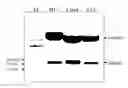
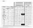

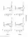
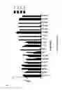

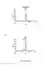
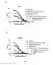
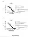

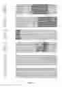


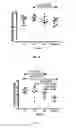
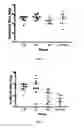

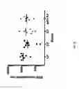


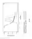
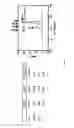

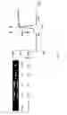
Sources:
- United States Patent and Trademark Office - verify current appl. status at the USPTO↗
Recent applications in this class:
- » 20250215048 2025-07-03
ASSEMBLED GLYCOPROTEINS - » 20250145661 2025-05-08
TARGET MOLECULE REDUCTION METHOD, REDUCTION DEVICE, AND ELECTRODE - » 20250042937 2025-02-06
RAPID, GREEN, DISULFIDE BOND FORMATION IN WATER USING DICYANOCOBINAMIDE - » 20240391951 2024-11-28
NOVEL METHODS FOR PRODUCING PEPTIDE THIOESTER AND PEPTIDE - » 20230357314 2023-11-09
METHODS FOR ULTRAFAST FORMATION OF DISULFIDE BONDS IN PEPTIDES AND PROTEINS - » 20230227495 2023-07-20
PRODUCTION OF HETERODIMERIC PROTEINS - » 20230151054 2023-05-18
SULFUR EXTRUSION FROM DISULFIDES BY CARBENES - » 20230143844 2023-05-11
Methods for producing peptide thioester and peptide - » 20230119996 2023-04-20
ALLERGEN INACTIVATION METHOD AND ALLERGEN INACTIVATION DEVICE - » 20220213139 2022-07-07
MANUFACTURE OF DISULFIDE BONDED PEPTIDES