Feeder cell and method for growing gamma delta T cells by using same
US20190284532A1
2019-09-19
16/349,225
2017-11-10
✅ Patent granted
US 12,077,779 B2
2024-09-03
WO; PCT/KR2017/012701; 20171110
WO; WO2018/088829; 20180517
Maria G Leavitt
Meunier Carlin & Curfman LLC
2039-08-03
Abstract:
The present invention relates to a novel feeder cell and a method for growing gamma delta T cells using the same. More specifically, a large amount of gamma delta T cells may be grown in vitro with high purity and without simulation of a T cell receptor by using a feeder cell into which costimulatory molecules are introduced and a low concentration of IL-2, differentiation into central memory cells may be possible when stimulated, activity by the feeder cell, and cytolytic against tumour cells is provided.
Inventors:
- Hyun-Woo Cho 2 🇰🇷 Gyeonggi-do, South Korea
- Tai Gyu Kim 19 🇰🇷 Seoul, South Korea
- Su Yeon KIM 4 🇰🇷 Seoul, South Korea
- Hyun Jung SOHN 5 🇰🇷 Seoul, South Korea
Assignee:
- The Catholic University of Korea Industry-Academic Cooperation Foundation 150 🇰🇷 Seoul, South Korea
Applicant:
Interested in similar patents?
Get notified when new applications in this technology area are published.
Classification:
C12N2501/52 » CPC further
Active agents used in cell culture processes, e.g. differentation; Cell markers; Cell surface determinants CD40, CD40-ligand (CD154)
C12N5/0638 » CPC main
Undifferentiated human, animal or plant cells, e.g. cell lines; Tissues; Cultivation or maintenance thereof; Culture media therefor; Animal cells or tissues; Human cells or tissues; Vertebrate cells; Cells from the blood or the immune system; T lymphocytes Cytotoxic T lymphocytes [CTL], lymphokine activated killer cells [LAK]
C07K14/70578 » CPC further
Peptides having more than 20 amino acids; Gastrins; Somatostatins; Melanotropins; Derivatives thereof from animals; from humans; Receptors; Cell surface antigens; Cell surface determinants NGF-receptor/TNF-receptor superfamily, e.g. CD27, CD30, CD40, CD95
C07K14/70596 » CPC further
Peptides having more than 20 amino acids; Gastrins; Somatostatins; Melanotropins; Derivatives thereof from animals; from humans; Receptors; Cell surface antigens; Cell surface determinants Molecules with a "CD"-designation not provided for elsewhere
C12N5/0603 » CPC further
Undifferentiated human, animal or plant cells, e.g. cell lines; Tissues; Cultivation or maintenance thereof; Culture media therefor; Animal cells or tissues; Human cells or tissues; Vertebrate cells Embryonic cells ; Embryoid bodies
C12N5/0636 » CPC further
Undifferentiated human, animal or plant cells, e.g. cell lines; Tissues; Cultivation or maintenance thereof; Culture media therefor; Animal cells or tissues; Human cells or tissues; Vertebrate cells; Cells from the blood or the immune system T lymphocytes
C12N5/0694 » CPC further
Undifferentiated human, animal or plant cells, e.g. cell lines; Tissues; Cultivation or maintenance thereof; Culture media therefor; Animal cells or tissues; Human cells or tissues; Vertebrate cells; Tumour cells; Cancer cells Cells of blood, e.g. leukemia cells, myeloma cells
G01N33/56972 » CPC further
Investigating or analysing materials by specific methods not covered by groups -; Biological material, e.g. blood, urine ; Haemocytometers; Chemical analysis of biological material, e.g. blood, urine; Testing involving biospecific ligand binding methods; Immunological testing; Immunoassay; Biospecific binding assay; Materials therefor for microorganisms, e.g. protozoa, bacteria, viruses; Animal cells White blood cells
C12N2501/2302 » CPC further
Active agents used in cell culture processes, e.g. differentation; Cytokines; Chemokines; Interleukins [IL] Interleukin-2 (IL-2)
C12N2501/51 » CPC further
Active agents used in cell culture processes, e.g. differentation; Cell markers; Cell surface determinants B7 molecules, e.g. CD80, CD86, CD28 (ligand), CD152 (ligand)
C12N2510/00 » CPC further
Genetically modified cells
A61K48/00 IPC
Medicinal preparations containing genetic material which is inserted into cells of the living body to treat genetic diseases; Gene therapy
A61K35/17 » CPC further
Medicinal preparations containing materials or reaction products thereof with undetermined constitution; Materials from mammals; Compositions comprising non-specified tissues or cells; Compositions comprising non-embryonic stem cells; Genetically modified cells; Blood; Artificial blood Lymphocytes; B-cells; T-cells; Natural killer cells; Interferon-activated or cytokine-activated lymphocytes
A61K39/39 » CPC further
Medicinal preparations containing antigens or antibodies characterised by the immunostimulating additives, e.g. chemical adjuvants
A61K51/10 IPC
Preparations containing radioactive substances for use in therapy or testing characterised by the carrier, i.e. characterised by the agent or material covalently linked or complexing the radioactive nucleus; Organic compounds; Peptides, e.g. proteins, carriers being peptides, polyamino acids, proteins Antibodies or immunoglobulins; Fragments thereof, the carrier being an antibody, an immunoglobulin or a fragment thereof, e.g. a camelised human single domain antibody or the Fc fragment of an antibody
C07K14/705 IPC
Peptides having more than 20 amino acids; Gastrins; Somatostatins; Melanotropins; Derivatives thereof from animals; from humans Receptors; Cell surface antigens; Cell surface determinants
C12N5/10 » CPC further
Undifferentiated human, animal or plant cells, e.g. cell lines; Tissues; Cultivation or maintenance thereof; Culture media therefor Cells modified by introduction of foreign genetic material
G01N33/50 IPC
Investigating or analysing materials by specific methods not covered by groups -; Biological material, e.g. blood, urine ; Haemocytometers Chemical analysis of biological material, e.g. blood, urine; Testing involving biospecific ligand binding methods; Immunological testing
G01N33/569 IPC
Investigating or analysing materials by specific methods not covered by groups -; Biological material, e.g. blood, urine ; Haemocytometers; Chemical analysis of biological material, e.g. blood, urine; Testing involving biospecific ligand binding methods; Immunological testing; Immunoassay; Biospecific binding assay; Materials therefor for microorganisms, e.g. protozoa, bacteria, viruses
C07K14/70532 » CPC further
Peptides having more than 20 amino acids; Gastrins; Somatostatins; Melanotropins; Derivatives thereof from animals; from humans; Receptors; Cell surface antigens; Cell surface determinants; Immunoglobulin superfamily B7 molecules, e.g. CD80, CD86
C12N2501/515 » CPC further
Active agents used in cell culture processes, e.g. differentation; Cell markers; Cell surface determinants CD3, T-cell receptor complex
Description
BACKGROUND
1. Field of the Invention
The present invention relates to novel feeder cells into which a costimulatory molecule is introduced, and a method of mass-proliferating high purity gamma-delta (γδ) T cells in vitro using the same in the presence of a low concentration of IL-2 without T cell receptor stimulation.
2. Discussion of Related Art
Recently, significant processes for understanding the role of human γδ T cells have been made in immune responses against infections, tumors and autoimmune diseases. Human γδ T cells expressing Vγ9Vδ2 TCR have been known as the major subset which can be activated in peripheral blood in a human leukocyte antigen (HLA)-unrestricted manner by small non-peptidic phosphoantigens. Vγ9Vδ2 T cells include NKG2D and ULBP4, which are expressed in tumor cells, and are capable of recognizing their ligands, thereby exhibiting strong cytotoxicity against tumor cells. In addition, both manipulations of the activation of Vγ9Vδ2 T cells in vivo and the activation of autologous Vγ9Vδ2 T cells expanded ex vivo and adoptively transferred are being considered as new immunotherapeutic approaches. Many protocols for the ex vivo expansion of Vγ9Vδ2 T cells using a natural antigen, i. e., isopentenyl pyrophosphate, against human Vγ9Vδ2 T cells have been developed. Particularly, Correia et al. have reported that a phosphoantigen alone cannot activate Vγ9Vδ2 T cells, and the addition of a high concentration of IL-2 is significant for completing the activation of Vγ9Vδ2 T cells. More significantly, mature dendritic cells pretreated with aminobisphosphate are able to induce the expansion of high-purity Vγ9Vδ2 T cells in peripheral blood mononuclear cells (PBMCs), which does not correspond to a conventional culture protocol. Since the mature dendritic cells express a variety of costimulatory molecules, including CD80, CD83 and 4-1BBL, data on these molecules shows that signaling using a large number of costimulatory receptors activates Vγ9Vδ2 T cells. Even though it has been proven that various costimulatory molecules including CD80 and a 4-1BB ligand (4-1BBL) participate in promotion of the proliferation and survival of peripheral Vγ9Vδ2 T cells, the functions of CD83 and its ligand for the activation of γδ T cells are little known.
SUMMARY OF THE INVENTION
An object of the present invention is directed to providing feeder cells that are able to express a costimulatory molecule group and stimulate γδ T cells.
Another object of the present invention is also directed to providing a composition and method for proliferating high purity γδ T cells in vitro using the feeder cells in the presence of a low concentration of IL-2 without T cell receptor stimulation.
To attain the objects of the present invention, the present invention provides feeder cells for stimulating γδ T cells, which express a costimulatory molecule group including 4-1BBL, CD80 and CD83, but do not express HLA.
The present invention provides a composition for in vitro proliferating γδ T cells including the feeder cells for stimulating γδ T cells.
The present invention also provides a method of in vitro proliferating γδ T cells, which includes ex vivo co-culturing the feeder cells for stimulating γδ T cells and γδ T cells without T cell receptor stimulation.
According to the present invention, high purity γδ T cells can be mass-proliferated in vitro by stimulating γδ T cells using feeder cells in the presence of a low concentration of IL-2 without T cell receptor stimulation, and the γδ T cells can differentiate into central memory cells and have cytolytic activity against tumor cells.
BRIEF DESCRIPTION OF THE DRAWINGS
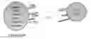
FIG. 1 is a diagram illustrating a feeder cell expressing costimulatory molecules for in vitro proliferating γδ T cells according to the present invention.
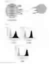
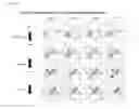
FIG. 2 shows the results of confirming the expression of costimulatory molecules 4-1BBL, CD80 and CD83, expressed in feeder cells, according to the present invention.
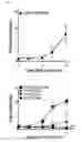
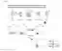
FIG. 3 shows the result of the proliferation of γδ T cells using feeder cells expressing 4-1BBL/CD80/CD83 according to the present invention.
FIG. 4 shows the result of the proliferation of γδ T cells using feeder cells expressing a combination of different types of costimulatory molecules.
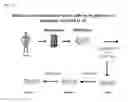
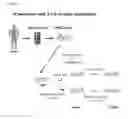
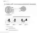
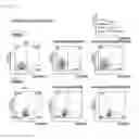
FIG. 5A is a diagram illustrating the process of stimulating feeder cells from human PBMCs in the presence of zoledronic acid and IL-2, and FIG. 5B is a graph showing the proliferation rate of γδ T cells obtained by stimulating enriched γδ T cells obtained by stimulating human PBMCs with zoledronic acid and IL-2 (1000 IU/mL) with feeder cells expressing 4-1BBL/CD80/CD83 in the presence of a low concentration of IL-2 (20 IU/mL).
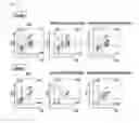
FIG. 6 is the FACS analysis result showing the proliferation rate of γδ T cells obtained by stimulating enriched γδ T cells obtained by stimulating human PBMCs with zoledronic acid and IL-2 (1000 IU/mL) with feeder cells expressing 4-1BBL/CD80/CD83 in the presence of a low concentration of IL-2 (20 IU/mL).
FIG. 7 is the FACS analysis (subsets analysis) result of a differentiation marker for γδ T cells obtained by stimulating enriched γδ T cells obtained by stimulating human PBMCs with zoledronic acid and IL-2 (1000 IU/mL) with feeder cells expressing 4-1BBL/CD80/CD83 in the presence of a low concentration of IL-2 (20 IU/mL).
FIG. 8A is the diagram of the comparison with T cell receptor stimulation, and FIG. 8B is the FACS analysis (subsets analysis) result of γδ T cells proliferated in vitro by stimulating enriched γδ T cells obtained by stimulating human PBMCs with zoledronic acid and IL-2 (1000 IU/mL) with feeder cells expressing 4-1BBL/CD80/CD83 in the presence of a low concentration of IL-2 (20 IU/mL) according to the absence or presence of an anti-CD3 antibody.
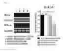
FIG. 9 is the RT-PCR result showing that anti-apoptotic molecule BCLA2A1 is expressed in γδ T cells proliferated using feeder cells expressing 4-1BBL/CD80/CD83.
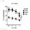
FIG. 10 is the result of confirming the cytotoxicity of γδ T cells proliferated using feeder cells expressing 4-1BBL/CD80/CD83 against tumor cells.
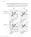
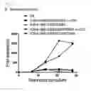
FIG. 11A is the FACS analysis result showing the expression of each costimulatory molecule in feeder cells expressing costimulatory molecules 4-1BBL/CD80/CD83/CD40L, and FIG. 11B is the result of proliferating γδ T cells in the absence or presence of an anti-CD3-antibody in the stimulation with feeder cells expressing 4-1BBL/CD80/CD83 and feeder cells expressing 4-1BBL/CD80/CD83/CD40L.
FIG. 12 is the result of the analysis of the purity of γδ T cells at day 14 and day 28 of the proliferation by the stimulation with feeder cells expressing 4-1BBL/CD80/CD83 and feeder cells expressing 4-1BBL/CD80/CD83/CD40L.
FIG. 13 is the result of analyzing the differentiation ability of γδ T cells into central memory cells at day 14 and day 28 of the proliferation by the stimulation with feeder cells expressing 4-1BBL/CD80/CD83 and feeder cells expressing 4-1BBL/CD80/CD83/CD40L.
DETAILED DESCRIPTION OF EXEMPLARY EMBODIMENTS
The inventors investigated the roles of costimulatory molecules including CD83 for the survival, proliferation and cytokine formation of human γδ T cells in vitro. First, since K562 cells do not express HLA molecules, but have an advantage of expressing adhesion molecules, for example, ICAM, LFA-3 and NKG2D ligands and MHC class I-related chain A, a stable K562-based transformant expressing a costimulatory molecule group consisting of 4-1BBL/CD80/CD83 or a costimulatory molecule group consisting of 4-1BBL/CD80/CD83/CD40L was prepared. These transformants were further transduced to raise the expression rate(s) of Fc receptor(s) such as CD32 and/or CD64.
It has been known that the 4-1BB/4-1BBL interaction is independent from B7/CD28 signaling, and increases the survival of cells activated in vivo. Particularly, the participation of CD83 noticeably upregulated during the maturation of dendritic cells has been known to inhibit apoptosis, thereby improving the long-term survival of T cells.
In addition, stimulation may be performed by additionally transferring costimulatory molecules such as γδ T cell-stimulatory molecules, i.e., MIC and ULBP to the cells. The inventors confirmed that K562-based feeder cells allow the long-term growth of functional effector-memory Vγ9Vδ2 T cells, and the proliferation rate of high purity Vγ9Vδ2 T cells is increased in vitro in the presence of a low concentration of IL-2 without T cell receptor stimulation.
From the above-described result, it was found that the costimulatory signals involved in the activation and survival of human γδ T cells eventually improve γδ T cell-based immunotherapeutic strategies for cancer, infections and autoimmune diseases.
Therefore, the present invention relates to feeder cells for stimulating γδ T cells, which express a costimulatory molecule group including 4-1BBL, CD80 and CD83 and do not express HLA.
The term “feeder cell” used herein refers to artificially manufactured antigen-presenting cells, and non-immune cells modified to express an immune molecule. Feeder cells expressing an MHC class I or II (MHC I or II) molecule alone or in combination with other accessory molecules (costimulatory molecules and/or adhesion molecules) may be used to study various aspects of T cell-activating cells which can be easily cultured, such as tumor cells or a fibroblast cell line in vivo. For the purpose of the present invention, the feeder cell means a cell in which cDNA of costimulatory molecules is introduced into any one of human cells without HLA expressing capacity, such as K562 cells and recombinant HEK 293T cells from which an HLA gene is artificially removed, but the present invention is not limited thereto.
The feeder cells of the present invention are prepared from cells without an HLA expressing capacity, and prepared by introducing respective nucleic acids encoding a costimulatory molecule group including costimulatory molecules such as 4-1BBL, CD80 and CD83, or a costimulatory molecule group including 4-1BBL, CD80, CD83 and CD40L into cells having no HLA expressing capacity or from which HLA expressing capacity is removed. Compared with conventional feeder cells, the feeder cells of the present invention are able to stimulate cells without the stimulation with an immunostimulatory ligand, that is, a T cell receptor.
Therefore, the feeder cells may mass-produce high purity γδ T cells in vitro only with the combination of costimulatory molecules without T cell receptor stimulation.
In the feeder cells, Fc receptor(s), that is, CD32 and/or CD64, other than the costimulatory molecules, are/is further transduced to increase the expression rate(s) of CD32 and/or CD64.
The feeder cells may stimulate any one type of naive γδ T cells isolated from human PBMCs and enriched γδ T cells obtained by stimulating human PBMCs with zoledronic acid.
The naive γδ T cells may be isolated from human PBMCs by FACS sorting or using anti-γδ antibody-conjugated microspheres, thereby exhibiting Vγ9Vδ2 subsets and having CD83/4-1BBL/anti-CD28 signals.
The enriched γδ T cells may be obtained by stimulating human PBMCs with zoledronic acid, or by being isolated by FACS sorting or using anti-γδ antibody-conjugated microspheres after stimulation with zoledronic acid. The enriched γδ T cells may exhibit Vγ9Vδ2 subsets the same as the naive γδ T cells, and have CD83/4-1BBL/anti-CD28 signals.
To provide antigenic specificity, the feeder cells of the present invention may be further transduced with nucleic acid(s) encoding any one or more antigens selected from the group consisting of a tumor antigen, a pathogenic antigen and an autoantibody.
The term “antigen” used herein is well known in the art, and includes an epitope, a peptide fragment of an antigen capable of binding to an MHC molecule, and an immunogen, as well as all molecules capable of binding to an antibody. In the present invention, as an antigen, a tumor antigen, a pathogenic antigen or an autoantibody (normal or pathological) may be used, but the present invention is not limited thereto.
The tumor antigen refers to a tumor-associated antigen (TAA), which is an antigen associated with a tumor. Examples of well-known TAAs include ovalbumin, survivin, gp75, gp1OO, MDM2, MART-1, MAGE-1, MAGE-3, tyrosinase, telomerase, her-2/neu, α-1 fetoprotein, G250, and NY-ESO-1. Partial peptide fragment sequences of TAAs binding to an MHC molecule include Ova257 (SIINFEKL; SEQ ID NO: 25), tyrosinase-related protein 1455 (Trp1455; TAPDNLGYA; SEQ ID NO: 26), Trp2180 (SVYDFFVWL; SEQ ID NO: 27), gp10025 (gp10025; EGSRNQDWL; SEQ ID NO: 28), a MAGE 1 nonapeptide (EADPTGHSY; SEQ ID NO: 29), a MART-APL peptide (LAGIGILTV; SEQ ID NO: 30), a natural peptide (AAGIGILTV; SEQ ID NO: 31), and a PSA-1 peptide (FLTPKKLQCV; SEQ ID NO: 32). The sequences of additional tumor-associated peptides and antigens are known to those of ordinary skill in the art.
The pathogenic antigen refers to an organism or virus that causes a disease, or an attenuated derivative thereof. The term “pathogen” refers to any virus or organism, or an attenuated derivative thereof, which is involved in the occurrence of a disease. The pathogens may include bacterial, protozoan, fungal and viral pathogens, for example, Helicobacters such as Helicobacter pylori, Salmonella sp., Shigella sp., Enterobacter sp., and Campylobacter sp., various mycobacteria such as Mycobacterium leprae and Mycobacterium tuberculosis, Bacillus anthracis, Yersinia pestis, Francisella tularensis, Brucella sp., Leptospira interrogans, Staphylococcus sp. such as S. aureus, Streptococcus sp., Clostridum sp., Candida albicans, Plasmodium sp., Leishmania sp., Trypanosoma sp., human immunodeficient virus (HIV), hepatitis C virus (HCV), human papilloma virus (HPV), cytomegalovirus (CMV), HTLV, herpes viruses (e.g., Type 1 herpes simplex virus, Type 2 herpes simplex virus, Corona virus, Varicella-Joster virus, and Epstein-Barr virus), papilloma virus, influenza virus, hepatitis B virus, poliovirus, measles virus, mumps virus, and rubella virus, but the present invention is not limited thereto.
The autoantibody is an antinuclear antibody, an anti-γ globulin antibody, an antibody against a self-hemocyte component or an antibody against a self-organ, but the present invention is not particularly limited thereto. When the autoantibody is used as a foreign antigen, a CD4 T cell vaccine may induce potent antitumor immunity, and thus may be effective for overcoming potential immune tolerance to an auto-antigen expressed in normal tissue.
The term “costimulatory molecule” used herein is a material participating in the interaction between a receptor-ligand pair expressed on the surface of an antigen-presenting cell and a T cell, and to induce the expression and proliferation of a cytokine gene, two or more signals are required for resting T cells. A first signal is a signal imparting specificity, generated by the interaction between an MHC/peptide complex and a TCR/CD3 complex, and a second signal is antigen-nonspecific, and called a “costimulatory” signal. These signals are known to have activity provided by bone marrow-derived helper cells such as macrophages and dendritic cells. The costimulatory molecule performs complete activation of γδ T cells by mediating a costimulatory signal required under a normal physiological condition. In the present invention, as such costimulatory ligands, a combination of 4-1BBL, CD80 and CD83 or a combination of 4-1BBL, CD80, CD83 and CD40L is used.
The feeder cells of the present invention may be prepared by transducing costimulatory molecules into human cells without HLA expressing capacity or from which HLA expressing capacity is removed using a known transformation technique. According to an exemplary embodiment of the present invention, costimulatory molecules may be transduced into K562 cells.
A nucleic acid encoding the costimulatory molecule may be DNA or RNA.
Preferably, 4-1BBL may be a human or mouse-derived nucleic acid sequence, for example, a base sequence set forth in SEQ ID NO: 1, but the present invention is not limited thereto.
CD80 may be a human or mouse-derived nucleic acid sequence. For example, CD80 may be a base sequence set forth in SEQ ID NO: 2, but the present invention is not particularly limited thereto.
CD83 may be a human or mouse-derived nucleic acid sequence, for example, a base sequence set forth in SEQ ID NO: 3, but the present invention is not particularly limited thereto.
CD40L may be a human or mouse-derived nucleic acid sequence, for example, a base sequence set forth in SEQ ID NO: 4, but the present invention is not particularly limited thereto.
When DNA is selected as a nucleic acid encoding the costimulatory molecule, it may be provided to human cells which have no HLA expressing capacity or from which HLA expressing capacity is removed in a form of being inserted into a vector.
The term “vector” used herein means a nucleic acid molecule capable of delivering a different nucleic acid linked to the nucleic acid itself. There is a type of vector, called “plasmid,” which is a circular double-stranded DNA loop capable of ligating an additional DNA segment. Another type of vector is a viral vector capable of ligating an additional DNA segment with a viral genome. Some vectors can be self-replicated in a host cell when being introduced into the host cell (e.g., a bacterial vector having a bacterial origin of replication and an episomal vector for a mammal). When being introduced into a host cell, a different vector (e.g., a non-episomal vector for a mammal) is able to be integrated into the genome of the host cell, and replicated with the host genome. In addition, some vectors may direct the expression of genes operably linked to each other. The vector of the specification refers to “recombinant expression vector” (or simply called “expression vector”). Generally, since an expression vector useful for a recombinant DNA technique is typically the most commonly-used vector type, the “plasmid” and “vector” may be used interchangeably. However, the present invention may include different types of expression vectors such as viral vectors providing equivalent functions (e.g., an adenovirus vector, an adeno-associated virus (AAV) vector, a herpes virus vector, a retrovirus vector, a lentivirus vector, and a baculovirus vector), and preferably, a retroviral vector.
The retrovirus is an RNA virus having a different life cycle from a lytic virus. In this regard, the retrovirus is an infectious agent that replicates through a DNA intermediate. When cells are infected by the retrovirus, the viral genome is converted into DNA due to reverse transcriptase. A DNA copy is used as a template for producing an encoded viral protein, which is necessary for a new RNA genome and the assembly of an infectious viral particle. There are many types of retroviruses, including all different families of retroviruses, for example, murine leukemia virus (MLV), human immunodeficiency virus (HIV), equine infectious anemia virus (EIAV), mouse mammary tumor-like virus (MMTV), Rouse sarcoma virus (RSV), Fujinami sarcoma virus (FuSV), Moloney murine leukemia virus (Mo-MLV), FBR murine osteosarcoma virus (FBR MSV), Moloney murine sarcoma virus (Mo-MSV), Abelson murine leukemia virus (A-MLV), avian myeloblastosis virus-29 (MC29), avian erythromblastasis virus (AEV) and lentivirus. Detailed lists of the retroviruses are shown in Coffin et al. (“Retroviruses” 1997 Cold Spring Harbour Laboratory Press Eds: J M Coffin, S M Hughes, H E Varmus pp 758-763).
Transformation includes any method for introducing a nucleic acid into an organism, a cell, tissue or an organ, and may be carried out by selecting a suitable standard technique according to host cells as known in the art. Such methods include electroporation, plasma fusion, calcium phosphate (CaPO4) precipitation, calcium chloride (CaCl2) precipitation, stirring with silicon carbide fibers, agrobacteria-mediated transformation, PEG, dextran sulfate, and Lipofectamine, but the present invention is not limited thereto.
According to an exemplary embodiment of the present invention, each cDNA of each of the genes of human costimulatory molecules 4-1BBL, CD80, CD83 and CD40L is prepared through PCR amplification, and inserted into a retrovirus vector. The pcDNA3/CD80, pLXSN/4-1BBL, pcDNA3/CD83, and pcDNA3/CD40L are sequentially introduced into K562 cells for transformation.
The term “gamma-delta T cells” used herein may be used interchangeably with “γδ T cells.”
The present invention also relates to a composition for in vitro proliferating γδ T cells including feeder cells for stimulating the γδ T cells.
The present invention also relates to a method of in vitro proliferating γδ T cells, which includes ex vivo co-culturing feeder cells for stimulating γδ T cells and γδ T cells without T cell receptor stimulation.
The γδ T cells used in the method of in vitro proliferating the γδ T cells according to the present invention may be any one of naive γδ T cells isolated from human PBMCs or enriched γδ T cells obtained by stimulating human PBMCs with zoledronic acid.
The co-culture of feeder cells and γδ T cells may be carried out without T cell receptor stimulation.
At this time, the T cell receptor stimulation means culturing in the presence of an anti-CD3 antibody or anti-γδ TCR antibody.
Therefore, the co-culture of feeder cells and γδ T cells are performed in a cell culture medium containing a low concentration of IL-2 in the absence of the anti-CD3 antibody or anti-γδ TCR antibody.
The cell culture medium may be a complete medium for animal cell culture. For example, Dulbecco's Modified Eagle's Medium (DMEM), Minimal Essential Medium (MEM), Basal Medium Eagle (BME), RPMI1640, F-10, F-12, α-Minimal Essential Medium (αMEM), Glasgow's Minimal Essential Medium (GMEM), or Iscove's Modified Dulbecco's Medium may be used, but the present invention is not limited thereto.
IL-2 may be added at a concentration of 20 to 100 IU/mL. Compared with a conventional method of in vitro proliferating γδ T cells, performed at a high concentration of 300 IU/mL or more, the method according to the present invention is performed with a considerably lower concentration of IL-2.
According to an exemplary embodiment of the present invention, the in vitro proliferation of γδ T cells may be performed by co-culturing feeder cells for stimulating γδ T cells, expressing a costimulatory molecule group consisting of 4-1BBL, CD80, CD83 and CD40L, and naive γδ T cells or enriched γδ T cells in the presence of 20 to 100 IU/mL of IL-2 without an anti-CD3 antibody or anti-γδ TCR antibody, or feeder cells for stimulating γδ T cells, expressing a costimulatory molecule group consisting of 4-1BBL, CD80 and CD83 and naive γδ T cells or enriched γδ T cells in the presence of 20 to 100 IU/mL of IL-2 without the anti-CD3 antibody or anti-γδ TCR antibody.
The simulation with feeder cells is repeated at intervals of 7 to 10 days, and the in vitro culture is able to be performed for 90 days or more, and more preferably, 14 to 100 days. Considering that the conventional method of in vitro proliferating γδ T cells is performed within a maximum of 14 days, long-term culture is possible.
The co-culture may be performed in a CO2 incubator with a CO2 flow amount of 5 to 15% at 35 to 37 r , but the present invention is not particularly limited.
In addition, according to an exemplary embodiment of the present invention, through the stimulation with feeder cells expressing 4-1BBL/CD80/CD83/CD40L or 4-1BBL/CD80/CD83, rather than the stimulation with zoledronic acid, high purity γδ T cells may be obtained, and the differentiation into central memory cells may last longer. Moreover, compared with the stimulation with feeder cells expressing 4-1BBL/CD80/CD83, the stimulation with feeder cells expressing 4-1BBL/CD80/CD83/CD40L enables long-term proliferation of γδ T cells, and thereby high purity γδ T cells may be obtained, and differentiation into central memory cells may last longer.
In addition, in the case of γδ T cells proliferated by the stimulation of feeder cells expressing 4-1BBL/CD80/CD83, an anti-apoptotic molecule is expressed such that cell proliferation is maintained for a long time, and the γδ T cells exhibit apoptotic capacity with respect to tumor cells.
Hereinafter, the present invention will be described in further detail with reference to examples according to the present invention, but the scope of the present invention is not limited by the following examples.
EXAMPLES
Example 1
In Vitro Proliferation of γδ T Cells Using Feeder Cells
(Cells)
Human samples were reviewed and approved by the Institutional Review Board of Catholic University, Korea. PBMCs were collected according to leukapheresis, and centrifuged using a Ficoll-Paque (GE Healthcare) density gradient. CD4− T cells and CD8+ T cells were isolated using CD4+ T cell and CD8+ T cell isolation kits (Miltenyi Biotec) according to magnetic cell sorting for positive selection (isolated to reach 95% or more purity). γδ T cells were isolated from PBMCs newly purified using an anti-TCRγ/δ MicroBead kit (Miltenyi Biotec) according to magnetic cell sorting (isolated to reach 60% or more purity). Particularly, to isolate Vγ9Vδ2 T cells (isolated to reach 95% or more purity), the γδ T cells were sorted using FACSAria (Becton Dickinson) so as to be further isolated into CD3+ and Vγ9+ subsets.
For cell surface staining, the cells were incubated with anti-TCRVγ9-PE (B3) and anti-CD3-FITC (OKT3) on ice for 30 minutes. A Burkitt's lymphoma cell line Daudi, an erythroid bone marrow cell line K562 and a leukemia cell line U937 were obtained from ATCC, and cultured in a complete medium. The complete medium was a 1640 RPMI (Lonza) medium containing 10% heat-inactivated FBS (Gibco), 2 mM L-glutamine (Lonza), 100 U/mL of penicillin (Lonza), and 100 μg/mL of streptomycin (Lonza).
(Establishment of Feeder Cells)
Human CD80, 4-1BBL and CD83 genes were amplified from cDNA through PCR, and cloned into 3.1-neo (Invitrogen) and a pLXSN retrovirus vector (Clontech). First, K32 was prepared by transducing CD80 into K562 cells. Then, K80/4-1BBL and K80/CD83 were produced by transducing 4-1BBL or CD83 into K80 cells. Finally, K80/4-1BBL/CD83 was produced by transducing CD83 into K80/4-1BBL. Human CD80 and 4-1BBL genes were cloned from a U937 cell line, and the CD83 gene was cloned from mature DC. pcDNA3/CD80 and/or pLXSN/4-1BBL and/or pcDNA3/CD83 plasmids were sequentially transformed into K562 cells using Nucleofector Kit V (Amaxa) according to the manufacturer's instructions. The transformants were cultured in the presence of 1 mg/mL of G-418 DISULPHATE (Duchefa Biochemie). The transformed cells were further selected by FACSAria. All artificial APCs were identified as a mycoplasma-free line, as determined by PCR (data not shown).
(FACS Analysis)
The following mAbs were used to test surface expression of costimulatory molecules and activation markers in K562 and Vγ9Vδ2 T cells: FITC-, PE-, Alexa Fluor 488-, APC-, or Cy5-conjugated mAbs: anti-CD3(OKT3), anti-TCRVδ2(B6), anti-HLA-A,B,C(W6/32), anti-CD27(O323), anti-CD27(0323), anti-CD45RA(HI100), anti-CD32(FUN-2), anti-CD25(BC96), anti-CD69(FN50), anti-CD28(CD28 .2), anti-CD86(IT 2.2), anti-137(4B4-1), anti-137L(5F4), anti-CD 83 (HB 15 e), anti-CD80(2D 10), anti-NKG2D(CD314), anti-TCRVγ9(B3), anti-TCRγ/6(B1), and anti-HLA-DR,DQ-PE Abs.
All antibodies were purchased from BioLegend or Ancell. Simply put, 1×10⁶ viable T cells in 100 pi of PBS (Lonza) were washed twice with 2% FBS-containing PBS. The cells stained with cell surface markers were fixed using 1% paraformaldehyde in PBS. Subsequently, the cells were analyzed using FACSCalibur (Becton Dickinson). Data was analyzed using FlowJo software (Tree Star).
(Expansion of Vγ9Vδ2 T Cells)
The Vγ9Vδ2 T cells used for expansion were prepared by isolating CD3+ and Vγ9+ cells from PBMCs in peripheral blood using FACSAria. Cell culture was performed (2 mL/well) in a 24-well plate (Nunc) containing a complete culture medium supplemented with 20 IU/mL of IL-2 (NOVARTIS). 1×10⁶ Vγ9Vδ2 T cells and 5×10⁵ irradiated (100 Gy) K80/4-1BBL/CD83, K80/4-1BBL or K80/CD83 cells were added to each well. And then, the cells were incubated at 37° C. under a 5% CO2 condition.
The growing Vγ9Vδ2 T cells were harvested every 3 or 4 days, counted, and stimulated again at 7 to 10-day intervals. The Vγ9Vδ2 T cells were plated again at 1 x 10⁶ cells/well in addition to 5 x 10⁵ irradiated artificial APCs, and loaded in a complete culture medium under conditions of 37° C. and 5% CO2.
PBMCs were stimulated with 5 μM zoledronic acid (Novartis) in a complete culture medium consisting of 1640 RPMI (Lonza) containing 10% inactivated FBS. An IL-2 (1000 IU/mL)-containing medium was added every 3 or 4 days, and the culture were transferred to a new 24-well plate under conditions of 37° C. and 5% CO2.
The Vγ9Vδ2 T cells were centrifuged with a Ficoll-Paque density gradient to remove dead Vγ9Vδ2 T cells and remaining artificial APCs, and then cryopreserved in liquid nitrogen until further use.
(ELISA)
Vγ9Vδ2 T cells were stimulated with K80/4-1BBL/CD83, K80/4-1BBL or K80/83, and 20 IU/mL of IL-2-loaded artificial APCs in a complete medium at 37° C. for 72 hours. A supernatant was collected, centrifuged to remove cell debris, and then stored at −20° C. . The concentrations of IL-2, IL-6, IFN-γ, TNF-α, GM-CSF, IL-4, IL-10, TGF-β, and IL-17a in the supernatant were measured through ELISA (BioLegend) according to the manufacturer's instructions.
(Analysis of RNA Expression By Reverse Transcription-PCR)
Vγ9Vδ2 T cells were stimulated with K80/4-1BBL/CD83, K80/4-1BBL or K80/83 and 20 IU/mL of IL-2-loaded artificial APCs in a complete medium at 37° C. for 7 days. The Vγ9Vδ2 T cells were harvested, and total RNA was extracted using an RNeasy Mini kit (QIAGEN). In a reverse transcription reaction, cDNA was synthesized using a Transcriptor First Strand cDNA Synthesis kit (Roche) with a 2.5 μM anchored-oligo (dT) primer and Transcriptor Reverse Transcriptase (Roche), and amplified by PCR using the following primers:
| GAPDH forward, | ||
| 5′-TGTTGCCATCAATGACCCCTT-3′, | (SEQ ID NO: 5) | |
| GAPDH reverse, | ||
| 5′-CTCCACGACGTACTCAGCG-3′; | (SEQ ID NO: 6) | |
| perforin forward, | ||
| 5′-GGCTGGACGTGACTCCTAAG-3′, | (SEQ ID NO: 7) | |
| perforin reverse, | ||
| 5′-CTGGGTGGAGGCGTTGAAG-3′; | (SEQ ID NO: 8) | |
| granzyme A forward, | ||
| 5′-GTGCTGGGGCTTTGATTGC-3′, | (SEQ ID NO: 9) | |
| granzyme A reverse: | ||
| 5′-GGGTCATAGCATGGATAGGGAAA-3′; | (SEQ ID NO: 10) | |
| granzyme B forward, | ||
| 5′-TGGGGGACCCAGAGATTAAAA-3′, | (SEQ ID NO: 11) | |
| granzyme B reverse, | ||
| 5′-TTTCGTCCATAGGAGACAATGC-3′; | (SEQ ID NO: 12) | |
| FasL forward, | ||
| 5′-GAACTCCGAGAGTCT ACCAGC-3′, | (SEQ ID NO: 13) | |
| FasL reverse, | ||
| 5′-TTGCCTGTTAAATGGGCCACT-3′; | (SEQ ID NO: 14) | |
| TNF-α forward, | ||
| 5′-ATGAGCACTGAAAGCATGATCC-3′, | (SEQ ID NO: 15) | |
| TNF-α reverse, | ||
| 5′-GAGGGCTGATTAGAGAGAGGTC-3′; | (SEQ ID NO: 16) | |
| IFN-γ forward, | ||
| 5′-CTCTTGGCTGTTACTGCCAGG-3′, | (SEQ ID NO: 17) | |
| IFN-γ reverse, | ||
| 5′-CTCCACACTCTTTTGGATGCT-3′; | (SEQ ID NO: 18) | |
| BCL2 forward, | ||
| 5′-GGTGGGGTCATGTGTGTGG-3′, | (SEQ ID NO: 19) | |
| BCL2 reverse, | ||
| 5′-CGGTTCAGGTACTCAGTCATCC-3′; | (SEQ ID NO: 20) | |
| BCL2A 1 forward, | ||
| 5′-TTACAGGCTGGCTCAGGACT-3′, | (SEQ ID NO: 21) | |
| BCL2A 1 reverse, | ||
| 5′-CCCAGTTAATGATGCCGTCT-3′; | (SEQ ID NO: 22) | |
| Bcl-xL forward, | ||
| 5′-AGCCTTGGATCCAGGAGAAC-3′, | (SEQ ID NO: 23) | |
| Bcl-xL reverse, | ||
| 5′-AGCGGTTGAAGCGTTCCT-3′ | (SEQ ID NO: 24) |
All of the above primers were disclosed previously (DeBarros, A. et al. Eur J Immunol 2011. 41: 195-201; Dokouhaki, P. et al. Cancer Lett 2010. 297: 126-136).
For each specimen, 20 μL of a reaction mixture contained a reaction buffer, 1 mM of each deoxynucleotide mixture, 20U of Protector RNase Inhibitor, 10 pM of each primer, 10U of Transcriptor Reverse Transcriptase. The thermocycling program was as follows: 50° C. for 1 hour and 83° C. for 10 minutes for cDNA amplification. The housekeeping gene GAPDH was used as a reference gene for quantification. The product was isolated from a 1.2% agarose gel at 100 volts for 25 minutes. mRNA was analyzed using Image Lab software (BIO-RAD).
(Cytotoxicity Assay)
A standard 51Cr-release assay was performed, as described in Park, M. Y. et al. Eur J Immunol 2008. 38: 2106-2117. Simply put, K562 and Daudi cell lines were incubated with 100 mCi [51Cr] sodium chromate/1×106 cells under conditions of 37° C. and 5% CO2 for 1 hour. The 51Cr-labeled target cells were incubated with effector cells, i.e., Vγ9Vδ2 T cells, at 37° C. for 4 hours. The supernatant (100 μL) was harvested, and radioactivity was counted using a gamma counter (Packard). The percent specific incorporation was calculated by the formula: [(experimental release-spontaneous release)/(maximum release-spontaneous release)]×100. The spontaneous release and the maximum release were measured in media and in the presence of 2% Triton-X100.
(Statistical Analysis)
All data was analyzed using a statistic program GraphPad Prism 5.0 (GraphPad Software Inc.). The difference between values was evaluated using a Student's t-test. Significance was determined when p<0.05.
Experimental Example 1>
Proliferation of γδ T Cells Using Feeder Cells
FIG. 1 is a diagram illustrating the proliferation of human γδ T cells in vitro by establishing stimulatory cells expressing costimulatory molecules CD83/4-1BBL/CD80, which are an immunostimulatory ligand-free K562 cell line, for the in vitro proliferation of γδ T cells.
As shown in FIG. 2, the expression of costimulatory molecules CD83, 4-1BBL and CD80 in feeder cells established in a K562 cell line was confirmed.
FIG. 3 shows the result of in vitro proliferation of γδ T cells stimulated with feeder cells, demonstrating the proliferation of γδ T cells until 14 days after co-culture.
FIG. 4 shows the result of proliferating γδ T cells stimulated by feeder cells expressing a combination of different types of costimulatory molecules, demonstrating that the proliferation rate of γδ T cells when being stimulated with feeder cells expressing CD83, 4-1BBL and CD80 is the highest.
Experimental Example 2
Experiments for Proliferation and Differentiation Marker of γδ T Cells Using Feeder Cells 7 Days After Stimulation with Zoledronic Acid and IL-2
According to the stimulation process diagram shown in FIG. 5A, after the number of γδ T cells was increased by treating four types of human peripheral blood with zoledronic acid and IL-2 (1000 IU/mL) for 7 days, 7 days later, γδ T cells were stimulated using feeder cells and a low concentration of IL-2 (20 IU/mL) for 3 weeks without a separate purification process, for example, using microspheres or by FACS sorting.
As shown in FIGS. 5B and 6, it was shown that the proliferation of γδ T cells was insignificant during 7 days of the treatment of four types of human peripheral blood with zoledronic acid and a high concentration of IL-2, but was rapidly increased during three weeks of the stimulation with stimulatory cells and a low concentration of IL-2.
In addition, most of the differentiation markers were identified as effector memory types in 7-day in vitro proliferation after the treatment of four types of human peripheral blood with zoledronic acid and a high concentration of IL-2 for 7 days (FIG. 7).
However, after the treatment with zoledronic acid and a high concentration of IL-2 for 7 days, γδ T cells were stimulated using feeder cells and a low concentration of IL-2 (20 IU/mL), and as a result of subsets analysis the γδ T cells, it was confirmed that, unlike γδ T cells stimulated only with zoledronic acid, there was subsets of central memory cells (FIG. 7).
Experimental Example 3
Experiment for Proliferation of γδ T Cells Using T Cell Receptor Stimulation
After the number of γδ T cells was increased through the treatment with zoledronic acid and IL-2 (1000 IU/mL) for 7 days, 7 days later, the γδ T cells were stimulated using feeder cells and a low concentration of IL-2 (20 IU/mL) without a separate purification process, for example, by using microspheres or through FACS sorting, and then subsets analysis was performed for the γδ T cells proliferated in vitro according to the presence or absence of an immunostimulatory ligand, which is an anti-CD3 antibody (refer to FIG. 8A).
As shown in FIG. 8B, it was confirmed that, when the anti-CD3 antibody was not used, the purity of Vγ9+/Vδ2+T cells was higher.
As described above, it was found that the in vitro proliferation of γδ T cells using the stimulation of feeder cells expressing the costimulatory molecules such as CD80, CD83 and 4-1BBL is performed for a long time, differentiation into central memory cells, rather than γδ T cells induced by a phosphoantigen, lasts longer, and high purity γδ T cells were able to be obtained.
Experimental Example 4
Effect of Proliferating γδ T Cells Using Feeder Cells Expressing Various Costimulatory Molecules
Among feeder cells expressing various types of costimulatory molecules, γδ T cells proliferated using feeder cells expressing all of 4-1BBL/CD80/CD83 exhibited the highest proliferation rate, the reason of which was proved by confirming the expression of an apoptosis-inhibiting molecule.
FIG. 9 is that the RT-PCR result showing that anti-apoptotic molecule BCLA2A1 is expressed in γδ T cells proliferated using feeder cells expressing all of 4-1BBL/CD80/CD83.
FIG. 10 is the result of confirming the cytotoxicity of γδ T cells proliferated using feeder cells expressing all of 4-1BBL/CD80/CD83 in Daudi and Raji tumor cell lines, confirming that the γδ T cells proliferated using feeder cells expressing all of 4-1BBL/CD80/CD83 will be used as cells killing allogenic tumor cells.
Experimental Example 5
Experiment for Proliferation and Differentiation Marker of γδ T Cells Using Feeder Cells Expressing 4-1BBL/CD80/CD83/CD40L
For in vitro proliferation of γδ T cells, feeder cells expressing costimulatory molecules 4-1BBL/CD80/CD83/CD40L, which are an immunostimulatory ligand-free K562 cell line, were established (FIG. 11A). After the number of γδ T cells was increased through the treatment of one type of human peripheral blood with zoledronic acid and IL-2 (1000 IU/mL) for 7 days, 7 days later, the γδ T cells were stimulated for 4 weeks using feeder cells and a low concentration of IL-2 (20 IU/mL) without a separate purification process, for example, by using microspheres or by FACS sorting. In addition to a proliferation rate, the purity and subsets analysis of the γδ T cells at day 14 and day 28 of the proliferation were analyzed.
FIG. 11B shows the result of proliferation using zoledronic acid and a high concentration of IL-2 (1000 IU/mL) in the same human peripheral blood, the result of proliferation using feeder cells expressing 4-1BBL/CD80/CD83, and the result of in vitro proliferation of γδ T cells stimulated by feeder cells expressing 4-1BBL/CD80/CD83/CD40L until day 28.
Unlike the case using zoledronic acid, when the feeder cells expressing 4-1BBL/CD80/CD83/CD40L were used, it can be seen that, the cells were continuously proliferated over 21 days without a decrease in proliferation rate, and the proliferation rate was about 16.5 fold higher than when feeder cells expressing 4-1BBL/CD80/CD83 were used.
In addition, in the case of the feeder cells expressing 4-1BBL/CD80/CD83/CD40L, the γδ T cells were able to be proliferated when a T cell receptor was not stimulated with anti-CD3.
FIG. 12 shows the result of analyzing the purity of γδ T cells at day 14 and day 28 of proliferation, confirming that, when zoledronic acid and a high concentration of IL-2 (1000 IU/mL) were used, or when feeder cells expressing 4-1BBL/CD80/CD83/CD40L, rather than feeder cells expressing 4-1BBL/CD80/CD83, were used, the purity of Vγ9+/Vδ2+T cells was higher.
FIG. 13 shows the result of subsets analysis γδ T cells at day 14 and day of proliferation, confirming that the feeder cells expressing 4-1BBL/CD80/CD83/CD40L exhibited lower differentiation into central memory cells and maintenance than the case using the feeder cells expressing 4-1BBL/CD80/CD83, but higher differentiation into central memory cells and maintenance than the case using zoledronic acid and a high concentration of IL-2 (1000 IU/mL).
As described above, it was found that the in vitro proliferation of γδ T cells using the stimulation with the feeder cells expressing 4-1BBL/CD80/CD83/CD40L was able to be performed for a long time, and obtained higher purity γδ T cells than those induced by a phosphoantigen or the stimulation with the feeder cells expressing 4-1BBL/CD80/CD83, and compared to the γδ T cells induced by phosphoantigen stimulation, the differentiation of the γδ T cells proliferated using the feeder cells expressing 4-1BBL/CD80/CD83/CD40L into central memory cells is longer maintained.
The present invention may be applied to the field of adoptive immunotherapy.
Claims
1. Feeder cells for stimulating γδ T cells, which express a costimulatory molecule group comprising 4-1BBL, CD80 and CD83, and do not express a human leukocyte antigen (HLA).
2. The feeder cells according to claim 1, wherein the costimulatory molecule group further comprises CD40L.
3. The feeder cells according to claim 1, wherein the feeder cells are further transduced with one or more Fc receptors selected from the group consisting of CD32 and CD64.
4. The feeder cells according to claim 1, wherein the feeder cells are derived from K562 cells, or recombinant HEK 293T cells from which an HLA gene is artificially removed.
5. The feeder cells according to claim 1, wherein the feeder cells stimulate any one of naive γδ T cells isolated from human PBMCs or enriched γδ T cells obtained by stimulating human PBMCs with zoledronic acid.
6. A composition for in vitro proliferation of γδ T cells, comprising the feeder cells for stimulating γδ T cells according to claim 1.
7. The composition according to claim 6, further comprising IL-2.
8. The composition according to claim 7, wherein IL-2 is contained at a concentration of 20 to 100 IU/mL.
9. The composition according to claim 6, further comprising one or more selected from the group consisting of an anti-CD3 antibody and an anti-γδ TCR antibody.
10. A method of in vitro proliferating the feeder cells, comprising:
ex vivo co-culturing the feeder cells for stimulating γδ T cells according to claim 1 and γδ T cells without T cell receptor stimulation.
11. The method according to claim 10, wherein the T cell receptor stimulation is culturing in the presence of an anti-CD3 antibody or an anti-γδ TCR antibody.
12. The method according to claim 10, wherein the in vitro proliferation of γδ T cells is performed by co-culturing feeder cells for stimulating γδ T cells and naive γδ T cells or enriched γδ T cells without an anti-CD3 antibody or an anti-γδ TCR antibody in the presence of 20 to 100 IU/mL of IL-2.
13. The method according to claim 10, wherein the co-culture is performed for 14 to 100 days.
Images & Drawings included:

Sources:
- United States Patent and Trademark Office - verify current appl. status at the USPTO↗
Recent applications in this class:
- » 20250230412 2025-07-17
METHODS AND COMPOSITIONS FOR TREATING CANCER - » 20250179433 2025-06-05
Cancer Neoepitopes - » 20250136937 2025-05-01
NOVEL TUMOR-SPECIFIC ANTIGENS FOR COLORECTAL CANCER AND USES THEREOF - » 20250115867 2025-04-10
AUGMENTATION OF CELL THERAPY EFFICACY INCLUDING TREATMENT WITH ALPHA, 1,3 FUCOSYLTRANSFERASE - » 20250109380 2025-04-03
METHOD FOR CULTURING NATURAL KILLER CELL, USING TRANSFORMED T CELL - » 20250101380 2025-03-27
TUMOR INFILTRATING LYMPHOCYTES ENGINEERED TO EXPRESS PAYLOADS - » 20250066730 2025-02-27
PROCESSES FOR GENERATING TIL PRODUCTS ENRICHED FOR TUMOR ANTIGEN-SPECIFIC T-CELLS - » 20250002855 2025-01-02
METHODS FOR EXPANDING T CELLS FOR THE TREATMENT OF CANCER AND RELATED MALIGNANCIES - » 20250002854 2025-01-02
METHODS OF GENERATING POPULATIONS OF TUMOUR-INFILTRATING T CELLS - » 20240376431 2024-11-14
MICROBIAL CROSS-REACTIVE ANTIGENS FOR USE IN THE STIMULATION OF T-CELLS
Recent applications for this Assignee:
- » 20250213680 2025-07-03
LIPID NANOPARTICLE COMPOSITION COMPRISING GALLIC ACID DERIVATIVE LIPID AND USE THEREOF - » 20250171857 2025-05-29
BIOMARKERS FOR DIAGNOSING OR PREDICTING PROGNOSIS OF NON-INVASIVE FOLLICULAR THYROID NEOPLASM WITH PAPILLARY-LIKE NUCLEAR FEATURES AND METHOD FOR TREATMENT OF THYROID NODULE - » 20250160882 2025-05-22
CANNULA FIXING DEVICE FOR INSERTION INTO BODY - » 20250154595 2025-05-15
SUBMANDIBULAR GLAND TISSUE BIOMARKER FOR DIAGNOSIS, PROGNOSIS PREDICTION, OR TREATMENT OF PARKINSON'S DISEASE, METHOD FOR DIAGNOSING PARKINSON'S DISEASE, OR PREDICTING PROGNOSIS USING THE SAME, AND METHOD FOR SCREENING SUBSTANCES FOR TREATING PARKINSON'S DISEASE - » 20250154594 2025-05-15
BLOOD BIOMARKER FOR DIAGNOSIS, PROGNOSIS PREDICTION, OR TREATMENT OF PARKINSON’S DISEASE, METHOD FOR DIAGNOSING PARKINSON’S DISEASE, OR PREDICTING PROGNOSIS USING THE SAME, AND METHOD FOR SCREENING SUBSTANCES FOR TREATING PARKINSON’S DISEASE - » 20250127502 2025-04-24
EYELID SPECULUM - » 20250079024 2025-03-06
METHOD FOR GENERATING INFECTIOUS DISEASE PREDICTION KEYWORD THAT CHANGES OVER TIME BASED ON WORD EMBEDDING AND APPARATUS PERFORMING THE SAME - » 20250029721 2025-01-23
APPARATUS AND METHOD FOR PREDICTING TENDON RE-RUPTURE BASED ON ARTIFICIAL INTELLIGENCE - » 20250009817 2025-01-09
IMMUNOMODULATING COMPOSITION CONTAINING GASSERI - » 20240415448 2024-12-19
METHOD FOR PREDICTING SURVIVAL OF NON SMALL CELL LUNG CANCER PATIENTS WITH BRAIN METASTASIS