HIGH THROUGHPUT ASSAY FOR MEASURING ADENOVIRUS REPLICATION KINETICS
US20250146013A1
2025-05-08
18/809,603
2024-08-20
Smart Summary: A new method has been developed to study how quickly adenoviruses replicate. This involves using special adenovirus genomes that contain additional genetic information and a sequence that helps them break apart. These modified viruses can be tested in large numbers to see how fast they multiply. The technique allows researchers to measure the speed of virus replication effectively. Overall, it helps scientists understand adenovirus behavior better, which could be useful for medical research. 🚀 TL;DR
Abstract:
Recombinant adenovirus genomes that include a heterologous open reading frame (ORF) and a self-cleaving peptide coding sequence are described. The recombinant adenovirus genomes and recombinant adenoviruses produced by the disclosed genomes can be used, for example, in high-throughput assays to measure virus replication kinetics. Methods for measuring replication kinetics of a recombinant adenovirus are also described.
Inventors:
- Colin Powers 23 🇺🇸 San Diego, CA, United States
- Clodagh O'Shea 28 🇺🇸 San Diego, CA, United States
- William Partlo 7 🇺🇸 San Diego, CA, United States
Assignee:
- Salk Institute for Biological Studies 212 🇺🇸 La Jolla, CA, United States
Applicant:
Interested in similar patents?
Get notified when new applications in this technology area are published.
Classification:
C07K2319/01 » CPC further
Fusion polypeptide containing a localisation/targetting motif
C07K2319/60 » CPC further
Fusion polypeptide containing spectroscopic/fluorescent detection, e.g. green fluorescent protein [GFP]
C07K2319/80 » CPC further
Fusion polypeptide containing a DNA binding domain, e.g. Lacl or Tet-repressor
C12N2320/12 » CPC further
Applications; Uses in screening processes in functional genomics, i.e. for the determination of gene function
C12N2320/52 » CPC further
Applications; Uses; Methods for regulating/modulating their activity modulating the physical stability, e.g. GC-content
C12N2710/10331 » CPC further
dsDNA viruses; Details; Adenoviridae; Mastadenovirus, e.g. human or simian adenoviruses Uses of virus other than therapeutic or vaccine, e.g. disinfectant
C12N15/85 » CPC main
Mutation or genetic engineering; DNA or RNA concerning genetic engineering, vectors, e.g. plasmids, or their isolation, preparation or purification; Use of hosts therefor; Recombinant DNA-technology; Introduction of foreign genetic material using vectors; Vectors; Use of hosts therefor; Regulation of expression; Vectors or expression systems specially adapted for eukaryotic hosts for animal cells
C07K14/005 » CPC further
Peptides having more than 20 amino acids; Gastrins; Somatostatins; Melanotropins; Derivatives thereof from viruses
C07K14/075 » CPC further
Peptides having more than 20 amino acids; Gastrins; Somatostatins; Melanotropins; Derivatives thereof from viruses; DNA viruses Adenoviridae
C12N7/00 » CPC further
Viruses; Bacteriophages; Compositions thereof; Preparation or purification thereof
C12N15/62 » CPC further
Mutation or genetic engineering; DNA or RNA concerning genetic engineering, vectors, e.g. plasmids, or their isolation, preparation or purification; Use of hosts therefor; Recombinant DNA-technology; DNA or RNA fragments; Modified forms thereof DNA sequences coding for fusion proteins
Description
CROSS REFERENCE TO RELATED APPLICATIONS
This application is a continuation of U.S. application Ser. No. 17/459,095, filed Aug. 27, 2021, which is a divisional of U.S. application Ser. No. 16/109,513, filed Aug. 22, 2018, issued as U.S. Pat. No. 11,130,968 on Sep. 28, 2021, which is a continuation of PCT International Application No. PCT/US2017/019082, filed Feb. 23, 2017, published in English under PCT Article 21 (2), which claims the benefit of U.S. Provisional Application No. 62/298,649, filed Feb. 23, 2016. The above-referenced applications are herein incorporated by reference in their entirety.
FIELD
This disclosure concerns the optimal placement of exogenous open reading frames in recombinant adenovirus constructs and use of the recombinant viruses in assays for measuring adenovirus replication kinetics.
INCORPORATION OF ELECTRONIC SEQUENCE LISTING
The electronic sequence listing, submitted herewith as an XML file named 7158-96018-16.xml (503,575 bytes), created on Aug. 19, 2024, is herein incorporated by reference in its entirety.
BACKGROUND
Adenovirus serotype 5 (Ad5) is the vector of choice in basic research applications, murine lung cancer models, and human gene therapy trials. Adenoviruses have a stable 36 kb double-stranded DNA genome protected by a protein capsid decorated with Ad fiber protein spikes that target infection to receptors on specific cell types. Adenoviruses do not integrate into host DNA, can be produced to high titers using established protocols, and have proven safety in human gene therapy and cancer applications. Thus, Ad-based vectors have enormous promise for cancer diagnostics and therapies. However, a need exists for a rapid and high-throughput means of evaluating replication kinetics of recombinant adenoviruses designed for clinical and therapeutic use.
SUMMARY
Disclosed herein are recombinant adenovirus genomes that include a heterologous open reading frame (ORF) and a self-cleaving peptide coding sequence. The recombinant adenovirus genomes and recombinant adenoviruses produced by the disclosed genomes can be used, for example, in assays to measure virus replication kinetics.
Provided herein are recombinant adenovirus genomes that include a heterologous ORF and a self-cleaving peptide coding sequence, both operably linked to and in the same reading frame as an endogenous adenovirus ORF. The self-cleaving peptide coding sequence is located between the heterologous ORF and the endogenous ORF. In some embodiments, the endogenous ORF is E1B-55k and the heterologous ORF is 3′ of E1B-55k; the endogenous ORF is DNA polymerase and the heterologous ORF is 5′ of DNA polymerase; the endogenous ORF is DNA-binding protein (DBP) and the heterologous ORF is 3′ of DBP; the endogenous ORF is adenovirus death protein (ADP) and the heterologous ORF is 5′ of ADP; the endogenous ORF is E3-14.7k and the heterologous ORF is 3′ of E3-14.7k; or the endogenous ORF is E4-ORF2 and the heterologous ORF is 5′ of E4-ORF2.
Further provided herein are recombinant adenoviruses that include a recombinant adenovirus genome disclosed herein.
Also provided are methods for measuring replication kinetics of a recombinant adenovirus. In some embodiments, the genome of the recombinant adenovirus comprises a heterologous ORF encoding a fluorescent protein and a self-cleaving peptide coding sequence, both operably linked to and in the same reading frame as an endogenous adenovirus ORF selected from E1B-55k, DNA polymerase, DBP, ADP, E3-14.7k and E4-ORF2. The self-cleaving peptide coding sequence is located between the heterologous ORF and the endogenous adenovirus ORF. In some examples, the method includes transfecting cells with the genome of the recombinant adenovirus, or infecting cells with particles of the recombinant adenovirus; culturing the transfected cells or infected cells for at least two days; measuring fluorescence at regular intervals throughout the culture period; and calculating log-slope from the fluorescence measurements. The method can be used, for example, to select an appropriate therapeutic adenovirus (such as an oncolytic adenovirus) for treatment of a tumor by obtaining tumor cells (such as from a biopsy) and measuring replication kinetics in the tumor cells of a recombinant adenovirus that corresponds to the therapeutic adenovirus, except that a therapeutic ORF of the therapeutic adenovirus is replaced with an ORF encoding a fluorescent protein. Similarly, the method can be used to select cancer patients that would respond to treatment with a particular therapeutic adenovirus or to identify the most efficacious therapeutic adenovirus for a particular tumor.
The foregoing and other objects and features of the disclosure will become more apparent from the following detailed description, which proceeds with reference to the accompanying figures.
BRIEF DESCRIPTION OF THE DRAWINGS
FIG. 1 is a schematic of an exemplary work-flow for testing adenoviral constructs. Whole virus genome plasmid is produced and transfected into suitable cells, such as 293-E4 cells, in a multi-well plate. As transfected cells expand, they are subjected to freeze/thaw to release viral particles, followed by centrifugation to pellet cell debris. The supernatant (containing the viral particles) is transferred to multiple, larger culture plates. Viral particles are harvested from transfected cells, CsCl purified and infectious virus titer is measured by ELISA. The cell type of interest is then infected with a known MOI of purified virus. At 48 or 72 hours post-infection, adenovirus late proteins, adenovirus genomes or plaques are measured by Western blot, q-PCR or plaque assay, respectively.
FIG. 2 is a schematic showing exponential viral growth. Oncolytic killing of all cells within a tumor requires exponential viral growth. However, in most instances, only a small percentage of tumor cells are initially infected. Thus, a small difference in the number of progeny per round of replication leads to large differences in the total number of particles after just a few rounds of replication. Shown is a comparison between a virus that produces 3 virions per cycle and a virus that produces 5 virions per cycle. As shown in the graph, after 5-6 rounds of replication, viral titers of the two viruses are significantly different.
FIG. 3 is a schematic showing the work-flow of the fluorescence-based viral kinetic (FBVK) assay disclosed herein. Whole virus genome plasmid is produced (such as by Adsembly or AdSLIC) and used to transfect a cell type of interest in a multi-well plate. Alternatively, cells are infected with recombinant adenovirus particles. The adenovirus genome comprises at least one open reading frame (ORF) encoding a fluorescent protein in a location within the viral genome that does not substantially alter viral replication kinetics. Fluorescence is monitored over time to calculate viral replication kinetics.
FIGS. 4A-4B outline an exemplary kinetic assay setup when starting with adenovirus genome plasmids. This assay does not require knowledge of initial transfection efficiency. Transfection conditions are selected to result in approximately 5-10% of cells initially transfected. In the example shown, a 48-well plate is used, which allows for the testing of 14 different virus constructs in triplicate, along with three mock-infected wells and three wells with FLUORESBRITE™ beads to compensate for tool sensitivity drift. (FIG. 4A) The wells of the upper half of the 48-well plate contain cells transfected with the genome plasmids of 6 different viruses, mock-infected cells, and blanks (FLUORESBRITE™ beads), each in triplicate. (FIG. 4B) The wells of the lower half of the 48-well plate contain cells transfected with the genome plasmids of 8 different viruses in triplicate. The multi-well plate is placed on a plate reader (such as a TECAN plate reader) for continuous fluorescence monitoring.
FIG. 5 outlines an exemplary kinetic assay setup when starting with recombinant virus. This assay does not require knowledge of virus titer. Recombinant virus is serially diluted and used to infect cells plated in a multi-well plate. In the example shown, a 96-well plate is used and each virus is diluted 1:100, 1:300, 1:900, 1:2700, 1:8100, 1:24,300, 1:72,900 and 1:218,700, allowing for the testing of 11 viruses simultaneously. Four wells are mock-infected and FLUORESBRITE™ beads are placed in four wells to compensate for tool sensitivity and drift. The multi-well plate is placed on a plate reader (such as a TECAN plate reader) for continuous fluorescence monitoring.
FIGS. 6A-6C provide a schematic overview of the Adsembly and AdSLIC techniques for the combinatorial assembly of recombinant adenoviruses. (FIG. 6A) The adenovirus genome is separated into four modules—E1, core, E3 and E4. (FIG. 6B) Adsembly involves genome reassembly using multi-site Gateway reactions. (FIG. 6C) AdSLIC utilizes sequence and ligation independent cloning (SLIC) to assemble adenovirus modules.
FIG. 7 is a bar graph showing ln-slope values for recombinant adenoviruses encoding a fluorescent protein in the E1 region. Shown are the values for the direct fusion construct YPet-E1A, and the YPet-P2A-E1A, E1A-P2A-mCherry and E1B-55k-P2A-YPet constructs, which each contain a P2A site. The YPet-P2A-ADP construct is shown for comparison.
FIG. 8 is a schematic of kinetic data analysis and interpretation for the fluorescence-based viral kinetic assay.
FIGS. 9A-9C are bar graphs showing ln-slope values for recombinant adenoviruses derived from Ad5, Ad9 or Ad34 and containing a heterologous ORF 3′ of the E3-14.7k ORF (or equivalent thereof in Ad9 and Ad34). Shown are the values for Ad5 (E3-14.7k-P2A-YPet; PCMN-887), Ad9 (E3-15k-P2A-YPet; PCMN-888) and Ad34 (E3-14.8k-P2A-YPet; PCMN-889) in 293 cells (FIG. 9A), A549 cells (FIG. 9B) and U2OS cells (FIG. 9C). Also shown in each figure are values for chimeric viruses comprising an Ad5 core (including E3-14.7k-P2 A-YPet) and fiber shaft/knob from either Ad9 (Ad5/Ad9) or Ad34 (Ad5/Ad34).
SEQUENCE LISTING
The nucleic and amino acid sequences listed in the accompanying sequence listing are shown using standard letter abbreviations for nucleotide bases, and three letter code for amino acids, as defined in 37 C.F.R. 1.822. In the accompanying sequence listing:
-
- SEQ ID NO: 1 is the nucleotide sequence of synthetic adenovirus genome CMBT-379 (YPet-P2A-E1A).
- SEQ ID NO: 2 is the nucleotide sequence of synthetic adenovirus genome CMBT-432 (E1A-P2A-YPet).
- SEQ ID NO: 3 is the nucleotide sequence of synthetic adenovirus genome CMBT-456 (E1B-55k-P2A-YPet).
- SEQ ID NO: 4 is the nucleotide sequence of synthetic adenovirus genome CMBT-499 (E1B-55k-P2A-mCherry).
- SEQ ID NO: 5 is the nucleotide sequence of synthetic adenovirus genome CMBT-530 (YPet-P2A-(DNA Poly)).
- SEQ ID NO: 6 is the nucleotide sequence of synthetic adenovirus genome CMBT-886 (DBP-P2A-YPet).
- SEQ ID NO: 7 is the nucleotide sequence of synthetic adenovirus genome CMBT-403 (YPet-P2A-ADP).
- SEQ ID NO: 8 is the nucleotide sequence of synthetic adenovirus genome CMBT-429 (ADP-P2A-YPet).
- SEQ ID NO: 9 is the nucleotide sequence of synthetic adenovirus genome PCMN-887 (E3-14.7k-P2A-YPet).
- SEQ ID NO: 10 is the nucleotide sequence of synthetic adenovirus genome CMBT-457 (YPet-P2A-E4-ORF2).
- SEQ ID NO: 11 is the nucleotide sequence of synthetic adenovirus genome CMBT-633 (mCherry-P2A-E4-ORF2).
- SEQ ID NO: 12 is the amino acid sequence of P2A.
- SEQ ID NO: 13 is the amino acid sequence of F2A.
- SEQ ID NO: 14 is the amino acid sequence of E2A.
- SEQ ID NO: 15 is the amino acid sequence of T2A.
- SEQ ID NO: 16 is the amino acid sequence of a modified P2A comprising GSG at the N-terminus.
- SEQ ID NO: 17 is the amino acid sequence of a modified F2A comprising GSG at the N-terminus.
- SEQ ID NO: 18 is the amino acid sequence of a modified E2A comprising GSG at the N-terminus.
- SEQ ID NO: 19 is the amino acid sequence of a modified T2A comprising GSG at the N-terminus.
- SEQ ID NO: 20 is the nucleotide sequence of synthetic adenovirus genome PCMN-888 (Ad9 E3-15k-P2A-YPet).
- SEQ ID NO: 21 is the nucleotide sequence of synthetic adenovirus genome PCMN-889 (Ad34 E3-14.8k-P2A-YPet).
DETAILED DESCRIPTION
I. Abbreviations
-
- Ad adenovirus
- ADP adenovirus death protein
- BFP blue fluorescent protein
- DBP DNA-binding protein
- E2A equine rhinitis A virus 2A
- ELISA enzyme-linked immunosorbent assay
- ERAV equine rhinitis A virus
- F2A foot and mouth disease virus 2A
- FACS fluorescence activated cells sorting
- FMDV food and mouth disease virus
- GFP green fluorescent protein
- MOI multiplicity of infection
- OD optical density
- ORF open reading frame
- P2A porcine teschovirus-1 2A
- pIX protein IX
- PTV1 porcine teschovirus-1
- RFP red fluorescent protein
- SLIC sequence and ligation independent cloning
- T2A Thosea asigna virus 2A
- TaV Thosea asigna virus
- YFP yellow fluorescent protein
II. Terms and Methods
Unless otherwise noted, technical terms are used according to conventional usage. Definitions of common terms in molecular biology may be found in Benjamin Lewin, Genes V, published by Oxford University Press, 1994 (ISBN 0-19-854287-9); Kendrew et al. (eds.), The Encyclopedia of Molecular Biology, published by Blackwell Science Ltd., 1994 (ISBN 0-632-02182-9); and Robert A. Meyers (ed.), Molecular Biology and Biotechnology: a Comprehensive Desk Reference, published by VCH Publishers, Inc., 1995 (ISBN 1-56081-569-8).
In order to facilitate review of the various embodiments of the disclosure, the following explanations of specific terms are provided:
-
- 2A peptide: A type of self-cleaving peptide encoded by some RNA viruses, such as picornaviruses. 2A peptides function by making the ribosome skip the synthesis of a peptide bond at the C-terminus of a 2A element, leading to separation between the end of the 2A sequence and the downstream peptide (Kim et al., PLoS One 6 (4):e18556, 2011). The “cleavage” occurs between the glycine and proline residues found on the C-terminus of the 2A peptide. Exemplary 2A peptides include, but are not limited to, the 2A peptides encoded by Thosea asigna virus (TaV), equine rhinitis A virus (ERAV), porcine teschovirus-1 (PTV1) and foot and mouth disease virus (FMDV), which are set forth herein as SEQ ID NOs: 12-15. In some embodiments, the 2A peptide comprises Gly-Ser-Gly at the N-terminus to improve cleavage efficiency (SEQ ID NOs: 16-19).
- Adenovirus: A non-enveloped virus with a linear, double-stranded DNA genome and an icosahedral capsid. There are currently 68 known serotypes of human adenovirus, which are divided into seven species (species A, B, C, D, E, F and G). Different serotypes of adenovirus are associated with different types of disease, with some serotypes causing respiratory disease (primarily species B and C), conjunctivitis (species B and D) and/or gastroenteritis (species F and G).
- Adenovirus death protein (ADP): A protein synthesized in the late stages of adenovirus infection that mediates lysis of cells and release of adenovirus to infect other cells. ADP is an integral membrane glycoprotein of 101 amino acids that localizes to the nuclear membrane, endoplasmic reticulum and Golgi. ADP was previously named E3-11.6K).
- Chimeric: Composed of at least two parts having different origins. In the context of the present disclosure, a “chimeric adenovirus” is an adenovirus having genetic material and/or proteins derived from at least two different serotypes (such as from Ad5 and a second serotype of adenovirus). In this context, a “capsid-swapped” adenovirus refers to a chimeric adenovirus in which the capsid proteins are derived from one serotype of adenovirus and the remaining proteins are derived from another adenovirus serotype. Similarly, a “chimeric fiber” is a fiber protein having amino acid sequence derived from at least two different serotypes of adenovirus. For example, a chimeric fiber can be composed of a fiber shaft from Ad5 and a fiber knob from a second serotype of adenovirus. In another example, a chimeric fiber is composed of an Ad5 tail and a fiber shaft and knob from a second serotype of adenovirus (such as Ad9 or Ad34).
- Contacting: Placement in direct physical association; includes both in solid and liquid form.
- Degenerate variant: In the context of the present disclosure, a “degenerate variant” refers to a polynucleotide encoding a peptide that includes a sequence that is degenerate as a result of the genetic code. There are 20 natural amino acids, most of which are specified by more than one codon. Therefore, all degenerate nucleotide sequences encoding a peptide are included as long as the amino acid sequence of the peptide encoded by the nucleotide sequence is unchanged.
- Deleted: An adenovirus genome encoding a “deleted” protein (such as the E4orf1 or E4orf6/7 protein) refers to an adenovirus having a complete deletion of the protein coding sequence, or a partial deletion that results in the absence of protein expression.
- Deregulation of E2F: Refers to an increase in activity of the E2F transcription factor and downstream target genes, which occurs in nearly all types of human cancer. Deregulation of the E2F pathway activity and transcription can result from a variety of different mutations in any upstream component of the pathway, such as loss of function mutations and deletions in Rb, p107 and p130 tumor suppressors. Rb was the first tumor suppressor to be identified and is absent or mutated in at least one third of human tumors. In addition, p16 mutations and/or epigenetic silencing can activate E2F in tumor cells. Cyclin D and CDK4 mutations, gene amplifications or over-expression can also result in deregulated E2F activity in human tumors. In addition E2F is activated by growth factor receptor pathway mutations including EGFR, RTKs, RAS, RAF, PI-3K, PTEN, RAF, MYC. Mutations in the p16INK4a-Cyclin D: cdk4/6-RB-E2F pathway generally occur in a mutually exclusive fashion, so that one ‘hit’ (for example, p16) is unaccompanied by others (for example, Rb mutation or cyclin D:cdk over-expression). However, most current chemotherapies are proliferative poisons that inhibit E2F transcriptional targets, but are also toxic to normal cells and have often devastating iatrogenic complications. As disclosed herein, an alternative therapeutic approach is to use a virus that undergoes selective lytic replication in cancer cell lesions that have deregulated the p16-cyclin D: cdk4-RB-E2F pathway.
- DNA-binding protein (DBP): This adenovirus protein binds to single-stranded DNA and RNA, as well as double-stranded DNA. DBP, a 72-kilodalton protein, is essential for replication of adenoviral DNA.
- E1A: The adenovirus early region 1A (E1A) gene and polypeptides expressed from the gene. The E1A protein plays a role in viral genome replication by driving cells into the cell cycle. As used herein, the term “E1A protein” refers to the proteins expressed from the E1A gene and the term includes E1A proteins produced by any adenovirus serotype.
- E3-RIDα/RIDβ and E3-14.7k: Early-expressed proteins produced from the E3 gene. The E3-RIDα, E3-RIDβ, and E3-14.7k proteins make up the receptor internalization and degradation complex (RID), which localizes to the nuclear membrane and causes the endocytosis and degradation of a variety of receptors including CD95 (FasL receptor), and TNER1 and 2 (TNF/TRAIL receptors) to protect infected cells from host antiviral responses. The E3-RIDα, E3-RIDβ, and E3-14.7k coding sequences are next to each other, in this order.
- E4orf1: An adenovirus protein produced from the E4 gene. The term “E4orf1 protein” includes E4orf1 proteins produced by the E4 gene from any adenovirus serotype.
- E4orf6/7: A protein encoded by the adenovirus E4 gene. The term “E4orf6/7 protein” includes E4orf6/7 proteins produced by the E4 gene from any adenovirus serotype.
- Fluorescent protein: A protein that emits light of a certain wavelength when exposed to a particular wavelength of light. Fluorescent proteins include, but are not limited to, green fluorescent proteins (such as GFP, EGFP, AcGFP1, Emerald, Superfolder GFP, Azami Green, mWasabi, TagGFP, TurboGFP and ZsGreen), blue fluorescent proteins (such as EBFP, EBFP2, Sapphire, T-Sapphire, Azurite and mTagBFP), cyan fluorescent proteins (such as ECFP, mECFP, Cerulean, CyPet, AmCyan1, Midori-Ishi Cyan, mTurquoise and mTFP1), yellow fluorescent proteins (EYFP, Topaz, Venus, mCitrine, YPet, TagYFP, PhiYFP, ZsYellow1 and mBanana), orange fluorescent proteins (Kusabira Orange, Kusabira Orange2, mOrange, mOrange2 and mTangerine), red fluorescent proteins (mRuby, mApple, mStrawberry, AsRed2, mRFP1, JRed, mCherry, HcRed1, mRaspberry, dKeima-Tandem, HcRed-Tandem, mPlum, AQ143, tdTomato and E2-Crimson), orange/red fluorescence proteins (dTomato, dTomato-Tandem, TagRFP, TagRFP-T, DsRed, DsRed2, DsRed-Express (T1) and DsRed-Monomer) and modified versions thereof.
- Fusion protein: A protein containing amino acid sequence from at least two different (heterologous) proteins or peptides. Fusion proteins can be generated, for example, by expression of a nucleic acid sequence engineered from nucleic acid sequences encoding at least a portion of two different (heterologous) proteins. To create a fusion protein, the nucleic acid sequences must be in the same reading frame and contain no internal stop codons. Fusion proteins, particularly short fusion proteins, can also be generated by chemical synthesis.
- Heterologous: A heterologous protein or polypeptide refers to a protein or polypeptide derived from a different source or species.
- Hexon: A major adenovirus capsid protein.
- Isolated: An “isolated” biological component (such as a nucleic acid molecule, protein, virus or cell) has been substantially separated or purified away from other biological components in the cell or tissue of the organism, or the organism itself, in which the component naturally occurs, such as other chromosomal and extra-chromosomal DNA and RNA, proteins and cells. Nucleic acid molecules and proteins that have been “isolated” include those purified by standard purification methods. The term also embraces nucleic acid molecules and proteins prepared by recombinant expression in a host cell as well as chemically synthesized nucleic acid molecules and proteins.
- Modification: A change in the sequence of a nucleic acid or protein sequence. For example, amino acid sequence modifications include, for example, substitutions, insertions and deletions, or combinations thereof. Insertions include amino and/or carboxyl terminal fusions as well as intrasequence insertions of single or multiple amino acid residues. Deletions are characterized by the removal of one or more amino acid residues from the protein sequence. In some embodiments herein, the modification (such as a substitution, insertion or deletion) results in a change in function, such as a reduction or enhancement of a particular activity of a protein. As used herein, “Δ” or “delta” refer to a deletion. Substitutional modifications are those in which at least one residue has been removed and a different residue inserted in its place. Amino acid substitutions are typically of single residues, but can occur at a number of different locations at once. Substitutions, deletions, insertions or any combination thereof may be combined to arrive at a final mutant sequence. These modifications can be prepared by modification of nucleotides in the DNA encoding the protein, thereby producing DNA encoding the modification. Techniques for making insertion, deletion and substitution mutations at predetermined sites in DNA having a known sequence are well known in the art. A “modified” protein, nucleic acid or virus is one that has one or more modifications as outlined above.
- Neoplasia, malignancy, cancer and tumor: A neoplasm is an abnormal growth of tissue or cells that results from excessive cell division. Neoplastic growth can produce a tumor. The amount of a tumor in an individual is the “tumor burden” which can be measured as the number, volume, or weight of the tumor. A tumor that does not metastasize is referred to as “benign.” A tumor that invades the surrounding tissue and/or can metastasize is referred to as “malignant.” Malignant tumors are also referred to as “cancer.”
- Hematologic cancers are cancers of the blood or bone marrow. Examples of hematological (or hematogenous) cancers include leukemias, including acute leukemias (such as acute lymphocytic leukemia, acute myelocytic leukemia, acute myelogenous leukemia and myeloblastic, promyelocytic, myelomonocytic, monocytic and erythroleukemia), chronic leukemias (such as chronic myelocytic (granulocytic) leukemia, chronic myelogenous leukemia, and chronic lymphocytic leukemia), polycythemia vera, lymphoma, Hodgkin's disease, non-Hodgkin's lymphoma (indolent and high grade forms), multiple myeloma, Waldenstrom's macroglobulinemia, heavy chain disease, myelodysplastic syndrome, hairy cell leukemia and myelodysplasia. In some cases, lymphomas are considered solid tumors.
- Solid tumors are abnormal masses of tissue that usually do not contain cysts or liquid areas. Solid tumors can be benign or malignant. Different types of solid tumors are named for the type of cells that form them (such as sarcomas, carcinomas, and lymphomas). Examples of solid tumors, such as sarcomas and carcinomas, include fibrosarcoma, myxosarcoma, liposarcoma, chondrosarcoma, osteosarcoma, and other sarcomas, synovioma, mesothelioma, Ewing's tumor, leiomyosarcoma, rhabdomyosarcoma, colon carcinoma, lymphoid malignancy, pancreatic cancer, breast cancer, lung cancers, ovarian cancer, prostate cancer, hepatocellular carcinoma, squamous cell carcinoma, basal cell carcinoma, adenocarcinoma, sweat gland carcinoma, medullary thyroid carcinoma, papillary thyroid carcinoma, pheochromocytomas sebaceous gland carcinoma, papillary carcinoma, human papilloma virus (HPV)-infected neoplasias, papillary adenocarcinomas, medullary carcinoma, bronchogenic carcinoma, renal cell carcinoma, hepatoma, bile duct carcinoma, choriocarcinoma, Wilms' tumor, cervical cancer, testicular tumor, seminoma, bladder carcinoma, melanoma, and CNS tumors (such as a glioma (such as brainstem glioma and mixed gliomas), glioblastoma (also known as glioblastoma multiforme) astrocytoma, CNS lymphoma, germinoma, medulloblastoma, Schwannoma craniopharyogioma, ependymoma, pinealoma, hemangioblastoma, acoustic neuroma, oligodendroglioma, menangioma, neuroblastoma, retinoblastoma and brain metastasis).
- Oncolytic virus: A virus that selectively kills cells of a proliferative disorder, e.g., cancer/tumor cells. Killing of the cancer cells can be detected by any method, such as determining viable cell count, or detecting cytopathic effect, apoptosis, or synthesis of viral proteins in the cancer cells (e.g., by metabolic labeling, immunoblot, or RT-PCR of viral genes necessary for replication), or reduction in size of a tumor.
- Operably linked: A first nucleic acid sequence is operably linked with a second nucleic acid sequence when the first nucleic acid sequence is placed in a functional relationship with the second nucleic acid sequence. For instance, a promoter is operably linked to a coding sequence if the promoter affects the transcription or expression of the coding sequence. Generally, operably linked DNA sequences are contiguous and, where necessary to join two protein-coding regions, in the same reading frame.
- Polypeptide, peptide or protein: A polymer in which the monomers are amino acid residues which are joined together through amide bonds. When the amino acids are alpha-amino acids, either the L-optical isomer or the D-optical isomer can be used. The terms “polypeptide,” “peptide” and “protein” are used interchangeably herein. These terms apply to amino acid polymers in which one or more amino acid residue is an artificial chemical mimetic of a corresponding naturally occurring amino acid, as well as to naturally occurring amino acid polymers and non-naturally occurring amino acid polymers. The term “residue” or “amino acid residue” includes reference to an amino acid that is incorporated into a protein, polypeptide, or peptide.
A conservative substitution in a polypeptide is a substitution of one amino acid residue in a protein sequence for a different amino acid residue having similar biochemical properties. Typically, conservative substitutions have little to no impact on the activity of a resulting polypeptide. For example, a protein or peptide including one or more conservative substitutions (for example no more than 1, 2, 3, 4 or 5 substitutions) retains the structure and function of the wild-type protein or peptide. A polypeptide can be produced to contain one or more conservative substitutions by manipulating the nucleotide sequence that encodes that polypeptide using, for example, standard procedures such as site-directed mutagenesis or PCR. In one example, such variants can be readily selected by testing antibody cross-reactivity or its ability to induce an immune response. Examples of conservative substitutions are shown below.
| Original Residue | Conservative Substitutions | |
| Ala | Ser | |
| Arg | Lys | |
| Asn | Gln, His | |
| Asp | Glu | |
| Cys | Ser | |
| Gln | Asn | |
| Glu | Asp | |
| His | Asn; Gln | |
| Ile | Leu, Val | |
| Leu | Ile; Val | |
| Lys | Arg; Gln; Glu | |
| Met | Leu; Ile | |
| Phe | Met; Leu; Tyr | |
| Ser | Thr | |
| Thr | Ser | |
| Trp | Tyr | |
| Tyr | Trp; Phe | |
| Val | Ile; Leu | |
Conservative substitutions generally maintain (a) the structure of the polypeptide backbone in the area of the substitution, for example, as a sheet or helical conformation, (b) the charge or hydrophobicity of the molecule at the target site, or (c) the bulk of the side chain.
The substitutions which in general are expected to produce the greatest changes in protein properties will be non-conservative, for instance changes in which (a) a hydrophilic residue, for example, seryl or threonyl, is substituted for (or by) a hydrophobic residue, for example, leucyl, isoleucyl, phenylalanyl, valyl or alanyl; (b) a cysteine or proline is substituted for (or by) any other residue; (c) a residue having an electropositive side chain, for example, lysyl, arginyl, or histadyl, is substituted for (or by) an electronegative residue, for example, glutamyl or aspartyl; or (d) a residue having a bulky side chain, for example, phenylalanine, is substituted for (or by) one not having a side chain, for example, glycine.
-
- Promoter: A region of DNA that directs/initiates transcription of a nucleic acid (e.g. a gene). A promoter includes necessary nucleic acid sequences near the start site of transcription. Typically, promoters are located near the genes they transcribe. A promoter also optionally includes distal enhancer or repressor elements which can be located as much as several thousand base pairs from the start site of transcription. A “constitutive promoter” is a promoter that is continuously active and is not subject to regulation by external signals or molecules. In contrast, the activity of an “inducible promoter” is regulated by an external signal or molecule (for example, a transcription factor or tetracycline).
- Protein IX (pIX): A minor component of the adenovirus capsid that associates with the hexon protein.
- Purified: The term “purified” does not require absolute purity; rather, it is intended as a relative term. Thus, for example, a purified peptide, protein, virus, or other active compound is one that is isolated in whole or in part from naturally associated proteins and other contaminants. In certain embodiments, the term “substantially purified” refers to a peptide, protein, virus or other active compound that has been isolated from a cell, cell culture medium, or other crude preparation and subjected to fractionation to remove various components of the initial preparation, such as proteins, cellular debris, and other components.
- Recombinant: A recombinant nucleic acid molecule, protein or virus is one that has a sequence that is not naturally occurring or has a sequence that is made by an artificial combination of two otherwise separated segments of sequence. This artificial combination can be accomplished by chemical synthesis or by the artificial manipulation of isolated segments of nucleic acid molecules, such as by genetic engineering techniques. The term “recombinant” also includes nucleic acids, proteins and viruses that have been altered solely by addition, substitution, or deletion of a portion of the natural nucleic acid molecule, protein or virus.
- Replication defects: An adenovirus that exhibits “replication defects” in a non-tumor cell (compared to a tumor cell) refers to an adenovirus that exhibits reduced viral replication in normal cells compared to tumor cells. Replication defects are evidenced by, for example, a lack of viral late protein expression, a reduction in viral DNA synthesis, a reduced ability to induce E2F target genes (e.g. cyclin A and B), a reduced ability to elicit S phase entry and/or a reduced ability to induce cell killing in normal cells compared to tumor cells.
- Replication deficient virus: A virus that preferentially inhibits cell proliferation, causes cell lysis, or induces apoptosis (collectively considered killing) in a predetermined cell population with a given phenotype (e.g., tumor cells with a deregulated E2F pathway). Such viruses are unable to or are limited in the ability to reduce or inhibit cell proliferation, cause cell lysis, induce apoptosis, or otherwise replicate in cells that do not have the predetermined cell phenotype (such as normal, non-tumor cells).
- Self-cleaving peptides: Peptides that induce the ribosome to skip the synthesis of a peptide bond at the C-terminus, leading to separation of the peptide sequence and a downstream polypeptide. Virally encoded 2A peptides are a type of self-cleaving peptide. Virally encoded 2A peptides include, for example, 2A peptides from porcine teschovirus-1 (PTV1), foot and mouth disease virus (FMDV), equine rhinitis A virus (ERAV) and Thosea asigna virus (TaV).
- Sequence identity: The identity or similarity between two or more nucleic acid sequences, or two or more amino acid sequences, is expressed in terms of the identity or similarity between the sequences. Sequence identity can be measured in terms of percentage identity; the higher the percentage, the more identical the sequences are. Sequence similarity can be measured in terms of percentage similarity (which takes into account conservative amino acid substitutions); the higher the percentage, the more similar the sequences are. Homologs or orthologs of nucleic acid or amino acid sequences possess a relatively high degree of sequence identity/similarity when aligned using standard methods. This homology is more significant when the orthologous proteins or cDNAs are derived from species which are more closely related (such as human and mouse sequences), compared to species more distantly related (such as human and C. elegans sequences).
Methods of alignment of sequences for comparison are well known in the art. Various programs and alignment algorithms are described in: Smith & Waterman, Adv. Appl. Math. 2:482, 1981; Needleman & Wunsch, J. Mol. Biol. 48:443, 1970; Pearson & Lipman, Proc. Natl. Acad. Sci. USA 85:2444, 1988; Higgins & Sharp, Gene, 73:237-44, 1988; Higgins & Sharp, CABIOS 5:151-3, 1989; Corpet et al., Nuc. Acids Res. 16:10881-90, 1988; Huang et al. Computer Appls. in the Biosciences 8, 155-65, 1992; and Pearson et al., Meth. Mol. Bio. 24:307-31, 1994. Altschul et al., J. Mol. Biol. 215:403-10, 1990, presents a detailed consideration of sequence alignment methods and homology calculations.
The NCBI Basic Local Alignment Search Tool (BLAST) (Altschul et al., J. Mol. Biol. 215:403-10, 1990) is available from several sources, including the National Center for Biological Information (NCBI) and on the internet, for use in connection with the sequence analysis programs blastp, blastn, blastx, tblastn and tblastx. Additional information can be found at the NCBI web site.
-
- Serotype: A group of closely related microorganisms (such as viruses) distinguished by a characteristic set of antigens.
- Subject: Living multi-cellular vertebrate organisms, a category that includes human and non-human mammals.
- Synthetic: Produced by artificial means in a laboratory, for example a synthetic nucleic acid or protein can be chemically synthesized in a laboratory.
- Uexon: An open reading frame located on the l strand (leftward transcription) between the early E3 region and the fiber gene (Tollefson et al., J Virol 81 (23):12918-12926).
Unless otherwise explained, all technical and scientific terms used herein have the same meaning as commonly understood by one of ordinary skill in the art to which this disclosure belongs. The singular terms “a,” “an,” and “the” include plural referents unless context clearly indicates otherwise. “Comprising A or B” means including A, or B, or A and B. It is further to be understood that all base sizes or amino acid sizes, and all molecular weight or molecular mass values, given for nucleic acids or polypeptides are approximate, and are provided for description. Although methods and materials similar or equivalent to those described herein can be used in the practice or testing of the present disclosure, suitable methods and materials are described below. All publications, patent applications, patents, and other references mentioned herein are incorporated by reference in their entirety. In case of conflict, the present specification, including explanations of terms, will control. In addition, the materials, methods, and examples are illustrative only and not intended to be limiting.
III. Overview of Embodiments
Disclosed herein are recombinant adenovirus genomes that include a heterologous open reading frame (ORF) and a self-cleaving peptide coding sequence. The recombinant adenovirus genomes and recombinant adenoviruses produced by the disclosed genomes can be used, for example, in high-throughput assays to measure virus replication kinetics.
Provided herein are recombinant adenovirus genomes that include a heterologous ORF and a self-cleaving peptide coding sequence, both operably linked to and in the same reading frame as an endogenous adenovirus ORF. The self-cleaving peptide coding sequence is located between the heterologous ORF and the endogenous ORF. In some embodiments, the endogenous ORF is E1B-55k and the heterologous ORF is 3′ of E1B-55k; the endogenous ORF is DNA polymerase and the heterologous ORF is 5′ of DNA polymerase; the endogenous ORF is DNA-binding protein (DBP) and the heterologous ORF is 3′ of DBP; the endogenous ORF is adenovirus death protein (ADP) and the heterologous ORF is 5′ of ADP; the endogenous ORF is E3-14.7k and the heterologous ORF is 3′ of E3-14.7k; or the endogenous ORF is E4-ORF2 and the heterologous ORF is 5′ of E4-ORF2.
In some embodiments, the self-cleaving peptide is a 2A peptide or variant thereof. In some examples, the 2A peptide includes a porcine teschovirus-1 (PTV1) 2A (P2A) peptide, a foot and mouth disease virus (FMDV) 2A (F2A) peptide, an equine rhinitis A virus (ERAV) 2A (E2A) peptide or a Thosea asigna virus (TaV) 2A (T2A) peptide, or a variant thereof. In particular examples, the P2A peptide sequence is at least 80%, at least 85%, at least 90%, at least 95%, at least 96%, at least 97%, at least 98% or at least 99% identical to the amino acid sequence of SEQ ID NO: 12 or SEQ ID NO: 16. In some examples, the 2A peptide variant comprises additional amino acid sequence (such as GSG) at the N-terminus.
In particular examples, the F2A peptide sequence is at least 80%, at least 85%, at least 90%, at least 95%, at least 96%, at least 97%, at least 98% or at least 99% identical to the amino acid sequence of SEQ ID NO: 13 or SEQ ID NO: 17. In particular examples, the E2A peptide sequence is at least 80%, at least 85%, at least 90%, at least 95%, at least 96%, at least 97%, at least 98% or at least 99% identical to the amino acid sequence of SEQ ID NO: 14 or SEQ ID NO: 18. In particular examples, the T2A peptide sequence is at least 80%, at least 85%, at least 90%, at least 95%, at least 96%, at least 97%, at least 98% or at least 99% identical to the amino acid sequence of SEQ ID NO: 15 or SEQ ID NO: 19. In specific non-limiting examples, the self-cleaving peptide comprises or consists of the amino acid sequence of any one of SEQ ID NOs: 12-19.
In some embodiments, the heterologous ORF encodes a fluorescent protein, such as, but not limited to a green fluorescent protein (GFP) a yellow fluorescent protein (YFP), a red fluorescent protein (RFP) or a blue fluorescent protein (BFP). Exemplary fluorescent proteins are known in the art and include, but are not limited to, the following:
-
- BFPs—EBFP, EBFP2, Sapphire, T-Sapphire, Azurite, mTagBFP;
- Cyan fluorescent proteins—ECFP, mECFP, Cerulean, CyPet, AmCyan1, Midori-Ishi Cyan, mTurquoise, mTFP1;
- GFPs—GFP, EGFP, AcGFP1, Emerald, Superfolder GFP, Azami Green, mWasabi, TagGFP, TurboGFP, ZsGreen;
- YFPs—EYFP, Topaz, Venus, mCitrine, YPet, TagYFP, PhiYFP, ZsYellow1, mBanana;
- Orange fluorescent proteins—Kusabira Orange, Kusabira Orange2, mOrange, mOrange2, mTangerine;
- Orange or Red fluorescent proteins—dTomato, dTomato-Tandem, TagRFP, TagRFP-T, DsRed, DsRed2, DsRed-Express (T1), DsRed-Monomer; and
- RFPs—mRuby, mApple, mStrawberry, AsRed2, mRFP1, JRed, mCherry, HcRed1, mRaspberry, dKeima-Tandem, HcRed-Tandem, mPlum, AQ143, tdTomato, E2-Crimson.
In specific non-limiting examples, the YFP is YPet or the RFP is mCherry.
In some embodiments, the recombinant adenovirus genome includes, in the 5′ to 3′ direction: E1B-55K-P2A-YPet; E1B-55K-P2A-mCherry; YPet-P2A-(DNA polymerase); DBP-P2A-YPet; YPet-P2A-ADP; E3-14.7k-P2A-YPet; YPet-P2A-E4-ORF2; or mCherry-P2A-E4-ORF2. In some examples, the nucleotide sequence of the recombinant adenovirus genome is at least 80%, at least 85%, at least 90%, at least 95%, at least 96%, at least 97%, at least 98% or at least 99% identical to any one of SEQ ID NOs: 3-7, 9-11, 20 and 21. In specific non-limiting examples, the nucleotide sequence of the recombinant adenovirus genome comprises or consists of any one of SEQ ID NOs: 3-7, 9-11, 20 and 21.
In some embodiments, the adenovirus is an adenovirus type 5 (Ad5). In other embodiments, the adenovirus is an Ad2, Ad3, Ad9, Ad11, Ad12 or Ad34. In yet other embodiments, the adenovirus is a chimeric adenovirus, such as, but not limited to, an Ad5/Ad9 or Ad5/Ad34 chimeric adenovirus.
Further provided herein are recombinant adenoviruses that include a recombinant adenovirus genome disclosed herein.
Also provided are methods for measuring replication kinetics of a recombinant adenovirus, such as a recombinant adenovirus disclosed herein. In some embodiments, the genome of the recombinant adenovirus includes a heterologous ORF encoding a fluorescent protein and a self-cleaving peptide coding sequence, both operably linked to and in the same reading frame as an endogenous adenovirus ORF selected from E1B-55k, DNA polymerase, DNA-binding protein (DBP), adenovirus death protein (ADP), E3-14.7k and E4-ORF2. The self-cleaving peptide coding sequence is located between the heterologous ORF and the endogenous adenovirus ORF. In some embodiments, the method includes transfecting cells with the genome of the recombinant adenovirus, or infecting cells with particles of the recombinant adenovirus; culturing the transfected cells or infected cells for at least two days; measuring fluorescence at regular intervals throughout the culture period; and calculating log-slope from the fluorescence measurements. In some examples, the cells are cultured in a multi-well plate.
In some embodiments, the endogenous ORF is E1B-55k and the heterologous ORF is 3′ of E1B-55k; the endogenous ORF is DNA polymerase and the heterologous ORF is 5′ of DNA polymerase; the endogenous ORF is DNA-binding protein (DBP) and the heterologous ORF is 3′ of DBP; the endogenous ORF is adenovirus death protein (ADP) and the heterologous ORF is 5′ of ADP; the endogenous ORF is E3-14.7k and the heterologous ORF is 3′ of E3-14.7k; or the endogenous ORF is E4-ORF2 and the heterologous ORF is 5′ of E4-ORF2. In some examples, the recombinant adenovirus further includes a second heterologous ORF.
In some embodiments, the replication kinetics of the recombinant adenovirus is measured in a first cell type and a second cell type. In some examples, the first cell type is a tumor cell (such as from any of the tumor types listed above) and the second cell type is a non-tumor cell (such as a normal mammalian cell).
In some embodiments, the transfected cells or infected cells are cultured for at least two days, at least three days, at least four days, at least five days, at least six days or at least 7 days. In some examples, the transfected cells or infected cells are cultured for about 2 days to about 14 days, such as about 4 days to about 12, or about 6 days to about 10 days. In specific non-limiting examples, the transfected cells or infected cells are cultured for about 2, about 3, about 4, about 5, about 6, about 7, about 8, about 8, about 9, about 10, about 11, about 12, about 13 or about 14 days.
In some embodiments, fluorescence is measured approximately every 2 minutes, every 4, minutes, every 6 minutes, every 8 minutes, every 10 minutes, every 15 minutes, every 20 minutes, every 30 minutes, every 45 minutes, every 60 minutes, every 90 minutes, or every 120 minutes. In some examples, fluorescence is measured using a fluorescence plate reader, such as a TECAN™ fluorescence plate reader.
In some embodiments of the virus replication kinetics assay, the method includes transfecting cells with the genome of the recombinant adenovirus. In some examples, transfection results in approximately 5-10% of cells transfected.
In other embodiments of the virus replication kinetics assay, the method includes infecting cells with particles of the recombinant adenovirus. In some examples, the cells are infected with serial dilutions of the recombinant adenovirus particles. A suitable number of virus dilutions can be selected by one of skill in the art. In some examples, about 4 to about 24 dilutions of virus are used in the assay, such as about 4 to about 20, about 6 to about 16 or about 8 to about 12 dilutions. In particular examples, at least 4, at least 5, about 6, about 7 or at least 8 dilutions are used in the assay. In specific non-limiting examples, the dilutions are 1:100, 1:300, 1:900, 1:2700, 1:8100, 1:24,300, 1:72,900 and 1:218,700.
In some embodiments, the method includes selecting an appropriate therapeutic adenovirus for treatment of a patient's tumor by measuring replication kinetics of a recombinant adenovirus in tumor cells obtained from the patient, wherein the recombinant adenovirus corresponds to the therapeutic adenovirus, except that a therapeutic ORF of the therapeutic adenovirus is replaced with an ORF encoding a fluorescent protein. In some examples, the therapeutic adenovirus is an oncolytic adenovirus. In some examples, the tumor cells are obtained from a biopsy.
In some embodiments, the method includes selecting a cancer patient that would respond to treatment with a therapeutic adenovirus by measuring replication kinetics of a recombinant adenovirus in tumor cells obtained from the patient, wherein the recombinant adenovirus corresponds to the therapeutic adenovirus, except that a therapeutic ORF of the therapeutic adenovirus is replaced with an ORF encoding a fluorescent protein. This method can be used, for example, to stratify cancer patients as predicted responders and predicted non-responders to a particular therapeutic adenovirus. In some examples, the therapeutic adenovirus is an oncolytic adenovirus. In some examples, the tumor cells are obtained from a biopsy.
In some embodiments, the method includes identifying the most efficacious therapeutic adenovirus for a patient's tumor by measuring replication kinetics of a panel of recombinant adenoviruses in tumor cells obtained from the patient, wherein the recombinant adenoviruses correspond to candidate therapeutic adenoviruses, except that a therapeutic ORF of the therapeutic adenoviruses is replaced with an ORF encoding a fluorescent protein. In some examples, the therapeutic adenoviruses are oncolytic adenoviruses. In some examples, the tumor cells are obtained from a biopsy.
Further provided herein are kits that include a recombinant adenovirus genome or a recombinant adenovirus disclosed herein; and cells, cell culture media and/or a multi-well plate. In some embodiments, the cells are tumor cells (such as cells from any of the tumor types listed herein). In some embodiments, the cells are non-tumor cells. In some embodiments, the cell culture media is selected such that it provides a high signal-to-background ratio. In some examples, the cell culture media is free of phenol red. In some embodiments, the multi-well plate is a 48-well, a 96-well or a 384-well plate. In particular examples, the multi-well plate is any plate that can be read on a fluorescence plate reader, such as a TECAN™ fluorescence plate reader.
IV. Optimal Placement of Exogenous ORFs
The 36 kb Adenovirus genome is compact, using both the top and bottom strands for coding of various genes. At many locations within the adenovirus genome, both the top and bottom strand are used simultaneously for coding separate genes. The genome size has evolved to be optimal for insertion into its capsid. As a result, the insertion of exogenous genes is limited by the size capacity of the capsid as excessive addition of exogenous nucleic acid leads to incomplete genome loading into the capsid and reduced viral kinetics.
A solution to the challenge presented by the limited available space in the adenovirus genome is to locate exogenous open reading frames (ORFs) as fusion products within native adenovirus ORFs. This strategy makes use of adenovirus promoters, 5′UTRs, and polyA tails already encoded in the genome. However, expression of a fusion between a native adenovirus protein and an exogenous protein can be deleterious to one or both protein functions and lead to a significant decrease in adenovirus replication kinetics.
The present disclosure provides a solution to this problem by using a self-cleaving peptide sequence placed between the native ORF and the exogenous ORF. When placed between the two ORFs on a single mRNA, the presence of the self-cleaving peptide sequence leads to ribosome skipping and release of the first protein separate from the second protein. In some embodiments disclosed herein, the self-cleaving peptide is a 2A peptide (P2A).
Also disclosed herein is the identification of optimal placement sites for exogenous ORFs within the adenovirus genome. The combination of the self-cleaving peptide sequence and the judicious placement of the exogenous ORF leads to high expression and minimal to no impact on viral kinetics. Further disclosed herein is use of the recombinant adenoviruses expressing exogenous genes in a high throughput assay for measuring viral replication kinetics.
As described in Example 1 below, several sites within the adenovirus genome were identified that upon insertion of a heterologous ORF, did not inhibit adenovirus replication kinetics. In particular, it was determined that a heterologous ORF could be inserted C-terminal to the E1B-55k ORF, N-terminal to the DNA polymerase ORF, C-terminal to the DBP ORF, N-terminal to the ADP ORF, C-terminal to the E3-14.7k ORF or N-terminal to E4-ORF2. In each instance, a self-cleaving peptide sequence (P2A site) was inserted between the adenovirus ORF and the heterologous ORF. Therefore, the present disclosure contemplates the use of the following recombinant adenovirus in assays to measure replication kinetics (where “SC” refers to a sequence encoding a self-cleaving peptide, such as P2A):
-
- E1B-55k-SC-heterologous ORF
- heterologous ORF-SC-(DNA polymerase)
- DBP-SC-heterologous ORF
- heterologous ORF-SC-ADP
- E3-14.7k-SC-heterologous ORF
- heterologous ORF-SC-E4-ORF2
In some embodiments herein, the self-cleaving peptide is a virally encoded 2A peptide, or a modified version thereof as described further below.
V. Self-Cleaving Peptide Sequences
Self-cleaving peptides are peptides that induce the ribosome to skip the synthesis of a peptide bond at the C-terminus, leading to separation of the peptide sequence and a downstream polypeptide. The use of self-cleaving peptides allows for expression of multiple proteins flanking the self-cleaving peptide from a single ORF. Virally encoded 2A peptides are one type of self-cleaving peptide.
As with other self-cleaving peptides, 2A peptides function by making the ribosome skip the synthesis of a peptide bond at the C-terminus of a 2A element, leading to separation between the end of the 2A sequence and the downstream peptide (Kim et al., PLoS One 6 (4):e18556, 2011). The “cleavage” occurs between the glycine and proline residues found on the C-terminus of the 2A peptide. Exemplary 2A peptides include, but are not limited to, the 2A peptides encoded by Thosea asigna virus (TaV), equine rhinitis A virus (ERAV), porcine teschovirus-1 (PTV1) and foot and mouth disease virus (FMDV), or modified versions thereof
In particular examples herein, the 2A peptide comprises PTV1 2A (P2A), FMDV 2A (F2A), ERAV 2A (E2A) or TaV 2A (T2A), the sequences of which are show below and are set forth herein as SEQ ID NOs: 12-15.
| P2A: | |
| (SEQ ID NO: 12) | |
| ATNFSLLKQAGDVEENPGP | |
| F2A: | |
| (SEQ ID NO: 13) | |
| VKQTLNFDLLKLAGDVESNPGP | |
| E2A: | |
| (SEQ ID NO: 14) | |
| QCTNYALLKLAGDVESNPGP | |
| T2A: | |
| (SEQ ID NO: 15) | |
| EGRGSLLTCGDVEENPGP |
In some examples, the 2A peptide is modified to include Gly-Ser-Gly at the N-terminus to improve cleavage efficiency. The sequences of modified P2A, F2A, E2A and T2A are shown below and are set forth herein as SEQ ID NOs: 16-19.
| Modified P2A: | |
| (SEQ ID NO: 16) | |
| GSGATNFSLLKQAGDVEENPGP | |
| Modified F2A: | |
| (SEQ ID NO: 17) | |
| GSGVKQTLNFDLLKLAGDVESNPGP | |
| Modified E2A: | |
| (SEQ ID NO: 18) | |
| GSGQCTNYALLKLAGDVESNPGP | |
| Modified T2A: | |
| (SEQ ID NO: 19) | |
| GSGEGRGSLLTCGDVEENPGP |
In some embodiments, the 2A polypeptide is a variant of a 2A polypeptide disclosed herein. Variants can include polypeptide sequences having at least 80%, at least 85%, at least 90%, at least 95%, at least 96%, at least 97%, at least 98%, at least 99%, or more, sequence identity to a wild-type or modified 2A polypeptide disclosed herein. Variants can include, for example, a deletion of at least one N-terminal amino acid from the 2A polypeptide of any one of SEQ ID NOs: 12-19, for example a deletion of 1, 2, 3, 4 or 5 amino acids, including ranges between any two of the listed values. Variants can include a deletion of at least one C-terminal amino acid from the 2A polypeptide of any one of SEQ ID NOs: 12-19, for example a deletion of 1, 2, 3, 4 or 5 amino acids, including ranges between any two of the listed values. Variants can also include, for example, at least 1, 2, 3, 4 or 5 amino acid substitutions, such as conservative amino acid substitutions.
VI. Method for Monitoring Viral Kinetics in Tissue Culture
The critical criteria for assessment of selectively replicating viruses is comparing viral growth kinetics between cancer and normal cells over multiple rounds of replication. Subtle differences in virus replication can be masked at high MOIs. Measuring multiple rounds of virus replication can overcome this problem.
To address the need for a rapid virus kinetics assay, a systematic high throughput screen for viral replication kinetics is needed. Current methods of evaluating virus replication often rely on specific cell lines that have incorporated luciferase or a reporter. However, the activity and level of transgene expression conferred by the encoded reporter measures cell viability, not viral replication per se. Furthermore, adenovirus proteins disrupt global gene expression (such as p300, E2F, CBP, mediator, splicing etc.).
Current methods of assessing adenovirus replication are indirect, insensitive endpoint assays that can only be used in certain cell types; depend on Ad5-specific antibodies; do not measure an entire viral life cycle over multiple rounds; require knowledge of viral titer; cannot use transfection of viral plasmids; do not quantify viral replication; do not predict cell killing; and do not enable comparisons between different subgroups.
Assays currently in use include (1) measuring Ad5 late viral proteins via western blot; (2) measuring adenoviral genomes via q-PCR; (3) plaque assays in specialized and limited cell types; (4) indirectly measuring viral replication using cell viability assays (such as wst-1/mtt); and (5) ELISA using adenovirus-specific antibodies and/or FACS.
Each of these assays has significant disadvantages. The first two methods do not measure the entire viral life-cycle, which includes such steps as viral uptake, gene expression, viral gene replication, capsid assembly, genome loading into the capsid, lysis, spread, and productive secondary infection, thus significantly limiting the utility of these methods.
Plaque assays require specialized cell-lines and efficient viral infection and complementation, which makes it difficult to compare the replication for different Ad serotypes, of which there are 68. In addition, plaque assays require cells to survive an agar overlay, which is only possible with limited cell types. Furthermore, plaque assays are inherently subjective, highly laborious, and provide no insight as to where virus replication is selectively impaired or enhanced (such as at initial infection, gene expression, replication, lysis etc.). Moreover, determination of the proper titer of a capsid-swap virus by methods such as plaque assay or ELISA are not possible because the choice of cell type can effect virus entry. Also, Ad5 antibodies do not recognize fiber swaps employed for altering virus tropism.
In regard to ELISA and FACS assays, these methods depend on using specific antibodies to adenovirus proteins and quantifying titer by detecting antibody binding by FACS or ELISA. However, the antibodies used in traditional assays only recognize specific serotypes and cannot be used to compare viral kinetics or different adenoviruses as they are not recognized by the available antibodies.
As disclosed herein, the incorporation of a fluorescent reporter expressed coincident with one or more viral proteins allows one to measure viral kinetics using methods similar to those used to measure growth of bacteria or yeast. In the methods disclosed herein, fluorescent expression levels are monitored over time and fit to a log growth curve, similar to measuring optical density (OD) of a bacterial or yeast culture to determine log-slope growth rate. Since log-slope is the only pertinent parameter, this method is robust against variations or errors in initial infection titer and can even be employed with transfection of the whole-genome plasmid instead of infection with purified virions.
Monitoring of fluorophore expression over time in tissue culture provides a non-invasive, multi-time point measure of viral progression. These measurements provide detailed information regarding the viral kinetics over several rounds of replication and thus include all aspects of the viral life cycle.
The fluorescent-based assay disclosed herein is high throughput and is tolerant to variations in initial virus titer and viral entry. This assay is so tolerant to initial conditions that it is possible to skip virion production and purification and simply use direct transfection of whole-genome plasmids produced by the previously described Adsembly and AdSLIC protocols (see WO2012/024351, which is incorporated by reference herein). Several weeks of time and a large volume of reagents, media, and tissue culture supplies are saved in the process. The assays disclosed herein are an indispensable tool for the rapid and accurate assessment of viral constructs.
In addition, the methods of assessing viral kinetics can be applied to any adenovirus serotype as well as any cell line, and is independent of the starting virus titer, the type of fluorophore selected and the viral protein half-life.
Viral kinetics is determined from the log-slope of measured fluorescence over multiple time points, in some instances covering up to about 10 days. This length of time is often optimal to capture several viral lifecycles, each of which last approximately 48 hours. In some embodiments, fluorescence is measured for at least two days, at least three days, at least four days, at least five days, at least six days or at least 7 days. In some examples, fluorescence is measured for about 2 days to about 14 days, such as about 4 days to about 12, or about 6 days to about 10 days. In specific non-limiting examples, fluorescence is measured for about 2, about 3, about 4, about 5, about 6, about 7, about 8, about 8, about 9, about 10, about 11, about 12, about 13 or about 14 days.
Comparing kinetics between different viral constructs, each with a potentially different fluorophore and signal level, can be addressed by use of log-slope. Taking the slope of the logarithm of the exponential growth in fluorescence signal vs. time results in a single value for each viral construct that can be cross-compared regardless of signal magnitudes or any initial time delay that might occur before exponential growth begins. This feature of data interpretation makes the assay insensitive to initial starting points. Poor control or even knowledge of initial viral titer has no impact on the log-slope during exponential growth. All that is necessary is an initial infection (or transfection) that results in transduction of a small fraction of cells in the tissue culture dish. The remaining, unaffected cells are available for secondary and tertiary infection.
Since this assay requires fluorescence measurements made at multiple time points over a period of days, a reference standard must be found that allows normalization across data points. This reference standard must be stable over time, temperature, humidity, and exposure to the excitation radiation used for fluorescence measurements. In some embodiments, the reference standard is background fluorescence from the polystyrene of empty wells. In other embodiments, a commercially available latex bead with embedded fluorophore is the reference standard.
The cell culture media used for the assay disclosed herein ideally provides a high signal-to-background ratio. Factors that lead to high background include phenol red or FBS in the media. Thus, in some embodiments, the culture media used in viral kinetic assays is media free of phenol red. The selection of fluorophore can also be selected to overcome background fluorescence from media. For examples, YPet is 2× brighter than enhanced GFP (eGFP). Thus, in some embodiments, the fluorescent protein is YPet. In other embodiments, the fluorescent protein is mCherry.
VI. Adsembly and AdSLIC
The adenovirus genome is organized into several functional groups, labeled E1, E2, E3, E4, and L1-5. The E1 region encodes proteins that control the transcription of all other viral genes and induces S-phase in the host cell. The E2 region encodes proteins that drive viral DNA replication. The E3 region proteins modulate host cell immune response and are dispensable in cell culture. The E4 region contains genes for a disparate set of functions. And the L1-5 region encodes the viral particle structural proteins.
Taking advantage of this natural segregation of functionality, the inventors previously developed a method of recombinant adenovirus assembly that allows quick and easy manipulation of the 36 kb Ad genome by separating it into 4 plasmids, E1, E3, E4, and Core, as shown in FIG. 6A (Adsembly and AdSLIC; see WO2012/024351, which is incorporated herein by reference). Because of their more reasonable size, manipulation of these smaller plasmids is straightforward using standard techniques.
Adsembly and AdSLIC enable the combinatorial in vitro assembly of adenoviruses with novel properties from compatible genomic library parts in 4 hours. Adsembly and AdSLIC provide a common genome design platform that enables synthetic viruses with novel properties to be assembled using four libraries of functional parts (FIG. 6A). These libraries of parts can be re-assembled in all possible combinations using either multi-site specific recombination sites (Adsembly; FIG. 6B) or sequence independent seamless cloning (AdSLIC; FIG. 6C).
The Adsembly and AdSLIC technologies enable the modular design and production of adenoviruses with unique capabilities. Developing the capability to design, manufacture, and test viruses in an automated, high-throughput manner will accelerate and expand the development of new viruses for therapeutic, diagnostic, and research studies.
While the cloning step was once the bottleneck for producing new viral constructs, the advent of Adsembly and AdSLIC have made it such that the ability to construct viral genomes has outpaced the ability to test them. An equally high throughput kinetics assay is critical to exploit the full potential and high content assembly of synthetic and personalized viral therapies and diagnostics using the Adsembly and AdSLIC methods.
The following examples are provided to illustrate certain particular features and/or embodiments. These examples should not be construed to limit the disclosure to the particular features or embodiments described.
EXAMPLES
Example 1: Identification of Optimal Locations in the Adenovirus Genome for Exogenous ORFs
This example describes the identification of specific locations within the adenovirus genome where exogenous ORFs can be inserted, along with a self-cleaving peptide sequence, without disrupting virus kinetics.
The insertion of exogenous genes in adenovirus vectors is limited by the size capacity of the adenovirus capsid. Excessive addition of exogenous nucleic acid leads to incomplete genome loading into the capsid and reduced viral kinetics. A solution to the challenge presented by the limited available space in the adenovirus genome is to locate exogenous open reading frames (ORFs) as fusion products within native adenovirus ORFs. This strategy makes use of adenovirus promoters, 5′UTRs, and polyA tails already encoded in the genome. However, expression of a fusion between a native adenovirus protein and an exogenous protein can be deleterious to one or both protein functions and lead to a significant decrease in adenovirus replication kinetics. In fact, studies disclosed herein demonstrate that direct fusion of an exogenous ORF to the adenovirus E1A, DNA polymerase or ADP ORFs significantly inhibits adenovirus replication kinetics. In addition, the inventors previously tried using an internal ribosomal entry site (IRES) to insert exogenous ORFs, which also failed to produce recombinant virus with wild-type kinetics.
This example describes a solution to this problem by using a self-cleaving peptide sequence placed between the native adenovirus ORF and the exogenous ORF. When placed between the two ORFs on a single mRNA, the presence of the self-cleaving peptide sequence leads to ribosome skipping and release of the first protein separate from the second protein. The adenovirus constructs generated in this example using the self-cleaving peptide P2A and a fluorescent protein (e.g. YPet, mCherry) as the exogenous ORF.
The table below provides a list of the constructs that were generated and indicates the expression level of the exogenous ORF (low, medium or high) and the level of virus replication kinetics (low, medium or high) in two different cells lines—293-E4 cells and A549 cells.
| SEQ ID | Expression | Kinetics in | Kinetics in | ||
| Construct | Designation | NO: | Level | 293-E4 cells | A549 cells |
| YPet-GS-E1A | CMBT-352 | Low | High | Low | |
| YPet-P2A-E1A | CMBT-379 | 1 | High | High | Medium |
| E1A-P2A-YPet | CMBT-432 | 2 | Medium | High | Medium |
| E1A-P2A-YPet-PEST | CMBT-569 | Medium | High | Medium | |
| E1A-P2A-mCherry | CMBT-455 | Medium | High | Medium | |
| E1B-55k-P2A-YPet | CMBT-456 | 3 | High | High | High |
| E1B-55k-P2A-mCherry | CMBT-499 | 4 | High | High | High |
| YPet-P2A-(DNA Poly) | CMBT-530 | 5 | Medium | High | High |
| YPet-(DNA Poly) | CMBT-590 | Medium | None | Not tested | |
| DBP-GS-BFP | CMBT-612 | High | High | Not tested | |
| DBP-P2A-YPet | CMBT-886 | 6 | High | High | High |
| mCherry-GS-ADP | CMBT-402 | High | Medium | Not tested | |
| ΔADP[mCherry] | CMBT-599 | High | High | Medium | |
| YPet-P2A-ADP | CMBT-403 | 7 | High | High | High |
| ADP-P2A-YPet | CMBT-429 | 8 | High | Low | None |
| E3-14.7k-P2A-YPet | PCMN-887 | 9 | High | High | High |
| YPet-P2A-E4-ORF2 | CMBT-457 | 10 | Medium | High | High |
| mCherry-P2A-E4-ORF2 | CMBT-633 | 11 | Medium | High | High |
Constructs exhibiting “high” replication kinetics (i.e. replication kinetics that are comparable to wild-type adenovirus) in both cell types are considered candidates for use in the virus replication kinetics assays described in Example 2 (candidate constructs are shown in bold).
Comparison of Direct Fusion and Insertion of a P2A Site
Several constructs were generated in which a fluorescent protein was fused directly to an adenovirus ORF. In particular, the following direct fusions were generated: YPet-E1A, YPet-(DNA polymerase) and mCherry-ADP.
YPet-E1A adenovirus exhibited a significant impairment in virus kinetics. Insertion of the P2A site between YPet and E1A (YPet-P2A-E1A) improved virus kinetics, but did not restore virus kinetics to wild-type level. Another construct was then generated to test fusion of P2A and YPet to the C-terminal end of E1A (E1A-P2A-YPet). This construct further improved virus kinetics, but again did not restore kinetics to the level of wild-type adenovirus.
Multiple attempts at transfecting the YPet-(DNA-poly) genome plasmid failed to produce viable virus (no plaques were formed). However, fusion of YPet-P2A to the N-terminus of DNA polymerase (YPet-P2A-(DNA poly)) produced a virus with wild-type kinetics, as shown in the table above.
Finally, the direct fusion of mCherry to ADP (mCherry-ADP) produced a virus with significantly impaired kinetics. However, insertion of the P2A site between the mCherry ORF and the ADP ORF resulted in a virus with wild-type kinetics (mCherry-P2A-ADP). The same result was obtained using a different fluorescent protein; the YPet-P2A-ADP construct exhibited wild-type virus kinetics. However, placement of P2A and the heterologous ORF on the C-terminal side of ADP produced a virus that did not replicate. Thus, for the ADP, the heterologous ORF must be placed at the N-terminus.
Additional Constructs With Wild-Type Virus Kinetics
FIG. 7 shows a comparison of Ln-Slope of five different constructs: YPet-E1A, YPet-P2A-E1A, E1A-P2A-mCherry, E1B-55k-P2A-YPet and YPet-P2A-ADP. As discussed above, direct fusion of YPet to E1A produced a virus with significantly impaired kinetics, and addition of the P2A site at either the N-terminus (YPet-P2A-E1A) or the C-terminus (E1A-P2A-mCherry) improved virus kinetics but not to wild-type levels. However, inserting the P2A site and a heterologous ORF at the C-terminus of E1B-55k (E1B-55k-P2A-YPet) or the N-terminus of ADP (YPet-P2A-ADP) generated a recombinant virus with wild-type virus kinetics.
Evaluation of viral kinetics for constructs having a P2A site and heterologous ORF on the C-terminus of DBP (DBP-P2A-YPet) or the C-terminus of E3-14.7k (E3-14.7k-P2A-YPet), or having a P2A site and heterologous ORF on the N-terminus of E4-ORF2 (YPet-P2A-E4-ORF2 and mCherry-P2A-E4-ORF2) produced viruses with wild-type replication kinetics.
The results of these data demonstrate that at least the following adenovirus genome constructs can be used on the viral replication assays described in Example 2:
-
- E1B-55k-SC-heterologous ORF
- heterologous ORF-SC-(DNA polymerase)
- DBP-SC-heterologous ORF
- heterologous ORF-SC-ADP
- E3-14.7k-SC-heterologous ORF
- heterologous ORF-SC-E4-ORF2
For use in the virus replication assays disclosed herein, the heterologous ORF encodes a fluorescent protein, such as (but not limited to) YPet or mCherry.
Other Adenovirus Serotypes
Previously described methods of measuring viral kinetics are all highly dependent upon cell-type specific assays and are thus serotype specific due to the divergent tropism of each adenovirus serotype. The adenovirus kinetic assay disclosed herein is not dependent upon any one cell type and so can be extended to serotypes other than Ad5. All adenovirus serotypes contain an ORF equivalent to Ad5 E3-14.7k. Therefore, viruses equivalent to Ad5 E3-14.7k-P2A-YPet (PCMN-887; SEQ ID NO: 9) were generated using Ad9 (containing E3- 15k) and Ad34 (containing E3-14.8k): PCMN-888 (Ad9 E3-15k-P2A-YPet; SEQ ID NO: 20) and PCMN-889 (Ad34 E3-14.8k-P2A-YPet; SEQ ID NO: 21). Chimeric viruses containing the Ad5 core and a fiber shaft and knob from either Ad9 or Ad34 were also generated. The four recombinant viruses were then tested in the FBVK assay using 293 cells (FIG. 9A), A549 cells (FIG. 9B) and U2OS cells (FIG. 9C). All four recombinant viruses exhibited high levels of YPet expression with minimal impact on viral kinetics resulting from insertion of the exogenous ORF.
Example 2: Methods for Evaluating Adenovirus Replication Kinetics
The Adsembly and AdSLIC methods for assembling recombinant adenoviruses provide a means for generating large numbers of recombinant virus genomes and viruses in a short period of time. However, a need exists for a rapid and high-throughput method for evaluating replication kinetics of recombinant adenoviruses designed for clinical and therapeutic use. This example describes a fluorescence-based viral kinetics assay that can be used to test virus replication kinetics of recombinant adenoviruses (FIG. 3). The assay can be performed with either recombinant adenovirus genome plasmids or recombinant adenovirus particles as the starting material.
When starting with a recombinant adenovirus genome, the assay includes transfecting cells with adenovirus genome plasmids (such as those described above in Example 1) and monitoring fluorophore expression over time (FIGS. 4A-4B). Transfection conditions are selected such that about 5-10% of the cells are initially transfected. Cells that are not initially transfected are available for secondary infection by virus particles produced from the initial transfection. Log-slope is used as a measure of kinetics based on secondary, tertiary, and quaternary (etc.) infections, thus it is not necessary to know the percentage of cells that are initially transfected. FIGS. 4A and 4B show an exemplary virus-based kinetics assay starting with recombinant adenovirus genome plasmids. In this example, a 48-well plate is used, which allows for testing of 14 different virus constructs (in triplicate) simultaneously. The upper half of the 48-well plate (FIG. 4A) includes triplicate wells of six different viruses, 3 mock-infected wells and 3 “blank” wells with FLUORESBRITE™ beads, which compensate for tool sensitivity drift. The lower half of the 48-well plate (FIG. 4B) includes triplicate wells of eight different virus constructs. Once cells are transfected, the plate is placed in a TECAN™ plate reader for continuous fluorescence monitoring. The data collected is used to calculate ln-slope for each construct (FIG. 8).
The assay can also be carried out by infecting cells with recombinant virus particles. In this version of the assay, cells are infected with recombinant virus particles and fluorophore expression is monitored over time (FIG. 5). As with the genome plasmid version of the assay, it is not necessary to know the exact titer of the starting virus stock. Typically, a dilution series is used for initial infection, such as a dilution series ranging from 1:100 to 1:218,700, as shown in FIG. 5. A dilution of 1:100 generally leads to infection of all cells, whereas a dilution of 1:218,700 generally leads to initial infection of very few cells. In this example, a 96-well plate is used and 11 different virus constructs are tested simultaneously at eight different dilutions (1:100, 1:300, 1:900, 1:2700, 1:8100, 1:24,300, 1:72,900 and 1:218,700). The plate also includes four wells of mock-infected cells and four wells of FLUORESBRITE™ beads. Once the cells are infected, the plate is placed in a TECAN™ plate reader for continuous fluorescence monitoring. The data collected is used to calculate ln-slope for each construct (FIG. 8).
The TECAN™ plate readers also provide incubation functions (maintaining an appropriate temperature as well as CO2 and O2 levels). Data points are taken every 15 minutes to calculate the ln-slope. Using these methods, it is possible to rapidly and efficiently compare the kinetics between a number of different viruses and between different cell types. For example, to evaluate whether particular recombinant adenoviruses could be used therapeutically as oncolytic viruses, this assay could be employed to find viruses that exhibit high replication kinetics in tumor cells, but slow virus kinetics in non-tumor cells. Furthermore, the virus kinetics of the recombinant viruses can be evaluated by infecting or transfecting the tumor cell type of interest in this assay.
Calculating Log-Slope
To measure log-slope, the linear plot of fluorescence intensity versus time is converted to a semi-log plot by taking the natural logarithm of the measured fluorescence intensity at each time point. Since the fluorescence intensity exhibits exponential growth during viral replication, this conversion results in a straight line when plotting ln(fluorescence intensity) vs. time. This straight line is then fit using standard least-squares methods. The resulting slope produced by this fit is the ln-slope of the fluorescence vs. time and thus the ln-slope of the viral growth vs. time. Equations are shown below.
FI(t)=F0eα(t-t0); where FI is measured fluorescence intensity, t is time, F0 is the initial fluorescence intensity at time=t0, and α is the ln-slope.
Take natural logarithm of both sides:
ln [ FI ( t ) ] = ln [ F 0 e α ( t - t 0 ) ] = ln ( F 0 ) + α ( t - t 0 )
The right hand side is now a linear equation with a ln-slope of α.
In view of the many possible embodiments to which the principles of the disclosure may be applied, it should be recognized that the illustrated embodiments are only examples of the disclosure and should not be taken as limiting the scope of the disclosure. Rather, the scope of the disclosure is defined by the following claims. We therefore claim all that comes within the scope and spirit of these claims.
Claims
1. A recombinant adenovirus genome, comprising a heterologous open reading frame (ORF) and a self-cleaving peptide coding sequence, both operably linked to and in the same reading frame as an endogenous adenovirus ORF, wherein the self-cleaving peptide coding sequence is located between the heterologous ORF and the endogenous ORF, and wherein:
the endogenous ORF is E1B-55k and the heterologous ORF is 3′ of E1B-55k;
the endogenous ORF is DNA polymerase and the heterologous ORF is 5′ of DNA polymerase;
the endogenous ORF is DNA-binding protein (DBP) and the heterologous ORF is 3′ of DBP;
the endogenous ORF is adenovirus death protein (ADP) and the heterologous ORF is 5′ of ADP;
the endogenous ORF is E3-14.7k and the heterologous ORF is 3′ of E3-14.7k; or
the endogenous ORF is E4-ORF2 and the heterologous ORF is 5′ of E4-ORF2.
2. The recombinant adenovirus genome of claim 1, wherein the self-cleaving peptide is a 2A peptide or variant thereof.
3. The recombinant adenovirus genome of claim 2, wherein the 2A peptide comprises a porcine teschovirus-1 (PTV1) 2A (P2A) peptide, a foot and mouth disease virus (FMDV) 2A (F2A) peptide, an equine rhinitis A virus (ERAV) 2A (E2A) peptide or a Thosea asigna virus (TaV) 2A (T2A) peptide, or a variant thereof.
4. The recombinant adenovirus genome of claim 3, wherein the amino acid sequence of the self-cleaving peptide is at least 80% identical to the amino acid sequence of any one of SEQ ID NOs: 12-19.
5. The recombinant adenovirus genome of claim 3, wherein the self-cleaving peptide comprises the amino acid sequence of any one of SEQ ID NOs: 12-19.
6. The recombinant adenovirus genome of claim 1, wherein the heterologous ORF encodes a fluorescent protein.
7. The recombinant adenovirus genome of claim 6, wherein the fluorescent protein is a green fluorescent protein (GFP) a yellow fluorescent protein (YFP), a red fluorescent protein (RFP) or a blue fluorescent protein (BFP).
8. The recombinant adenovirus genome of claim 7, wherein the YFP is YPet or the RFP is mCherry.
9. The recombinant adenovirus genome of claim 1, comprising in the 5′ to 3′ direction:
E1B-55K-P2A-YPet;
E1B-55K-P2A-mCherry;
YPet-P2A-(DNA polymerase);
DBP-P2A-YPet;
YPet-P2A-ADP;
E3-14.7k-P2A-YPet;
YPet-P2A-E4-ORF2; or
mCherry-P2A-E4-ORF2.
10. The recombinant adenovirus genome of claim 1, comprising the nucleotide sequence of any one of SEQ ID NOs: 3-7, 9-11, 20 and 21.
11. A recombinant adenovirus comprising the recombinant adenovirus genome of claim 1.
12. A kit comprising:
(i) the recombinant adenovirus genome of claim 1; and
(ii) cells, cell culture media and/or a multi-well plate.
13. A method for measuring replication kinetics of a recombinant adenovirus, the method comprising:
(i) transfecting cells with a genome of the recombinant adenovirus, or infecting cells with particles of the recombinant adenovirus comprising a genome, wherein the recombinant adenovirus genome comprises a heterologous open reading frame (ORF) encoding a fluorescent protein, and a self-cleaving peptide coding sequence, both operably linked to and in the same reading frame as an endogenous adenovirus ORF, wherein the self-cleaving peptide coding sequence is located between the heterologous ORF and the endogenous ORF;
(ii) culturing the transfected cells or infected cells for at least two days;
(ii) measuring fluorescence at regular intervals throughout the culture period; and
(iv) calculating log-slope from the fluorescence measurements, thereby measuring replication kinetics of the recombinant adenovirus.
Images & Drawings included:

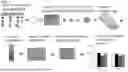


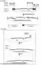


Sources:
- United States Patent and Trademark Office - verify current appl. status at the USPTO↗
Similar patent applications:
Recent applications in this class:
- » 20250171800 2025-05-29
Materials and Methods for Treatment of Hemoglobinopathies - » 20250163455 2025-05-22
INTRON FRAGMENTS - » 20250163454 2025-05-22
IN VITRO AND IN VIVO PROTEIN TRANSLATION VIA IN SITU CIRCULARIZED RNAS - » 20250154525 2025-05-15
PLANT VIRUS MOVEMENT PROTEINS AND METHODS OF USING SAME - » 20250154524 2025-05-15
EUKARYOTIC CELLS COMPRISING ADENOVIRUS-ASSOCIATED VIRUS POLYNUCLEOTIDES - » 20250154523 2025-05-15
CELL-SPECIFIC EXPRESSION OF modRNA - » 20250146016 2025-05-08
NON-IMMUNOGENIC CIRCULAR, NON-VIRAL DNA VECTORS - » 20250146015 2025-05-08
NON-IMMUNOGENIC CIRCULAR, NON-VIRAL DNA VECTORS - » 20250146014 2025-05-08
PLASMID FOR PRODUCTION OF RECOMBINANT PROTEIN IN MAMMALIAN CELL - » 20250122526 2025-04-17
CHIMERIC REPRESSORS AND METHODS OF USING THE SAME
Recent applications for this Assignee:
- » 20250171794 2025-05-29
COMPOSITIONS AND METHODS FOR INCREASING PERIDERM IN PLANT ROOTS - » 20250163450 2025-05-22
IRON MAN (IMA) OVEREXPRESSION INCREASES DISEASE RESISTANCE - » 20250134941 2025-05-01
HSV-1 ONCOLYTIC VIRUS THERAPIES THAT SPECIFICALLY KILL ALT DEPENDENT CANCERS - » 20250122570 2025-04-17
EPIGENOME BIOMARKERS FOR IDENTIFYING ALZHEIMER'S DISEASE - » 20250101393 2025-03-27
RNA TARGETING METHODS AND COMPOSITIONS - » 20250082781 2025-03-13
METHODS OF LOWERING BLOOD GLUCOSE AND TREATING TYPE 2 DIABETES BY ACTIVATION OF PDE4D3 - » 20250049032 2025-02-13
ETHYLENE SIGNALING ACTIVATOR MODULATES ROOT SYSTEM ARCHITECTURE - » 20240409961 2024-12-12
METHODS AND COMPOSITIONS FOR GENOME EDITING - » 20240342231 2024-10-17
SELECTIVE CELL TARGETING USING ADENOVIRUS AND CHEMICAL DIMERS - » 20240216544 2024-07-04
METHODS AND COMPOSITIONS FOR EXPRESSION OF EDITING PROTEINS