GAG binding proteins
US20080112926A1
2008-05-15
11/422,169
2006-06-05
✅ Patent granted
US 7,585,937 B2
2009-09-08
-
-
Prema Mertz
2027-02-20
Abstract:
A method is provided for introducing a GAG binding site into a protein comprising the steps:
-
- identifying a region in a protein which is not essential for structure maintenance
- introducing at least one basic amino acid into said site and/or deleting at least one bulky and/or acidic amino acid in said site,
whereby said GAG binding site has a GAG binding affinity of Kd≦10 μM, preferably ≦1 μM, still preferred ≦0.1 μM, as well as modified GAG binding proteins.
Assignee:
- PROTAFFIN BIOTECHNOLOGIE AG 11 🇦🇹 Graz, Austria
Interested in similar patents?
Get notified when new applications in this technology area are published.
Classification:
C07K14/54 IPC
Peptides having more than 20 amino acids; Gastrins; Somatostatins; Melanotropins; Derivatives thereof from animals; from humans; Cytokines; Lymphokines; Interferons Interleukins [IL]
C07K14/523 » CPC main
Peptides having more than 20 amino acids; Gastrins; Somatostatins; Melanotropins; Derivatives thereof from animals; from humans; Cytokines; Lymphokines; Interferons; Chemokines Beta-chemokines, e.g. RANTES, I-309/TCA-3, MIP-1alpha, MIP-1beta/ACT-2/LD78/SCIF, MCP-1/MCAF, MCP-2, MCP-3, LDCF-1, LDCF-2
A61P11/06 » CPC further
Drugs for disorders of the respiratory system Antiasthmatics
A61P17/06 » CPC further
Drugs for dermatological disorders Antipsoriatics
A61P19/02 » CPC further
Drugs for skeletal disorders for joint disorders, e.g. arthritis, arthrosis
A61P19/10 » CPC further
Drugs for skeletal disorders for bone diseases, e.g. rachitism, Paget's disease for osteoporosis
A61P25/00 » CPC further
Drugs for disorders of the nervous system
A61P25/28 » CPC further
Drugs for disorders of the nervous system for treating neurodegenerative disorders of the central nervous system, e.g. nootropic agents, cognition enhancers, drugs for treating Alzheimer's disease or other forms of dementia
A61P37/08 » CPC further
Drugs for immunological or allergic disorders Antiallergic agents
C07K14/5421 » CPC further
Peptides having more than 20 amino acids; Gastrins; Somatostatins; Melanotropins; Derivatives thereof from animals; from humans; Cytokines; Lymphokines; Interferons; Interleukins [IL] IL-8
A61K31/70 IPC
Medicinal preparations containing organic active ingredients Carbohydrates; Sugars; Derivatives thereof
A61K38/00 » CPC further
Medicinal preparations containing peptides
A61K38/21 IPC
Medicinal preparations containing peptides; Peptides having more than 20 amino acids; Gastrins; Somatostatins; Melanotropins; Derivatives thereof from animals; from humans; Cytokines; Lymphokines; Interferons Interferons [IFN]
A61P43/00 » CPC further
Drugs for specific purposes, not provided for in groups -
C07K14/00 IPC
Peptides having more than 20 amino acids; Gastrins; Somatostatins; Melanotropins; Derivatives thereof
C07K7/00 IPC
Peptides having 5 to 20 amino acids in a fully defined sequence; Derivatives thereof
C12N15/00 IPC
Mutation or genetic engineering; DNA or RNA concerning genetic engineering, vectors, e.g. plasmids, or their isolation, preparation or purification; Use of hosts therefor
C12N15/11 IPC
Mutation or genetic engineering; DNA or RNA concerning genetic engineering, vectors, e.g. plasmids, or their isolation, preparation or purification; Use of hosts therefor; Recombinant DNA-technology DNA or RNA fragments; Modified forms thereof
C12N5/06 IPC
Undifferentiated human, animal or plant cells, e.g. cell lines; Tissues; Cultivation or maintenance thereof; Culture media therefor Animal cells or tissues; Human cells or tissues
Description
This application is a national stage of PCT/EP2004/013670 filed on Dec. 2, 2004. The entire contents of the above-identified application are hereby incorporated by reference.
The present invention relates to methods and tools for the inhibition of the interaction of chemokines and their high-affinity receptors on leukocytes and methods for the therapeutic treatment of inflammatory diseases.
Chemokines, originally derived from chemoattractant cytokines, actually comprise more than 50 members and represent a family of small, inducible, and secreted proteins of low molecular weight (6-12 kDa in their monomeric form) that play a decisive role during immunosurveillance and inflammatory processes. Depending on their function in immunity and inflammation, they can be distinguished into two classes. Inflammatory chemokines are produced by many different tissue cells as well as by immigrating leukocytes in response to bacterial toxins and inflammatory cytokines like IL-1, TNF and interferons. Their main function is to recruit leukocytes for host defence and in the process of inflammation. Homing chemokines, on the other hand, are expressed constitutively in defined areas of the lymphoid tissues. They direct the traffic and homing of lymphocytes and dendritic cells within the immune system. These chemokines, as illustrated by BCA-1, SDF-1 or SLC, control the relocation and recirculation of lymphocytes in the context of maturation, differentiation, activation and ensure their correct homing within secondary lymphoid organs.
Despite the large number of representatives, chemokines show remarkably similar structural folds although the sequence homology varies between 20 to 70 percent. Chemokines consist of roughly 70-130 amino acids with four conserved cysteine residues. The cysteines form two disulphide bonds (Cys 1→Cys 3, Cys 2→Cys 4) which are responsible for their characteristic three-dimensional structure. Chemotactic cytokines consist of a short amino terminal domain (3-10 amino acids) preceding the first cysteine residue, a core made of β-strands and connecting loops found between the second and the fourth cysteine residue, as well as a carboxy-terminal α-helix of 20-60 amino acids. The protein core has a well ordered structure whereas the N- and C-terminal parts are disordered. As secretory proteins they are synthesised with a leader sequence of 20-25 amino acids which is cleaved off before release.
The chemokines have been subdivided into four families on the basis of the relative position of their cysteine residues in the mature protein. In the α-chemokine subfamily, the first two of the four cysteines are separated by a single amino acid (CXC), whereas in the β-chemokines the corresponding cysteine residues are adjacent to each other (CC). The α-chemokines can be further classified into those that contain the ELR sequence in the N-terminus, thereby being chemotactic for neutrophils (IL-8 for example), and those that lack the ELR motif and act on lymphocytes (1-TAC for example). Structurally the β-chemokines can be subdivided into two families: the monocyte-chemoattractant protein eotaxin family, containing the five monocyte chemoattractant proteins (MCP) and eotaxin which are approximately 65 percent identical to each other, and the remaining β-chemokines. As with the CXC-family, the N-terminal amino acids preceding the CC-residues are critical components for the biologic activity and leukocyte selectivity of the chemokines. The β-chemokines, in general, do not act on neutrophils but attract monocytes, eosinophils, basophils and lymphocytes with variable selectivity.
Only a few chemokines do not fit into the CC- or the CXC-family. Lymphotactin is so far the only chemokine which shows just two instead of the four characteristic cysteines in its primary structure, and is thus classified as γ- or C-chemokine. On the other hand, by concluding this classification, fractalkine has to be mentioned as the only representative of the δ- or CXXXC-subfamily with three amino acids separating the first two cysteines. Both of them, Lymphotaxin and fractalkine, induce chemotaxis of T-cells and natural killer cells.
Chemokines induce cell migration and activation by binding to specific cell surface, seven transmembrane-spanning (7TM) G-protein-coupled receptors on target cells. Eighteen chemokine receptors have been cloned so far including six CXC, ten CC, one CX3C and one XC receptor. Chemokine receptors are expressed on different types of leukocytes, some of them are restricted to certain cells (e.g. CXCR1 is restricted to neutrophils) whereas others are more widely expressed (e.g. CCR2 is expressed on monocytes, T cells, natural killer cells and basophils). Similar to chemokines, the receptors can be constitutively expressed on certain cells, whereas some are inducible. Some of them can even be down-regulated making the cells unresponsive to a certain chemokine but remaining responsive to another. Most receptors recognise more than one chemokine and vice versa but recognition is restricted to chemokines of the corresponding subfamily (see Table 1).
| TABLE 1 | |||
| Inflammatory | |||
| Chemokine | Receptor | Chemotactic for | Diseases |
| CXC- | IL-8 | CXCR1 | Neutrophils | Acute respiratory distress |
| Chemokine | CXCR2 | syndrome [71]; | ||
| (+ELR-motif) | Bacterial pneumonia [72]; | |||
| Rheumathoid | ||||
| arthritis [73]; | ||||
| Inflammatory bowel | ||||
| disease [74]; | ||||
| Psoriasis [75]; | ||||
| Bacterial meninegitis [76] | ||||
| CC- | MCP-1 | CCR2 | Basophils; Monocytes; | Asthma [77]; |
| Chemokine | Activated T cells; | Glomerulonephritis [78]; | ||
| Dentritic cells; Natural | Atheroscleosis [79]; | |||
| killer cells | Inflammatory bowel | |||
| disease [80]; | ||||
| Psoriasis [81]; | ||||
| Bacterial and viral | ||||
| meningitis [82, 83] | ||||
| RANTES | CCR1 | Eosinophils; Monocytes; | Asthma [84]; | |
| Activated T cells; | Glomerulonephritis [85] | |||
| Dentritic cells | ||||
| CCR3 | Eosinophils; Basophils; | |||
| Dentritic cells | ||||
| CCR5 | Monocytes; Activated T | |||
| cells; Dentritic cells; | ||||
| Natural killer cells | ||||
Chemokines have two main sites of interaction with their receptors, one in the amino-terminal domain and the other within an exposed loop of the backbone that extends between the second and the third cysteine residue. Both sites are kept in close proximity by the disulphide bonds. The receptor recognises first the binding site within the loop region which appears to function as a docking domain. This interaction restricts the mobility of the chemokine thus facilitating the proper orientation of the amino-terminal domain. Studies have been performed with mutant chemokines that still bound effectively to their receptors but did not signal. These mutants were obtained by amino acid deletion or modification within the N-termini of, for example, IL-8, RANTES and MCP-1.
Multiple intracellular signalling pathways occur after receptor activation as a result of chemokine binding. Chemokines also interact with two types of nonsignalling molecules. One is the DARC receptor which is expressed on erythrocytes and on endothelial cells and which binds CC- as well as CXC-chemokines to prevent them from circulation. The second type are heparan sulphate glycosaminoglycans (GAGs) which are part of proteoglycans and which serve as co-receptors of chemokines. They capture and present chemokines on the surface of the homing tissue (e.g. endothelial cells) in order to establish a local concentration gradient. In an inflammatory response, such as in rheumatoid arthritis, leukocytes rolling on the endothelium in a selectin-mediated process are brought into contact with the chemokines presented by the proteoglycans on the cell surface. Thereby, leukocyte integrins become activated which leads to firm adherence and extravasation. The recruited leukocytes are activated by local inflammatory cytokines and may become desensitised to further chemokine signalling because of high local concentration of chemokines. For maintaining a tissue bloodstream chemokine gradient, the DARC receptor functions as a sink for surplus chemokines.
Heparan sulphate (HS) proteoglycans, which consist of a core protein with covalently attached glycosaminoglycan sidechains (GAGs), are found in most mammalian cells and tissues. While the protein part determines the localisation of the proteoglycan in the cell membrane or in the extracellular matrix, the glycosaminoglycan component mediates interactions with a variety of extracellular ligands, such as growth factors, chemokines and adhesions molecules. The biosynthesis of proteoglycans has previously been extensively reviewed. Major groups of the cell surface proteoglycans are the syndecan family of transmembrane proteins (four members in mammals) and the glypican family of proteins attached to the cell membrane by a glycosylphosphatidylinositol (GPI) tail (six members in mammals). While glypicans are expressed widely in the nervous system, in kidney and, to a lesser extent, in skeletal and smooth muscle, syndecan-1 is the major HSPG in epithelial cells, syndecan-2 predominates in fibroblasts and endothelial cells, syndecan-3 abounds in neuronal cells and syndecan-4 is widely expressed. The majority of the GAG chains added to the syndecan core proteins through a tetrasaccharide linkage region onto particular serines are HS chains. Although the amino acid sequences of the extracellular domains of specific syndecan types are not conserved among different species, contrary to the transmembrane and the cytoplasmic domains, the number and the positions of the GAG chains are highly conserved. The structure of the GAGs, however, is species-specific and is, moreover, dependent upon the nature of the HSPG-expressing tissue.
Heparan sulphate (HS) is the most abundant member of the glycosaminoglycan (GAG) family of linear polysaccharides which also includes heparin, chondroitin sulphate, dermatan sulphate and keratan sulphate. Naturally occurring HS is characterised by a linear chain of 20-100 disaccharide units composed of N-acetyl-D-glucosamine (GlcNAc) and D-glucuronic acid (GlcA) which can be modified to include N- and O-sulphation (6-O and 3-O sulphation of the glucosamine and 2-O sulphation of the uronic acid) as well as epimerisation of β-D-gluronic acid to α-L-iduronic acid (IdoA).
Clusters of N- and O-sulphated sugar residues, separated by regions of low sulphation, are assumed to be mainly responsible for the numerous protein binding and regulatory properties of HS. In addition to the electrostatic interactions of the HS sulphate groups with basic amino acids, van der Waals and hydrophobic interactions are also thought to be involved in protein binding. Furthermore, the presence of the conformationally flexible iduronate residues seems to favour GAG binding to proteins. Other factors such as the spacing between the protein binding sites play also a critical role in protein-GAG binding interactions: For example γ-interferon dimerisation induced by HS requires GAG chains with two protein binding sequences separated by a 7 kDa region with low sulphation. Additional sequences are sometimes required for full biological activity of some ligands: in order to support FGF-2 signal transduction, HS must have both the minimum binding sequence as well as additional residues that are supposed to interact with the FGF receptor.
Heparin binding proteins often contain consensus sequences consisting of clusters of basic amino acid residues. Lysine, arginine, asparagine, histidine and glutamine are frequently involved in electrostatic contacts with the sulphate and carboxyl groups on the GAG. The spacing of the basic amino acids, sometimes determined by the proteins 3-D structure, are assumed to control the GAG binding specificity and affinity. The biological activity of the ligand can also be affected by the kinetics of HS-protein interaction. Reducing the dimension of growth factor diffusion is one of the suggested HSPG functions for which the long repetitive character of the GAG chains as well as their relatively fast on and off rates of protein binding are ideally suited. In some cases, kinetics rather than thermodynamics drives the physiological function of HS-protein binding. Most HS ligands require GAG sequences of well-defined length and structure. Heparin, which is produced by mast cells, is structurally very similar to heparan sulphate but is characterised by higher levels of post-polymerisation modifications resulting in a uniformly high degree of sulphation with a relatively small degree of structural diversity. Thus, the highly modified blocks in heparan sulphate are sometimes referred to as “heparin-like”. For this reason, heparin can be used as a perfect HS analogue for protein biophysical studies as it is, in addition, available in larger quantities and therefore less expensive than HS. Different cell types have been shown to synthesise proteoglycans with different glycosaminoglycan structure which changes during pathogenesis, during development or in response to extracellular signals such as growth factors. This structural diversity of HSPGs leads to a high binding versatility emphasising the great importance of proteoglycans.
Since the demonstration that heparan sulphate proteoglycans are critical for FGF signalling, several investigations were performed showing the importance of chemokine-GAG binding for promoting chemokine activity. First, almost all chemokines studied to date appear to bind HS in vitro, suggesting that this represents a fundamental property of these proteins. Second, the finding that in vivo T lymphocytes secrete CC-chemokines as a complex with glycosaminoglycans indicates that this form of interaction is physiologically relevant. Furthermore, it is known that the association of chemokines with HS helps to stabilise concentration gradients across the endothelial surface thereby providing directional information for migrating leukocytes. HS is also thought to protect chemokines from proteolytic degradation and to induce their oligomerisation thus promoting local high concentrations in the vicinity of the G-coupled signalling receptors. The functional relevance of oligomerisation, however, remains controversial although all chemokines have a clear structural basis for multimerisation. Dimerisation through association of the β-sheets is observed for all chemokines of the CXC-family (e.g. IL-8), contrary to most members of the CC-chemokine family (e.g. RANTES), which dimerise via their N-terminal strands.
A wealth of data has been accumulated on the inhibition of the interaction of chemokines and their high-affinity receptors on leukocytes by low molecular weight compounds. However, there has been no breakthrough in the therapeutic treatment of inflammatory diseases by this approach.
Interleukin-8 (IL-8) is a key molecule involved in neutrophil attraction during chronic and acute inflammation. Several approaches have been undertaken to block the action of IL-8 so far, beginning with inhibition of IL-8 production by for example glucocorticoids, Vitamin D3, cyclosporin A, transforming growth factor β, interferons etc., all of them inhibiting IL-8 activity at the level of production of IL-8 mRNA. A further approach previously used is to inhibit the binding of IL-8 to its receptors by using specific antibodies either against the receptor on the leukocyte or against IL-8 itself in order to act as specific antagonists and therefore inhibiting the IL-8 activity.
The aim of the present invention is therefore to provide an alternative strategy for the inhibition or disturbance of the interaction of chemokines/receptors on leukocytes. Specifically the action of IL-8, RANTES or MCP-1 should be targetted by such a strategy.
Subject matter of the present invention is therefore a method to produce new GAG binding proteins as well as alternative GAG binding proteins which show a high(er) affinity to a GAG co-receptor (than the wild type). Such modified GAG binding proteins can act as competitors with wild-type GAG binding proteins and are able to inhibit or down-regulate the activity of the wild-type GAG binding protein, however without the side effects which occur with the known recombinant proteins used in the state of the art. The molecules according to the present invention do not show the above mentioned disadvantages. The present modified GAG binding proteins can be used in drugs for various therapeutical uses, in particular—in the case of chemokines—for the treatment of inflammation diseases without the known disadvantages which occur in recombinant chemokines known in the state of the art. The modification of the GAG binding site according to the present invention turned out to be a broadly applicable strategy for all proteins which activity is based on the binding event to this site, especially chemokines with a GAG site. The preferred molecules according to the present invention with a higher GAG binding affinity proved to be specifically advantageous with respect to their biological effects, especially with respect to their anti-inflammatory activity by their competition with wild type molecules for the GAG site.
Therefore, the present invention provides a method for introducing a GAG binding site into a protein characterised in that it comprises the steps:
-
- identifying a region in a protein which is not essential for structure maintenance
- introducing at least one basic amino acid into said site and/or deleting at least one bulky and/or acidic amino acid in said site,
whereby said GAG binding site has a GAG binding affinity of Kd≦10 μM, preferably ≦1 μM, still preferred ≦0.1 μM. By introducing at least one basic amino acid and/or deleting at least one bulky and/or acidic amino acid in said region, a novel, improved “artificial” GAG binding site is introduced in said protein. This comprises the new, complete introduction of a GAG binding site into a protein which did not show a GAG binding activity before said modification. This also comprises the introduction of a GAG binding site into a protein which already showed GAG binding activity. The new GAG binding site can be introduced into a region of the protein which did not show GAG binding affinity as well as a region which did show GAG binding affinity. However, with the most preferred embodiment of the present invention, a modification of the GAG binding affinity of a given GAG binding protein is provided, said modified protein's GAG binding ability is increased compared to the wild-type protein. The present invention relates to a method of introducing a GAG binding site into a protein, a modified GAG binding protein as well as to an isolated DNA molecule, a vector, a recombinant cell, a pharmaceutical composition and the use of said modified protein.
BRIEF DESCRIPTION OF THE DRAWINGS
FIG. 1 shows a CD spectra.
FIG. 2 shows secondary structure contents of various mutants.
FIG. 3 shows graphics of results from fluorescence anisotropy tests of various mutants.
Figure shows graphics of results from fluorescence anisotropy tests of two mutants.
Figure shows the graphic of results from isothermal fluorescence titrations.
Figure shows the graphic of results from unfolding experiments of various mutants.
Figure shows chemotaxis index of IL-8 mutants.
Figure shows the results of the RANTES chemotaxis assay.”
The term “introducing at least one basic amino acid” relates to the introduction of additional amino acids as well as the substitution of amino acids. The main purpose is to increase the relative amount of basic amino acids, preferably Arg, Lys, H is, Asn and/or Gln, compared to the total amount of amino acids in said site, whereby the resulting GAG binding site should preferably comprise at least 3 basic amino acids, still preferred 4, most preferred 5 amino acids.
The GAG binding site is preferably at a solvent exposed position, e.g. at a loop. This will assure an effective modification.
Whether or not a region of a protein is essential for structure maintenance, can be tested for example by computational methods with specific programmes known to the person skilled in the art. After modification of the protein, the conformational stability is preferably tested in silico.
The term “bulky amino acid” refers to amino acids with long or sterically interfering side chains; these are in particular Trp, Ile, Leu, Phe, Tyr. Acidic amino acids are in particular Glu and Asp. Preferably, the resulting GAG binding site is free of bulky and acidic amino acids, meaning that all bulky and acidic amino acids are removed.
The GAG binding affinity is determined—for the scope of protection of the present application—over the dissociation constant Kd. One possibility is to determine the dissociation constant (Kd) values of any given protein by the structural change in ligand binding. Various techniques are well known to the person skilled in the art, e.g. isothermal fluorescence titrations, isothermal titration calorimetry, surface plasmon resonance, gel mobility assay, and indirectly by competition experiments with radioactively labelled GAG ligands. A further possibility is to predict binding regions by calculation with computational methods also known to the person skilled in the art, whereby several programmes may be used.
A protocol for introducing a GAG binding site into a protein is for example as follows:
-
- Identify a region of the protein which is not essential for overall structural maintenance and which might be suitable for GAG binding
- Design a new GAG binding site by introducing (replacement or insertion) basic Arg, Lys, His, Asp and Gln residues at any position or by deleting amino acids which interfere with GAG binding
- Check the conformational stability of the resulting mutant protein in silico
- Clone the wild-type protein cDNA (alternatively: purchase the cDNA)
- Use this as template for PCR-assisted mutagenesis to introduce the above mentioned changes into the amino acid sequence
- Subclone the mutant gene into a suitable expression system (prokaryotic or eukaryotic dependent upon biologically required post-translational modifications)
- Expression, purification and characterisation of the mutant protein in vitro
- Criterion for an introduced GAG binding affinity: KdGAG(mutant)<10 μM.
Examples of said engineered proteins with new GAG binding sites are for example the Fc part of IgG as well as the complement factors C3 and C4 modified as follows:
| Fc: (439)KSLSLS(444)-> KSKKLS | (SEQ ID NOS 1 & 2) | |
| C3: (1297)WIASHT(1302)-> WKAKHK | (SEQ ID NOS 3 & 4) | |
| C4: (1)MLDAERLK(8)-> MKKAKRLK | (SEQ ID NOS 5 & 6) |
A further aspect of the present invention is a protein obtainable by the inventive method as described above. The inventive protein therefore comprises a—compared to the wild-type protein—new GAG binding site as defined above and will therefore act as competitor with natural GAG binding proteins, in particular since the GAG binding affinity of the inventive protein is very high, e.g. Kd≦10 μM.
A further aspect of the present invention is a modified GAG binding protein, whereby a GAG binding region in said protein is modified by substitution, insertion, and/or deletion of at least one amino acid in order to increase the relative amount of basic amino acids in said GAG binding region, and/or reduce the amount of bulky and/or acidic amino acids in said GAG binding region, preferably at a solvent exposed position, and in that the GAG binding affinity of said protein is increased compared to the GAG binding affinity of a respective wild-type protein.
It has been surprisingly shown that by increasing the relative amount of basic amino acids, in particular Arg, Lys, His, Asn and Gln, in the GAG binding region, the modified GAG binding protein shows increased GAG binding affinity compared to the wild-type proteins, in particular when the relative amount of basic amino acids is increased at a solvent exposed position, since a positively charged area on the protein surface has shown to enhance the binding affinity. Preferably, at least 3, still preferred 4, most preferred 5, basic amino acids are present in the GAG binding region.
The term “GAG binding protein” relates to any protein which binds to a GAG co-receptor. Whether or not a protein binds to a GAG co-receptor can be tested with the help of known protocols as mentioned above. Hileman et al. (BioEssays 20 (1998), 156-167) disclose consensus sites in glycosaminoglycan binding proteins. The information disclosed in this article is also useful as starting information for the present invention. The term “protein” makes clear that the molecules provided by the present invention are at least 80 amino acids in length. This is required for making them suitable candidates for the present anti-inflammation strategy. Smaller molecules interacting with a GAG binding site and being physiologically or pathologically relevant due to such an interaction are not known and therefore not relevant for the present invention. Preferably, the molecules according to the present invention are composed of at least 90, at least 100, at least 120, at least 150, at least 200, at least 300, at least 400 or at least 500 amino acid residues.
In the scope of the present application the term “GAG binding region” is defined as a region which binds to GAG with a dissociation constant (Kd-value) of under 100 μM, preferably under 50 μM, still preferred under 20 μM, as determined by isothermal fluorescence titration (see examples below).
Any modifications mentioned in the present application can be carried out with known biochemical methods, for example site-directed mutagenesis. It should also be noted that molecular cloning of GAG binding sites is, of course, prior art (see e.g. WO96/34965 A, WO 92/07935 A, Jayaraman et al. (FEBS Letters 482 (2000), 154-158), WO02/20715 A, Yang et al. (J. Cell. Biochem. 56 (1994), 455-468), wherein molecular shuffling or de novo syntesis of GAG regions are described; Butcher et al., (FEBS Letters 4009 (1997), 183-187) (relates to artificial peptides, not proteins); Jinno-Oue et al, (J. Virol. 75 (2001), 12439-12445)de novo synthesis)).
The GAG binding region can be modified by substitution, insertion and/or deletion. This means that a non-basic amino acid may be substituted by a basic amino acid, a basic amino acid may be inserted into the GAG binding region or a non-basic amino acid may be deleted. Furthermore, an amino acid which interferes with GAG binding, preferably all interfering amino acids binding is deleted. Such amino acids are in particular bulky amino acids as described above as well as acidic amino acids, for example Glu and Asp. Whether or not an amino acid interferes with GAG binding may be examined with for example mathematical or computational methods. The result of any of these modifications is that the relative amount of basic amino acids in said GAG binding region is increased, whereby “relative” refers to the amount of basic amino acids in said GAG binding region compared to the number of all amino acids in said GAG binding region. Furthermore, amino acids which interfere sterically or electrostatically with GAG binding are deleted.
Whether or not an amino acid is present in a solvent exposed position, can be determined for example with the help of the known three dimensional structure of the protein or with the help of computational methods as mentioned above.
Whether or not the GAG binding affinity of said modified protein is increased compared to the GAG binding affinity of the respective wild-type protein, can be determined as mentioned above with the help of, for example, fluorescence titration experiments which determine the dissociation constants. The criterion for improved GAG binding affinity will be Kd (mutant)<Kd (wild-type), preferably by at least 100%. Specifically improved modified proteins have—compared with wild-type Kd—a GAG binding affinity which is higher by a factor of minimum 5, preferably of minimum 10, still preferred of minimum 100. The increased GAG binding affinity will therefore preferably show a Kd of under 10 μM, preferred under 1 μM, still preferred under 0.1 μM.
By increasing the GAG binding affinity the modified protein will act as a specific antagonist and will compete with the wild-type GAG binding protein for the GAG binding.
Preferably, at least one basic amino acid selected from the group consisting of Arg, Lys, and His is inserted into said GAG binding region. These amino acids are easily inserted into said GAG binding region, whereby the term “inserted” relates to an insertion as such as well as substituting any non-basic amino acid with arginine, lysine or histidine. Of course, it is possible to insert more than one basic amino acid whereby the same basic amino acid may be inserted or also a combination of two or three of the above mentioned amino acids.
Still preferred, the protein is a chemokine, preferably IL-8, RANTES or MCP-1. Chemokines are known to have a site of interaction with co-receptor GAG whereby this chemokine binding is often a condition for further receptor activation as mentioned above. Since chemokines are often found in inflammatory diseases, it is of major interest to block the chemokine receptor activation. Such chemokines are preferably IL-8, RANTES or MCP-1, which are well characterised molecules and of which the GAG binding regions are well known (see, for example, Lortat-Jacob et al., PNAS 99 (3) (2002), 1229-1234). By increasing the amount of basic amino acids in the GAG binding region of these chemokines, their binding affinity is increased and therefore the wild-type chemokines will bind less frequently or not at all, depending on the concentration of the modified protein in respect to the concentration of the wild-type protein.
According to an advantageous aspect, said GAG binding region is a C terminal α-helix. A typical chemical monomer is organised around a triple stranded anti-parallel β-sheet overlaid by a C-terminal α-helix. It has been shown that this C-terminal α-helix in chemokines is to a major part involved in the GAG binding, so that modification in this C-terminal α-helix in order to increase the amount of basic amino acids results in a modified chemokine with an increased GAG binding affinity.
Advantageously, positions 17, 21, 70, and/or 71 in IL-8 are substituted by Arg, Lys, His, Asn and/or Gln. Here it is possible that only one of these aforementioned sites is modified. However, also more than one of these sites may be modified as well as all, whereby all modifications may be either Arg or Lys or His or Asn or Gln or a mixture of those. In IL-8 these positions have shown to highly increase the GAG binding affinity of IL-8 and therefore these positions are particularly suitable for modifications.
Preferably the increased binding affinity is an increased binding affinity to heparan sulphate and/or heparin. Heparan sulphate is the most abundant member of the GAG family of linear polysaccharides which also includes heparin. Heparin is structurally very similar to heparan sulphate characterised by higher levels of post-polymerisation modifications resulting in a uniformly high degree of sulphation with a relatively small degree of structural diversity. Therefore, the highly modified blocks in heparan sulphate are sometimes referred to as heparin-like and heparin can be used as a heparan sulphate analogue for protein biophysical studies. In any case, both, heparan sulphate and heparin are particularly suitable.
Still preferred, a further biologically active region is modified thereby inhibiting or down-regulating a further biological activity of said protein. This further biological activity is known for most GAG binding proteins, for example for chemokines. This will be the binding region to a receptor, for example to the 7TM receptor. The term “further” defines a biologically active region which is not the GAG binding region which, however, binds to other molecules, cells or receptors and/or activates them. By modifying this further biologically active region the further biological activity of this protein is inhibited or down-regulated and thereby a modified protein is provided which is a strong antagonist to the wild-type protein. This means that on the one hand the GAG binding affinity is higher than in the wild-type GAG binding protein, so that the modified protein will to a large extent bind to the GAG instead of the wild-type protein. On the other hand, the further activity of the wild-type protein which mainly occurs when the protein is bound to GAG, is inhibited or down-regulated, since the modified protein will not carry out this specific activity or carries out this activity to a lesser extent. With this modified protein an effective antagonist for wild-type GAG binding proteins is provided which does not show the side effects known from other recombinant proteins as described in the state of the art. This further biologically active region can for example be determined in vitro by receptor competition assays (using fluorescently labelled wt chemokines, calcium influx, and cell migration (performed on native leukocytes or on 7TM stably-transfected cell lines). Examples of such further biologically active regions are, in addition to further receptor binding sites (as in the growth factor family), enzymatic sites (as in hydrolases, lyases, sulfotransferases, N-deacetylases, and copolymerases), protein interaction sites (as in antithrombin III), and membrane binding domains (as in the herpes simplex virus gD protein). With this preferred embodiment of double-modified proteins therefore dominant (concerning GAG binding) negative (concerning receptor) mutants are provided which are specifically advantageous with respect to the objectives set for the present invention.
Still preferred, said further biologically active region is modified by deletion, insertion, and/or substitution, preferably with alanine, a sterically and/or electrostatically similar residue. It is, of course, possible to either delete or insert or substitute at least one amino acid in said further biologically active region. However, it is also possible to provide a combination of at least two of these modifications or all three of them. By substituting a given amino acid with alanine or a sterically/electronically similar residue—“similar” meaning similar to the amino acid being substituted—the modified protein is not or only to a lesser extent modified sterically/electrostatically. This is particularly advantageous, since other activities of the modified protein, in particular the affinity to the GAG binding region, are not changed.
Advantageously, said protein is a chemokine and said further biological activity is leukocyte activation. As mentioned above, chemokines are involved in leukocyte attraction during chronic and acute inflammation. Therefore, by inhibiting or down-regulating leukocyte activation inflammation is decreased or inhibited which makes this particular modified protein an important tool for studying, diagnosing and treating inflammatory diseases.
According to an advantageous aspect, said protein is IL-8 and said further biologically active region is located within the first 10 N-terminal amino acids. The first N-terminal amino acids are involved in leukocyte activation, whereby in particular Glu-4, Leu-5 and Arg-6 were identified to be essential for receptor binding and activation. Therefore, either these three or even all first 10 N-terminal amino acids can be substituted or deleted in order to inhibit or down-regulate the receptor binding and activation.
A further advantageous protein is an IL-8 mutant with the first 6 N-terminal amino acids deleted. As mentioned above, this mutant will not or to a lesser extent bind and activate leukocytes, so that it is particularly suitable for studying, diagnosing and treating inflammatory diseases.
Preferably, said protein is an IL-8 mutant selected from the group consisting of del6F17RE70KN71R, del6F17RE70RN71K and del6E70KN71K. These mutants have shown to be particularly advantageous, since the deletion of the first 6 N-terminal amino acids inhibits or down-regulates receptor binding and activation. Furthermore, the two phenylalanines in position 17 and 21 were found to make first contact with the receptor on its N-terminal extracellular domain to facilitate the later activation of the receptor. In order to prevent any neutrophil contact, these two amino acids 17 and 21 are exchanged, whereby they are exchanged to basic amino acids, since they are in close proximity to the GAG binding motif of the C-terminal α-helix as can be seen on a three dimensional model of a protein. By exchanging the position 17 and/or 21 to either arginine or lysine the GAG binding affinity is therefore increased.
A further aspect of the present invention is an isolated polynucleic acid molecule which codes for the inventive protein as described above. The polynucleic acid may be DNA or RNA. Thereby the modifications which lead to the inventive modified protein are carried out on DNA or RNA level. This inventive isolated polynucleic acid molecule is suitable for diagnostic methods as well as gene therapy and the production of inventive modified protein on a large scale.
Still preferred, the isolated polynucleic acid molecule hybridises to the above defined inventive polynucleic acid molecule under stringent conditions. Depending on the hybridisation conditions complementary duplexes form between the two DNA or RNA molecules, either by perfectly being matched or also comprising mismatched bases (see Sambrook et al., Molecular Cloning: A laboratory manual, 2nd ed., Cold Spring Harbor, N.Y. 1989). Probes greater in length than about 50 nucleotides may accommodate up to 25 to 30% mismatched bases. Smaller probes will accommodate fewer mismatches. The tendency of a target and probe to form duplexes containing mismatched base pairs is controlled by the stringency of the hybridisation conditions which itself is a function of factors, such as the concentration of salt or formamide in the hybridisation buffer, the temperature of the hybridisation and the post-hybridisation wash conditions. By applying well-known principles that occur in the formation of hybrid duplexes conditions having the desired stringency can be achieved by one skilled in the art by selecting from among a variety of hybridisation buffers, temperatures and wash conditions. Thus, conditions can be selected that permit the detection of either perfectly matched or partially mismatched hybrid duplexes. The melting temperature (Tm) of a duplex is useful for selecting appropriate hybridisation conditions. Stringent hybridisation conditions for polynucleotide molecules over 200 nucleotides in length typically involve hybridising at a temperature 15-25° C. below the melting temperature of the expected duplex. For oligonucleotide probes over 30 nucleotides which form less stable duplexes than longer probes, stringent hybridisation usually is achieved by hybridising at 5 to 10° C. below the Tm. The Tm of a nucleic acid duplex can be calculated using a formula based on the percent G+C contained in the nucleic acids and that takes chain lengths into account, such as the formula Tm=81.5-16.6 (log [Na+)])+0.41 (% G+C)−(600/N), where N=chain length.
A further aspect of the present invention relates to a vector which comprises an isolated DNA molecule according to the present invention as defined above. The vector comprises all regulatory elements necessary for efficient transfection as well as efficient expression of proteins. Such vectors are well known in the art and any suitable vector can be selected for this purpose.
A further aspect of the present application relates to a recombinant cell which is stably transfected with an inventive vector as described above. Such a recombinant cell as well as any therefrom descendant cell comprises the vector. Thereby a cell line is provided which expresses the modified protein either continuously or upon activation depending on the vector.
A further aspect of the present invention relates to a pharmaceutical composition which comprises a protein, a polynucleic acid or a vector according to the present invention as defined above and a pharmaceutically acceptable carrier. Of course, the pharmaceutical composition may further comprise additional substances which are usually present in pharmaceutical compositions, such as salts, buffers, emulgators, colouring agents, etc.
A further aspect of the present invention relates to the use of the modified protein, a polynucleic acid or a vector according to the present invention as defined above in a method for inhibiting or suppressing the biological activity of the respective wild-type protein. As mentioned above, the modified protein will act as an antagonist whereby the side effects which occur with known recombinant proteins will not occur with the inventive modified protein. In the case of chemokines this will be in particular the biological activity involved in inflammatory reactions.
Therefore, a further use of the modified protein, polynucleic acid or vector according to the present invention is in a method for producing a medicament for the treatment of an inflammatory condition. In particular, if the modified protein is a chemokine, it will act as antagonist without side effects and will be particularly suitable for the treatment of an inflammatory condition. Therefore, a further aspect of the present application is also a method for the treatment of an inflammatory condition, wherein a modified protein according to the present invention, the isolated polynucleic acid molecule or vector according to the present invention or a pharmaceutical composition according to the present invention is administered to a patient.
Preferably, the inflammatory condition is selected from a group comprising rheumatoid arthritis, psoriasis, osteoarthritis, asthma, Alzheimer's disease, and multiple sclerosis. Since the activation through chemokines can be inhibited with a modified protein according to the present invention, inflammatory reactions can be inhibited or down-regulated whereby the above mentioned inflammatory conditions can be prevented or treated.
The present invention is described in further detail with the help of the following examples and figures to which the invention is, however, not limited whereby FIG. 1 is a CD spectra; FIG. 2 shows secondary structure contents of various mutants; FIGS. 3 and 4 show graphics of results from fluorescence anisotropy tests of various mutants; FIG. 5 shows the graphic of results from isothermal fluorescence titrations; FIG. 6 shows the graphic of results from unfolding experiments of various mutants, FIG. 7 shows chemotaxis index of IL-8 mutants (SEQ ID NOS 1070-1074 are disclosed respectively in order of appearance), and FIG. 8 shows the results of the RANTES chemotaxis assay.
EXAMPLES
Example 1
Generation of Recombinant IL-8 Genes and Cloning of the Mutants
Polymerase chain reaction (PCR) technique was used to generate the desired cDNAs for the mutants by introducing the mutations using sense and antisense mutagenesis primers. A synthetic plasmid containing the cDNA for wtIL-8 was used as template, Clontech Advantage®2 Polymerase Mix applied as DNA polymerase and the PCR reaction performed using a Mastergradient Cycler of Eppendorf. The mutagenesis primers used are summarised in the table below beginning with
| the forward sequences (5′to 3′): | |
| (SEQ ID NO: 7) |
| CACC ATG TGT CAG TGT ATA AAG ACA TAC TCC | |
| (primer for the mutation Δ6) | |
| (SEQ ID NO: 8) |
| CACC ATG TGT CAG TGT ATA AAG ACA TAC TCC AAA CCT | |
| AGG CAC CCC AAA AGG ATA | |
| (primer for the mutation Δ6 F17R F21R) | |
| The reverse sequences are (5′ to 3′): |
| (SEQ ID NO: 9) |
| TTA TGA ATT CCT AGC CCT CTT | |
| (primer for the mutation E70R) | |
| (SEQ ID NO: 10) |
| TTA TGA ATT CTT AGC CCT CTT | |
| (primer for the mutation E70K) | |
| (SEQ ID NO: 11) |
| TTA TGA CTT CTC AGC CCT CTT | |
| (primer for the mutation N71K) | |
| (SEQ ID NO: 12) |
| TTA TGA CTT CTT AGC CCT CTT | |
| (primer for the mutation E70K N71K) | |
| (SEQ ID NO: 13) |
| TTA TGA CTT CCT AGC CCT CTT | |
| (primer for the mutation E70R N71K) | |
| (SEQ ID NO: 14) |
| TTA TGA CCT CTT AGC CCT CTT | |
| (primer for the mutation E70K N71R) | |
| (SEQ ID NO: 15) |
| TTA TGA CCT CCT AGC CCT CTT | |
| (primer for the mutation E70R N71R) |
The PCR products were purified, cloned into the pCR®T7/NT-TOPO®TA (Invitrogen) vector and transformed into TOP10F competent E. coli (Invitrogen). As a next step a confirmation of the sequence was carried out by double-stranded DNA sequencing using a ABI PRISM CEl Sequencer.
Example 2
Expression and Purification of the Recombinant Proteins
Once the sequences were confirmed, the constructs were transformed into calcium-competent BL21(DE3) E. coli for expression. Cells were grown under shaking in 1 l Lennox Broth (Sigma) containing 100 μg/ml Ampicillin at 37° C. until an OD600 of about 0.8 was reached. Induction of protein expression was accomplished by addition of isopropyl-β-D-thiogalactopyranoside (IPTG) to a final concentration of 1 mM. Four hours later the cells were harvested by centrifugation at 6000 g for 20 minutes. The cell pellet was then resuspended in a buffer containing 20 mM TRIS/HCl, S0 mM NaCl, pH 8, sonicated at 100 watts for 5×20 s and finally centrifuged again for 20 min at 10,000 g. Since the main fraction of the recombinant IL-8 proteins was found in inclusion bodies, denaturing conditions were chosen for further purification. So the cell pellet was resuspended in a buffer of 6M Gua/HCl and 50 mM MES, pH 6.5. The suspension was then stirred at 4° C. for 4 hours, followed by a dialysis step against 50 mM MES, pH 6.5. The resulting suspension was then centrifuged and filtered to be loaded on a strong cation exchange column (SP Sepharose® from Pharmacia Biotech). The elution was accomplished by a linear gradient from OM-LM NaCl in a 50 mM MES buffer, pH 6.5 over 60 minutes. After lyophilisation of the fractions containing the desired protein, a second purification step was carried out by reversed-phase HPLC using a C18 column. In this case a non-linear gradient from 10%-90% Acetonitril was chosen to elute the desired protein. Refolding of the denatured protein was finally accomplished by the same cation exchange column under the same conditions as described above.
The protein was then checked for purity and identity by silver stain analysis in the first case and Western Blot analysis, using a specific monoclonal antibody against wtIL-8, in the second. Refolding of the proteins was also confirmed by Circular Dichroism (CD) measurements.
Example 3
Biophysical Characterisation of the Mutants
3.1 Circular Dicroism Measurements and Analysis
In order to investigate secondary structure changes of the mutant protein in the presence and absence of heparan sulphate (HS), CD spectroscopy was carried out. Measurements were recorded on a Jasco J-710 spectropolarimeter over a range of 195-250 nm, and a cell of 0.1 cm path length was used. Spectra of the protein solutions with a concentration of 5 μM were recorded with a response time of 1 s, step resolution of 0.2 nm, speed of 50 nm/min, band width of 1 nm and a sensitivity of 20 mdeg. Three scans were averaged to yield smooth spectra. The protein spectra were then background-corrected relating to the CD-signal either of the buffer itself or buffer/HS. Secondary structure analysis of the protein in the presence and absence of HS was finally accomplished using the programme SELCON.
Since a great number of amino acids were changed in a number of novel combinations, it was tried to find out the dimension of the resulting secondary structure changes by circular dichroism methods.
Different structures were obtained depending on the mutations introduced. Except for one mutant expressed (Δ6 F17R F21R E70K N71R) which didn't show any structure, all mutants exhibited measurable α-helices, β-sheets and loops. Compared to IL-8 wt only one mutant (Δ6 E70R) showed nearly similar structure whereas the others differed mainly in their α-helix which ranged from 17.2% to 45.2% out of the total structure. Nevertheless, this fact suggests that the overall structure of IL-8 wt was maintained despite many changes within the proteins sequence. This could not have been previously predicted. Having already found that heparan sulphate oligosaccharides only, and not heparin, were able to affect IL-8 wt secondary structure, attention was focused on the effects induced by unfractionated heparan sulphate. All examined mutants showed structural changes upon HS binding which can be seen as evidence of complex formation.
To demonstrate the structural changes upon introduced mutations and heparan sulphate addition, some of the data obtained are summarised in the graphs above and below.
3.2 Fluorescence Measurements
For studying concentration and ligand dependent quaternary structure changes fluorescence spectroscopy was performed. Due to its high sensitivity, requiring only nanogram quantities of protein, fluorescence technique was the method of choice for carrying out the desired investigations. Measurements were undertaken using a Perkin-Elmer (Beaconsfield, England) LS50B fluorometer.
3.3 Fluorescence Anisotropy
By recording the concentration dependent fluorescence anisotropy of the chemokine resulting from the extrinsic chromophore bisANS it was aimed to find out the dimerisation constant of the mutants. Measurements were performed in PBS starting with high concentrations (up to 4 μM protein) followed by stepwise dilution. For each data point, the anisotropy signal (r) recorded at 507 nm was averaged over 60 sec.
IL-8 oligomerisation has been reported to relevantly influence the proteins GAG binding properties. Set at monomeric concentration, IL-8 bound size defined oligosaccharides 1000-fold tighter than at dimeric concentration. Therefore, the oligomerisation properties of IL-8 mutants were investigated by fluorescence anisotropy. Since the IL-8 intrinsic fluorophore (Trp57) was not sensitive enough for all of the mutants, the extrinsic fluorophore bis-ANS was used for these measurements. Again, as already noticed for the secondary structure, the mutant Δ6 E70R showed high resemblance also in the oligomerisation constant (koligo=350 nM) to IL-8 wt (koligo=379M). The mutant with the highest koligo(koligo=460 nM), which therefore dimerised worst, was Δ6 F17RF21R E70RN71K. Previous studies identified the β-sheets to be mainly involved in the dimerisation process, a fact, which correlates with the results for this mutants' secondary structure, which showed a very low share of β-sheet of only 11.4%. The mutant with the lowest koligo(koligo=147 nM), was found to be Δ6 F17RF21R E70K, which again showed the highest share of β-sheet structure (29.8%) of all mutants investigated. Also the impact of heparan sulphate addition was observed. As for IL-8 wt, where heparan sulphate caused a shift of the oligomerisation constant to much higher levels (koligo=1.075 μM), this was also found for the IL-8 mutants investigated. Δ6 F17RF21R E70K shifted from 0.147 μM to 1.162 μM, and the mutant Δ6 E70R from 0.350 μM to 1.505 μM in the presence of heparan sulphate. Some of the results obtained are demonstrated in FIGS. 3 and 4, whereby FIG. 3 shows the dependence of the fluorescence anisotropy of IL-8 mutants in PBS on the chemokine concentration and FIG. 4 shows the dependence of the fluorescence anisotropy of Δ6 F17RF21R E70K in PBS on the chemokine concentration in the presence (ten fold excess) and absence of HS ((pc=10 xy excess) protein concentration).
3.4 Isothermal Fluorescence Titration (IFT) Experiments
Dissociation constants (Kd values) are a measure for the binding affinity of a ligand to a protein and therefore concentration-dependent change in the fluorescence emission properties of the protein (fluorescence quenching) upon ligand binding was used for the determination of Kd. Since these mutants contain an intrinsic tryptophan chromophore which is located at or near the proposed GAG binding site and therefore should be sensitive to structural changes upon ligand binding, IFT experiments seemed to be suitable for this kind of investigation. Fluorescence intensity titration was performed in PBS using a protein concentration of 700 nM. The emission of the protein solution upon excitation at 282 nm was recorded over a range of 300-400 nm following the addition of an aliquot of the respective GAG ligand and an equilibration period of 60 sec.
Binding to unfractionated heparin and heparan sulphate was investigated. The mutants were set at dimeric concentration to assure sufficient sensitivity. A quenching of Trp57 fluorescence intensity upon GAG binding was registered within a range of 25-35%. Significant improvement of ligand binding was observed, especially for heparin binding. Δ6 F17RN71R E70K (Kd=14 nM) and Δ6 F17RF21R N71K (Kd=14.6 nM) showed 2600-fold better binding, and Δ6 E70K N71K (Kd=74 nM) 1760-fold better binding compared to IL-8 wt (Kd=37 μM). Good results were also obtained for heparan sulphate binding. For Δ6 F17RN71R E70K a Kd of 107 nM was found, for Δ6 F17RF21R N71K the Kd was 95 nM and the mutant Δ6 E70K N71K showed a Kd of 34 nM. As IL-8 wt binds with a Kd of 4.2 μM, the KdS found for the mutants represent an extraordinary improvement in binding, see FIG. 5.
3.5 Unfolding Experiments
In order to obtain information about the proteins stability and whether this stability would be changed upon GAG ligand binding, unfolding experiments were undertaken. As mentioned above fluorescence techniques are very sensitive for observing quaternary structure changes and therefore are also the method of choice to investigate thermal structural changes of the protein. Measurements were undertaken as described for the IFT in which not the ligand concentration was changed but the temperature. Protein structure was observed at a concentration of 0.7 μM from temperatures of 15-85° C. in the absence and the presence of a 10 fold excess of heparan sulphate or heparin.
The emission maximum of the proteins ranged from 340 nm to 357 nm, values which are typical for a solvent exposed tryptophan residue. Beginning with the unfolding experiments at 15° C., the emission maximum of the mutants varied between 340 nm-351 nm. Compared to IL-8 wt, whose emission maximum was observed at 340 nm, this means slightly higher values. Upon an increase in temperature, the intensity of emission maximum decreased, accompanied by a shift of the maximum to either a higher or lower wavelength. The emission maximum of Δ6 E70R and Δ6 E70K N71K shifted from 352.5 nm-357 nm and 343 nm-345 nm, which is typical for a further exposure of the Trp57 residue to the solvent trough temperature increase, but interestingly the mutants Δ6 F17RN71R E70K and Δ6 F17RF21R E70R N71K showed a blue shift, ranging from 350 nm-343 nm and, less pronounced, from 350 nm-348 nm (see FIG. 6). By slowly decreasing the temperature, the process of unfolding was partially reversible regarding both the wavelength shift and changes of intensity. Addition of a 5 fold excess of heparan sulphate led to an increase of stability of the proteins, probably through complex formation. This could be observed on the one hand by a shift of the melting point to higher temperature, and on the other hand by a significantly less pronounced shift of emission maximum upon temperature increase.
Example 4
Cell-Based Assay of the Receptor-“Negative” Function of the Dominant-Negative IL-8 Mutants
In order to characterise the impaired receptor function of the IL-8 mutants with respect to neutrophil attraction, transfilter-based chemotaxis of neutrophils in response to IL-8 mutants was assayed in a microchemotaxis chamber equipped with a 5 μm PVP-free polycarbonate membrane.
Cell Preparation:
Briefly, a neutrophil fraction was prepared from freshly collected human blood. This was done by adding a 6% dextran solution to heparin-treated blood (1:2) which was then left for sedimentation for 45 min. The upper clear cell solution was collected and washed twice with HBSS w/o Ca and Mg. Cells were counted and finally diluted with HBSS at 2 Mio/ml cell suspension, taking into account that only 60% of the counted cells were neutrophils.
Chemotaxis Assay:
IL-8 mutants were diluted at concentrations of 10 μg/ml, 1 μg/ml and 0.1 μg/ml and put in triplicates in the lower compartment of the chamber (26 μl per well). The freshly prepared neutrophils were seeded in the upper chamber (50 μl per well) and incubated for 30 minutes at 37° C. in a 5% CO2 humidified incubator. After incubation, the chamber was disassembled, the upper side of the filter was washed and wiped off and cells attached to the lower side were fixed with methanol and stained with Hemacolor solutions (Merck). Cells were then counted at 400× magnifications in 4 randomly selected microscopic fields per well. Finally, the mean of three independent experiments was plotted against the chemokine concentration. In FIG. 7, the chemotaxis index for various IL-8 mutants is shown. As expected, all mutants showed significantly decreased receptor binding activity.
Example 5
Generation of Recombinant Rantes Genes, Expression, Biophysical and Activity Characterisation of the Mutants
The concept of dominant-negative “GAG-masking” chemokine mutants was also employed to RANTES, a chemokine involved in type IV hypersensitivity reactions like transplant rejection, atopic dermatitis as well as in other inflammatory disorders like arthritis, progressive glomerulonephritis and inflammatory lung disease.
The receptor binding capability was impaired by introducing into the wt protein an initiating methionine residue. Expression of the wt RANTES in E. Coli lead to the retention of this methionine residue, which renders wt RANTES to a potent inhibitor of monocyte migration, the so-called Met-RANTES. Different mutations enhancing the GAG binding affinity were introduced via PCR-based site-directed mutagenesis methods.
By these means 9 RANTES mutants have so far been cloned, expressed and purified, Met-RANTES A22K, Met-RANTES H23K, Met-RANTES T43K, Met-RANTES N46R, Met-RANTES N46K, Met-RANTES Q48K, Met-RANTES A22K/N46R, Met-RANTES V49R/E66S and Met-RANTES 15LSLA18 V49R/E66S.
Isothermal fluorescence titration experiments were carried out to measure the relative affinity constants (Kd values) of the RANTES mutants for size defined heparin. As can be seen in the table all RANTES mutant proteins showed higher affinities for this heparin, with Met-RANTES A22K, Met-RANTES H23K, Met-RANTES T43K and Met-RANTES A22K/N46R showing the most promising results.
| Kd in nM | |
| Wt Rantes | 456.2 ± 8.5 | |
| Met-Rantes V49R/E66S | 345.5 ± 21.7 | |
| Rantes 15LSLA18 V49R/66S | 297.3 ± 14.1 | |
| Rantes N46R | 367.7 ± 11.7 | |
| Rantes N46K | 257.4 ± 10.2 | |
| Rantes H23K | 202.5 ± 12.8 | |
| Rantes Q48K | 383.4 ± 39.6 | |
| Rantes T43K | 139.2 ± 30.1 | |
| Rantes A22K | 202.1 ± 9.8 | |
| Rantes A22K/N46R | 164.0 ± 16.6 | |
RANTES Chemotaxis Assay
RANTES mutant directed cell migration was investigated using the 48-well Boyden chamber system equipped with 5 μm PVP-coated polycarbonate membranes. RANTES and RANTES mutant dilutions in RPMI 1640 containing 20 mM HEPES pH 7.3 and 1 mg/ml BSA were placed in triplicates in the lower wells of the chamber. 50 μl of THP-1 cell suspensions (promonocytic cell line from the European collection of cell cultures) in the same medium at 2×10⁶ cells/ml were placed in the upper wells. After a 2 h incubation period at 37° C. in 5% CO2 the upper surface of the filter was washed in HBSS solution. The migrated cells were fixed in methanol and stained with Hemacolor solution (Merck). Five 400× magnifications per well were counted and the mean of three independently conducted experiments was plotted against the chemokine concentration in FIG. 8. The error bars represent the standard error of the mean of the three experiments. Again, as in the case of the IL-8 mutants, all RANTES mutants showed significantly reduced receptor binding activity.
Example 6
Proteins with GAG Binding Regions
By bioinformatical and by proteomical means GAG binding proteins were characterised together with their GAG binding regions. In the following tables 2 and 3 chemokines are shown with their GAG binding regions (table 2) and examples of other proteins are given also with their GAG binding regions (table 3).
| TABLE 2 |
| Chemokines and their GAG binding domains |
| CXC - chemokines |
| IL-8: 18HPK20, (R47) 60RVVEKFLKR68 | |
| (residues 60-68 of SEQ ID NO: 16) | |
| (SEQ ID NO: 16) |
| SAKELRCQCIKTYSKPFHPKFIKELRVIESGPHCANTEIIVKLSDGRELC | |
| LDPKENWVQRVVEKFLKRAENS | |
| MGSA/GROα: 19HPK21, 45KNGR48 | |
| (residues 45-48 of SEQ ID NO: 17), | |
| 60KKIIEK66 | |
| (residues 60-66 of SEQ ID NO: 17) | |
| (SEQ ID NO: 17) |
| ASVATELRCQCLQTLQGIHPKNIQSVNVKSPGPHCAQTEVIATLKNGRKA | |
| CLNPASPIVKKIIEKMLNSDKSN | |
| MIP-2α/GROβ: 19HLK21, K45, 60KKIIEKMLK68 | |
| (residues 60-68 of SEQ ID NO: 18) | |
| (SEQ ID NO: 18) |
| APLATELRCQCLQTLQGIHLKNIQSVKVKSPGPHCAQTEVIATLKNGQKA | |
| CLNPASPMVKKIIEKMLKNGKSN | |
| NAP-2: 15HPK18, 42KDGR45 | |
| (residues 42-45 of SEQ ID NO: 19), | |
| 57KKIVQK62 | |
| (residues 57-62 of SEQ ID NO: 19) | |
| (SEQ ID NO: 19) |
| AELRCLCIKTTSGIHPKNIQSLEVIGKGTHCNQVEVIATLKDGRKICLDP | |
| DAPRIKKIVQKKLAGDESAD | |
| PF-4: 20RPRH23 | |
| (residues 20-23 of SEQ ID NO: 20), | |
| 46KNGR49 | |
| (residues 46-49 of SEQ ID NO: 20), | |
| 61KKIIKK66 | |
| (residues 61-66 of SEQ ID NO: 20) | |
| (SEQ ID NO: 20) |
| EAEEDGDLQCLCVKTTSQVRPRHITSLEVIKAGPHCPTAQLIATLKNGRK | |
| ICLDLQAPLYKKIIKKLLES | |
| SDF-1α: K1, 24KHLK27 | |
| (residues 24-27 of SEQ ID NO: 21), | |
| 41RLK43 | |
| (SEQ ID NO: 21) |
| KPVSLSYRCPCRFFESHVARANVKHLKILNTPNCALQIVARLKNNNRQVC | |
| IDPKLKWIQEYLEKALN | |
| CC - chemokines | |
| RANTES: (17RPLPRAH23 (residues 17-23 of SEQ ID | |
| NO: 22)) | |
| 44RKNR47 | |
| (residues 44-47 of SEQ ID NO: 22) | |
| (SEQ ID NO: 22) |
| SPYSSDTTPCCFAYIARPLPRAHIKEYFYTSGKCSNPAVFVTRKNRQVCA | |
| NPEKKWVREYINSLEMS | |
| MCP-2: 18RKIPIQR24 | |
| (residues 18-24 of SEQ ID NO: 23), | |
| 46KRGK49 | |
| (residues 46-49 of SEQ ID NO: 23) | |
| (SEQ ID NO: 23) |
| QPDSVSIPITCCFNVINRKIPIQRLESYTRITNIQCPKEAVIFKTKRGKE | |
| VCADPKERWVRDSMKHLDQIFQNLKP | |
| MCP-3: 22KQR24, 47KLDK50 | |
| (residues 47-50 of SEQ ID NO: 24), | |
| 66KHLDKK71 | |
| (residues 66-71 of SEQ ID NO: 24) | |
| (SEQ ID NO: 24) |
| QPVGINTSTTCCYRFINKKIPKQRLESYRRTTSSHCPREAVIFKTKLDKE | |
| ICADPTQKWVQDFMKHLDKKTQTPKL | |
| MIP-1α: R17, 44KRSR47 | |
| (residues 44-47 of SEQ ID NO: 25) | |
| (SEQ ID NO: 25) |
| SLAADTPTACCFSYTSRQIPQNFIADYFETSSQCSKPGVIFLTKRSRQVC | |
| ADPSEEWVQKYVSDLELSA | |
| MIP-1β: R18, 45KRSK48 | |
| (residues 45-48 of SEQ ID NO: 26) | |
| (SEQ ID NO: 26) |
| APMGSDPPTACCFSYTARKLPRNFVVDYYETSSLCSQPAVVFQTKRSKQV | |
| CADPSESWVQEYVYDLELN | |
| MPIF-1: R18, 45KKGR48 | |
| (residues 45-48 of SEQ ID NO: 27) | |
| (SEQ ID NO: 27) |
| MDRFHATSADCCISYTPRSIPCSLLESYFETNSECSKPGVIFLTKKGRRF | |
| CANPSDKQVQVCMRMLKLDTRIKTRKN | |
| MIP-5/HCC-2: 40KKGR43 | |
| (residues 40-43 of SEQ ID NO: 28) | |
| (SEQ ID NO: 28) |
| HFAADCCTSYISQSIPCSLMKSYFETSSECSKPGVIFLTKKGRQVCAKPS | |
| GPGVQDCMKKLKPYSI | |
| TABLE 3 | |||
| SEQ | |||
| ID | |||
| NO: | |||
| Peroxisome biogenesis | 29 | 181 TRRAKE 186 | |
| factor 1 | 30 | 367 QKKIRS 372 | |
| 31 | 1263 PKRRKN 1268 | ||
| 32 | 181 TRRAKE 186 | ||
| 33 | 367 QKKIRS 372 | ||
| 34 | 1263 PKRRKN 1268 | ||
| MLTK-beta | 35 | 415 SKRRGKKV 422 | |
| 36 | 312 ERRLKM 317 | ||
| 37 | 416 KRRGKK 421 | ||
| 38 | 312 ERRLKM 317 | ||
| 39 | 416 KRRGKK 421 | ||
| BHLH factor Hes4 | 40 | 43 EKRRRARI 50 | |
| 41 | 43 EKRRRA 48 | ||
| 42 | 43 EKRRRA 48 | ||
| Protocadherin 11 | 43 | 867 MKKKKKKK 874 | |
| 44 | 867 MKKKKK 872 | ||
| 45 | 867 MKKKKK 872 | ||
| 46 | 899 MKKKKKKK 906 | ||
| 47 | 899 MKKKKK 904 | ||
| 48 | 899 MKKKKK 904 | ||
| catenin (cadherin-associ- | 49 | 315 RRRLRS 320 | |
| ated protein) delta 1 | 50 | 404 VRKLKG 409 | |
| 51 | 460 LRKARD 465 | ||
| 52 | 545 RRKLRE 550 | ||
| 53 | 621 AKKGKG 626 | ||
| 54 | 787 AKKLRE 792 | ||
| 55 | 315 RRRLRS 320 | ||
| 56 | 404 VRKLKG 409 | ||
| 57 | 460 LRKARD 465 | ||
| 58 | 545 RRKLRE 550 | ||
| 59 | 621 AKKGKG 626 | ||
| 60 | 787 AKKLRE 792 | ||
| Muscarinic acetylcholine | 61 | 221 EKRTKD 226 | |
| receptor M5 | 62 | 427 TKRKRV 432 | |
| 63 | 514 WKKKKV 519 | ||
| 64 | 221 EKRTKD 226 | ||
| 65 | 427 TKRKRV 432 | ||
| 66 | 514 WKKKKV 519 | ||
| Alpha-2A adrenergic | 67 | 147 PRRIKA 152 | |
| receptor | 68 | 224 KRRTRV 229 | |
| 69 | 147 PRRIKA 152 | ||
| 70 | 224 KRRTRV 229 | ||
| IL-5 promoter REII-region- | 71 | 440 TKKKTRRR 447 | |
| binding protein | 72 | 569 GKRRRRRG 576 | |
| 73 | 38 ARKGKR 43 | ||
| 74 | 437 GKKTKK 442 | ||
| 75 | 444 TRRRRA 449 | ||
| 76 | 569 GKRRRR 574 | ||
| 77 | 38 ARKGKR 43 | ||
| 78 | 437 GKKTKK 442 | ||
| 79 | 444 TRRRRA 449 | ||
| 80 | 569 GKRRRR 574 | ||
| Mitofusin 1 | 81 | 291 ARKQKA 296 | |
| 82 | 395 KKKIKE 400 | ||
| 83 | 291 ARKQKA 296 | ||
| 84 | 395 KKKIKE 400 | ||
| N-cym protein | 85 | 71 VRRCKI 76 | |
| 86 | 71 VRRCKI 76 | ||
| Smad ubiquitination regula- | 87 | 672 ERRARL 677 | |
| tory factor 1 | 88 | 672 ERRARL 677 | |
| CUG-BP and ETR-3 like | 89 | 468 MKRLKV 473 | |
| factor 5 | 90 | 475 LKRPKD 480 | |
| 91 | 468 MKRLKV 473 | ||
| 92 | 475 LKRPKD 480 | ||
| Ewings sarcoma EWS-Fli1 | 93 | 347 QRKSKP 352 | |
| 94 | 347 QRKSKP 352 | ||
| NUF2R | 95 | 455 LKRKMFKM 462 | |
| 96 | 331 LKKLKT 336 | ||
| 97 | 347 VKKEKL 352 | ||
| 98 | 331 LKKLKT 336 | ||
| 99 | 347 VKKEKL 352 | ||
| Kruppel-like zinc finger | 100 | 22 EKRERT 27 | |
| protein GLIS2 | 101 | 22 EKRERT 27 | |
| FKSG32 | 102 | 15 LKRVRE 20 | |
| 103 | 431 VRRGRI 436 | ||
| 104 | 15 LKRVRE 20 | ||
| 105 | 431 VRRGRI 436 | ||
| BARH-LIKE 1 PROTEIN | 106 | 175 LKKPRK 180 | |
| 107 | 228 NRRTKW 233 | ||
| 108 | 175 LKKPRK 180 | ||
| 109 | 228 NRRTKW 233 | ||
| Nucleolar GTP-binding pro- | 110 | 393 SRKKRERD 400 | |
| tein 1 | 111 | 624 GKRKAGKK 631 | |
| 112 | 48 MRKVKF 53 | ||
| 113 | 141 IKRQKQ 146 | ||
| 114 | 383 ARRKRM 388 | ||
| 115 | 393 SRKKRE 398 | ||
| 116 | 490 KKKLKI 495 | ||
| 117 | 543 ARRSRS 548 | ||
| 118 | 550 TRKRKR 555 | ||
| 119 | 586 VKKAKT 591 | ||
| 120 | 629 GKKDRR 634 | ||
| 121 | 48 MRKVKF 53 | ||
| 122 | 141 IKRQKQ 146 | ||
| 123 | 383 ARRKRM 388 | ||
| 124 | 393 SRKKRE 398 | ||
| 125 | 490 KKKLKI 495 | ||
| 126 | 543 ARRSRS 548 | ||
| 127 | 550 TRKRKR 555 | ||
| 128 | 586 VKKAKT 591 | ||
| 129 | 629 GKKDRR 634 | ||
| EVG1 | 130 | 17 RRRPKT 22 | |
| 131 | 138 ERKRKA 143 | ||
| 132 | 17 RRRPKT 22 | ||
| 133 | 138 ERKRKA 143 | ||
| ASPL | 134 | 282 PKKSKS 287 | |
| 135 | 282 PKKSKS 287 | ||
| Zinc transporter 1 | 136 | 477 EKKPRR 482 | |
| 137 | 477 EKKPRR 482 | ||
| Uveal autoantigen | 138 | 603 EKKGRK 608 | |
| 139 | 995 ERKFKA 1000 | ||
| 140 | 1023 VKKNKQ 1028 | ||
| 141 | 603 EKKGRK 608 | ||
| 142 | 995 ERKFKA 1000 | ||
| 143 | 1023 VKKNKQ 1028 | ||
| RAB39 | 144 | 7 VRRDRV 12 | |
| 145 | 7 VRRDRV 12 | ||
| Down syndrome cell adhesion | 146 | 320 PRKVKS 325 | |
| molecule | 147 | 387 VRKDKL 392 | |
| 148 | 320 PRKVKS 325 | ||
| 149 | 387 VRKDKL 392 | ||
| Protein-tyrosine phospha- | 150 | 139 GRKKCERY 146 | |
| tase, non-receptor type 12 | 151 | 59 VKKNRY 64 | |
| 152 | 59 VKKNRY 64 | ||
| WD-repeat protein 11 | 153 | 752 VRKIRF 757 | |
| 154 | 752 VRKIRF 757 | ||
| Gastric cancer-related pro- | 155 | 20 SRKRQTRR 27 | |
| tein VRG107 | 156 | 25 TRRRRN 30 | |
| 157 | 25 TRRRRN 30 | ||
| Early growth response pro- | 158 | 356 ARRKGRRG 363 | |
| tein 4 | 159 | 452 EKKRHSKV 459 | |
| 160 | 357 RRKGRR 362 | ||
| 161 | 357 RRKGRR 362 | ||
| Vesicle transport-related | 162 | 309 PKRKNKKS 316 | |
| protein | 163 | 226 DKKLRE 231 | |
| 164 | 310 KRKNKK 315 | ||
| 165 | 355 VKRLKS 360 | ||
| 166 | 226 DKKLRE 231 | ||
| 167 | 310 KRKNKK 315 | ||
| 168 | 355 VKRLKS 360 | ||
| UPF3X | 169 | 140 AKKKTKKR 147 | |
| 170 | 141 KKKTKK 146 | ||
| 171 | 217 ERRRRE 222 | ||
| 172 | 225 RKRQRE 230 | ||
| 173 | 233 RRKWKE 238 | ||
| 174 | 240 EKRKRK 245 | ||
| 175 | 296 DKREKA 301 | ||
| 176 | 373 RRRQKE 378 | ||
| 177 | 393 MKKEKD 398 | ||
| 178 | 426 VKRDRI 431 | ||
| 179 | 140 AKKKTKKRD 148 | ||
| 180 | 141 KKKTKK 146 | ||
| 181 | 217 ERRRRE 222 | ||
| 182 | 225 RKRQRE 230 | ||
| 183 | 233 RRKWKE 238 | ||
| 184 | 240 EKRKRK 245 | ||
| 185 | 296 DKREKA 301 | ||
| 186 | 373 RRRQKE 378 | ||
| 187 | 393 MKKEKD 398 | ||
| 188 | 426 VKRDRI 431 | ||
| CGI-201 protein, type IV | 189 | 49 ARRTRS 54 | |
| 190 | 49 ARRTRS 54 | ||
| RING finger protein 23 | 191 | 98 KRKIRD 103 | |
| 192 | 98 KRKIRD 103 | ||
| FKSG17 | 193 | 72 EKKARK 77 | |
| 194 | 95 IRKSKN 100 | ||
| 195 | 72 EKKARK 77 | ||
| 196 | 95 IRKSKN 100 | ||
| P83 | 197 | 681 ARKERE 686 | |
| 198 | 681 ARKERE 686 | ||
| Ovarian cancer-related pro- | 199 | 62 LKRDRF 67 | |
| tein 1 | 200 | 62 LKRDRF 67 | |
| MHC class II transactivator | 201 | 407 HRRPRE 412 | |
| CIITA | 202 | 741 PRKKRP 746 | |
| 203 | 783 DRKQKV 788 | ||
| 204 | 407 HRRPRE 412 | ||
| 205 | 741 PRKKRP 746 | ||
| 206 | 783 DRKQKV 788 | ||
| Platelet glycoprotein VI-2 | 207 | 275 SRRKRLRH 282 | |
| 208 | 275 SRRKRL 280 | ||
| 209 | 275 SRRKRL 280 | ||
| Ubiquitin-like 5 protein | 210 | 11 GKKVRV 16 | |
| 211 | 11 GKKVRV 16 | ||
| Protein kinase D2 | 212 | 191 ARKRRL 196 | |
| 213 | 191 ARKRRL 196 | ||
| Homeobox protein GSH-2 | 214 | 202 GKRMRT 207 | |
| 215 | 252 NRRVKH 257 | ||
| 216 | 202 GKRMRT 207 | ||
| 217 | 252 NRRVKH 257 | ||
| ULBP3 protein | 218 | 166 ARRMKE 171 | |
| 219 | 201 HRKKRL 206 | ||
| 220 | 166 ARRMKE 171 | ||
| 221 | 201 HRKKRL 206 | ||
| Type II iodothyronine | 222 | 87 SKKEKV 92 | |
| deiodinase | 223 | 87 SKKEKV 92 | |
| 224 | 299 SKRCKK 304 | ||
| 225 | 299 SKRCKK 304 | ||
| Sperm antigen | 226 | 160 LKKYKE 165 | |
| 227 | 478 IKRLKE 483 | ||
| 228 | 160 LKKYKEKRT 168 | ||
| 229 | 160 LKKYKE 165 | ||
| 230 | 478 IKRLKE 483 | ||
| UDP-GalNAc: polypeptide N- | 231 | 4 ARKIRT 9 | |
| acetylgalactosaminyl- | 232 | 44 DRRVRS 49 | |
| transferase | 233 | 138 PRKCRQ 143 | |
| 234 | 4 ARKIRT 9 | ||
| 235 | 44 DRRVRS 49 | ||
| 236 | 138 PRKCRQ 143 | ||
| NCBE | 237 | 62 HRRHRH 67 | |
| 238 | 73 RKRDRE 78 | ||
| 239 | 1012 SKKKKL 1017 | ||
| 240 | 62 HRRHRH 67 | ||
| 241 | 73 RKRDRE 78 | ||
| 242 | 1012 SKKKKL 1017 | ||
| WD repeat protein | 243 | 372 LKKKEERL 379 | |
| 244 | 384 EKKQRR 389 | ||
| 245 | 400 AKKMRP 405 | ||
| 246 | 384 EKKQRR 389 | ||
| 247 | 400 AKKMRP 405 | ||
| Phosphodiesterase 11A | 248 | 27 MRKGKQ 32 | |
| 249 | 27 MRKGKQ 32 | ||
| Probable cation-trans- | 250 | 891 ERRRRPRD 898 | |
| porting ATPase 2 | 251 | 306 SRKWRP 311 | |
| 252 | 891 ERRRRP 896 | ||
| 253 | 306 SRKWRP 311 | ||
| 254 | 891 ERRRRP 896 | ||
| HMG-box transcription | 255 | 420 GKKKKRKR 427 | |
| factor TCF-3 | 256 | 399 ARKERQ 404 | |
| 257 | 420 GKKKKR 425 | ||
| 258 | 420 GKKKKRKRE 428 | ||
| 259 | 399 ARKERQ 404 | ||
| 260 | 420 GKKKKR 425 | ||
| HVPS11 | 261 | 793 VRRYRE 798 | |
| 262 | 793 VRRYRE 798 | ||
| PIST | 263 | 165 NKKEKM 170 | |
| 264 | 165 NKKEKM 170 | ||
| FYN-binding protein | 265 | 473 KKREKE 478 | |
| 266 | 501 KKKFKL 506 | ||
| 267 | 682 LKKLKK 687 | ||
| 268 | 696 RKKFKY 701 | ||
| 269 | 473 KKREKE 478 | ||
| 270 | 501 KKKFKL 506 | ||
| 271 | 682 LKKLKK 687 | ||
| 272 | 696 RKKFKY 701 | ||
| Clorf25 | 273 | 620 GKKQKT 625 | |
| 274 | 620 GKKQKT 625 | ||
| Clorf14 | 275 | 441 LRRRKGKR 448 | |
| 276 | 70 LRRWRR 75 | ||
| 277 | 441 LRRRKG 446 | ||
| 278 | 70 LRRWRR 75 | ||
| 279 | 441 LRRRKG 446 | ||
| T-box transcription factor | 280 | 144 DKKAKY 149 | |
| TBX3 | 281 | 309 GRREKR 314 | |
| 282 | 144 DKKAKY 149 | ||
| 283 | 309 GRREKR 314 | ||
| Mitochondrial 39S ribosomal | 284 | 121 AKRQRL 126 | |
| protein L47 | 285 | 216 EKRARI 221 | |
| 286 | 230 RKKAKI 235 | ||
| 287 | 121 AKRQRL 126 | ||
| 288 | 216 EKRARI 221 | ||
| 289 | 230 RKKAKI 235 | ||
| CGI-203 | 290 | 33 VRRIRD 38 | |
| 291 | 33 VRRIRD 38 | ||
| Jagged1 | 292 | 1093 LRKRRK 1098 | |
| 293 | 1093 LRKRRK 1098 | ||
| Secretory carrier-associ- | 294 | 102 DRRERE 107 | |
| ated membrane protein 1 | 295 | 102 DRRERE 107 | |
| Vitamin D receptor-inter- | 296 | 673 KKKKSSRL 680 | |
| acting protein complex | 297 | 672 TKKKKS 677 | |
| component DRIP205 | 298 | 954 QKRVKE 959 | |
| 299 | 978 GKRSRT 983 | ||
| 300 | 995 PKRKKA 1000 | ||
| 301 | 1338 GKREKS 1343 | ||
| 302 | 1482 HKKHKK 1487 | ||
| 303 | 1489 KKKVKD 1494 | ||
| 304 | 672 TKKKKS 677 | ||
| 305 | 954 QKRVKE 959 | ||
| 306 | 978 GKRSRT 983 | ||
| 307 | 995 PKRKKA 1000 | ||
| 308 | 1338 GKREKS 1343 | ||
| 309 | 1482 HKKHKK 1487 | ||
| 310 | 1489 KKKVKD 1494 | ||
| Secretory carrier-associ- | 311 | 100 ERKERE 105 | |
| ated membrane protein 2 | 312 | 100 ERKERE 105 | |
| Nogo receptor | 313 | 420 SRKNRT 425 | |
| 314 | 420 SRKNRT 425 | ||
| FLAMINGO 1 | 315 | 169 GRRKRN 174 | |
| 316 | 2231 ARRQRR 2236 | ||
| 317 | 169 GRRKRN 174 | ||
| 318 | 2231 ARRQRR 2236 | ||
| CC-chemokine receptor | 319 | 58 CKRLKS 63 | |
| 320 | 58 CKRLKS 63 | ||
| Prolactin regulatory | 321 | 271 HKRLRQ 276 | |
| element-binding protein | 322 | 271 HKRLRQ 276 | |
| Kappa B and V(D)J recombi- | 323 | 17 PRKRLTKG 24 | |
| nation signal sequences | 324 | 713 RKRRKEKS 720 | |
| binding protein | 325 | 903 PKKKRLRL 910 | |
| 326 | 180 HKKERK 185 | ||
| 327 | 629 TKKTKK 634 | ||
| 328 | 712 LRKRRK 717 | ||
| 329 | 903 PKKKRL 908 | ||
| 330 | 1447 QKRVKE 1452 | ||
| 331 | 1680 SRKPRM 1685 | ||
| 332 | 180 HKKERK 185 | ||
| 333 | 629 TKKTKK 634 | ||
| 334 | 712 LRKRRK 717 | ||
| 335 | 903 PKKKRL 908 | ||
| 336 | 1447 QKRVKE 1452 | ||
| 337 | 1680 SRKPRM 1685 | ||
| Breast cancer metastasis- | 338 | 200 SKRKKA 205 | |
| suppressor 1 | 339 | 229 IKKARA 234 | |
| 340 | 200 SKRKKA 205 | ||
| 341 | 229 IKKARA 234 | ||
| Forkhead box protein P3 | 342 | 414 RKKRSQRP 421 | |
| 343 | 413 FRKKRS 418 | ||
| 344 | 413 FRKKRS 418 | ||
| FAS BINDING PROTEIN | 345 | 228 LKRKLIRL 235 | |
| 346 | 391 RKKRRARL 398 | ||
| 347 | 358 ARRLRE 363 | ||
| 348 | 390 ERKKRR 395 | ||
| 349 | 629 CKKSRK 634 | ||
| 350 | 358 ARRLRE 363 | ||
| 351 | 390 ERKKRR 395 | ||
| 352 | 629 CKKSRK 634 | ||
| Ubiquitin carboxyl-terminal | 353 | 228 HKRMKV 233 | |
| hydrolase 12 | 354 | 244 LKRFKY 249 | |
| 355 | 228 HKRMKV 233 | ||
| 356 | 244 LKRFKY 249 | ||
| KIAA0472 protein | 357 | 110 HRKPKL 115 | |
| 358 | 110 HRKPKL 115 | ||
| PNAS-101 | 359 | 68 LKRSRP 73 | |
| 360 | 106 PRKSRR 111 | ||
| 361 | 68 LKRSRP 73 | ||
| 362 | 106 PRKSRR 111 | ||
| PNAS-26 | 363 | 118 DRRTRL 123 | |
| 364 | 118 DRRTRL 123 | ||
| Myslin transcription factor | 365 | 176 GRRKSERQ 183 | |
| 2 sodium/potassium-trans- | 366 | 47 SRRFRC 52 | |
| porting ATPase gamma chain | 367 | 55 NKKRRQ 60 | |
| 368 | 47 SRRFRC 52 | ||
| 369 | 55 NKKRRQ 60 | ||
| Mdm4 protein | 370 | 441 EKRPRD 446 | |
| 371 | 464 ARRLKK 469 | ||
| 372 | 441 EKRPRD 446 | ||
| 373 | 464 ARRLKK 469 | ||
| G antigen family D 2 | 374 | 87 QKKIRI 92 | |
| protein | 375 | 87 QKKIRI 92 | |
| NipSnap2 protein | 376 | 153 FRKARS 158 | |
| 377 | 153 FRKARS 158 | ||
| Stannin | 378 | 73 ERKAKL 78 | |
| 379 | 73 ERKAKL 78 | ||
| Sodium bicarbonate | 380 | 973 EKKKKKKK 980 | |
| cotransporter | 381 | 165 LRKHRH 170 | |
| 382 | 666 LKKFKT 671 | ||
| 383 | 966 DKKKKE 971 | ||
| 384 | 973 EKKKKK 978 | ||
| 385 | 165 LRKHRH 170 | ||
| 386 | 666 LKKFKT 671 | ||
| 387 | 966 DKKKKE 971 | ||
| 388 | 973 EKKKKK 978 | ||
| Myosin X | 389 | 683 YKRYKV 688 | |
| 390 | 828 EKKKRE 833 | ||
| 391 | 1653 LKRIRE 1658 | ||
| 392 | 1676 LKKTKC 1681 | ||
| 393 | 683 YKRYKV 688 | ||
| 394 | 828 EKKKRE 833 | ||
| 395 | 1653 LKRIRE 1658 | ||
| 396 | 1676 LKKTKC 1681 | ||
| PNAS-20 | 397 | 21 RKRKSVRG 28 | |
| 398 | 20 ERKRKS 25 | ||
| 399 | 20 ERKRKS 25 | ||
| Pellino | 400 | 36 RRKSRF 41 | |
| 401 | 44 FKRPKA 49 | ||
| 402 | 36 RRKSRF 41 | ||
| 403 | 44 FKRPKA 49 | ||
| Hyaluronan mediated | 404 | 66 ARKVKS 71 | |
| motility receptor | 405 | 66 ARKVKS 71 | |
| Short transient receptor | 406 | 753 FKKTRY 758 | |
| potential channel 7 | 407 | 753 FKKTRY 758 | |
| Liprin-alpha2 | 408 | 825 PKKKGIKS 832 | |
| 409 | 575 IRRPRR 580 | ||
| 410 | 748 LRKHRR 753 | ||
| 411 | 839 GKKEKA 844 | ||
| 412 | 875 DRRLKK 880 | ||
| 413 | 575 IRRPRR 580 | ||
| 414 | 748 LRKHRR 753 | ||
| 415 | 839 GKKEKA 844 | ||
| 416 | 875 DRRLKK 880 | ||
| Transcription intermediary | 417 | 904 DKRKCERL 911 | |
| factor 1-alpha | 418 | 1035 PRKKRLKS 1042 | |
| 419 | 321 NKKGKA 326 | ||
| 420 | 1035 PRKKRL 1040 | ||
| 421 | 321 NKKGKA 326 | ||
| 422 | 1035 PRKKRL 1040 | ||
| CARTILAGE INTERMEDIATE | 423 | 719 QRRNKR 724 | |
| LAYER PROTEIN | 424 | 719 QRRNKR 724 | |
| UBX domain-containing | 425 | 194 YRKIKL 199 | |
| protein 1 | 426 | 194 YRKIKL 199 | |
| Arachidonate 12-lipoxy- | 427 | 166 VRRHRN 171 | |
| genase, 12R type | 428 | 233 WKRLKD 238 | |
| 429 | 166 VRRHRN 171 | ||
| 430 | 233 WKRLKD 238 | ||
| Hematopoietic PBX-inter- | 431 | 159 LRRRRGRE 166 | |
| acting protein | 432 | 698 LKKRSGKK 705 | |
| 433 | 159 LRRRRG 164 | ||
| 434 | 703 GKKDKH 708 | ||
| 435 | 159 LRRRRG 164 | ||
| 436 | 703 GKKDKH 708 | ||
| NAG18 | 437 | 28 LKKKKK 33 | |
| 438 | 28 LKKKKK 33 | ||
| POU 5 domain protein | 439 | 222 ARKRKR 227 | |
| 440 | 222 ARKRKR 227 | ||
| NRCAM PROTEIN | 441 | 2 PKKKRL 7 | |
| 442 | 887 SKRNRR 892 | ||
| 443 | 1185 IRRNKG 1190 | ||
| 444 | 1273 GKKEKE 1278 | ||
| 445 | 2 PKKKRL 7 | ||
| 446 | 887 SKRNRR 892 | ||
| 447 | 1185 IRRNKG 1190 | ||
| 448 | 1273 GKKEKE 1278 | ||
| protocadherin gamma cluster | 449 | 11 TRRSRA 16 | |
| 450 | 11 TRRSRA 16 | ||
| SKD1 protein | 451 | 288 IRRRFEKR 295 | |
| 452 | 251 ARRIKT 256 | ||
| 453 | 362 FKKVRG 367 | ||
| 454 | 251 ARRIKT 256 | ||
| 455 | 362 FKKVRG 367 | ||
| ANTI-DEATH PROTEIN | 456 | 58 HRKRSRRV 65 | |
| 457 | 59 RKRSRR 64 | ||
| 458 | 59 RKRSRR 64 | ||
| Centrin 3 | 459 | 14 TKRKKRRE 21 | |
| 460 | 14 TKRKKR 19 | ||
| 461 | 14 TKRKKR 19 | ||
| Ectonucleoside triphosphate | 462 | 512 TRRKRH 517 | |
| diphosphohydrolase 3 | 463 | 512 TRRKRH 517 | |
| Homeobox protein prophet of | 464 | 12 PKKGRV 17 | |
| PIT-1 | 465 | 69 RRRHRT 74 | |
| 466 | 119 NRRAKQ 124 | ||
| 467 | 12 PKKGRV 17 | ||
| 468 | 69 RRRHRT 74 | ||
| 469 | 119 NRRAKQ 124 | ||
| PROSTAGLANDIN EP3 RECEPTOR | 470 | 77 YRRRESKR 84 | |
| 471 | 389 MRKRRLRE 396 | ||
| 472 | 82 SKRKKS 87 | ||
| 473 | 389 MRKRRL 394 | ||
| 474 | 82 SKRKKS 87 | ||
| 475 | 389 MRKRRL 394 | ||
| Pituitary homeobox 3 | 476 | 58 LKKKQRRQ 65 | |
| 477 | 59 KKKQRR 64 | ||
| 478 | 112 NRRAKW 117 | ||
| 479 | 118 RKRERS 123 | ||
| 480 | 59 KKKQRR 64 | ||
| 481 | 112 NRRAKW 117 | ||
| 482 | 118 RKRERS 123 | ||
| HPRL-3 | 483 | 136 KRRGRI 141 | |
| 484 | 136 KRRGRI 141 | ||
| Advillin | 485 | 812 MKKEKG 817 | |
| 486 | 812 MKKEKG 817 | ||
| Nuclear LIM interactor- | 487 | 32 GRRARP 37 | |
| interacting factor 1 | 488 | 109 LKKQRS 114 | |
| 489 | 32 GRRARP 37 | ||
| 490 | 109 LKKQRS 114 | ||
| Core histone macro-H2A.1 | 491 | 5 GKKKSTKT 12 | |
| 492 | 114 AKKRGSKG 121 | ||
| 493 | 70 NKKGRV 75 | ||
| 494 | 132 AKKAKS 137 | ||
| 495 | 154 ARKSKK 159 | ||
| 496 | 302 DKKLKS 307 | ||
| 497 | 70 NKKGRV 75 | ||
| 498 | 132 AKKAKS 137 | ||
| 499 | 154 ARKSKK 159 | ||
| 500 | 302 DKKLKS 307 | ||
| Villin-like protein | 501 | 180 KRRRNQKL 187 | |
| 502 | 179 EKRRRN 184 | ||
| 503 | 179 EKRRRN 184 | ||
| BETA-FILAMIN | 504 | 254 PKKARA 259 | |
| 505 | 2002 ARRAKV 2007 | ||
| 506 | 254 PKKARA 259 | ||
| 507 | 2002 ARRAKV 2007 | ||
| Tripartite motif protein | 508 | 290 LKKFKD 295 | |
| TRIM31 alpha | 509 | 290 LKKFKD 295 | |
| Nuclear receptor co- | 510 | 106 SKRPRL 111 | |
| repressor 1 | 511 | 299 ARKQRE 304 | |
| 512 | 330 RRKAKE 335 | ||
| 513 | 349 IRKQRE 354 | ||
| 514 | 412 QRRVKF 417 | ||
| 515 | 497 KRRGRN 502 | ||
| 516 | 580 RRKGRI 585 | ||
| 517 | 687 SRKPRE 692 | ||
| 518 | 2332 SRKSKS 2337 | ||
| 519 | 106 SKRPRL 111 | ||
| 520 | 299 ARKQRE 304 | ||
| 521 | 330 RRKAKE 335 | ||
| 522 | 349 IRKQRE 354 | ||
| 523 | 412 QRRVKF 417 | ||
| 524 | 497 KRRGRN 502 | ||
| 525 | 580 RRKGRI 585 | ||
| 526 | 687 SRKPRE 692 | ||
| 527 | 2332 SRKSKS 2337 | ||
| BRAIN EXPRESSED RING | 528 | 432 KRRVKS 437 | |
| FINGER PROTEIN | 529 | 432 KRRVKS 437 | |
| PB39 | 530 | 231 TKKIKL 236 | |
| 531 | 231 TKKIKL 236 | ||
| Sperm acrosomal protein | 532 | 48 FRKRMEKE 55 | |
| 533 | 24 RRKARE 29 | ||
| 534 | 135 KRKLKE 140 | ||
| 535 | 213 KKRLRQ 218 | ||
| 536 | 24 RRKARE 29 | ||
| 537 | 135 KRKLKE 140 | ||
| 538 | 213 KKRLRQ 218 | ||
| VESICLE TRAFFICKING PROTEIN | 539 | 177 SKKYRQ 182 | |
| SEC22B | 540 | 177 SKKYRQ 182 | |
| Nucleolar transcription | 541 | 79 VRKFRT 84 | |
| factor 1 | 542 | 102 GKKLKK 107 | |
| 543 | 125 EKRAKY 130 | ||
| 544 | 147 SKKYKE 152 | ||
| 545 | 156 KKKMKY 161 | ||
| 546 | 240 KKRLKW 245 | ||
| 547 | 451 KKKAKY 456 | ||
| 548 | 523 EKKEKL 528 | ||
| 549 | 558 SKKMKF 563 | ||
| 550 | 79 VRKFRT 84 | ||
| 551 | 102 GKKLKK 107 | ||
| 552 | 125 EKRAKY 130 | ||
| 553 | 147 SKKYKE 152 | ||
| 554 | 156 KKKMKY 161 | ||
| 555 | 240 KKRLKW 245 | ||
| 556 | 451 KKKAKY 456 | ||
| 557 | 523 EKKEKL 528 | ||
| 558 | 558 SKKMKF 563 | ||
| Plexin-B3 | 559 | 248 FRRRGARA 255 | |
| Junctophilin type3 | 560 | 626 QKRRYSKG 633 | |
| Plaucible mixed-lineage | 561 | 773 YRKKPHRP 780 | |
| kinase protein | 562 | 312 ERRLKM 317 | |
| 563 | 312 ERRLKM 317 | ||
| fatty acid binding protein | 564 | 78 DRKVKS 83 | |
| 4, adipocyte | 565 | 105 IKRKRE 110 | |
| 566 | 78 DRKVKS 83 | ||
| 567 | 105 IKRKRE 110 | ||
| exostoses (multiple) 1 | 568 | 78 SKKGRK 83 | |
| 569 | 78 SKKGRK 83 | ||
| DHHC-domain-containing | 570 | 64 HRRPRG 69 | |
| cysteine-rich protein | 571 | 64 HRRPRG 69 | |
| Myb proto-oncogene protein | 572 | 2 ARRPRH 7 | |
| 573 | 292 EKRIKE 297 | ||
| 574 | 523 LKKIKQ 528 | ||
| 575 | 2 ARRPRH 7 | ||
| 576 | 292 EKRIKE 297 | ||
| 577 | 523 LKKIKQ 528 | ||
| Long-chain-fatty-acid--COA | 578 | 259 RRKPKP 264 | |
| ligase 2 | 579 | 259 RRKPKP 264 | |
| syntaxin1B2 | 580 | 260 ARRKKI 265 | |
| 581 | 260 ARRKKI 265 | ||
| Dachshund 2 | 582 | 162 ARRKRQ 167 | |
| 583 | 516 QKRLKK 521 | ||
| 584 | 522 EKKTKR 527 | ||
| 585 | 162 ARRKRQ 167 | ||
| 586 | 516 QKRLKK 521 | ||
| 587 | 522 EKKTKR 527 | ||
| DEAD/DEXH helicase DDX31 | 588 | 344 EKRKSEKA 351 | |
| 589 | 760 TRKKRK 765 | ||
| 590 | 760 TRKKRK 765 | ||
| Androgen receptor | 591 | 628 ARKLKK 633 | |
| 592 | 628 ARKLKK 633 | ||
| Retinoic acid receptor | 593 | 364 RKRRPSRP 371 | |
| alpha | 594 | 163 NKKKKE 168 | |
| 595 | 363 VRKRRP 368 | ||
| 596 | 163 NKKKKE 168 | ||
| 597 | 363 VRKRRP 368 | ||
| Kinesin heavy chain | 598 | 340 WKKKYEKE 347 | |
| 599 | 605 VKRCKQ 610 | ||
| 600 | 864 EKRLRA 869 | ||
| 601 | 605 VKRCKQ 610 | ||
| 602 | 864 EKRLRA 869 | ||
| DIUBIQUITIN | 603 | 30 VKKIKE 35 | |
| 604 | 30 VKKIKE 35 | ||
| BING1 PROTEIN | 605 | 519 NKKFKM 524 | |
| 606 | 564 ERRHRL 569 | ||
| 607 | 519 NKKFKM 524 | ||
| 608 | 564 ERRHRL 569 | ||
| Focal adhesion kinase 1 | 609 | 664 SRRPRF 669 | |
| 610 | 664 SRRPRF 669 | ||
| EBN2 PROTEIN | 611 | 20 TKRKKPRR 27 | |
| 612 | 13 PKKDKL 18 | ||
| 613 | 20 TKRKKP 25 | ||
| 614 | 47 NKKNRE 52 | ||
| 615 | 64 LKKSRI 69 | ||
| 616 | 76 PKKPRE 81 | ||
| 617 | 493 SRKQRQ 498 | ||
| 618 | 566 VKRKRK 571 | ||
| 619 | 13 PKKDKL 18 | ||
| 620 | 20 TKRKKP 25 | ||
| 621 | 47 NKKNRE 52 | ||
| 622 | 64 LKKSRI 69 | ||
| 623 | 76 PKKPRE 81 | ||
| 624 | 493 SRKQRQ 498 | ||
| 625 | 566 VKRKRK 571 | ||
| CO16 PROTEIN | 626 | 33 ARRLRR 38 | |
| 627 | 115 PRRCKW 120 | ||
| 628 | 33 ARRLRR 38 | ||
| 629 | 115 PRRCKW 120 | ||
| KYNURENINE 3-MONOOXYGENASE | 630 | 178 MKKPRF 183 | |
| 631 | 178 MKKPRF 183 | ||
| MLN 51 protein | 632 | 4 RRRQRA 9 | |
| 633 | 255 PRRIRK 260 | ||
| 634 | 407 ARRTRT 412 | ||
| 635 | 4 RRRQRA 9 | ||
| 636 | 255 PRRIRK 260 | ||
| 637 | 407 ARRTRT 412 | ||
| MHC class II antigen | 638 | 99 QKRGRV 104 | |
| MHC class II antigen | 639 | 99 QKRGRV 104 | |
| Transforming acidic coiled- | 640 | 225 SRRSKL 230 | |
| coil-containing protein 1 | 641 | 455 PKKAKS 460 | |
| 642 | 225 SRRSKL 230 | ||
| 643 | 455 PKKAKS 460 | ||
| Neuro-endocrine specific | 644 | 479 EKRNRK 484 | |
| protein VGF | 645 | 479 EKRNRK 484 | |
| Organic cation transporter | 646 | 230 GRRYRR 235 | |
| 647 | 535 PRKNKE 540 | ||
| 648 | 230 GRRYRR 235 | ||
| 649 | 535 PRKNKE 540 | ||
| DNA polymerase theta | 650 | 215 KRRKHLKR 222 | |
| 651 | 214 WKRRKH 219 | ||
| 652 | 220 LKRSRD 225 | ||
| 653 | 1340 GRKLRL 1345 | ||
| 654 | 1689 SRKRKL 1694 | ||
| 655 | 214 WKRRKH 219 | ||
| 656 | 220 LKRSRD 225 | ||
| 657 | 1340 GRKLRL 1345 | ||
| 658 | 1689 SRKRKL 1694 | ||
| CDC45-related protein | 659 | 169 MRRRQRRE 176 | |
| 660 | 155 EKRTRL 160 | ||
| 661 | 170 RRRQRR 175 | ||
| 662 | 483 NRRCKL 488 | ||
| 663 | 155 EKRTRL 160 | ||
| 664 | 170 RRRQRR 175 | ||
| 665 | 483 NRRCKL 488 | ||
| Chloride intracellular | 666 | 197 AKKYRD 202 | |
| channel protein 2 | 667 | 197 AKKYRD 202 | |
| Methyl-CpG binding protein | 668 | 85 KRKKPSRP 92 | |
| 669 | 83 SKKRKK 88 | ||
| 670 | 318 QKRQKC 323 | ||
| 671 | 354 YRRRKR 359 | ||
| 672 | 83 SKKRKK 88 | ||
| 673 | 318 QKRQKC 323 | ||
| 674 | 354 YRRRKR 359 | ||
| Protein kinase C, eta type | 675 | 155 RKRQRA 160 | |
| 676 | 155 RKRQRA 160 | ||
| Heterogeneous nuclear | 677 | 71 LKKDRE 76 | |
| ribonucleoprotein H | 678 | 169 LKKHKE 174 | |
| 679 | 71 LKKDRE 76 | ||
| 680 | 169 LKKHKE 174 | ||
| ORF2 | 681 | 11 SRRTRW 16 | |
| 682 | 155 ERRRKF 160 | ||
| 683 | 185 LRRCRA 190 | ||
| 684 | 530 SRRSRS 535 | ||
| 685 | 537 GRRRKS 542 | ||
| 686 | 742 ERRAKQ 747 | ||
| 687 | 11 SRRTRW 16 | ||
| 688 | 155 ERRRKF 160 | ||
| 689 | 185 LRRCRA 190 | ||
| 690 | 530 SRRSRS 535 | ||
| 691 | 537 GRRRKS 542 | ||
| 692 | 742 ERRAKQ 747 | ||
| F-box only protein 24 | 693 | 9 LRRRRVKR 16 | |
| 694 | 9 LRRRRV 14 | ||
| 695 | 29 EKRGKG 34 | ||
| 696 | 9 LRRRRV 14 | ||
| 697 | 29 EKRGKG 34 | ||
| Leucin rich neuronal | 698 | 51 NRRLKH 56 | |
| protein | 699 | 51 NRRLKH 56 | |
| RER1 protein | 700 | 181 KRRYRG 186 | |
| 701 | 181 KRRYRG 186 | ||
| Nephrocystin | 702 | 3 ARRQRD 8 | |
| 703 | 430 PKKPKT 435 | ||
| 704 | 557 NRRSRN 562 | ||
| 705 | 641 EKRDKE 646 | ||
| 706 | 3 ARRQRD 8 | ||
| 707 | 430 PKKPKT 435 | ||
| 708 | 557 NRRSRN 562 | ||
| 709 | 641 EKRDKE 646 | ||
| Adenylate kinase isoenzyme | 710 | 60 GKKLKA 65 | |
| 2, mitochondrial | 711 | 116 KRKEKL 121 | |
| 712 | 60 GKKLKA 65 | ||
| 713 | 116 KRKEKL 121 | ||
| Chlordecone reductase | 714 | 245 AKKHKR 250 | |
| 715 | 245 AKKHKR 250 | ||
| Metaxin 2 | 716 | 166 KRKMKA 171 | |
| 717 | 166 KRKMKA 171 | ||
| Paired mesoderm homeobox | 718 | 89 KKKRKQRR 96 | |
| protein 1 | 719 | 88 EKKKRK 93 | |
| 720 | 94 QRRNRT 99 | ||
| 721 | 144 NRRAKF 149 | ||
| 722 | 88 EKKKRK 93 | ||
| 723 | 94 QRRNRT 99 | ||
| 724 | 144 NRRAKF 149 | ||
| Ring finger protein | 725 | 174 LKRKWIRC 181 | |
| 726 | 8 TRKIKL 13 | ||
| 727 | 95 MRKQRE 100 | ||
| 728 | 8 TRKIKL 13 | ||
| 729 | 95 MRKQRE 100 | ||
| Ataxin 7 | 730 | 55 PRRTRP 60 | |
| 731 | 377 GRRKRF 382 | ||
| 732 | 704 GKKRKN 709 | ||
| 733 | 834 GKKRKC 839 | ||
| 734 | 55 PRRTRP 60 | ||
| 735 | 377 GRRKRF 382 | ||
| 736 | 704 GKKRKN 709 | ||
| 737 | 834 GKKRKC 839 | ||
| Growth-arrest-specific | 738 | 169 ARRRCDRD 176 | |
| protein 1 SKAP55 protein | 739 | 115 EKKSKD 120 | |
| 740 | 115 EKKSKD 120 | ||
| Serine palmitoyltransferase | 741 | 232 PRKARV 237 | |
| 1 | 742 | 232 PRKARV 237 | |
| Serine palmitoyltransferase | 743 | 334 KKKYKA 339 | |
| 2 | 744 | 450 RRRLKE 455 | |
| 745 | 334 KKKYKA 339 | ||
| 746 | 450 RRRLKE 455 | ||
| Synaptopodin | 747 | 405 KRRQRD 410 | |
| 748 | 405 KRRQRD 410 | ||
| Alpha-tectorin | 749 | 1446 TRRCRC 1451 | |
| 750 | 2080 IRRKRL 2085 | ||
| 751 | 1446 TRRCRC 1451 | ||
| 752 | 2080 IRRKRL 2085 | ||
| LONG FORM TRANSCRIPTION | 753 | 291 QKRRTLKN 298 | |
| FACTOR C-MAF | |||
| Usher syndrome type IIa | 754 | 1285 MRRLRS 1290 | |
| protein | 755 | 1285 MRRLRS 1290 | |
| MSin3A associated polypep- | 756 | 95 QKKVKI 100 | |
| tide p30 | 757 | 124 NRRKRK 129 | |
| 758 | 158 LRRYKR 163 | ||
| 759 | 95 QKKVKI 100 | ||
| 760 | 124 NRRKRK 129 | ||
| 761 | 158 LRRYKR 163 | ||
| Ig delta chain C region | 762 | 142 KKKEKE 147 | |
| 763 | 142 KKKEKE 147 | ||
| THYROID HORMONE RECEPTOR- | 764 | 383 AKRKADRE 390 | |
| ASSOCIATED PROTEIN COMPLEX | 765 | 833 KKRHRE 838 | |
| COMPONENT TRAP100 | 766 | 833 KKRHRE 838 | |
| P60 katanin | 767 | 369 LRRRLEKR 376 | |
| 768 | 326 SRRVKA 331 | ||
| 769 | 326 SRRVKA 331 | ||
| Transcription factor jun-D | 770 | 286 RKRKLERI 293 | |
| 771 | 273 RKRLRN 278 | ||
| 772 | 285 CRKRKL 290 | ||
| 773 | 273 RKRLRN 278 | ||
| 774 | 285 CRKRKL 290 | ||
| Sterol/retinol dehydro- | 775 | 152 VRKARG 157 | |
| genase | 776 | 152 VRKARG 157 | |
| Glycogen [starch] synthase, | 777 | 554 DRRFRS 559 | |
| liver | 778 | 578 SRRQRI 583 | |
| 779 | 554 DRRFRS 559 | ||
| 780 | 578 SRRQRI 583 | ||
| Estrogen-related receptor | 781 | 173 TKRRRK 178 | |
| gamma | 782 | 353 VKKYKS 358 | |
| 783 | 173 TKRRRK 178 | ||
| 784 | 353 VKKYKS 358 | ||
| Neural retina-specific | 785 | 162 QRRRTLKN 169 | |
| leucine zipper protein | |||
| Cytosolic phospholipase A2- | 786 | 514 NKKKILRE 521 | |
| gamma | 787 | 31 LKKLRI 36 | |
| 788 | 218 FKKGRL 223 | ||
| 789 | 428 CRRHKI 433 | ||
| 790 | 31 LKKLRI 36 | ||
| Cytosolic phospholipase A2- | 791 | 218 FKKGRL 223 | |
| gamma | 792 | 428 CRRHKI 433 | |
| GLE1 | 793 | 415 AKKIKM 420 | |
| 794 | 415 AKKIKM 420 | ||
| Multiple exostoses type II | 795 | 296 VRKRCHKH 303 | |
| protein EXT2.I | 796 | 659 RKKFKC 664 | |
| 797 | 659 RKKFKC 664 | ||
| Cyclic-AMP-dependent | 798 | 86 EKKARS 91 | |
| transcription factor ATF-7 | 799 | 332 GRRRRT 337 | |
| 800 | 344 ERRQRF 349 | ||
| 801 | 86 EKKARS 91 | ||
| 802 | 332 GRRRRT 337 | ||
| 803 | 344 ERRQRF 349 | ||
| Protein kinase/endoribo- | 804 | 886 LRKFRT 891 | |
| nulcease | 805 | 886 LRKFRT 891 | |
| Transcription factor E2F6 | 806 | 23 RRRCRD 28 | |
| 807 | 59 VKRPRF 64 | ||
| 808 | 98 VRKRRV 103 | ||
| 809 | 117 EKKSKN 122 | ||
| 810 | 23 RRRCRD 28 | ||
| 811 | 59 VKRPRF 64 | ||
| 812 | 98 VRKRRV 103 | ||
| 813 | 117 EKKSKN 122 | ||
| MAP kinase-activating death | 814 | 1333 IRKKVRRL 1340 | |
| domain protein | 815 | 160 KRRAKA 165 | |
| 816 | 943 MKKVRR 948 | ||
| 817 | 1034 DKRKRS 1039 | ||
| 818 | 1334 RKKVRR 1339 | ||
| 819 | 1453 TKKCRE 1458 | ||
| 820 | 160 KRRAKA 165 | ||
| 821 | 943 MKKVRR 948 | ||
| 822 | 1034 DKRKRS 1039 | ||
| 823 | 1334 RKKVRR 1339 | ||
| 824 | 1453 TKKCRE 1458 | ||
| Orphan nuclear receptor PXR | 825 | 126 KRKKSERT 133 | |
| 826 | 87 TRKTRR 92 | ||
| 827 | 125 IKRKKS 130 | ||
| 828 | 87 TRKTRR 92 | ||
| 829 | 125 IKRKKS 130 | ||
| LENS EPITHELIUM-DERIVED | 830 | 149 RKRKAEKQ 156 | |
| GROWTH FACTOR | 831 | 286 KKRKGGRN 293 | |
| 832 | 145 ARRGRK 150 | ||
| 833 | 178 PKRGRP 183 | ||
| 834 | 285 EKKRKG 290 | ||
| 835 | 313 DRKRKQ 318 | ||
| 836 | 400 LKKIRR 405 | ||
| 837 | 337 VKKVEKKRE 345 | ||
| 838 | 145 ARRGRK 150 | ||
| 839 | 178 PKRGRP 183 | ||
| 840 | 285 EKKRKG 290 | ||
| 841 | 313 DRKRKQ 318 | ||
| 842 | 400 LKKIRR 405 | ||
| LIM homeobox protein co- | 843 | 255 TKRRKRKN 262 | |
| factor | 844 | 255 TKRRKR 260 | |
| 845 | 255 TRRRKR 260 | ||
| MULTIPLE MEMBRANE SPANNING | 846 | 229 WKRIRF 234 | |
| RECEPTOR TRC8 | 847 | 229 WKRIRF 234 | |
| Transcription factor SUPT3H | 848 | 172 DKKKLRRL 179 | |
| 849 | 169 MRKDKK 174 | ||
| 850 | 213 NKRQKI 218 | ||
| 851 | 169 MRKDKK 174 | ||
| 852 | 213 NKRQKI 218 | ||
| GEMININ | 853 | 50 KRKHRN 55 | |
| 854 | 104 EKRRKA 109 | ||
| 855 | 50 KRKHRN 55 | ||
| 856 | 104 EKRRKA 109 | ||
| Cell cycle-regulated factor | 857 | 165 EKKKVSKA 172 | |
| p78 | 858 | 124 IKRKKF 129 | |
| 859 | 188 TKRVKK 193 | ||
| 860 | 381 DRRQKR 386 | ||
| 861 | 124 IKRKKF 129 | ||
| 862 | 188 TKRVKK 193 | ||
| 863 | 381 DRRQKR 386 | ||
| lymphocyte antigen 6 | 864 | 61 QRKGRK 66 | |
| complex, locus D | 865 | 85 ARRLRA 90 | |
| 866 | 61 QRKGRK 66 | ||
| 867 | 85 ARRLRA 90 | ||
| Delta 1-pyrroline-5- | 868 | 455 LRRTRI 460 | |
| carboxylate synthetase | 869 | 455 LRRTRI 460 | |
| B CELL LINKER PROTEIN BLNK | 870 | 36 IKKLKV 41 | |
| 871 | 36 IKKLKV 41 | ||
| B CELL LINKER PROTEIN | 872 | 36 IKKLKV 41 | |
| BLNK-S | 873 | 36 IKKLKV 41 | |
| fetal Alzheimer antigen | 874 | 5 ARRRRKRR 12 | |
| 875 | 16 PRRRRRRT 23 | ||
| 876 | 93 WKKKTSRP 100 | ||
| 877 | 5 ARRRRK 10 | ||
| 878 | 16 PRRRRR 21 | ||
| 879 | 26 PRRPRI 31 | ||
| 880 | 35 TRRMRW 40 | ||
| 881 | 5 ARRRRK 10 | ||
| 882 | 16 PRRRRR 21 | ||
| 883 | 26 PRRPRI 31 | ||
| 884 | 35 TRRMRW 40 | ||
| Transient receptor poten- | 885 | 505 CKKKMRRK 512 | |
| tial channel 4 zeta splice | 886 | 506 KKKMRR 511 | |
| variant | 887 | 676 HRRSKQ 681 | |
| 888 | 506 KKKMRR 511 | ||
| 889 | 676 HRRSKQ 681 | ||
| Myofibrillogenesis regu- | 890 | 65 RKRGKN 70 | |
| lator MR-2 | 891 | 65 RKRGKN 70 | |
| SH2 domain-containing | 892 | 269 IKKRSLRS 276 | |
| phosphatase anchor protein | |||
| 2c | |||
| immunoglobulin superfamily, | 893 | 394 SKRPKN 399 | |
| member 3 | 894 | 394 SKRPKN 399 | |
| Meis (mouse) homolog 3 | 895 | 112 PRRSRR 117 | |
| 896 | 120 WRRTRG 125 | ||
| 897 | 112 PRRSRR 117 | ||
| 898 | 120 WRRTRG 125 | ||
| Deleted in azoospermia 2 | 899 | 105 GKKLKL 110 | |
| 900 | 114 IRKQKL 119 | ||
| 901 | 105 GKKLKL 110 | ||
| 902 | 114 IRKQKL 119 | ||
| Centaurin gamma3 | 903 | 543 NRKKHRRK 550 | |
| 904 | 544 RKKHRR 549 | ||
| 905 | 544 RKKHRR 549 | ||
| Pre-B-cell leukemia | 906 | 233 ARRKRR 238 | |
| transcription factor-1 | 907 | 286 NKRIRY 291 | |
| 908 | 233 ARRKRR 238 | ||
| 909 | 286 NKRIRY 291 | ||
| 60S ribosomal protein L13a | 910 | 112 DKKKRM 117 | |
| 911 | 158 KRKEKA 163 | ||
| 912 | 167 YRKKKQ 172 | ||
| 913 | 112 DKKKRM 117 | ||
| 914 | 158 KRKEKA 163 | ||
| 915 | 167 YRKKKQ 172 | ||
| WD40-and FYVE-domain con- | 916 | 388 IKRLKI 393 | |
| taining protein 3 | 917 | 388 IKRLKI 393 | |
| LENG1 protein | 918 | 34 RKRRGLRS 41 | |
| 919 | 84 SRKKTRRM 91 | ||
| 920 | 1 MRRSRA 6 | ||
| 921 | 33 ERKRRG 38 | ||
| 922 | 85 RKKTRR 90 | ||
| 923 | 1 MRRSRA 6 | ||
| 924 | 33 ERKRRG 38 | ||
| 925 | 85 RKKTRR 90 | ||
| MRIP2 | 926 | 375 NKKKHLKK 382 | |
| G protein-coupled receptor | 927 | 430 EKKKLKRH 437 | |
| 928 | 290 WKKKRA 295 | ||
| 929 | 395 RKKAKF 400 | ||
| 930 | 431 KKKLKR 436 | ||
| 931 | 290 WKKKRA 295 | ||
| 932 | 395 RKKAKF 400 | ||
| 933 | 431 KKKLKR 436 | ||
| 934 | 143 LKKFRQ 148 | ||
| 935 | 228 LRKIRT 233 | ||
| 936 | 143 LKKFRQ 148 | ||
| 937 | 228 LRKIRT 233 | ||
| 938 | 232 QKRRRHRA 239 | ||
| 939 | 232 QKRRRH 237 | ||
| 940 | 232 QKRRRH 237 | ||
| Sperm ion channel | 941 | 402 QKRKTGRL 409 | |
| A-kinase anchoring protein | 942 | 2232 KRKKLVRD 2239 | |
| 943 | 2601 EKRRRERE 2608 | ||
| 944 | 2788 EKKKKNKT 2795 | ||
| 945 | 370 RKKNKG 375 | ||
| 946 | 1763 SKKSKE 1768 | ||
| 947 | 2200 EKKVRL 2205 | ||
| 948 | 2231 LKRKKL 2236 | ||
| 949 | 2601 EKRRRE 2606 | ||
| 950 | 2785 EKKEKK 2790 | ||
| 951 | 1992 QKKDVVKRQ 2000 | ||
| 952 | 370 RKKNKG 375 | ||
| 953 | 1763 SKKSKE 1768 | ||
| 954 | 2200 EKKVRL 2205 | ||
| 955 | 2231 LKRKKL 2236 | ||
| 956 | 2601 EKRRRE 2606 | ||
| 957 | 2785 EKKEKK 2790 | ||
| Lymphocyte-specific protein | 958 | 315 GKRYKF 320 | |
| LSP1 | 959 | 315 GKRYKF 320 | |
| similar to signaling | 960 | 261 RRRGKT 266 | |
| lymphocytic activation | 961 | 261 RRRGKT 266 | |
| molecule (H. sapiens) | |||
| Dermatan-4-sulfo- | 962 | 242 VRRYRA 247 | |
| transferase-1 | 963 | 242 VRRYRA 247 | |
| Moesin | 964 | 291 MRRRKP 296 | |
| 965 | 325 EKKKRE 330 | ||
| 966 | 291 MRRRKP 296 | ||
| 967 | 325 EKKKRE 330 | ||
| A-Raf proto-oncogene | 968 | 288 KKKVKN 293 | |
| serine/threonine-protein | 969 | 358 LRKTRH 363 | |
| kinase | 970 | 288 KKKVKN 293 | |
| 971 | 358 LRKTRH 363 | ||
| Cytochrome P450 2C18 | 972 | 117 GKRWKE 122 | |
| 973 | 117 GKRWKE 122 | ||
| 974 | 117 GKRWKE 122 | ||
| 975 | 156 LRKTKA 161 | ||
| 976 | 117 GKRWKE 122 | ||
| 977 | 156 LRKTKA 161 | ||
| Protein tyrosine phospha- | 978 | 594 IRRRAVRS 601 | |
| tase, non-receptor type 3 | 979 | 263 FKRKKF 268 | |
| 980 | 388 IRKPRH 393 | ||
| 981 | 874 VRKMRD 879 | ||
| 982 | 263 FKRKKF 268 | ||
| 983 | 388 IRKPRH 393 | ||
| 984 | 874 VRKMRD 879 | ||
| similar to kallikrein 7 | 985 | 15 VKKVRL 20 | |
| (chymotryptic, stratum | 986 | 15 VKKVRL 20 | |
| corneum) | |||
| Hormone sensitive lipase | 987 | 703 ARRLRN 708 | |
| 988 | 703 ARRLRN 708 | ||
| 40S ribosomal protein S30 | 989 | 25 KKKKTGRA 32 | |
| 990 | 23 EKKKKK 28 | ||
| 991 | 23 EKKKKK 28 | ||
| Zinc finger protein 91 | 992 | 617 LRRHKR 622 | |
| 993 | 617 LRRHKR 622 | ||
| NNP-1 protein | 994 | 320 NRKRLYKV 327 | |
| 995 | 387 ERKRSRRR 394 | ||
| 996 | 432 QRRRTPRP 439 | ||
| 997 | 454 EKKKKRRE 461 | ||
| 998 | 29 VRKLRK 34 | ||
| 999 | 355 GRRQKK 360 | ||
| 1000 | 361 TKKQKR 366 | ||
| 1001 | 388 RKRSRR 393 | ||
| 1002 | 454 EKKKKR 459 | ||
| 1003 | 29 VRKLRK 34 | ||
| 1004 | 355 GRRQKK 360 | ||
| 1005 | 361 TKKQKR 366 | ||
| 1006 | 388 RKRSRR 393 | ||
| 1007 | 454 EKKKKR 459 | ||
| Methionyl-tRNA synthetase | 1008 | 725 WKRIKG 730 | |
| 1009 | 725 WKRIKG 730 | ||
| ELMO2 | 1010 | 560 NRRRQERF 567 | |
| Meningioma-expressed | 1011 | 432 RKRAKD 437 | |
| antigen 6/11 | 1012 | 432 RKRAKD 437 | |
| Inositol polyphosphate | 1013 | 375 LRKKLHKF 382 | |
| 4-phosphatase type I-beta | 1014 | 829 ARKNKN 834 | |
| 1015 | 829 ARKNKN 834 | ||
| 1016 | 815 SKKRKN 820 | ||
| 1017 | 815 SKKRKN 820 | ||
| C7ORF12 | 1018 | 40 SRRYRG 45 | |
| 1019 | 338 HRKNKP 343 | ||
| 1020 | 40 SRRYRG 45 | ||
| 1021 | 338 HRKNKP 343 | ||
| Rap guanine nucleotide | 1022 | 138 SRRRFRKI 145 | |
| exchange factor | 1023 | 1071 QRKKRWRS 1078 | |
| 1024 | 1099 HKKRARRS 1106 | ||
| 1025 | 139 RRRFRK 144 | ||
| 1026 | 661 SKKVKA 666 | ||
| 1027 | 930 LKRMKI 935 | ||
| 1028 | 1071 QRKKRW 1076 | ||
| 1029 | 1100 KKRARR 1105 | ||
| 1030 | 1121 ARKVKQ 1126 | ||
| 1031 | 139 RRRFRK 144 | ||
| 1032 | 661 SKKVKA 666 | ||
| 1033 | 930 LKRMKI 935 | ||
| 1034 | 1071 QRKKRW 1076 | ||
| 1035 | 1100 KKRARR 1105 | ||
| 1036 | 1121 ARKVKQ 1126 | ||
| Sigma 1C adaptin | 1037 | 27 ERKKITRE 34 | |
| Alsin | 1038 | 883 GRKRKE 888 | |
| 1039 | 883 GRKRKE 888 | ||
| NOPAR2 | 1040 | 14 LKRPRL 19 | |
| 1041 | 720 VKREKP 725 | ||
| 1042 | 14 LKRPRL 19 | ||
| 1043 | 720 VKREKP 725 | ||
| AT-binding transcription | 1044 | 294 SKRPKT 299 | |
| factor 1 | 1045 | 961 EKKNKL 966 | |
| 1046 | 1231 NKRPRT 1236 | ||
| 1047 | 1727 DKRLRT 1732 | ||
| 1048 | 2032 QKRFRT 2037 | ||
| 1049 | 2087 EKKSKL 2092 | ||
| 1050 | 2317 QRKDKD 2322 | ||
| 1051 | 2343 PKKEKG 2348 | ||
| 1052 | 294 SKRPKT 299 | ||
| 1053 | 961 EKKNKL 966 | ||
| 1054 | 1231 NKRPRT 1236 | ||
| 1055 | 1727 DKRLRT 1732 | ||
| 1056 | 2032 QKRFRT 2037 | ||
| 1057 | 2087 EKKSKL 2092 | ||
| 1058 | 2317 QRKDKD 2322 | ||
| 1059 | 2343 PKKEKG 2348 | ||
| Suppressin | 1060 | 232 YKRRKK 237 | |
| 1061 | 232 YKRRKK 237 | ||
| Midline 1 protein | 1062 | 100 TRRERA 105 | |
| 1063 | 494 HRKLKV 499 | ||
| 1064 | 100 TRRERA 105 | ||
| 1065 | 494 HRKLKV 499 | ||
| High mobility group protein | 1066 | 6 PKKPKG 11 | |
| 2a | 1067 | 84 GKKKKD 89 | |
| 1068 | 6 PKKPKG 11 | ||
| 1069 | 84 GKKKKD 89 | ||
This application claims priority to A 1952/2003 filed on Dec. 4, 2003, the entirety of which is hereby incorporated by reference.
Claims
1. A method for introducing a GAG binding site into a protein, wherein the method comprises the steps of:
identifying a region in a protein which is not essential for structure maintenance
introducing at least one basic amino acid into said site and/or deleting at least one bulky and/or acidic amino acid in said site,
whereby the GAG binding site has a GAG binding affinity of Kd≦10 μM.
2. A protein obtainable by a method according to claim 1.
3. A modified GAG binding protein, wherein a GAG binding region in said protein is modified by substitution, insertion, and/or deletion of at least one amino acid in order to increase the relative amount of basic amino acids in said GAG binding region, and/or reduce the amount of bulky and/or acidic amino acids in said GAG binding region preferably at a solvent exposed position, and in that the GAG binding affinity of said protein is increased compared to the GAG binding affinity of a respective wild-type protein.
4. A modified protein according to claim 2, wherein at least one basic amino acid selected from the group consisting of Arg, Lys, and His is inserted into said GAG binding region.
5. A modified protein according to claim 2, wherein the protein is a chemokine, preferably IL-8, PANTES or MCP-1.
6. A modified protein according to claim 5, wherein the GAG binding region is a C terminal a-helix.
7. A modified protein according to claim 6, wherein positions 17, 21, 70, 71 in are substituted by Arg, Lys, His, Asn and/or Gin.
8. A modified protein according to claim 2, wherein the increased GAG binding affinity is an increased binding affinity to heparan sulphate and/or heparin.
9. A modified protein according to claim 1, wherein a further biologically active region is modified thereby inhibiting or down-regulating a further biological activity of said protein.
10. A modified protein according to claim 9, wherein the further biologically active region is modified by deletion, insertion, and/or substitution, preferably with alanine, a sterically and/or electrostatically similar residue.
11. A modified protein according to claim 9, wherein the said protein is a chemokine and the further biological activity is leukocyte activation.
12. A modified protein according to claim 1, wherein the protein is IL-8 and the further biologically active region is located within the first 10 N-terminal amino acids.
13. A modified protein according to claim 12, wherein the said protein is a mutant with the first 6 N-terminal amino acids deleted.
14. A modified protein according to claim 13, wherein the said protein is a mutant selected from the group consisting of del6F17RE70KN71R, del16F17RE70RN71K and del16E70KN71K.
15. An isolated polynucleic acid molecule it that codes for a protein according to claim 2.
16. An isolated polynucleic acid molecule that hybridizes to the DNA molecule according to claim 15 under stringent conditions.
17. A vector that it comprises an isolated DNA molecule according to claim 15.
18. A recombinant cell that is stably transfected with a vector according to claim 17.
19. A pharmaceutical composition, characterized in that it comprises a modified GAG binding protein, characterized in that a GAG binding region in said protein is modified by substitution, insertion, and/or deletion of at least one amino acid in order to increase the relative amount of basic amino acids in said GAG binding region, and/or reduce the amount of bulky and/or acidic amino acids in said GAG binding region preferably at a solvent exposed position, and in that the GAG binding affinity of said protein is increased compared to the GAG binding affinity of a respective wild-type protein, or a polynucleic acid or vector encoding modified GAG binding protein, and a pharmaceutically acceptable carrier.
20. A method for inhibiting or suppressing the biological activity of the respective wild-type protein comprising administering a pharmaceutical composition according to claim 19.
21. A method of treating an inflammatory condition comprising administering a pharmaceutical composition according to claim 19.
22. A method according to claim 21, wherein the inflammatory condition is selected from a group comprising rheumatoid arthritis, psoriasis, osteoarthritis, asthma, Alzheimer's disease, and multiple sclerosis.
23. The modified protein according to claim 3, wherein at least one basic amino acid selected from the group consisting of Arg, Lys, and His is inserted into said GAG binding region.
24. The modified protein according to claim 3, wherein the protein is a chemokine, preferably IL-8, RANTES or MCP-1.
25. The modified protein according to claim 24, wherein the GAG binding region is a C terminal a-helix.
26. The modified protein according to claim 25, wherein positions 17, 21, 70, 71 in are substituted by Arg, Lys, His, Asn and/or Gin.
27. The modified protein according to claim 3, wherein the increased GAG binding affinity is an increased binding affinity to heparan sulphate and/or heparin.
28. The modified protein according to claim 3, wherein a further biologically active region is modified thereby inhibiting or down-regulating a further biological activity of said protein.
29. The modified protein according to claim 28, wherein the further biologically active region is modified by deletion, insertion, and/or substitution, preferably with alanine, a sterically and/or electrostatically similar residue.
30. The modified protein according to claim 28, wherein the protein is a chemokine and said further biologically activity is leukocyte activation.
31. The modified protein according to claim 30, wherein the protein is IL-8 and the further biologically active region is located within the first 10 N-terminal amino acids.
32. The modified protein according to claim 31, wherein the protein is a mutant with the first 6 N-terminal amino acids deleted.
33. The modified protein according to claim 32, wherein the protein is a mutant seltected from the group consisting of del6F17RE70KN71R, del16F17RE70RN71K and del16E70KN71K.
34. An isolated polynucleic acid molecule that codes for a protein according to claim 3.
35. An isolated polynucleic acid molecule that hybridizes to the DNA molecule according to claim 34 under stringent conditions.
36. A vector that it comprises an isolated DNA molecule according to claim 34.
37. A recombinant cell that is stably transfected with a vector according to claim 36.
38. The method according to claim 1, wherein the GAG binding site has a GAG binding affinity of ≦1 μM.
39. The method according to claim 1, wherein the GAG binding site has a GAG binding affinity of ≦0.1 μM.
40. A pharmaceutical composition comprising a protein obtainable by the steps of:
identifying a region in a protein which is not essential for structure maintenance
introducing at least one basic amino acid into said site and/or deleting at least one bulky and/or acidic amino acid in said site,
whereby the GAG binding site has a GAG binding affinity of Kd≦10 μM.
41. A method for inhibiting or suppressing the biological activity of the respective wild-type protein comprising administering a pharmaceutical composition according to claim 40.
42. A method of treating an inflammatory condition comprising administering a pharmaceutical composition according to claim 40.
43. A method according to claim 42, wherein the inflammatory condition is selected from a group comprising rheumatoid arthritis, psoriasis, osteoarthritis, asthma, Alzheimer's diseases, and multiple sclerosis.
Images & Drawings included:
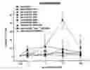
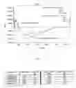


Sources:
- United States Patent and Trademark Office - verify current appl. status at the USPTO↗
Similar patent applications:
- » 20090005541
GAG binding protein - » 20100331237
GAG BINDING PROTEINS - » 20110144305
GAG BINDING PROTEIN - » 10090378
Method of identifying modulators of HIV-1 Vpu and Gag interaction with U binding protein (Ubp) - » 20090023161
Antiviral screening method to identify inhibitors of phosphatidylinositol 4,5-bisphosphate [PI(4,5)P] binding to the HIV gag matrix (MA) protein
Recent applications in this class:
- » 20250188140 2025-06-12
CCL5 MRNA NANOPARTICLE AND METHODS OF USE THEREOF - » 20230303647 2023-09-28
CYCLIC CHEMERIN-9 DERIVATIVES - » 20230141043 2023-05-11
NOVEL IMMUNOMODULATOR - » 20220112257 2022-04-14
MUL TISPECIFIC PROCHEMOKINE THERAPEUTIC PROTEINS (PARK) AND METHOD OF MAKING AND USING THEREOF - » 20220064245 2022-03-03
FUSION PROTEINS COMPRISING A CYTOKINE AND SCAFFOLD PROTEIN - » 20190153055 2019-05-23
Fusion protein comprising CCL3 variant and use thereof - » 20140017790 2014-01-16
Control agent for controlling undifferentiated state and use thereof - » 20140005096 2014-01-02
METHODS OF TREATING INFLAMMATION - » 20130330305 2013-12-12
Cytokine derivatives - » 20130150303 2013-06-13
GLYCOSAMINOGLYCAN-ANTAGONISING MCP-1 MUTANTS AND METHODS OF USING SAME
Recent applications for this Assignee:
- » 20140073557 2014-03-13
FGFRI-BASED ANTAGONISTS WITH IMPROVED GLYCOSAMINOGLYCAN AFFINITY AND METHODS OF USING SAME - » 20130150303 2013-06-13
GLYCOSAMINOGLYCAN-ANTAGONISING MCP-1 MUTANTS AND METHODS OF USING SAME - » 20120288474 2012-11-15
COMPOSITION FOR TREATMENT OF CXCL8-MEDIATED LUNG INFLAMMATION - » 20120046218 2012-02-23
GLYCOSAMINOGLYCAN-ANTAGONISING MCP-I MUTANTS AND METHODS OF USING SAME - » 20110144305 2011-06-16
GAG BINDING PROTEIN - » 20100331237 2010-12-30
GAG BINDING PROTEINS - » 20100298203 2010-11-25
SDF-I-BASED GLYCOSAMINOGLYCAN ANTAGONISTS AND METHODS OF USING SAME - » 20100197583 2010-08-05
Glycosaminoglycan-antagonising MCP-1 mutants - » 20100035294 2010-02-11
SYSTEM TO ASSESS THE BIOLOGICAL ACTIVITY OF CHEMOATTRACTANTS - » 20090005541 2009-01-01
GAG binding protein