GAG BINDING PROTEINS
US20100331237A1
2010-12-30
12/858,456
2010-08-18
Abstract:
A method is provided for introducing a GAG binding site into a protein comprising the steps:
-
- identifying a region in a protein which is not essential for structure maintenance
- introducing at least one basic amino acid into said site and/or deleting at least one bulky and/or acidic amino acid in said site,
whereby said GAG binding site has a GAG binding affinity of Kd≦10 μM, preferably ≦1 μM, still preferred ≦0.1 μM, as well as modified GAG binding proteins.
Assignee:
- PROTAFFIN BIOTECHNOLOGIE AG 11 🇦🇹 Graz, Austria
Interested in similar patents?
Get notified when new applications in this technology area are published.
Classification:
C07K14/523 » CPC main
Peptides having more than 20 amino acids; Gastrins; Somatostatins; Melanotropins; Derivatives thereof from animals; from humans; Cytokines; Lymphokines; Interferons; Chemokines Beta-chemokines, e.g. RANTES, I-309/TCA-3, MIP-1alpha, MIP-1beta/ACT-2/LD78/SCIF, MCP-1/MCAF, MCP-2, MCP-3, LDCF-1, LDCF-2
A61P25/00 » CPC further
Drugs for disorders of the nervous system
A61P37/08 » CPC further
Drugs for immunological or allergic disorders Antiallergic agents
A61P43/00 » CPC further
Drugs for specific purposes, not provided for in groups -
C07K14/5421 » CPC further
Peptides having more than 20 amino acids; Gastrins; Somatostatins; Melanotropins; Derivatives thereof from animals; from humans; Cytokines; Lymphokines; Interferons; Interleukins [IL] IL-8
A61K38/00 » CPC further
Medicinal preparations containing peptides
A61K38/17 IPC
Medicinal preparations containing peptides; Peptides having more than 20 amino acids; Gastrins; Somatostatins; Melanotropins; Derivatives thereof from animals; from humans
C07K1/00 IPC
General methods for the preparation of peptides, i.e. processes for the organic chemical preparation of peptides or proteins of any length
C07K14/00 IPC
Peptides having more than 20 amino acids; Gastrins; Somatostatins; Melanotropins; Derivatives thereof
C07H21/00 IPC
Compounds containing two or more mononucleotide units having separate phosphate or polyphosphate groups linked by saccharide radicals of nucleoside groups, e.g. nucleic acids
A61K31/7088 IPC
Medicinal preparations containing organic active ingredients; Carbohydrates; Sugars; Derivatives thereof Compounds having three or more nucleosides or nucleotides
A61P11/06 » CPC further
Drugs for disorders of the respiratory system Antiasthmatics
A61P17/06 » CPC further
Drugs for dermatological disorders Antipsoriatics
A61P19/02 » CPC further
Drugs for skeletal disorders for joint disorders, e.g. arthritis, arthrosis
A61P19/10 » CPC further
Drugs for skeletal disorders for bone diseases, e.g. rachitism, Paget's disease for osteoporosis
A61P25/28 » CPC further
Drugs for disorders of the nervous system for treating neurodegenerative disorders of the central nervous system, e.g. nootropic agents, cognition enhancers, drugs for treating Alzheimer's disease or other forms of dementia
C12N15/63 IPC
Mutation or genetic engineering; DNA or RNA concerning genetic engineering, vectors, e.g. plasmids, or their isolation, preparation or purification; Use of hosts therefor; Recombinant DNA-technology Introduction of foreign genetic material using vectors; Vectors; Use of hosts therefor; Regulation of expression
C12N5/10 IPC
Undifferentiated human, animal or plant cells, e.g. cell lines; Tissues; Cultivation or maintenance thereof; Culture media therefor Cells modified by introduction of foreign genetic material
Description
This application is a continuation of U.S. application Ser. No. 12/131,311 filed Jun. 2, 2008, which is a divisional of U.S. application Ser. No. 11/422,169 filed Jun. 5, 2006, which is a 371 of PCT/EP2004/013670 filed Dec. 2, 2004. The entire contents of the above-identified applications are hereby incorporated by reference.
The present invention relates to methods and tools for the inhibition of the interaction of chemokines and their high-affinity receptors on leukocytes and methods for the therapeutic treatment of inflammatory diseases.
Chemokines, originally derived from chemoattractant cytokines, actually comprise more than 50 members and represent a family of small, inducible, and secreted proteins of low molecular weight (6-12 kDa in their monomeric form) that play a decisive role during immunosurveillance and inflammatory processes. Depending on their function in immunity and inflammation, they can be distinguished into two classes. Inflammatory chemokines are produced by many different tissue cells as well as by immigrating leukocytes in response to bacterial toxins and inflammatory cytokines like IL-1, TNF and interferons. Their main function is to recruit leukocytes for host defense and in the process of inflammation. Homing chemokines, on the other hand, are expressed constitutively in defined areas of the lymphoid tissues. They direct the traffic and homing of lymphocytes and dendritic cells within the immune system. These chemokines, as illustrated by BCA-1, SDF-1 or SLC, control the relocation and recirculation of lymphocytes in the context of maturation, differentiation, activation and ensure their correct homing within secondary lymphoid organs.
Despite the large number of representatives, chemokines show remarkably similar structural folds although the sequence homology varies between 20 to 70 percent. Chemokines consist of roughly 70-130 amino acids with four conserved cysteine residues. The cysteines form two disulphide bonds (Cys 1→Cys 3, Cys 2→Cys 4) which are responsible for their characteristic three-dimensional structure. Chemotactic cytokines consist of a short amino terminal domain (3-10 amino acids) preceding the first cysteine residue, a core made of β-strands and connecting loops found between the second and the fourth cysteine residue, as well as a carboxy-terminal α-helix of 20-60 amino acids. The protein core has a well ordered structure whereas the N- and C-terminal parts are disordered. As secretory proteins they are synthesised with a leader sequence of 20-25 amino acids which is cleaved off before release.
The chemokines have been subdivided into four families on the basis of the relative position of their cysteine residues in the mature protein. In the α-chemokine subfamily, the first two of the four cysteines are separated by a single amino acid (CXC), whereas in the β-chemokines the corresponding cysteine residues are adjacent to each other (CC). The α-chemokines can be further classified into those that contain the ELR sequence in the N-terminus, thereby being chemotactic for neutrophils (IL-8 for example), and those that lack the ELR motif and act on lymphocytes (I-TAC for example). Structurally the β-chemokines can be subdivided into two families: the monocyte-chemoattractant protein eotaxin family, containing the five monocyte chemoattractant proteins (MCP) and eotaxin which are approximately 65 percent identical to each other, and the remaining β-chemokines. As with the CXC-family, the N-terminal amino acids preceding the CC-residues are critical components for the biologic activity and leukocyte selectivity of the chemokines. The β-chemokines, in general, do not act on neutrophils but attract monocytes, eosinophils, basophils and lymphocytes with variable selectivity.
Only a few chemokines do not fit into the CC-or the CXC-family. Lymphotactin is so far the only chemokine which shows just two instead of the four characteristic cysteines in its primary structure, and is thus classified as γ- or C-chemokine. On the other hand, by concluding this classification, fractalkine has to be mentioned as the only representative of the δ- or CXXXC-subfamily with three amino acids separating the first two cysteines. Both of them, Lymphotaxin and fractalkine, induce chemotaxis of T-cells and natural killer cells.
Chemokines induce cell migration and activation by binding to specific cell surface, seven transmembrane-spanning (7TM) G-protein-coupled receptors on target cells. Eighteen chemokine receptors have been cloned so far including six CXC, ten CC, one CX3C and one XC receptor. Chemokine receptors are expressed on different types of leukocytes, some of them are restricted to certain cells (e.g. CXCR1 is restricted to neutrophils) whereas others are more widely expressed (e.g. CCR2 is expressed on monocytes, T cells, natural killer cells and basophils). Similar to chemokines, the receptors can be constitutively expressed on certain cells, whereas some are inducible. Some of them can even be down-regulated making the cells unresponsive to a certain chemokine but remaining responsive to another. Most receptors recognise more than one chemokine and vice versa but recognition is restricted to chemokines of the corresponding subfamily (see Table 1).
| TABLE 1 | |||
| Inflammatory | |||
| Chemokine | Receptor | Chemotactic for | Diseases |
| CXC- | IL-8 | CXCR1 | Neutrophils | Acute respiratory distress |
| Chemokine | CXCR2 | syndrome [71]; | ||
| (+ELR-motif) | Bacterial pneumonia [72]; | |||
| Rheumathoid | ||||
| arthritis [73]; | ||||
| Inflammatory bowel | ||||
| disease [74]; | ||||
| Psoriasis [75]; | ||||
| Bacterial meningitis [76] | ||||
| CC- | MCP-1 | CCR2 | Basophils; Monocytes; | Asthma [77]; |
| Chemokine | Activated T cells; | Glomerulonephritis [78]; | ||
| Dentritic cells; Natural | Atheroscleosis [79]; | |||
| killer cells | Inflammatory bowel | |||
| disease [80]; | ||||
| Psoriasis [81]; | ||||
| Bacterial and viral | ||||
| meningitis [82, 83] | ||||
| RANTES | CCR1 | Eosinophils; Monocytes; | Asthma [84]; | |
| Activated T cells; | Glomerulonephritis [85] | |||
| Dentritic cells | ||||
| CCR3 | Eosinophils; Basophils; | |||
| Dentritic cells | ||||
| CCR5 | Monocytes; Activated T | |||
| cells; Dentritic cells; | ||||
| Natural killer cells | ||||
Chemokines have two main sites of interaction with their receptors, one in the amino-terminal domain and the other within an exposed loop of the backbone that extends between the second and the third cysteine residue. Both sites are kept in close proximity by the disulphide bonds. The receptor recognises first the binding site within the loop region which appears to function as a docking domain. This interaction restricts the mobility of the chemokine thus facilitating the proper orientation of the amino-terminal domain. Studies have been performed with mutant chemokines that still bound effectively to their receptors but did not signal. These mutants were obtained by amino acid deletion or modification within the N-termini of, for example, IL-8, RANTES and MCP-1.
Multiple intracellular signaling pathways occur after receptor activation as a result of chemokine binding. Chemokines also interact with two types of nonsignaling molecules. One is the DARC receptor which is expressed on erythrocytes and on endothelial cells and which binds CC- as well as CXC-chemokines to prevent them from circulation. The second type are heparan sulphate glycosaminoglycans (GAGs) which are part of proteoglycans and which serve as co-receptors of chemokines. They capture and present chemokines on the surface of the homing tissue (e.g. endothelial cells) in order to establish a local concentration gradient. In an inflammatory response, such as in rheumatoid arthritis, leukocytes rolling on the endothelium in a selectin-mediated process are brought into contact with the chemokines presented by the proteoglycans on the cell surface. Thereby, leukocyte integrins become activated which leads to firm adherence and extravasation. The recruited leukocytes are activated by local inflammatory cytokines and may become desensitised to further chemokine signaling because of high local concentration of chemokines. For maintaining a tissue bloodstream chemokine gradient, the DARC receptor functions as a sink for surplus chemokines.
Heparan sulphate (HS) proteoglycans, which consist of a core protein with covalently attached glycosaminoglycan sidechains (GAGs), are found in most mammalian cells and tissues. While the protein part determines the localisation of the proteoglycan in the cell membrane or in the extracellular matrix, the glycosaminoglycan component mediates interactions with a variety of extracellular ligands, such as growth factors, chemokines and adhesions molecules. The biosynthesis of proteoglycans has previously been extensively reviewed. Major groups of the cell surface proteoglycans are the syndecan family of transmembrane proteins (four members in mammals) and the glypican family of proteins attached to the cell membrane by a glycosylphosphatidylinositol (GPI) tail (six members in mammals). While glypicans are expressed widely in the nervous system, in kidney and, to a lesser extent, in skeletal and smooth muscle, syndecan-1 is the major HSPG in epithelial cells, syndecan-2 predominates in fibroblasts and endothelial cells, syndecan-3 abounds in neuronal cells and syndecan-4 is widely expressed. The majority of the GAG chains added to the syndecan core proteins through a tetrasaccharide linkage region onto particular serines are HS chains. Although the amino acid sequences of the extracellular domains of specific syndecan types are not conserved among different species, contrary to the transmembrane and the cytoplasmic domains, the number and the positions of the GAG chains are highly conserved. The structure of the GAGs, however, is species-specific and is, moreover, dependent upon the nature of the HSPG-expressing tissue.
Heparan sulphate (HS) is the most abundant member of the glycosaminoglycan (GAG) family of linear polysaccharides which also includes heparin, chondroitin sulphate, dermatan sulphate and keratan sulphate. Naturally occurring HS is characterised by a linear chain of 20-100 disaccharide units composed of N-acetyl-D-glucosamine (GlcNAc) and D-glucuronic acid (GlcA) which can be modified to include N- and O-sulphation (6-O and 3-O sulphation of the glucosamine and 2-O sulphation of the uronic acid) as well as epimerisation of β-D-gluronic acid to α-L-iduronic acid (IdoA).
Clusters of N- and O-sulphated sugar residues, separated by regions of low sulphation, are assumed to be mainly responsible for the numerous protein binding and regulatory properties of HS. In addition to the electrostatic interactions of the HS sulphate groups with basic amino acids, van der Waals and hydrophobic interactions are also thought to be involved in protein binding. Furthermore, the presence of the conformationally flexible iduronate residues seems to favour GAG binding to proteins. Other factors such as the spacing between the protein binding sites play also a critical role in protein-GAG binding interactions: For example γ-interferon dimerisation induced by HS requires GAG chains with two protein binding sequences separated by a 7 kDa region with low sulphation. Additional sequences are sometimes required for full biological activity of some ligands: in order to support FGF-2 signal transduction, HS must have both the minimum binding sequence as well as additional residues that are supposed to interact with the FGF receptor.
Heparin binding proteins often contain consensus sequences consisting of clusters of basic amino acid residues. Lysine, arginine, asparagine, histidine and glutamine are frequently involved in electrostatic contacts with the sulphate and carboxyl groups on the GAG. The spacing of the basic amino acids, sometimes determined by the proteins 3-D structure, are assumed to control the GAG binding specificity and affinity. The biological activity of the ligand can also be affected by the kinetics of HS-protein interaction. Reducing the dimension of growth factor diffusion is one of the suggested HSPG functions for which the long repetitive character of the GAG chains as well as their relatively fast on and off rates of protein binding are ideally suited. In some cases, kinetics rather than thermodynamics drives the physiological function of HS-protein binding. Most HS ligands require GAG sequences of well-defined length and structure. Heparin, which is produced by mast cells, is structurally very similar to heparan sulphate but is characterised by higher levels of post-polymerisation modifications resulting in a uniformly high degree of sulphation with a relatively small degree of structural diversity. Thus, the highly modified blocks in heparan sulphate are sometimes referred to as “heparin-like”. For this reason, heparin can be used as a perfect HS analogue for protein biophysical studies as it is, in addition, available in larger quantities and therefore less expensive than HS. Different cell types have been shown to synthesise proteoglycans with different glycosaminoglycan structure which changes during pathogenesis, during development or in response to extracellular signals such as growth factors. This structural diversity of HSPGs leads to a high binding versatility emphasising the great importance of proteoglycans.
Since the demonstration that heparan sulphate proteoglycans are critical for FGF signaling, several investigations were performed showing the importance of chemokine-GAG binding for promoting chemokine activity. First, almost all chemokines studied to date appear to bind HS in vitro, suggesting that this represents a fundamental property of these proteins. Second, the finding that in vivo T lymphocytes secrete CC-chemokines as a complex with glycosaminoglycans indicates that this form of interaction is physiologically relevant. Furthermore, it is known that the association of chemokines with HS helps to stabilise concentration gradients across the endothelial surface thereby providing directional information for migrating leukocytes. HS is also thought to protect chemokines from proteolytic degradation and to induce their oligomerisation thus promoting local high concentrations in the vicinity of the G-coupled signaling receptors. The functional relevance of oligomerisation, however, remains controversial although all chemokines have a clear structural basis for multimerisation. Dimerisation through association of the β-sheets is observed for all chemokines of the CXC-family (e.g. IL-8), contrary to most members of the CC-chemokine family (e.g. RANTES), which dimerise via their N-terminal strands.
A wealth of data has been accumulated on the inhibition of the interaction of chemokines and their high-affinity receptors on leukocytes by low molecular weight compounds. However, there has been no breakthrough in the therapeutic treatment of inflammatory diseases by this approach.
Interleukin-8 (IL-8) is a key molecule involved in neutrophil attraction during chronic and acute inflammation. Several approaches have been undertaken to block the action of IL-8 so far, beginning with inhibition of IL-8 production by for example glucocorticoids, Vitamin D3, cyclosporin A, transforming growth factor β, interferons etc., all of them inhibiting IL-8 activity at the level of production of IL-8 mRNA. A further approach previously used is to inhibit the binding of IL-8 to its receptors by using specific antibodies either against the receptor on the leukocyte or against IL-8 itself in order to act as specific antagonists and therefore inhibiting the IL-8 activity.
The aim of the present invention is therefore to provide an alternative strategy for the inhibition or disturbance of the interaction of chemokines/receptors on leukocytes. Specifically the action of IL-8, RANTES or MCP-1 should be targeted by such a strategy.
Subject matter of the present invention is therefore a method to produce new GAG binding proteins as well as alternative GAG binding proteins which show a high(er) affinity to a GAG co-receptor (than the wild type). Such modified GAG binding proteins can act as competitors with wild-type GAG binding proteins and are able to inhibit or down-regulate the activity of the wild-type GAG binding protein, however without the side effects which occur with the known recombinant proteins used in the state of the art. The molecules according to the present invention do not show the above mentioned disadvantages. The present modified GAG binding proteins can be used in drugs for various therapeutical uses, in particular—in the case of chemokines—for the treatment of inflammation diseases without the known disadvantages which occur in recombinant chemokines known in the state of the art. The modification of the GAG binding site according to the present invention turned out to be a broadly applicable strategy for all proteins which activity is based on the binding event to this site, especially chemokines with a GAG site. The preferred molecules according to the present invention with a higher GAG binding affinity proved to be specifically advantageous with respect to their biological effects, especially with respect to their anti-inflammatory activity by their competition with wild type molecules for the GAG site.
Therefore, the present invention provides a method for introducing a GAG binding site into a protein characterised in that it comprises the steps:
-
- identifying a region in a protein which is not essential for structure maintenance
- introducing at least one basic amino acid into said site and/or deleting at least one bulky and/or acidic amino acid in said site,
whereby said GAG binding site has a GAG binding affinity of Kd=10 μM, preferably 1 μM, still preferred ≦0.1 μM. By introducing at least one basic amino acid and/or deleting at least one bulky and/or acidic amino acid in said region, a novel, improved “artificial” GAG binding site is introduced in said protein. This comprises the new, complete introduction of a GAG binding site into a protein which did not show a GAG binding activity before said modification. This also comprises the introduction of a GAG binding site into a protein which already showed GAG binding activity. The new GAG binding site can be introduced into a region of the protein which did not show GAG binding affinity as well as a region which did show GAG binding affinity. However, with the most preferred embodiment of the present invention, a modification of the GAG binding affinity of a given GAG binding protein is provided, said modified protein's GAG binding ability is increased compared to the wild-type protein. The present invention relates to a method of introducing a GAG binding site into a protein, a modified GAG binding protein as well as to an isolated DNA molecule, a vector, a recombinant cell, a pharmaceutical composition and the use of said modified protein.
BRIEF DESCRIPTION OF THE DRAWINGS:
FIG. 1 shows a CD spectra.
FIG. 2 shows secondary structure contents of various mutants.
FIG. 3 shows graphics of results from fluorescence anisotropy tests of various mutants.
FIG. 4 shows graphics of results from fluorescence anisotropy tests of two mutants.
FIG. 5 shows the graphic of results from isothermal fluorescence titrations.
FIG. 6 shows the graphic of results from unfolding experiments of various mutants.
FIG. 7 shows chemotaxis index of IL-8 mutants.
FIG. 8 shows the results of the RANTES chemotaxis assay.
The term “introducing at least one basic amino acid” relates to the introduction of additional amino acids as well as the substitution of amino acids. The main purpose is to increase the relative amount of basic amino acids, preferably Arg, Lys, His, Asn and/or Gln, compared to the total amount of amino acids in said site, whereby the resulting GAG binding site should preferably comprise at least 3 basic amino acids, still preferred 4, most preferred 5 amino acids.
The GAG binding site is preferably at a solvent exposed position, e.g. at a loop. This will assure an effective modification.
Whether or not a region of a protein is essential for structure maintenance, can be tested for example by computational methods with specific programmes known to the person skilled in the art. After modification of the protein, the conformational stability is preferably tested in silico.
The term “bulky amino acid” refers to amino acids with long or sterically interfering side chains; these are in particular Trp, Ile, Leu, Phe, Tyr. Acidic amino acids are in particular Glu and Asp. Preferably, the resulting GAG binding site is free of bulky and acidic amino acids, meaning that all bulky and acidic amino acids are removed.
The GAG binding affinity is determined—for the scope of protection of the present application—over the dissociation constant Kd. One possibility is to determine the dissociation constant (Kd) values of any given protein by the structural change in ligand binding. Various techniques are well known to the person skilled in the art, e.g. isothermal fluorescence titrations, isothermal titration calorimetry, surface plasmon resonance, gel mobility assay, and indirectly by competition experiments with radioactively labelled GAG ligands. A further possibility is to predict binding regions by calculation with computational methods also known to the person skilled in the art, whereby several programmes may be used.
A protocol for introducing a GAG binding site into a protein is for example as follows:
-
- Identify a region of the protein which is not essential for overall structural maintenance and which might be suitable for GAG binding
- Design a new GAG binding site by introducing (replacement or insertion) basic Arg, Lys, His, Asp and Gln residues at any position or by deleting amino acids which interfere with GAG binding
- Check the conformational stability of the resulting mutant protein in silico
- Clone the wild-type protein cDNA (alternatively: purchase the cDNA)
- Use this as template for PCR-assisted mutagenesis to introduce the above mentioned changes into the amino acid sequence
- Subclone the mutant gene into a suitable expression system (prokaryotic or eukaryotic dependent upon biologically required post-translational modifications)
- Expression, purification and characterisation of the mutant protein in vitro
- Criterion for an introduced GAG binding affinity: KdGAG(mutant)≦10 μM.
Examples of said engineered proteins with new GAG binding sites are for example the Fc part of IgG as well as the complement factors C3 and C4 modified as follows:
| Fc: (439)KSLSLS(444)-> KSKKLS | (SEQ ID NOS 1 & 2) |
| C3: (1297)WIASHT(1302)-> WKAKHK | (SEQ ID NOS 3 & 4) |
| C4: (1)MLDAERLK(8)-> MKKAKRLK | (SEQ ID NOS 5 & 6) |
A further aspect of the present invention is a protein obtainable by the inventive method as described above. The inventive protein therefore comprises a—compared to the wild-type protein—new GAG binding site as defined above and will therefore act as competitor with natural GAG binding proteins, in particular since the GAG binding affinity of the inventive protein is very high, e.g. Kd≦10 μM.
A further aspect of the present invention is a modified GAG binding protein, whereby a GAG binding region in said protein is modified by substitution, insertion, and/or deletion of at least one amino acid in order to increase the relative amount of basic amino acids in said GAG binding region, and/or reduce the amount of bulky and/or acidic amino acids in said GAG binding region, preferably at a solvent exposed position, and in that the GAG binding affinity of said protein is increased compared to the the GAG binding affinity of a respective wild-type protein.
It has been surprisingly shown that by increasing the relative amount of basic amino acids, in particular Arg, Lys, His, Asn and Gln, in the GAG binding region, the modified GAG binding protein shows increased GAG binding affinity compared to the wild-type proteins, in particular when the relative amount of basic amino acids is increased at a solvent exposed position, since a positively charged area on the protein surface has shown to enhance the binding affinity. Preferably, at least 3, still preferred 4, most preferred 5, basic amino acids are present in the GAG binding region.
The term “GAG binding protein” relates to any protein which binds to a GAG co-receptor. Whether or not a protein binds to a GAG co-receptor can be tested with the help of known protocols as mentioned above. Hileman et al. (BioEssays 20 (1998), 156-167) disclose consensus sites in glycosaminoglycan binding proteins. The information disclosed in this article is also useful as starting information for the present invention. The term “protein” makes clear that the molecules provided by the present invention are at least 80 amino acids in length. This is required for making them suitable candidates for the present anti-inflammation strategy. Smaller molecules interacting with a GAG binding site and being physiologically or pathologically relevant due to such an interaction are not known and therefore not relevant for the present invention. Preferably, the molecules according to the present invention are composed of at least 90, at least 100, at least 120, at least 150, at least 200, at least 300, at least 400 or at least 500 amino acid residues.
In the scope of the present application the term “GAG binding region” is defined as a region which binds to GAG with a dissociation constant (Kd-value) of under 100 μM, preferably under 50 μM, still preferred under 20 μM, as determined by isothermal fluorescence titration (see examples below).
Any modifications mentioned in the present application can be carried out with known biochemical methods, for example site-directed mutagenesis. It should also be noted that molecular cloning of GAG binding sites is, of course, prior art (see e.g. WO96/34965 A, WO 92/07935 A, Jayaraman et al. (FEBS Letters 482 (2000), 154-158), WO02/20715 A, Yang et al. (J. Cell. Biochem. 56 (1994), 455-468), wherein molecular shuffling or de novo synthesis of GAG regions are described; Butcher et al., (FEBS Letters 4009 (1997), 183-187) (relates to artificial peptides, not proteins); Jinno-Oue et al, (J. Virol. 75 (2001), 12439-12445) de novo synthesis)).
The GAG binding region can be modified by substitution, insertion and/or deletion. This means that a non-basic amino acid may be substituted by a basic amino acid, a basic amino acid may be inserted into the GAG binding region or a non-basic amino acid may be deleted. Furthermore, an amino acid which interferes with GAG binding, preferably all interfering amino acids binding is deleted. Such amino acids are in particular bulky amino acids as described above as well as acidic amino acids, for example Glu and Asp. Whether or not an amino acid interferes with GAG binding may be examined with for example mathematical or computational methods. The result of any of these modifications is that the relative amount of basic amino acids in said GAG binding region is increased, whereby “relative” refers to the amount of basic amino acids in said GAG binding region compared to the number of all amino acids in said GAG binding region. Furthermore, amino acids which interfere sterically or electrostatically with GAG binding are deleted.
Whether or not an amino acid is present in a solvent exposed position, can be determined for example with the help of the known three dimensional structure of the protein or with the help of computational methods as mentioned above.
Whether or not the GAG binding affinity of said modified protein is increased compared to the GAG binding affinity of the respective wild-type protein, can be determined as mentioned above with the help of, for example, fluorescence titration experiments which determine the dissociation constants. The criterion for improved GAG binding affinity will be Kd (mutant)<Kd (wild-type), preferably by at least 100%. Specifically improved modified proteins have—compared with wild-type Kd—a GAG binding affinity which is higher by a factor of minimum 5, preferably of minimum 10, still preferred of minimum 100. The increased GAG binding affinity will therefore preferably show a Kd of under 10 μM, preferred under 1 μM, still preferred under 0.1 μM.
By increasing the GAG binding affinity the modified protein will act as a specific antagonist and will compete with the wild-type GAG binding protein for the GAG binding.
Preferably, at least one basic amino acid selected from the group consisting of Arg, Lys, and His is inserted into said GAG binding region. These amino acids are easily inserted into said GAG binding region, whereby the term “inserted” relates to an insertion as such as well as substituting any non-basic amino acid with arginine, lysine or histidine. Of course, it is possible to insert more than one basic amino acid whereby the same basic amino acid may be inserted or also a combination of two or three of the above mentioned amino acids.
Still preferred, the protein is a chemokine, preferably IL-8, RANTES or MCP-1. Chemokines are known to have a site of interaction with co-receptor GAG whereby this chemokine binding is often a condition for further receptor activation as mentioned above. Since chemokines are often found in inflammatory diseases, it is of major interest to block the chemokine receptor activation. Such chemokines are preferably IL-8, RANTES or MCP-1, which are well characterised molecules and of which the GAG binding regions are well known (see, for example, Lortat-Jacob et al., PNAS 99 (3) (2002), 1229-1234). By increasing the amount of basic amino acids in the GAG binding region of these chemokines, their binding affinity is increased and therefore the wild-type chemokines will bind less frequently or not at all, depending on the concentration of the modified protein in respect to the concentration of the wild-type protein.
According to an advantageous aspect, said GAG binding region is a C terminal α-helix. A typical chemical monomer is organised around a triple stranded anti-parallel β-sheet overlaid by a C-terminal α-helix. It has been shown that this C-terminal α-helix in chemokines is to a major part involved in the GAG binding, so that modification in this C-terminal α-helix in order to increase the amount of basic amino acids results in a modified chemokine with an increased GAG binding affinity.
Advantageously, positions 17, 21, 70, and/or 71 in IL-8 are substituted by Arg, Lys, His, Asn and/or Gln. Here it is possible that only one of these aforementioned sites is modified. However, also more than one of these sites may be modified as well as all, whereby all modifications may be either Arg or Lys or His or Asn or Gln or a mixture of those. In IL-8 these positions have shown to highly increase the GAG binding affinity of IL-8 and therefore these positions are particularly suitable for modifications.
Preferably the increased binding affinity is an increased binding affinity to heparan sulphate and/or heparin. Heparan sulphate is the most abundant member of the GAG family of linear polysaccharides which also includes heparin. Heparin is structurally very similar to heparan sulphate characterised by higher levels of post-polymerisation modifications resulting in a uniformly high degree of sulphation with a relatively small degree of structural diversity. Therefore, the highly modified blocks in heparan sulphate are sometimes referred to as heparin-like and heparin can be used as a heparan sulphate analogue for protein biophysical studies. In any case, both, heparan sulphate and heparin are particularly suitable.
Still preferred, a further biologically active region is modified thereby inhibiting or down-regulating a further biological activity of said protein. This further biological activity is known for most GAG binding proteins, for example for chemokines. This will be the binding region to a receptor, for example to the 7TM receptor. The term “further” defines a biologically active region which is not the GAG binding region which, however, binds to other molecules, cells or receptors and/or activates them. By modifying this further biologically active region the further biological activity of this protein is inhibited or down-regulated and thereby a modified protein is provided which is a strong antagonist to the wild-type protein. This means that on the one hand the GAG binding affinity is higher than in the wild-type GAG binding protein, so that the modified protein will to a large extent bind to the GAG instead of the wild-type protein. On the other hand, the further activity of the wild-type protein which mainly occurs when the protein is bound to GAG, is inhibited or down-regulated, since the modified protein will not carry out this specific activity or carries out this activity to a lesser extent. With this modified protein an effective antagonist for wild-type GAG binding proteins is provided which does not show the side effects known from other recombinant proteins as described in the state of the art. This further biologically active region can for example be determined in vitro by receptor competition assays (using fluorescently labelled wt chemokines, calcium influx, and cell migration (performed on native leukocytes or on 7TM stably-transfected cell lines). Examples of such further biologically active regions are, in addition to further receptor binding sites (as in the growth factor family), enzymatic sites (as in hydrolases, lyases, sulfotransferases, N-deacetylases, and copolymerases), protein interaction sites (as in antithrombin III), and membrane binding domains (as in the herpes simplex virus gD protein). With this preferred embodiment of double-modified proteins therefore dominant (concerning GAG binding) negative (concerning receptor) mutants are provided which are specifically advantageous with respect to the objectives set for the present invention.
Still preferred, said further biologically active region is modified by deletion, insertion, and/or substitution, preferably with alanine, a sterically and/or electrostatically similar residue. It is, of course, possible to either delete or insert or substitute at least one amino acid in said further biologically active region. However, it is also possible to provide a combination of at least two of these modifications or all three of them. By substituting a given amino acid with alanine or a sterically/electronically similar residue—“similar” meaning similar to the amino acid being substituted—the modified protein is not or only to a lesser extent modified sterically/electrostatically. This is particularly advantageous, since other activities of the modified protein, in particular the affinity to the GAG binding region, are not changed.
Advantageously, said protein is a chemokine and said further biological activity is leukocyte activation. As mentioned above, chemokines are involved in leukocyte attraction during chronic and acute inflammation. Therefore, by inhibiting or down-regulating leukocyte activation inflammation is decreased or inhibited which makes this particular modified protein an important tool for studying, diagnosing and treating inflammatory diseases.
According to an advantageous aspect, said protein is IL-8 and said further biologically active region is located within the first 10 N-terminal amino acids. The first N-terminal amino acids are involved in leukocyte activation, whereby in particular Glu-4, Leu-5 and Arg-6 were identified to be essential for receptor binding and activation. Therefore, either these three or even all first 10 N-terminal amino acids can be substituted or deleted in order to inhibit or down-regulate the receptor binding and activation.
A further advantageous protein is an IL-8 mutant with the first 6 N-terminal amino acids deleted. As mentioned above, this mutant will not or to a lesser extent bind and activate leukocytes, so that it is particularly suitable for studying, diagnosing and treating inflammatory diseases.
Preferably, said protein is an IL-8 mutant selected from the group consisting of del6F17RE70KN71R, del6F17RE70RN71K and del6E70KN71K. These mutants have shown to be particularly advantageous, since the deletion of the first 6 N-terminal amino acids inhibits or down-regulates receptor binding and activation. Furthermore, the two phenylalanines in position 17 and 21 were found to make first contact with the receptor on its N-terminal extracellular domain to facilitate the later activation of the receptor. In order to prevent any neutrophil contact, these two amino acids 17 and 21 are exchanged, whereby they are exchanged to basic amino acids, since they are in close proximity to the GAG binding motif of the C-terminal α-helix as can be seen on a three dimensional model of a protein. By exchanging the position 17 and/or 21 to either arginine or lysine the GAG binding affinity is therefore increased.
A further aspect of the present invention is an isolated polynucleic acid molecule which codes for the inventive protein as described above. The polynucleic acid may be DNA or RNA. Thereby the modifications which lead to the inventive modified protein are carried out on DNA or RNA level. This inventive isolated polynucleic acid molecule is suitable for diagnostic methods as well as gene therapy and the production of inventive modified protein on a large scale.
Still preferred, the isolated polynucleic acid molecule hybridises to the above defined inventive polynucleic acid molecule under stringent conditions. Depending on the hybridisation conditions complementary duplexes form between the two DNA or RNA molecules, either by perfectly being matched or also comprising mismatched bases (see Sambrook et al., Molecular Cloning: A laboratory manual, 2nd ed., Cold Spring Harbor, N.Y. 1989). Probes greater in length than about 50 nucleotides may accommodate up to 25 to 30% mismatched bases. Smaller probes will accommodate fewer mismatches. The tendency of a target and probe to form duplexes containing mismatched base pairs is controlled by the stringency of the hybridisation conditions which itself is a function of factors, such as the concentration of salt or formamide in the hybridisation buffer, the temperature of the hybridisation and the post-hybridisation wash conditions. By applying well-known principles that occur in the formation of hybrid duplexes conditions having the desired stringency can be achieved by one skilled in the art by selecting from among a variety of hybridisation buffers, temperatures and wash conditions. Thus, conditions can be selected that permit the detection of either perfectly matched or partially mismatched hybrid duplexes. The melting temperature (Tm) of a duplex is useful for selecting appropriate hybridisation conditions. Stringent hybridisation conditions for polynucleotide molecules over 200 nucleotides in length typically involve hybridising at a temperature 15-25° C. below the melting temperature of the expected duplex. For oligonucleotide probes over 30 nucleotides which form less stable duplexes than longer probes, stringent hybridisation usually is achieved by hybridising at 5 to 10° C. below the Tm. The Tm of a nucleic acid duplex can be calculated using a formula based on the percent G+C contained in the nucleic acids and that takes chain lengths into account, such as the formula Tm=81.5-16.6 (log [Na+)])+0.41 (% G+C)−(600/N), where N=chain length.
A further aspect of the present invention relates to a vector which comprises an isolated DNA molecule according to the present invention as defined above. The vector comprises all regulatory elements necessary for efficient transfection as well as efficient expression of proteins. Such vectors are well known in the art and any suitable vector can be selected for this purpose.
A further aspect of the present application relates to a recombinant cell which is stably transfected with an inventive vector as described above. Such a recombinant cell as well as any therefrom descendant cell comprises the vector. Thereby a cell line is provided which expresses the modified protein either continuously or upon activation depending on the vector.
A further aspect of the present invention relates to a pharmaceutical composition which comprises a protein, a polynucleic acid or a vector according to the present invention as defined above and a pharmaceutically acceptable carrier. Of course, the pharmaceutical composition may further comprise additional substances which are usually present in pharmaceutical compositions, such as salts, buffers, emulgators, colouring agents, etc.
A further aspect of the present invention relates to the use of the modified protein, a polynucleic acid or a vector according to the present invention as defined above in a method for inhibiting or suppressing the biological activity of the respective wild-type protein. As mentioned above, the modified protein will act as an antagonist whereby the side effects which occur with known recombinant proteins will not occur with the inventive modified protein. In the case of chemokines this will be in particular the biological activity involved in inflammatory reactions.
Therefore, a further use of the modified protein, polynucleic acid or vector according to the present invention is in a method for producing a medicament for the treatment of an inflammatory condition. In particular, if the modified protein is a chemokine, it will act as antagonist without side effects and will be particularly suitable for the treatment of an inflammatory condition. Therefore, a further aspect of the present application is also a method for the treatment of an inflammatory condition, wherein a modified protein according to the present invention, the isolated polynucleic acid molecule or vector according to the present invention or a pharmaceutical composition according to the present invention is administered to a patient.
Preferably, the inflammatory condition is selected from a group comprising rheumatoid arthritis, psoriasis, osteoarthritis, asthma, Alzheimer's disease, and multiple sclerosis. Since the activation through chemokines can be inhibited with a modified protein according to the present invention, inflammatory reactions can be inhibited or down-regulated whereby the above mentioned inflammatory conditions can be prevented or treated.
The present invention is described in further detail with the help of the following examples and figures to which the invention is, however, not limited whereby FIG. 1 is a CD spectra; FIG. 2 shows secondary structure contents of various mutants; FIGS. 3 and 4 show graphics of results from fluorescence anisotropy tests of various mutants; FIG. 5 shows the graphic of results from isothermal fluorescence titrations; FIG. 6 shows the graphic of results from unfolding experiments of various mutants, FIG. 7 shows chemotaxis index of IL-8 mutants (SEQ ID NOS 1070-1074 are disclosed respectively in order of appearance), and FIG. 8 shows the results of the RANTES chemotaxis assay.
EXAMPLES
Example 1
Generation of Recombinant IL-8 Genes and Cloning of the Mutants
Polymerase chain reaction (PCR) technique was used to generate the desired cDNAs for the mutants by introducing the mutations using sense and antisense mutagenesis primers. A synthetic plasmid containing the cDNA for wtIL-8 was used as template, Clontech Advantage®2 Polymerase Mix applied as DNA polymerase and the PCR reaction performed using a Mastergradient Cycler of Eppendorf. The mutagenesis primers used are summarised in the table below beginning with the forward sequences (5″to 3″):
| (SEQ ID NO: 7) |
| CACC ATG TGT CAG TGT ATA AAG ACA TAC TCC | |
| (primer for the mutation Δ6) | |
| (SEQ ID NO: 8) |
| CACC ATG TGT CAG TGT ATA AAG ACA TAC TCC | |
| AAA CCT AGG CAC CCC AAA AGG ATA | |
| (primer for the mutation Δ6 F17R F21R) |
The reverse sequences are (5″to 3″):
| (SEQ ID NO: 9) |
| TTA TGA ATT CCT AGC CCT CTT | |
| (primer for the mutation E70R) | |
| (SEQ ID NO: 10) |
| TTA TGA ATT CTT AGC CCT CTT | |
| (primer for the mutation E70K) | |
| (SEQ ID NO: 11) |
| TTA TGA CTT CTC AGC CCT CTT | |
| (primer for the mutation N71K) | |
| (SEQ ID NO: 12) |
| TTA TGA CTT CTT AGC CCT CTT | |
| (primer for the mutation E70K N71K) | |
| (SEQ ID NO: 13) |
| TTA TGA CTT CCT AGC CCT CTT | |
| (primer for the mutation E70R N71K) | |
| (SEQ ID NO: 14) |
| TTA TGA CCT CTT AGC CCT CTT | |
| (primer for the mutation E70K N71R) | |
| (SEQ ID NO: 15) |
| TTA TGA CCT CCT AGC CCT CTT | |
| (primer for the mutation E70R N71R) |
The PCR products were purified, cloned into the pCR®T7/NT-TOPO®TA (Invitrogen) vector and transformed into TOP10F competent E. coli (Invitrogen). As a next step a confirmation of the sequence was carried out by double-stranded DNA sequencing using a ABI PRISM CE1 Sequencer.
Example 2
Expression and Purification of the Recombinant Proteins
Once the sequences were confirmed, the constructs were transformed into calcium-competent BL21(DE3) E. coli for expression. Cells were grown under shaking in 1 l Lennox Broth (Sigma) containing 100 μg/ml Ampicillin at 37° C. until an OD600 of about 0.8 was reached. Induction of protein expression was accomplished by addition of isopropyl-β-D-thiogalactopyranoside (IPTG) to a final concentration of 1 mM. Four hours later the cells were harvested by centrifugation at 6000 g for 20 minutes. The cell pellet was then resuspended in a buffer containing 20 mM TRIS/HCl, 50 mM NaCl, pH 8, sonicated at 100 watts for 5×20 s and finally centrifuged again for 20 min at 10,000 g. Since the main fraction of the recombinant IL-8 proteins was found in inclusion bodies, denaturing conditions were chosen for further purification. So the cell pellet was resuspended in a buffer of 6M Gua/HCl and 50 mM MES, pH 6.5. The suspension was then stirred at 4° C. for 4 hours, followed by a dialysis step against 50 mM MES, pH 6.5. The resulting suspension was then centrifuged and filtered to be loaded on a strong cation exchange column (SP Sepharose® from Pharmacia Biotech). The elution was accomplished by a linear gradient from 0M-1M NaCl in a 50 mM MES buffer, pH 6.5 over 60 minutes. After lyophilisation of the fractions containing the desired protein, a second purification step was carried out by reversed-phase HPLC using a C18 column. In this case a nonlinear gradient from 10%-90% Acetonitril was chosen to elute the desired protein. Refolding of the denatured protein was finally accomplished by the same cation exchange column under the same conditions as described above.
The protein was then checked for purity and identity by silver stain analysis in the first case and Western Blot analysis, using a specific monoclonal antibody against wtIL-8, in the second. Refolding of the proteins was also confirmed by Circular Dichroism (CD) measurements.
Example 3
Biophysical Characterisation of the Mutants
3.1 Circular Dichroism Measurements and Analysis
In order to investigate secondary structure changes of the mutant protein in the presence and absence of heparan sulphate (HS), CD spectroscopy was carried out. Measurements were recorded on a Jasco J-710 spectropolarimeter over a range of 195-250 nm, and a cell of 0.1 cm path length was used. Spectra of the protein solutions with a concentration of 5 μM were recorded with a response time of 1 s, step resolution of 0.2 nm, speed of 50 nm/min, band width of 1 nm and a sensitivity of 20 mdeg. Three scans were averaged to yield smooth spectra. The protein spectra were then background-corrected relating to the CD-signal either of the buffer itself or buffer/HS. Secondary structure analysis of the protein in the presence and absence of HS was finally accomplished using the programme SELCON.
Since a great number of amino acids were changed in a number of novel combinations, it was tried to find out the dimension of the resulting secondary structure changes by circular dichroism methods.
Different structures were obtained depending on the mutations introduced. Except for one mutant expressed (Δ6 F17R F21R E70K N71R) which didn't show any structure, all mutants exhibited measurable α-helices, β-sheets and loops. Compared to IL-8wt only one mutant (Δ6 E70R) showed nearly similar structure whereas the others differed mainly in their α-helix which ranged from 17.2% to 45.2% out of the total structure. Nevertheless, this fact suggests that the overall structure of IL-8wt was maintained despite many changes within the proteins sequence. This could not have been previously predicted. Having already found that heparan sulphate oligosaccharides only, and not heparin, were able to affect IL-8wt secondary structure, attention was focused on the effects induced by unfractionated heparan sulphate. All examined mutants showed structural changes upon HS binding which can be seen as evidence of complex formation.
To demonstrate the structural changes upon introduced mutations and heparan sulphate addition, some of the data obtained are summarised in the graphs above and below.
3.2 Fluorescence Measurements
For studying concentration and ligand dependent quaternary structure changes fluorescence spectroscopy was performed. Due to its high sensitivity, requiring only nanogram quantities of protein, fluorescence technique was the method of choice for carrying out the desired investigations. Measurements were undertaken using a Perkin-Elmer (Beaconsfield, England) LS50B fluorometer.
3.3 Fluorescence Anisotropy
By recording the concentration dependent fluorescence anisotropy of the chemokine resulting from the extrinsic chromophore bisANS it was aimed to find out the dimerisation constant of the mutants. Measurements were performed in PBS starting with high concentrations (up to 4 μM protein) followed by stepwise dilution. For each data point, the anisotropy signal (r) recorded at 507 nm was averaged over 60 sec.
IL-8 oligomerisation has been reported to relevantly influence the proteins GAG binding properties. Set at monomeric concentration, IL-8 bound size defined oligosaccharides 1000-fold tighter than at dimeric concentration. Therefore, the oligomerisation properties of IL-8 mutants were investigated by fluorescence anisotropy. Since the IL-8 intrinsic fluorophore (Trp57) was not sensitive enough for all of the mutants, the extrinsic fluorophore bis-ANS was used for these measurements. Again, as already noticed for the secondary structure, the mutant Δ6 E70R showed high resemblance also in the oligomerisation constant (koligo=350 nM) to IL-8wt (koligo=379 nM). The mutant with the highest koligo(koligo=460 nM), which therefore dimerised worst, was Δ6 F17RF21R E70RN71K. Previous studies identified the β-sheets to be mainly involved in the dimerisation process, a fact, which correlates with the results for this mutants' secondary structure, which showed a very low share of β-sheet of only 11.4%. The mutant with the lowest koligo (koligo=147 nM), was found to be Δ6 F17RF21R E70K, which again showed the highest share of β-sheet structure (29.8%) of all mutants investigated. Also the impact of heparan sulphate addition was observed. As for IL-8wt, where heparan sulphate caused a shift of the oligomerisation constant to much higher levels (koligo=1.075 μM), this was also found for the IL-8 mutants investigated. Δ6 F17RF21R E70K shifted from 0.147 μM to 1.162 μM, and the mutant Δ6 E70R from 0.350 μM to 1.505 μM in the presence of heparan sulphate. Some of the results obtained are demonstrated in FIGS. 3 and 4, whereby FIG. 3 shows the dependence of the fluorescence anisotropy of IL-8 mutants in PBS on the chemokine concentration and FIG. 4 shows the dependence of the fluorescence anisotropy of Δ6 F17RF21R E70K in PBS on the chemokine concentration in the presence (ten fold excess) and absence of HS ((pc=10 xy excess) protein concentration).
3.4 Isothermal Fluorescence Titration (IFT) Experiments
Dissociation constants (Kd values) are a measure for the binding affinity of a ligand to a protein and therefore concentration-dependent change in the fluorescence emission properties of the protein (fluorescence quenching) upon ligand binding was used for the determination of Kd. Since these mutants contain an intrinsic tryptophan chromophore which is located at or near the proposed GAG binding site and therefore should be sensitive to structural changes upon ligand binding, IFT experiments seemed to be suitable for this kind of investigation. Fluorescence intensity titration was performed in PBS using a protein concentration of 700 nM. The emission of the protein solution upon excitation at 282 nm was recorded over a range of 300-400 nm following the addition of an aliquot of the respective GAG ligand and an equilibration period of 60 sec.
Binding to unfractionated heparin and heparan sulphate was investigated. The mutants were set at dimeric concentration to assure sufficient sensitivity. A quenching of Trp57 fluorescence intensity upon GAG binding was registered within a range of 25-350. Significant improvement of ligand binding was observed, especially for heparin binding. Δ6 F17RN71R E7OK (Kd=14 nM) and Δ6 F17RF21R N71K (Kd=14.6 nM) showed 2600-fold better binding, and Δ6 E70K N71K (Kd=74 nM) 1760-fold better binding compared to IL-8wt (Kd=37 μM). Good results were also obtained for heparan sulphate binding. For Δ6 F17RN71R E70K a Kd of 107 nM was found, for Δ6 F17RF21R N71K the Kd was 95 nM and the mutant Δ6 E70K N71K showed a Kd of 34 nM. As IL-8wt binds with a Kd of 4.2 μM, the Kds found for the mutants represent an extraordinary improvement in binding, see FIG. 5.
3.5 Unfolding Experiments
In order to obtain information about the proteins stability and whether this stability would be changed upon GAG ligand binding, unfolding experiments were undertaken. As mentioned above fluorescence techniques are very sensitive for observing quaternary structure changes and therefore are also the method of choice to investigate thermal structural changes of the protein. Measurements were undertaken as described for the IFT in which not the ligand concentration was changed but the temperature. Protein structure was observed at a concentration of 0.7 μM from temperatures of 15-85° C. in the absence and the presence of a 10 fold excess of heparan sulphate or heparin.
The emission maximum of the proteins ranged from 340 nm to 357 nm, values which are typical for a solvent exposed tryptophan residue. Beginning with the unfolding experiments at 15° C., the emission maximum of the mutants varied between 340 nm-351 nm. Compared to IL-8wt, whose emission maximum was observed at 340 nm, this means slightly higher values. Upon an increase in temperature, the intensity of emission maximum decreased, accompanied by a shift of the maximum to either a higher or lower wavelength. The emission maximum of Δ6 E70R and Δ6 E70K N71K shifted from 352.5 nm-357 nm and 343 nm-345 nm, which is typical for a further exposure of the Trp57 residue to the solvent trough temperature increase, but interestingly the mutants Δ6 F17RN71R E70K and Δ6 F17RF21R E70R N71K showed a blue shift, ranging from 350 nm-343 nm and, less pronounced, from 350 nm-348 nm (see FIG. 6). By slowly decreasing the temperature, the process of unfolding was partially reversible regarding both the wavelength shift and changes of intensity. Addition of a 5 fold excess of heparan sulphate led to an increase of stability of the proteins, probably through complex formation. This could be observed on the one hand by a shift of the melting point to higher temperature, and on the other hand by a significantly less pronounced shift of emission maximum upon temperature increase.
Example 4
Cell-Based Assay of the Receptor-“Negative” Function of the Dominant-Negative IL-8 Mutants
In order to characterise the impaired receptor function of the IL-8 mutants with respect to neutrophil attraction, transfilter-based chemotaxis of neutrophils in response to IL-8 mutants was assayed in a microchemotaxis chamber equipped with a 5 μm PVP-free polycarbonate membrane.
Cell Preparation:
Briefly, a neutrophil fraction was prepared from freshly collected human blood. This was done by adding a 6% dextran solution to heparin-treated blood (1:2) which was then left for sedimentation for 45 min. The upper clear cell solution was collected and washed twice with HBSS w/o Ca and Mg. Cells were counted and finally diluted with HBSS at 2 Mio/ml cell suspension, taking into account that only 60% of the counted cells were neutrophils.
Chemotaxis Assay:
IL-8 mutants were diluted at concentrations of 10 μg/ml, 1 μg/ml and 0.1 μg/ml and put in triplicates in the lower compartment of the chamber (26 μl per well). The freshly prepared neutrophils were seeded in the upper chamber (50 μl per well) and incubated for 30 minutes at 37° C. in a 5% CO2 humidified incubator. After incubation, the chamber was disassembled, the upper side of the filter was washed and wiped off and cells attached to the lower side were fixed with methanol and stained with Hemacolor solutions (Merck). Cells were then counted at 400× magnifications in randomly selected microscopic fields per well. Finally, the mean of three independent experiments was plotted against the chemokine concentration. In FIG. 7, the chemotaxis index for various IL-8 mutants is shown. As expected, all mutants showed significantly decreased receptor binding activity.
Example 5
Generation of Recombinant RANTES Genes, Expression, Biophysical and Activity Characterisation of the Mutants
The concept of dominant-negative “GAG-masking” chemokine mutants was also employed to RANTES, a chemokine involved in type IV hypersensitivity reactions like transplant rejection, atopic dermatitis as well as in other inflammatory disorders like arthritis, progressive glomerulonephritis and inflammatory lung disease.
The receptor binding capability was impaired by introducing into the wt protein an initiating methionine residue. Expression of the wt RANTES in E. Coli lead to the retention of this methionine residue, which renders wt RANTES to a potent inhibitor of monocyte migration, the so-called Met-RANTES. Different mutations enhancing the GAG binding affinity were introduced via PCR-based site-directed mutagenesis methods.
By these means 9 RANTES mutants have so far been cloned, expressed and purified, Met-RANTES A22K, Met-RANTES H23K, Met-RANTES T43K, Met-RANTES N46R, Met-RANTES N46K, Met-RANTES Q48K, Met-RANTES A22K/N46R, Met-RANTES V49R/E66S and Met-RANTES 15LSLA18 V49R/E66S.
Isothermal fluorescence titration experiments were carried out to measure the relative affinity constants (Kd values) of the RANTES mutants for size defined heparin. As can be seen in the table all RANTES mutant proteins showed higher affinities for this heparin, with Met-RANTES A22K, Met-RANTES H23K, Met-RANTES T43K and Met-RANTES A22K/N46R showing the most promising results.
| Kd in nM | |
| Wt Rantes | 456.2 ± 8.5 | |
| Met-Rantes V49R/E66S | 345.5 ± 21.7 | |
| Rantes 15LSLA18 V49R/66S | 297.3 ± 14.1 | |
| Rantes N46R | 367.7 ± 11.7 | |
| Rantes N46K | 257.4 ± 10.2 | |
| Rantes H23K | 202.5 ± 12.8 | |
| Rantes Q48K | 383.4 ± 39.6 | |
| Rantes T43K | 139.2 ± 30.1 | |
| Rantes A22K | 202.1 ± 9.8 | |
| Rantes A22K/N46R | 164.0 ± 16.6 | |
RANTES Chemotaxis Assay
RANTES mutant directed cell migration was investigated using the 48-well Boyden chamber system equipped with 5 μm PVP-coated polycarbonate membranes. RANTES and RANTES mutant dilutions in RPMI 1640 containing 20 mM HEPES pH 7.3 and 1 mg/ml BSA were placed in triplicates in the lower wells of the chamber. 50 μl of THP-1 cell suspensions (promonocytic cell line from the European collection of cell cultures) in the same medium at 2×10⁶ cells/ml were placed in the upper wells. After a 2 h incubation period at 37° C. in 5% CO2 the upper surface of the filter was washed in HBSS solution. The migrated cells were fixed in methanol and stained with Hemacolor solution (Merck). Five 400× magnifications per well were counted and the mean of three independently conducted experiments was plotted against the chemokine concentration in FIG. 8. The error bars represent the standard error of the mean of the three experiments. Again, as in the case of the IL-8 mutants, all RANTES mutants showed significantly reduced receptor binding activity.
Example 6
Proteins with GAG Binding Regions
By bioinformatical and by proteomical means GAG binding proteins were characterised together with their GAG binding regions. In the following tables 2 and 3 chemokines are shown with their GAG binding regions (table 2) and examples of other proteins are given also with their GAG binding regions (table 3).
| TABLE 2 |
| Chemokines and their GAG binding domains |
| CXC-chemokines | |
| IL-8: 18HPK20, (R47) 60RVVEKFLKR68(residues 60-68 of SEQ ID NO: 16) | |
| SAKELRCQCIKTYSKPFHPKFIKELRVIESGPHCANTEIIVKLSDGRELCLDPKENWVQR | |
| VVEKFLKRAENS (SEQ ID NO: 16) | |
| MGSA/GROα: 19HPK21, 45KNGR48(residues 45-48 of SEQ ID NO: 17), | |
| 60KKIIEK66(residues 60-66 of SEQ ID NO: 17) | |
| ASVATELRCQCLQTLQGIHPKNIQSVNVKSPGPHCAQTEVIATLKNGRKACLNPASPIVK | |
| KIIEKMLNSDKSN (SEQ ID NO: 17) | |
| MIP-2α/GROβ: 19HLK21,K45, 60KKIIEKMLK68(residues 60-68 of SEQ ID NO: 18) | |
| APLATELRCQCLQTLQGIHLKNIQSVKVKSPGPHCAQTEVIATLKNGQKACLNPASPMVK | |
| KIIEKMLKNGKSN (SEQ ID NO: 18) | |
| NAP-2: 15HPK18, 42KDGR45(residues 42-45 of SEQ ID NO: 19), 57KKIVQK62(residues | |
| 57-62 of SEQ ID NO: 19) | |
| AELRCLCIKTTSGIHPKNIQSLEVIGKGTHCNQVEVIATLKDGRKICLDPDAPRIKKIVQ | |
| KKLAGDESAD (SEQ ID NO: 19) | |
| PF-4: 20RPRH23(residues 20-23 of SEQ ID NO: 20), 46KNGR49(residues 46-49 of | |
| SEQ ID NO: 20), 61KKIIKK66(residues 61-66 of SEQ ID NO: 20) | |
| EAEEDGDLQCLCVKTTSQVRPRHITSLEVIKAGPHCPTAQLIATLKNGRKICLDLQAPLY | |
| KKIIKKLLES (SEQ ID NO: 20) | |
| SDF-1α: K1, 24KHLK27(residues 24-27 of SEQ ID NO: 21), 41RLK43 | |
| KPVSLSYRCPCRFFESHVARANVKHLKILNTPNCALQIVARLKNNNRQVCIDPKLKWIQE | |
| YLEKALN (SEQ ID NO: 21) | |
| CC-chemokines | |
| RANTES: (17RPLPRAH23(residues 17-23 of SEQ ID NO: 22)) 44RKNR47(residues 44-47 | |
| of SEQ ID NO: 22) | |
| SPYSSDTTPCCFAYIARPLPRAHIKEYFYTSGKCSNPAVVEVTRKNRQVCANPEKKWVRE | |
| YINSLEMS (SEQ ID NO: 22) | |
| MCP-2: 18RKIPIQR24(residues 18-24 of SEQ ID NO: 23), 46KRGK49(residues 46-49 | |
| of SEQ ID NO: 23) | |
| QPDSVSIPITCCFNVINRKIPIQRLESYTRITNIQCPKEAVIEKTKRGKEVCADPKERWVRDSMKHLDQI | |
| FQNLKP (SEQ ID NO: 23) | |
| MCP-3: 22KQR24, 47KLDK50(residues 47-50 of SEQ ID NO: 24), | |
| 66KHLDKK71(residues 66-71 of SEQ ID NO: 24) | |
| QPVGINTSTTCCYRFINKKIPKQRLESYRRTTSSHCPREAVIFKTKLDKEICADPTQKWV | |
| QDFMKHLDKKTQTPKL (SEQ ID NO: 24) | |
| MIP-1α: R17, 44KRSR47(residues 44-47 of SEQ ID NO: 25) | |
| SLAADTPTACCFSYTSRQIPQNFIADYFETSSQCSKPGVIFLTKRSRQVCADPSEEWVQK | |
| YVSDLELSA (SEQ ID NO: 24) | |
| MIP-1β: R18, 45KRSK48(residues 45-48 of SEQ ID NO: 26) | |
| APMGSDPPTACCFSYTARKLPRNFVVDYYETSSLCSQPAVVFQTKRSKQVCADPSESWVQEYVYDLELN | |
| (SEQ ID NO: 26) | |
| MPIF-1: R18, 45KKGR48(residues 45-48 of SEQ ID NO: 27) | |
| MDRFHATSADCCISYTPRSIPCSLLESYFETNSECSKPGVIFLTKKGRRFCANPSDKQVQ | |
| VCMRMLKLDTRIKTRKN (SEQ ID NO: 27) | |
| MIP-5/HCC-2: 40KKGR43(residues 40-43 of SEQ ID NO: 28) | |
| HFAADCCTSYISQSIPCSLMKSYFETSSECSKPGVIFLTKKGRQVCAKPSGPGVQDCMKK | |
| LKPYSI (SEQ ID NO: 28) | |
| TABLE 3 | |
| SEQ ID NO: | |
| Peroxisome biogenesis factor 1 | 29 | 181 | TRRAKE | 186 | |
| 30 | 367 | QKKIRS | 372 | ||
| 31 | 1263 | PKRRKN | 1268 | ||
| 32 | 181 | TRRAKE | 186 | ||
| 33 | 367 | QKKIRS | 372 | ||
| 34 | 1263 | PKRRKN | 1268 | ||
| MLTK-beta | 35 | 415 | SKRRGKKV | 422 | |
| 36 | 312 | ERRLKM | 317 | ||
| 37 | 416 | KRRGKK | 421 | ||
| 38 | 312 | ERRLKM | 317 | ||
| 39 | 416 | KRRGKK | 421 | ||
| BHLH factor Hes4 | 40 | 43 | EKRRRARI | 50 | |
| 41 | 43 | EKRRRA | 48 | ||
| 42 | 43 | EKRRRA | 48 | ||
| Protocadherin 11 | 43 | 867 | MKKKKKKK | 874 | |
| 44 | 867 | MKKKKK | 872 | ||
| 45 | 867 | MKKKKK | 872 | ||
| 46 | 899 | MKKKKKKK | 906 | ||
| 47 | 899 | MKKKKK | 904 | ||
| 48 | 899 | MKKKKK | 904 | ||
| catenin (cadherin-associated protein), | 49 | 315 | RRRLRS | 320 | |
| delta 1 | 50 | 404 | VRKLKG | 409 | |
| 51 | 460 | LRKARD | 465 | ||
| 52 | 545 | RRKLRE | 550 | ||
| 53 | 621 | AKKGKG | 626 | ||
| 54 | 787 | AKKLRE | 792 | ||
| 55 | 315 | RRRLRS | 320 | ||
| 56 | 404 | VRKLKG | 409 | ||
| 57 | 460 | LRKARD | 465 | ||
| 58 | 545 | RRKLRE | 550 | ||
| 59 | 621 | AKKGKG | 626 | ||
| 60 | 787 | AKKLRE | 792 | ||
| Muscarinic acetylcholine receptor M5 | 61 | 221 | EKRTKD | 226 | |
| 62 | 427 | TKRKRV | 432 | ||
| 63 | 514 | WKKKKV | 519 | ||
| 64 | 221 | EKRTKD | 226 | ||
| 65 | 427 | TKRKRV | 432 | ||
| 66 | 514 | WKKKKV | 519 | ||
| Alpha-2A adrenergic receptor | 67 | 147 | PRRIKA | 152 | |
| 68 | 224 | KRRTRV | 229 | ||
| 69 | 147 | PRRIKA | 152 | ||
| 70 | 224 | KRRTRV | 229 | ||
| IL-5 promoter REII-region-binding | 71 | 440 | TKKKTRRR | 447 | |
| protein | 72 | 569 | GKRRRRRG | 576 | |
| 73 | 38 | ARKGKR | 43 | ||
| 74 | 437 | GKKTKK | 442 | ||
| 75 | 444 | TRRRRA | 449 | ||
| 76 | 569 | GKRRRR | 574 | ||
| 77 | 38 | ARKGKR | 43 | ||
| 78 | 437 | GKKTKK | 442 | ||
| 79 | 444 | TRRRRA | 449 | ||
| 80 | 569 | GKRRRR | 574 | ||
| Mitofusin 1 | 81 | 291 | ARKQKA | 296 | |
| 82 | 395 | KKKIKE | 400 | ||
| 83 | 291 | ARKQKA | 296 | ||
| 84 | 395 | KKKIKE | 400 | ||
| N-cym protein | 85 | 71 | VRRCKI | 76 | |
| 86 | 71 | VRRCKI | 76 | ||
| Smad ubiquitination regulatory | 87 | 672 | ERRARL | 677 | |
| factor 1 | 88 | 672 | ERRARL | 677 | |
| CUG-BP and ETR-3 like factor 5 | 89 | 468 | MKRLKV | 473 | |
| 90 | 475 | LKRPKD | 480 | ||
| 91 | 468 | MKRLKV | 473 | ||
| 92 | 475 | LKRPKD | 480 | ||
| Ewings sarcoma EWS-Fli1 | 93 | 347 | QRKSKP | 352 | |
| 94 | 347 | QRKSKP | 352 | ||
| NUF2R | 95 | 455 | LKRKMFKM | 462 | |
| 96 | 331 | LKKLKT | 336 | ||
| 97 | 347 | VKKEKL | 352 | ||
| 98 | 331 | LKKLKT | 336 | ||
| 99 | 347 | VKKEKL | 352 | ||
| Kruppel-like zinc finger protein | 100 | 22 | EKRERT | 27 | |
| GLIS2 | 101 | 22 | EKRERT | 27 | |
| FKSG32 | 102 | 15 | LKRVRE | 20 | |
| 103 | 431 | VRRGRI | 436 | ||
| 104 | 15 | LKRVRE | 20 | ||
| 105 | 431 | VRRGRI | 436 | ||
| BARH-LIKE 1 PROTEIN | 106 | 175 | LKKPRK | 180 | |
| 107 | 228 | NRRTKW | 233 | ||
| 108 | 175 | LKKPRK | 180 | ||
| 109 | 228 | NRRTKW | 233 | ||
| Nucleolar GTP-binding protein 1 | 110 | 393 | SRKKRERD | 400 | |
| 111 | 624 | GKRKAGKK | 631 | ||
| 112 | 48 | MRKVKF | 53 | ||
| 113 | 141 | IKRQKQ | 146 | ||
| 114 | 383 | ARRKRM | 388 | ||
| 115 | 393 | SRKKRE | 398 | ||
| 116 | 490 | KKKLKI | 495 | ||
| 117 | 543 | ARRSRS | 548 | ||
| 118 | 550 | TRKRKR | 555 | ||
| 119 | 586 | VKKAKT | 591 | ||
| 120 | 629 | GKKDRR | 634 | ||
| 121 | 48 | MRKVKF | 53 | ||
| 122 | 141 | IKRQKQ | 146 | ||
| 123 | 383 | ARRKRM | 388 | ||
| 124 | 393 | SRKKRE | 398 | ||
| 125 | 490 | KKKLKI | 495 | ||
| 126 | 543 | ARRSRS | 548 | ||
| 127 | 550 | TRKRKR | 555 | ||
| 128 | 586 | VKKAKT | 591 | ||
| 129 | 629 | GKKDRR | 634 | ||
| EVG1 | 130 | 17 | RRRPKT | 22 | |
| 131 | 138 | ERKRKA | 143 | ||
| 132 | 17 | RRRPKT | 22 | ||
| 133 | 138 | ERKRKA | 143 | ||
| ASPL | 134 | 282 | PKKSKS | 287 | |
| 135 | 282 | PKKSKS | 287 | ||
| Zinc transporter 1 | 136 | 477 | EKKPRR | 482 | |
| 137 | 477 | EKKPRR | 482 | ||
| Uveal autoantigen | 138 | 603 | EKKGRK | 608 | |
| 139 | 995 | ERKFKA | 1000 | ||
| 140 | 1023 | VKKNKQ | 1028 | ||
| 141 | 603 | EKKGRK | 608 | ||
| 142 | 995 | ERKFKA | 1000 | ||
| 143 | 1023 | VKKNKQ | 1028 | ||
| RAB39 | 144 | 7 | VRRDRV | 12 | |
| 145 | 7 | VRRDRV | 12 | ||
| Down syndrome cell adhesion molecule | 146 | 320 | PRKVKS | 325 | |
| 147 | 387 | VRKDKL | 392 | ||
| 148 | 320 | PRKVKS | 325 | ||
| 149 | 387 | VRKDKL | 392 | ||
| Protein-tyrosine phosphatase, non- | 150 | 139 | GRKKCERY | 146 | |
| receptor type 12 | 151 | 59 | VKKNRY | 64 | |
| 152 | 59 | VKKNRY | 64 | ||
| WD-repeat protein 11 | 153 | 752 | VRKIRF | 757 | |
| 154 | 752 | VRKIRF | 757 | ||
| Gastric cancer-related protein | 155 | 20 | SRKRQTRR | 27 | |
| VRG107 | 156 | 25 | TRRRRN | 30 | |
| 157 | 25 | TRRRRN | 30 | ||
| Early growth response protein 4 | 158 | 356 | ARRKGRRG | 363 | |
| 159 | 452 | EKKRHSKV | 459 | ||
| 160 | 357 | RRKGRR | 362 | ||
| 161 | 357 | RRKGRR | 362 | ||
| Vesicle transport-related protein | 162 | 309 | PKRKNKKS | 316 | |
| 163 | 226 | DKKLRE | 231 | ||
| 164 | 310 | KRKNKK | 315 | ||
| 165 | 355 | VKRLKS | 360 | ||
| 166 | 226 | DKKLRE | 231 | ||
| 167 | 310 | KRKNKK | 315 | ||
| 168 | 355 | VKRLKS | 360 | ||
| UPF3X | 169 | 140 | AKKKTKKR | 147 | |
| 170 | 141 | KKKTKK | 146 | ||
| 171 | 217 | ERRRRE | 222 | ||
| 172 | 225 | RKRQRE | 230 | ||
| 173 | 233 | RRKWKE | 238 | ||
| 174 | 240 | EKRKRK | 245 | ||
| 175 | 296 | DKREKA | 301 | ||
| 176 | 373 | RRRQKE | 378 | ||
| 177 | 393 | MKKEKD | 398 | ||
| 178 | 426 | VKRDRI | 431 | ||
| 179 | 140 | AKKKTKKRD | 148 | ||
| 180 | 141 | KKKTKK | 146 | ||
| 181 | 217 | ERRRRE | 222 | ||
| 182 | 225 | RKRQRE | 230 | ||
| 183 | 233 | RRKWKE | 238 | ||
| 184 | 240 | EKRKRK | 245 | ||
| 185 | 296 | DKREKA | 301 | ||
| 186 | 373 | RRRQKE | 378 | ||
| 187 | 393 | MKKEKD | 398 | ||
| 188 | 426 | VKRDRI | 431 | ||
| CGI-201 protein, type IV | 189 | 49 | ARRTRS | 54 | |
| 190 | 49 | ARRTRS | 54 | ||
| RING finger protein 23 | 191 | 98 | KRKIRD | 103 | |
| 192 | 98 | KRKIRD | 103 | ||
| FKSG17 | 193 | 72 | EKKARK | 77 | |
| 194 | 95 | IRKSKN | 100 | ||
| 195 | 72 | EKKARK | 77 | ||
| 196 | 95 | IRKSKN | 100 | ||
| P83 | 197 | 681 | ARKERE | 686 | |
| 198 | 681 | ARKERE | 686 | ||
| Ovarian cancer-related protein 1 | 199 | 62 | LKRDRF | 67 | |
| 200 | 62 | LKRDRF | 67 | ||
| MHC class II transactivator CIITA | 201 | 407 | HRRPRE | 412 | |
| 202 | 741 | PRKKRP | 746 | ||
| 203 | 783 | DRKQKV | 788 | ||
| 204 | 407 | HRRPRE | 412 | ||
| 205 | 741 | PRKKRP | 746 | ||
| 206 | 783 | DRKQKV | 788 | ||
| Platelet glycoprotein VI-2 | 207 | 275 | SRRKRLRH | 282 | |
| 208 | 275 | SRRKRL | 280 | ||
| 209 | 275 | SRRKRL | 280 | ||
| Ubiquitin-like 5 protein | 210 | 11 | GKKVRV | 16 | |
| 211 | 11 | GKKVRV | 16 | ||
| Protein kinase D2 | 212 | 191 | ARKRRL | 196 | |
| 213 | 191 | ARKRRL | 196 | ||
| Homeobox protein GSH-2 | 214 | 202 | GKRMRT | 207 | |
| 215 | 252 | NRRVKH | 257 | ||
| 216 | 202 | GKRMRT | 207 | ||
| 217 | 252 | NRRVKH | 257 | ||
| ULBP3 protein | 218 | 166 | ARRMKE | 171 | |
| 219 | 201 | HRKKRL | 206 | ||
| 220 | 166 | ARRMKE | 171 | ||
| 221 | 201 | HRKKRL | 206 | ||
| Type II iodothyronine deiodinase | 222 | 87 | SKKEKV | 92 | |
| 223 | 87 | SKKEKV | 92 | ||
| 224 | 299 | SKRCKK | 304 | ||
| 225 | 299 | SKRCKK | 304 | ||
| Sperm antigen | 226 | 160 | LKKYKE | 165 | |
| 227 | 478 | IKRLKE | 483 | ||
| 228 | 160 | LKKYKEKRT | 168 | ||
| 229 | 160 | LKKYKE | 165 | ||
| 230 | 478 | IKRLKE | 483 | ||
| UDP-GalNAc: polypeptide N- | 231 | 4 | ARKIRT | 9 | |
| acetylgalactosaminyltransferase | 232 | 44 | DRRVRS | 49 | |
| 233 | 138 | PRKCRQ | 143 | ||
| 234 | 4 | ARKIRT | 9 | ||
| 235 | 44 | DRRVRS | 49 | ||
| 236 | 138 | PRKCRQ | 143 | ||
| NCBE | 237 | 62 | HRRHRH | 67 | |
| 238 | 73 | RKRDRE | 78 | ||
| 239 | 1012 | SKKKKL | 1017 | ||
| 240 | 62 | HRRHRH | 67 | ||
| 241 | 73 | RKRDRE | 78 | ||
| 242 | 1012 | SKKKKL | 1017 | ||
| WD repeat protein | 243 | 372 | LKKKEERL | 379 | |
| 244 | 384 | EKKQRR | 389 | ||
| 245 | 400 | AKKMRP | 405 | ||
| 246 | 384 | EKKQRR | 389 | ||
| 247 | 400 | AKKMRP | 405 | ||
| Phosphodiesterase 11A | 248 | 27 | MRKGKQ | 32 | |
| 249 | 27 | MRKGKQ | 32 | ||
| Probable cation-transporting ATPase 2 | 250 | 891 | ERRRRPRD | 898 | |
| 251 | 306 | SRKWRP | 311 | ||
| 252 | 891 | ERRRRP | 896 | ||
| 253 | 306 | SRKWRP | 311 | ||
| 254 | 891 | ERRRRP | 896 | ||
| HMG-box transcription factor TCF-3 | 255 | 420 | GKKKKRKR | 427 | |
| 256 | 399 | ARKERQ | 404 | ||
| 257 | 420 | GKKKKR | 425 | ||
| 258 | 420 | GKKKKRKRE | 428 | ||
| 259 | 399 | ARKERQ | 404 | ||
| 260 | 420 | GKKKKR | 425 | ||
| HVPS11 | 261 | 793 | VRRYRE | 798 | |
| 262 | 793 | VRRYRE | 798 | ||
| PIST | 263 | 165 | NKKEKM | 170 | |
| 264 | 165 | NKKEKM | 170 | ||
| FYN-binding protein | 265 | 473 | KKREKE | 478 | |
| 266 | 501 | KKKFKL | 506 | ||
| 267 | 682 | LKKLKK | 687 | ||
| 268 | 696 | RKKFKY | 701 | ||
| 269 | 473 | KKREKE | 478 | ||
| 270 | 501 | KKKFKL | 506 | ||
| 271 | 682 | LKKLKK | 687 | ||
| 272 | 696 | RKKFKY | 701 | ||
| C1orf25 | 273 | 620 | GKKQKT | 625 | |
| 274 | 620 | GKKQKT | 625 | ||
| C1orf14 | 275 | 441 | LRRRKGKR | 448 | |
| 276 | 70 | LRRWRR | 75 | ||
| 277 | 441 | LRRRKG | 446 | ||
| 278 | 70 | LRRWRR | 75 | ||
| 279 | 441 | LRRRKG | 446 | ||
| T-box transcription factor TBX3 | 280 | 144 | DKKAKY | 149 | |
| 281 | 309 | GRREKR | 314 | ||
| 282 | 144 | DKKAKY | 149 | ||
| 283 | 309 | GRREKR | 314 | ||
| Mitochondrial 39S ribosomal protein | 284 | 121 | AKRQRL | 126 | |
| L47 | 285 | 216 | EKRARI | 221 | |
| 286 | 230 | RKKAKI | 235 | ||
| 287 | 121 | AKRQRL | 126 | ||
| 288 | 216 | EKRARI | 221 | ||
| 289 | 230 | RKKAKI | 235 | ||
| CGI-203 | 290 | 33 | VRRIRD | 38 | |
| 291 | 33 | VRRIRD | 38 | ||
| Jagged1 | 292 | 1093 | LRKRRK | 1098 | |
| 293 | 1093 | LRKRRK | 1098 | ||
| Secretory carrier-associated membrane | 294 | 102 | DRRERE | 107 | |
| protein 1 | 295 | 102 | DRRERE | 107 | |
| Vitamin D receptor-interacting protein | 296 | 673 | KKKKSSRL | 680 | |
| complex component DRIP205 | 297 | 672 | TKKKKS | 677 | |
| 298 | 954 | QKRVKE | 959 | ||
| 299 | 978 | GKRSRT | 983 | ||
| 300 | 995 | PKRKKA | 1000 | ||
| 301 | 1338 | GKREKS | 1343 | ||
| 302 | 1482 | HKKHKK | 1487 | ||
| 303 | 1489 | KKKVKD | 1494 | ||
| 304 | 672 | TKKKKS | 677 | ||
| 305 | 954 | QKRVKE | 959 | ||
| 306 | 978 | GKRSRT | 983 | ||
| 307 | 995 | PKRKKA | 1000 | ||
| 308 | 1338 | GKREKS | 1343 | ||
| 309 | 1482 | HKKHKK | 1487 | ||
| 310 | 1489 | KKKVKD | 1494 | ||
| Secretory carrier-associated membrane | 311 | 100 | ERKERE | 105 | |
| protein 2 | 312 | 100 | ERKERE | 105 | |
| Nogo receptor | 313 | 420 | SRKNRT | 425 | |
| 314 | 420 | SRKNRT | 425 | ||
| FLAMINGO 1 | 315 | 169 | GRRKRN | 174 | |
| 316 | 2231 | ARRQRR | 2236 | ||
| 317 | 169 | GRRKRN | 174 | ||
| 318 | 2231 | ARRQRR | 2236 | ||
| CC-chemokine receptor | 319 | 58 | CKRLKS | 63 | |
| 320 | 58 | CKRLKS | 63 | ||
| Prolactin regulatory element-binding | 321 | 271 | HKRLRQ | 276 | |
| protein | 322 | 271 | HKRLRQ | 276 | |
| Kappa B and V(D)J recombination signal | 323 | 17 | PRKRLTKG | 24 | |
| sequences binding protein | 324 | 713 | RKRRKEKS | 720 | |
| 325 | 903 | PKKKRLRL | 910 | ||
| 326 | 180 | HKKERK | 185 | ||
| 327 | 629 | TKKTKK | 634 | ||
| 328 | 712 | LRKRRK | 717 | ||
| 329 | 903 | PKKKRL | 908 | ||
| 330 | 1447 | QKRVKE | 1452 | ||
| 331 | 1680 | SRKPRM | 1685 | ||
| 332 | 180 | HKKERK | 185 | ||
| 333 | 629 | TKKTKK | 634 | ||
| 334 | 712 | LRKRRK | 717 | ||
| 335 | 903 | PKKKRL | 908 | ||
| 336 | 1447 | QKRVKE | 1452 | ||
| 337 | 1680 | SRKPRM | 1685 | ||
| Breast cancer metastasis-suppressor 1 | 338 | 200 | SKRKKA | 205 | |
| 339 | 229 | IKKARA | 234 | ||
| 340 | 200 | SKRKKA | 205 | ||
| 341 | 229 | IKKARA | 234 | ||
| Forkhead box protein P3 | 342 | 414 | RKKRSQRP | 421 | |
| 343 | 413 | FRKKRS | 418 | ||
| 344 | 413 | FRKKRS | 418 | ||
| FAS BINDING PROTEIN | 345 | 228 | LKRKLIRL | 235 | |
| 346 | 391 | RKKRRARL | 398 | ||
| 347 | 358 | ARRLRE | 363 | ||
| 348 | 390 | ERKKRR | 395 | ||
| 349 | 629 | CKKSRK | 634 | ||
| 350 | 358 | ARRLRE | 363 | ||
| 351 | 390 | ERKKRR | 395 | ||
| 352 | 629 | CKKSRK | 634 | ||
| Ubiquitin carboxyl-terminal | 353 | 228 | HKRMKV | 233 | |
| hydrolase 12 | 354 | 244 | LKRFKY | 249 | |
| 355 | 228 | HKRMKV | 233 | ||
| 356 | 244 | LKRFKY | 249 | ||
| KIAA0472 protein | 357 | 110 | HRKPKL | 115 | |
| 358 | 110 | HRKPKL | 115 | ||
| PNAS-101 | 359 | 68 | LKRSRP | 73 | |
| 360 | 106 | PRKSRR | 111 | ||
| 361 | 68 | LKRSRP | 73 | ||
| 362 | 106 | PRKSRR | 111 | ||
| PNAS-26 | 363 | 118 | DRRTRL | 123 | |
| 364 | 118 | DRRTRL | 123 | ||
| Myelin transcription factor 2 | 365 | 176 | GRRKSERQ | 183 | |
| Sodium/potassium-transporting ATPase | 366 | 47 | SRRFRC | 52 | |
| gamma chain | 367 | 55 | NKKRRQ | 60 | |
| 368 | 47 | SRRFRC | 52 | ||
| 369 | 55 | NKKRRQ | 60 | ||
| Mdm4 protein | 370 | 441 | EKRPRD | 446 | |
| 371 | 464 | ARRLKK | 469 | ||
| 372 | 441 | EKRPRD | 446 | ||
| 373 | 464 | ARRLKK | 469 | ||
| G antigen family D 2 protein | 374 | 87 | QKKIRI | 92 | |
| 375 | 87 | QKKIRI | 92 | ||
| NipSnap2 protein | 376 | 153 | FRKARS | 158 | |
| 377 | 153 | FRKARS | 158 | ||
| Stannin | 378 | 73 | ERKAKL | 78 | |
| 379 | 73 | ERKAKL | 78 | ||
| Sodium bicarbonate cotransporter | 380 | 973 | EKKKKKKK | 980 | |
| 381 | 165 | LRKHRH | 170 | ||
| 382 | 666 | LKKFKT | 671 | ||
| 383 | 966 | DKKKKE | 971 | ||
| 384 | 973 | EKKKKK | 978 | ||
| 385 | 165 | LRKHRH | 170 | ||
| 386 | 666 | LKKFKT | 671 | ||
| 387 | 966 | DKKKKE | 971 | ||
| 388 | 973 | EKKKKK | 978 | ||
| Myosin X | 389 | 683 | YKRYKV | 688 | |
| 390 | 828 | EKKKRE | 833 | ||
| 391 | 1653 | LKRIRE | 1658 | ||
| 392 | 1676 | LKKTKC | 1681 | ||
| 393 | 683 | YKRYKV | 688 | ||
| 394 | 828 | EKKKRE | 833 | ||
| 395 | 1653 | LKRIRE | 1658 | ||
| 396 | 1676 | LKKTKC | 1681 | ||
| PNAS-20 | 397 | 21 | RKRKSVRG | 28 | |
| 398 | 20 | ERKRKS | 25 | ||
| 399 | 20 | ERKRKS | 25 | ||
| Pellino | 400 | 36 | RRKSRF | 41 | |
| 401 | 44 | FKRPKA | 49 | ||
| 402 | 36 | RRKSRF | 41 | ||
| 403 | 44 | FKRPKA | 49 | ||
| Hyaluronan mediated motility | 404 | 66 | ARKVKS | 71 | |
| receptor | 405 | 66 | ARKVKS | 71 | |
| Short transient receptor potential | 406 | 753 | FKKTRY | 758 | |
| channel 7 | 407 | 753 | FKKTRY | 758 | |
| Liprin-alpha2 | 408 | 825 | PKKKGIKS | 832 | |
| 409 | 575 | IRRPRR | 580 | ||
| 410 | 748 | LRKHRR | 753 | ||
| 411 | 839 | GKKEKA | 844 | ||
| 412 | 875 | DRRLKK | 880 | ||
| 413 | 575 | IRRPRR | 580 | ||
| 414 | 748 | LRKHRR | 753 | ||
| 415 | 839 | GKKEKA | 844 | ||
| 416 | 875 | DRRLKK | 880 | ||
| Transcription intermediary factor 1- | 417 | 904 | DKRKCERL | 911 | |
| alpha | 418 | 1035 | PRKKRLKS | 1042 | |
| 419 | 321 | NKKGKA | 326 | ||
| 420 | 1035 | PRKKRL | 1040 | ||
| 421 | 321 | NKKGKA | 326 | ||
| 422 | 1035 | PRKKRL | 1040 | ||
| CARTILAGE INTERMEDIATE LAYER PROTEIN | 423 | 719 | QRRNKR | 724 | |
| 424 | 719 | QRRNKR | 724 | ||
| UBX domain-containing protein 1 | 425 | 194 | YRKIKL | 199 | |
| 426 | 194 | YRKIKL | 199 | ||
| Arachidonate 12-lipoxygenase, 12R | 427 | 166 | VRRHRN | 171 | |
| type | 428 | 233 | WKRLKD | 238 | |
| 429 | 166 | VRRHRN | 171 | ||
| 430 | 233 | WKRLKD | 238 | ||
| Hematopoietic PBX-interacting | 431 | 159 | LRRRRGRE | 166 | |
| protein | 432 | 698 | LKKRSGKK | 705 | |
| 433 | 159 | LRRRRG | 164 | ||
| 434 | 703 | GKKDKH | 708 | ||
| 435 | 159 | LRRRRG | 164 | ||
| 436 | 703 | GKKDKH | 708 | ||
| NAG18 | 437 | 28 | LKKKKK | 33 | |
| 438 | 28 | LKKKKK | 33 | ||
| POU 5 domain protein | 439 | 222 | ARKRKR | 227 | |
| 440 | 222 | ARKRKR | 227 | ||
| NRCAM PROTEIN | 441 | 2 | PKKKRL | 7 | |
| 442 | 887 | SKRNRR | 892 | ||
| 443 | 1185 | IRRNKG | 1190 | ||
| 444 | 1273 | GKKEKE | 1278 | ||
| 445 | 2 | PKKKRL | 7 | ||
| 446 | 887 | SKRNRR | 892 | ||
| 447 | 1185 | IRRNKG | 1190 | ||
| 448 | 1273 | GKKEKE | 1278 | ||
| protocadherin gamma cluster | 449 | 11 | TRRSRA | 16 | |
| 450 | 11 | TRRSRA | 16 | ||
| SKD1 protein | 451 | 288 | IRRRFEKR | 295 | |
| 452 | 251 | ARRIKT | 256 | ||
| 453 | 362 | FKKVRG | 367 | ||
| 454 | 251 | ARRIKT | 256 | ||
| 455 | 362 | FKKVRG | 367 | ||
| ANTI-DEATH PROTEIN | 456 | 58 | HRKRSRRV | 65 | |
| 457 | 59 | RKRSRR | 64 | ||
| 458 | 59 | RKRSRR | 64 | ||
| Centrin 3 | 459 | 14 | TKRKKRRE | 21 | |
| 460 | 14 | TKRKKR | 19 | ||
| 461 | 14 | TKRKKR | 19 | ||
| Ectonucleoside triphosphate | 462 | 512 | TRRKRH | 517 | |
| diphosphohydrolase 3 | 463 | 512 | TRRKRH | 517 | |
| Homeobox protein prophet of PIT-1 | 464 | 12 | PKKGRV | 17 | |
| 465 | 69 | RRRHRT | 74 | ||
| 466 | 119 | NRRAKQ | 124 | ||
| 467 | 12 | PKKGRV | 17 | ||
| 468 | 69 | RRRHRT | 74 | ||
| 469 | 119 | NRRAKQ | 124 | ||
| PROSTAGLANDIN EP3 RECEPTOR | 470 | 77 | YRRRESKR | 84 | |
| 471 | 389 | MRKRRLRE | 396 | ||
| 472 | 82 | SKRKKS | 87 | ||
| 473 | 389 | MRKRRL | 394 | ||
| 474 | 82 | SKRKKS | 87 | ||
| 475 | 389 | MRKRRL | 394 | ||
| Pituitary homeobox 3 | 476 | 58 | LKKKQRRQ | 65 | |
| 477 | 59 | KKKQRR | 64 | ||
| 478 | 112 | NRRAKW | 117 | ||
| 479 | 118 | RKRERS | 123 | ||
| 480 | 59 | KKKQRR | 64 | ||
| 481 | 112 | NRRAKW | 117 | ||
| 482 | 118 | RKRERS | 123 | ||
| HPRL-3 | 483 | 136 | KRRGRI | 141 | |
| 484 | 136 | KRRGRI | 141 | ||
| Advillin | 485 | 812 | MKKEKG | 817 | |
| 486 | 812 | MKKEKG | 817 | ||
| Nuclear LIM interactor-interacting | 487 | 32 | GRRARP | 37 | |
| factor 1 | 488 | 109 | LKKQRS | 114 | |
| 489 | 32 | GRRARP | 37 | ||
| 490 | 109 | LKKQRS | 114 | ||
| Core histone macro-H2A.1 | 491 | 5 | GKKKSTKT | 12 | |
| 492 | 114 | AKKRGSKG | 121 | ||
| 493 | 70 | NKKGRV | 75 | ||
| 494 | 132 | AKKAKS | 137 | ||
| 495 | 154 | ARKSKK | 159 | ||
| 496 | 302 | DKKLKS | 307 | ||
| 497 | 70 | NKKGRV | 75 | ||
| 498 | 132 | AKKAKS | 137 | ||
| 499 | 154 | ARKSKK | 159 | ||
| 500 | 302 | DKKLKS | 307 | ||
| Villin-like protein | 501 | 180 | KRRRNQKL | 187 | |
| 502 | 179 | EKRRRN | 184 | ||
| 503 | 179 | EKRRRN | 184 | ||
| BETA-FILAMIN | 504 | 254 | PKKARA | 259 | |
| 505 | 2002 | ARRAKV | 2007 | ||
| 506 | 254 | PKKARA | 259 | ||
| 507 | 2002 | ARRAKV | 2007 | ||
| Tripartite motif protein TRIM31 | 508 | 290 | LKKFKD | 295 | |
| alpha | 509 | 290 | LKKFKD | 295 | |
| Nuclear receptor co-repressor 1 | 510 | 106 | SKRPRL | 111 | |
| 511 | 299 | ARKQRE | 304 | ||
| 512 | 330 | RRKAKE | 335 | ||
| 513 | 349 | IRKQRE | 354 | ||
| 514 | 412 | QRRVKF | 417 | ||
| 515 | 497 | KRRGRN | 502 | ||
| 516 | 580 | RRKGRI | 585 | ||
| 517 | 687 | SRKPRE | 692 | ||
| 518 | 2332 | SRKSKS | 2337 | ||
| 519 | 106 | SKRPRL | 111 | ||
| 520 | 299 | ARKQRE | 304 | ||
| 521 | 330 | RRKAKE | 335 | ||
| 522 | 349 | IRKQRE | 354 | ||
| 523 | 412 | QRRVKF | 417 | ||
| 524 | 497 | KRRGRN | 502 | ||
| 525 | 580 | RRKGRI | 585 | ||
| 526 | 687 | SRKPRE | 692 | ||
| 527 | 2332 | SRKSKS | 2337 | ||
| BRAIN EXPRESSED RING FINGER PROTEIN | 528 | 432 | KRRVKS | 437 | |
| 529 | 432 | KRRVKS | 437 | ||
| PB39 | 530 | 231 | TKKIKL | 236 | |
| 531 | 231 | TKKIKL | 236 | ||
| Sperm acrosomal protein | 532 | 48 | FRKRMEKE | 55 | |
| 533 | 24 | RRKARE | 29 | ||
| 534 | 135 | KRKLKE | 140 | ||
| 535 | 213 | KKRLRQ | 218 | ||
| 536 | 24 | RRKARE | 29 | ||
| 537 | 135 | KRKLKE | 140 | ||
| 538 | 213 | KKRLRQ | 218 | ||
| VESICLE TRAFFICKING PROTEIN SEC22B | 539 | 177 | SKKYRQ | 182 | |
| 540 | 177 | SKKYRQ | 182 | ||
| Nucleolar transcription factor 1 | 541 | 79 | VRKFRT | 84 | |
| 542 | 102 | GKKLKK | 107 | ||
| 543 | 125 | EKRAKY | 130 | ||
| 544 | 147 | SKKYKE | 152 | ||
| 545 | 156 | KKKMKY | 161 | ||
| 546 | 240 | KKRLKW | 245 | ||
| 547 | 451 | KKKAKY | 456 | ||
| 548 | 523 | EKKEKL | 528 | ||
| 549 | 558 | SKKMKF | 563 | ||
| 550 | 79 | VRKFRT | 84 | ||
| 551 | 102 | GKKLKK | 107 | ||
| 552 | 125 | EKRAKY | 130 | ||
| 553 | 147 | SKKYKE | 152 | ||
| 554 | 156 | KKKMKY | 161 | ||
| 555 | 240 | KKRLKW | 245 | ||
| 556 | 451 | KKKAKY | 456 | ||
| 557 | 523 | EKKEKL | 528 | ||
| 558 | 558 | SKKMKF | 563 | ||
| Plexin-B3 | 559 | 248 | FRRRGARA | 255 | |
| Junctophilin type3 | 560 | 626 | QKRRYSKG | 633 | |
| Plaucible mixed-lineage kinase | 561 | 773 | YRKKPHRP | 780 | |
| protein | 562 | 312 | ERRLKM | 317 | |
| 563 | 312 | ERRLKM | 317 | ||
| fatty acid binding protein 4, adipocyte | 564 | 78 | DRKVKS | 83 | |
| 565 | 105 | IKRKRE | 110 | ||
| 566 | 78 | DRKVKS | 83 | ||
| 567 | 105 | IKRKRE | 110 | ||
| exostoses (multiple) 1 | 568 | 78 | SKKGRK | 83 | |
| 569 | 78 | SKKGRK | 83 | ||
| DHHC-domain-containing cysteine-rich | 570 | 64 | HRRPRG | 69 | |
| protein | 571 | 64 | HRRPRG | 69 | |
| Myb proto-oncogene protein | 572 | 2 | ARRPRH | 7 | |
| 573 | 292 | EKRIKE | 297 | ||
| 574 | 523 | LKKIKQ | 528 | ||
| 575 | 2 | ARRPRH | 7 | ||
| 576 | 292 | EKRIKE | 297 | ||
| 577 | 523 | LKKIKQ | 528 | ||
| Long-chain-fatty-acid--CoA ligase 2 | 578 | 259 | RRKPKP | 264 | |
| 579 | 259 | RRKPKP | 264 | ||
| syntaxin1B2 | 580 | 260 | ARRKKI | 265 | |
| 581 | 260 | ARRKKI | 265 | ||
| Dachshund 2 | 582 | 162 | ARRKRQ | 167 | |
| 583 | 516 | QKRLKK | 521 | ||
| 584 | 522 | EKKTKR | 527 | ||
| 585 | 162 | ARRKRQ | 167 | ||
| 586 | 516 | QKRLKK | 521 | ||
| 587 | 522 | EKKTKR | 527 | ||
| DEAD/DEXH helicase DDX31 | 588 | 344 | EKRKSEKA | 351 | |
| 589 | 760 | TRKKRK | 765 | ||
| 590 | 760 | TRKKRK | 765 | ||
| Androgen receptor | 591 | 628 | ARKLKK | 633 | |
| 592 | 628 | ARKLKK | 633 | ||
| Retinoic acid receptor alpha | 593 | 364 | RKRRPSRP | 371 | |
| 594 | 163 | NKKKKE | 168 | ||
| 595 | 363 | VRKRRP | 368 | ||
| 596 | 163 | NKKKKE | 168 | ||
| 597 | 363 | VRKRRP | 368 | ||
| Kinesin heavy chain | 598 | 340 | WKKKYEKE | 347 | |
| 599 | 605 | VKRCKQ | 610 | ||
| 600 | 864 | EKRLRA | 869 | ||
| 601 | 605 | VKRCKQ | 610 | ||
| 602 | 864 | EKRLRA | 869 | ||
| DIUBIQUITIN | 603 | 30 | VKKIKE | 35 | |
| 604 | 30 | VKKIKE | 35 | ||
| BING1 PROTEIN | 605 | 519 | NKKFKM | 524 | |
| 606 | 564 | ERRHRL | 569 | ||
| 607 | 519 | NKKFKM | 524 | ||
| 608 | 564 | ERRHRL | 569 | ||
| Focal adhesion kinase 1 | 609 | 664 | SRRPRF | 669 | |
| 610 | 664 | SRRPRF | 669 | ||
| EBN2 PROTEIN | 611 | 20 | TKRKKPRR | 27 | |
| 612 | 13 | PKKDKL | 18 | ||
| 613 | 20 | TKRKKP | 25 | ||
| 614 | 47 | NKKNRE | 52 | ||
| 615 | 64 | LKKSRI | 69 | ||
| 616 | 76 | PKKPRE | 81 | ||
| 617 | 493 | SRKQRQ | 498 | ||
| 618 | 566 | VKRKRK | 571 | ||
| 619 | 13 | PKKDKL | 18 | ||
| 620 | 20 | TKRKKP | 25 | ||
| 621 | 47 | NKKNRE | 52 | ||
| 622 | 64 | LKKSRI | 69 | ||
| 623 | 76 | PKKPRE | 81 | ||
| 624 | 493 | SRKQRQ | 498 | ||
| 625 | 566 | VKRKRK | 571 | ||
| CO16 PROTEIN | 626 | 33 | ARRLRR | 38 | |
| 627 | 115 | PRRCKW | 120 | ||
| 628 | 33 | ARRLRR | 38 | ||
| 629 | 115 | PRRCKW | 120 | ||
| KYNURENINE 3-MONOOXYGENASE | 630 | 178 | MKKPRF | 183 | |
| 631 | 178 | MKKPRF | 183 | ||
| MLN 51 protein | 632 | 4 | RRRQRA | 9 | |
| 633 | 255 | PRRIRK | 260 | ||
| 634 | 407 | ARRTRT | 412 | ||
| 635 | 4 | RRRQRA | 9 | ||
| 636 | 255 | PRRIRK | 260 | ||
| 637 | 407 | ARRTRT | 412 | ||
| MHC class II antigen | 638 | 99 | QKRGRV | 104 | |
| MHC class II antigen | 639 | 99 | QKRGRV | 104 | |
| Transforming acidic coiled-coil- | 640 | 225 | SRRSKL | 230 | |
| containing protein 1 | 641 | 455 | PKKAKS | 460 | |
| 642 | 225 | SRRSKL | 230 | ||
| 643 | 455 | PKKAKS | 460 | ||
| Neuro-endocrine specific protein VGF | 644 | 479 | EKRNRK | 484 | |
| 645 | 479 | EKRNRK | 484 | ||
| Organic cation transporter | 646 | 230 | GRRYRR | 235 | |
| 647 | 535 | PRKNKE | 540 | ||
| 648 | 230 | GRRYRR | 235 | ||
| 649 | 535 | PRKNKE | 540 | ||
| DNA polymerase theta | 650 | 215 | KRRKHLKR | 222 | |
| 651 | 214 | WKRRKH | 219 | ||
| 652 | 220 | LKRSRD | 225 | ||
| 653 | 1340 | GRKLRL | 1345 | ||
| 654 | 1689 | SRKRKL | 1694 | ||
| 655 | 214 | WKRRKH | 219 | ||
| 656 | 220 | LKRSRD | 225 | ||
| 657 | 1340 | GRKLRL | 1345 | ||
| 658 | 1689 | SRKRKL | 1694 | ||
| CDC45-related protein | 659 | 169 | MRRRQRRE | 176 | |
| 660 | 155 | EKRTRL | 160 | ||
| 661 | 170 | RRRQRR | 175 | ||
| 662 | 483 | NRRCKL | 488 | ||
| 663 | 155 | EKRTRL | 160 | ||
| 664 | 170 | RRRQRR | 175 | ||
| 665 | 483 | NRRCKL | 488 | ||
| Chloride intracellular channel | 666 | 197 | AKKYRD | 202 | |
| protein 2 | 667 | 197 | AKKYRD | 202 | |
| Methyl-CpG binding protein | 668 | 85 | KRKKPSRP | 92 | |
| 669 | 83 | SKKRKK | 88 | ||
| 670 | 318 | QKRQKC | 323 | ||
| 671 | 354 | YRRRKR | 359 | ||
| 672 | 83 | SKKRKK | 88 | ||
| 673 | 318 | QKRQKC | 323 | ||
| 674 | 354 | YRRRKR | 359 | ||
| Protein kinase C, eta type | 675 | 155 | RKRQRA | 160 | |
| 676 | 155 | RKRQRA | 160 | ||
| Heterogeneous nuclear | 677 | 71 | LKKDRE | 76 | |
| ribonucleoprotein H | 678 | 169 | LKKHKE | 174 | |
| 679 | 71 | LKKDRE | 76 | ||
| 680 | 169 | LKKHKE | 174 | ||
| ORF2 | 681 | 11 | SRRTRW | 16 | |
| 682 | 155 | ERRRKF | 160 | ||
| 683 | 185 | LRRCRA | 190 | ||
| 684 | 530 | SRRSRS | 535 | ||
| 685 | 537 | GRRRKS | 542 | ||
| 686 | 742 | ERRAKQ | 747 | ||
| 687 | 11 | SRRTRW | 16 | ||
| 688 | 155 | ERRRKF | 160 | ||
| 689 | 185 | LRRCRA | 190 | ||
| 690 | 530 | SRRSRS | 535 | ||
| 691 | 537 | GRRRKS | 542 | ||
| 692 | 742 | ERRAKQ | 747 | ||
| F-box only protein 24 | 693 | 9 | LRRRRVKR | 16 | |
| 694 | 9 | LRRRRV | 14 | ||
| 695 | 29 | EKRGKG | 34 | ||
| 696 | 9 | LRRRRV | 14 | ||
| 697 | 29 | EKRGKG | 34 | ||
| Leucin rich neuronal protein | 698 | 51 | NRRLKH | 56 | |
| 699 | 51 | NRRLKH | 56 | ||
| RER1 protein | 700 | 181 | KRRYRG | 186 | |
| 701 | 181 | KRRYRG | 186 | ||
| Nephrocystin | 702 | 3 | ARRQRD | 8 | |
| 703 | 430 | PKKPKT | 435 | ||
| 704 | 557 | NRRSRN | 562 | ||
| 705 | 641 | EKRDKE | 646 | ||
| 706 | 3 | ARRQRD | 8 | ||
| 707 | 430 | PKKPKT | 435 | ||
| 708 | 557 | NRRSRN | 562 | ||
| 709 | 641 | EKRDKE | 646 | ||
| Adenylate kinase isoenzyme 2, | 710 | 60 | GKKLKA | 65 | |
| mitochondrial | 711 | 116 | KRKEKL | 121 | |
| 712 | 60 | GKKLKA | 65 | ||
| 713 | 116 | KRKEKL | 121 | ||
| Chlordecone reductase | 714 | 245 | AKKHKR | 250 | |
| 715 | 245 | AKKHKR | 250 | ||
| Metaxin 2 | 716 | 166 | KRKMKA | 171 | |
| 717 | 166 | KRKMKA | 171 | ||
| Paired mesoderm homeobox protein 1 | 718 | 89 | KKKRKQRR | 96 | |
| 719 | 88 | EKKKRK | 93 | ||
| 720 | 94 | QRRNRT | 99 | ||
| 721 | 144 | NRRAKF | 149 | ||
| 722 | 88 | EKKKRK | 93 | ||
| 723 | 94 | QRRNRT | 99 | ||
| 724 | 144 | NRRAKF | 149 | ||
| Ring finger protein | 725 | 174 | LKRKWIRC | 181 | |
| 726 | 8 | TRKIKL | 13 | ||
| 727 | 95 | MRKQRE | 100 | ||
| 728 | 8 | TRKIKL | 13 | ||
| 729 | 95 | MRKQRE | 100 | ||
| Ataxin 7 | 730 | 55 | PRRTRP | 60 | |
| 731 | 377 | GRRKRF | 382 | ||
| 732 | 704 | GKKRKN | 709 | ||
| 733 | 834 | GKKRKC | 839 | ||
| 734 | 55 | PRRTRP | 60 | ||
| 735 | 377 | GRRKRF | 382 | ||
| 736 | 704 | GKKRKN | 709 | ||
| 737 | 834 | GKKRKC | 839 | ||
| Growth-arrest-specific protein 1 | 738 | 169 | ARRRCDRD | 176 | |
| SKAP55 protein | 739 | 115 | EKKSKD | 120 | |
| 740 | 115 | EKKSKD | 120 | ||
| Serine palmitoyltransferase 1 | 741 | 232 | PRKARV | 237 | |
| 742 | 232 | PRKARV | 237 | ||
| Serine palmitoyltransferase 2 | 743 | 334 | KKKYKA | 339 | |
| 744 | 450 | RRRLKE | 455 | ||
| 745 | 334 | KKKYKA | 339 | ||
| 746 | 450 | RRRLKE | 455 | ||
| Synaptopodin | 747 | 405 | KRRQRD | 410 | |
| 748 | 405 | KRRQRD | 410 | ||
| Alpha-tectorin | 749 | 1446 | TRRCRC | 1451 | |
| 750 | 2080 | IRRKRL | 2085 | ||
| 751 | 1446 | TRRCRC | 1451 | ||
| 752 | 2080 | IRRKRL | 2085 | ||
| LONG FORM TRANSCRIPTION FACTOR C-MAF | 753 | 291 | QKRRTLKN | 298 | |
| Usher syndrome type IIa protein | 754 | 1285 | MRRLRS | 1290 | |
| 755 | 1285 | MRRLRS | 1290 | ||
| MSin3A associated polypeptide p30 | 756 | 95 | QKKVKI | 100 | |
| 757 | 124 | NRRKRK | 129 | ||
| 758 | 158 | LRRYKR | 163 | ||
| 759 | 95 | QKKVKI | 100 | ||
| 760 | 124 | NRRKRK | 129 | ||
| 761 | 158 | LRRYKR | 163 | ||
| Ig delta chain C region | 762 | 142 | KKKEKE | 147 | |
| 763 | 142 | KKKEKE | 147 | ||
| THYROID HORMONE RECEPTOR-ASSOCIATED | 764 | 383 | AKRKADRE | 390 | |
| PROTEIN COMPLEX COMPONENT TRAP100 | 765 | 833 | KKRHRE | 838 | |
| 766 | 833 | KKRHRE | 838 | ||
| P60 katanin | 767 | 369 | LRRRLEKR | 376 | |
| 768 | 326 | SRRVKA | 331 | ||
| 769 | 326 | SRRVKA | 331 | ||
| Transcription factor jun-D | 770 | 286 | RKRKLERI | 293 | |
| 771 | 273 | RKRLRN | 278 | ||
| 772 | 285 | CRKRKL | 290 | ||
| 773 | 273 | RKRLRN | 278 | ||
| 774 | 285 | CRKRKL | 290 | ||
| Sterol/retinol dehydrogenase | 775 | 152 | VRKARG | 157 | |
| 776 | 152 | VRKARG | 157 | ||
| Glycogen [starch] synthase, liver | 777 | 554 | DRRFRS | 559 | |
| 778 | 578 | SRRQRI | 583 | ||
| 779 | 554 | DRRFRS | 559 | ||
| 780 | 578 | SRRQRI | 583 | ||
| Estrogen-related receptor gamma | 781 | 173 | TKRRRK | 178 | |
| 782 | 353 | VKKYKS | 358 | ||
| 783 | 173 | TKRRRK | 178 | ||
| 784 | 353 | VKKYKS | 358 | ||
| Neural retina-specific leucine zipper | 785 | 162 | QRRRTLKN | 169 | |
| protein | |||||
| Cytosolic phospholipase A2-gamma | 786 | 514 | NKKKILRE | 521 | |
| 787 | 31 | LKKLRI | 36 | ||
| 788 | 218 | FKKGRL | 223 | ||
| 789 | 428 | CRRHKI | 433 | ||
| 790 | 31 | LKKLRI | 36 | ||
| Cytosolic phospholipase A2-gamma | 791 | 218 | FKKGRL | 223 | |
| 792 | 428 | CRRHKI | 433 | ||
| GLE1 | 793 | 415 | AKKIKM | 420 | |
| 794 | 415 | AKKIKM | 420 | ||
| Multiple exostoses type II protein | 795 | 296 | VRKRCHKH | 303 | |
| EXT2.I | 796 | 659 | RKKFKC | 664 | |
| 797 | 659 | RKKFKC | 664 | ||
| Cyclic-AMP-dependent transcription | 798 | 86 | EKKARS | 91 | |
| factor ATF-7 | 799 | 332 | GRRRRT | 337 | |
| 800 | 344 | ERRQRF | 349 | ||
| 801 | 86 | EKKARS | 91 | ||
| 802 | 332 | GRRRRT | 337 | ||
| 803 | 344 | ERRQRF | 349 | ||
| Protein kinase/endoribonulcease | 804 | 886 | LRKFRT | 891 | |
| 805 | 886 | LRKFRT | 891 | ||
| Transcription factor E2F6 | 806 | 23 | RRRCRD | 28 | |
| 807 | 59 | VKRPRF | 64 | ||
| 808 | 98 | VRKRRV | 103 | ||
| 809 | 117 | EKKSKN | 122 | ||
| 810 | 23 | RRRCRD | 28 | ||
| 811 | 59 | VKRPRF | 64 | ||
| 812 | 98 | VRKRRV | 103 | ||
| 813 | 117 | EKKSKN | 122 | ||
| MAP kinase-activating death domain | 814 | 1333 | IRKKVRRL | 1340 | |
| protein | 815 | 160 | KRRAKA | 165 | |
| 816 | 943 | MKKVRR | 948 | ||
| 817 | 1034 | DKRKRS | 1039 | ||
| 818 | 1334 | RKKVRR | 1339 | ||
| 819 | 1453 | TKKCRE | 1458 | ||
| 820 | 160 | KRRAKA | 165 | ||
| 821 | 943 | MKKVRR | 948 | ||
| 822 | 1034 | DKRKRS | 1039 | ||
| 823 | 1334 | RKKVRR | 1339 | ||
| 824 | 1453 | TKKCRE | 1458 | ||
| Orphan nuclear receptor PXR | 825 | 126 | KRKKSERT | 133 | |
| 826 | 87 | TRKTRR | 92 | ||
| 827 | 125 | IKRKKS | 130 | ||
| 828 | 87 | TRKTRR | 92 | ||
| 829 | 125 | IKRKKS | 130 | ||
| LENS EPITHELIUM-DERIVED GROWTH FACTOR | 830 | 149 | RKRKAEKQ | 156 | |
| 831 | 286 | KKRKGGRN | 293 | ||
| 832 | 145 | ARRGRK | 150 | ||
| 833 | 178 | PKRGRP | 183 | ||
| 834 | 285 | EKKRKG | 290 | ||
| 835 | 313 | DRKRKQ | 318 | ||
| 836 | 400 | LKKIRR | 405 | ||
| 837 | 337 | VKKVEKKRE | 345 | ||
| 838 | 145 | ARRGRK | 150 | ||
| 839 | 178 | PKRGRP | 183 | ||
| 840 | 285 | EKKRKG | 290 | ||
| 841 | 313 | DRKRKQ | 318 | ||
| 842 | 400 | LKKIRR | 405 | ||
| LIM homeobox protein cofactor | 843 | 255 | TKRRKRKN | 262 | |
| 844 | 255 | TKRRKR | 260 | ||
| 845 | 255 | TKRRKR | 260 | ||
| MULTIPLE MEMBRANE SPANNING RECEPTOR | 846 | 229 | WKRIRF | 234 | |
| TRC8 | 847 | 229 | WKRIRF | 234 | |
| Transcription factor SUPT3H | 848 | 172 | DKKKLRRL | 179 | |
| 849 | 169 | MRKDKK | 174 | ||
| 850 | 213 | NKRQKI | 218 | ||
| 851 | 169 | MRKDKK | 174 | ||
| 852 | 213 | NKRQKI | 218 | ||
| GEMININ | 853 | 50 | KRKHRN | 55 | |
| 854 | 104 | EKRRKA | 109 | ||
| 855 | 50 | KRKHRN | 55 | ||
| 856 | 104 | EKRRKA | 109 | ||
| Cell cycle-regulated factor p78 | 857 | 165 | EKKKVSKA | 172 | |
| 858 | 124 | IKRKKF | 129 | ||
| 859 | 188 | TKRVKK | 193 | ||
| 860 | 381 | DRRQKR | 386 | ||
| 861 | 124 | IKRKKF | 129 | ||
| 862 | 188 | TKRVKK | 193 | ||
| 863 | 381 | DRRQKR | 386 | ||
| lymphocyte antigen 6 complex, locus D | 864 | 61 | QRKGRK | 66 | |
| 865 | 85 | ARRLRA | 90 | ||
| 866 | 61 | QRKGRK | 66 | ||
| 867 | 85 | ARRLRA | 90 | ||
| Delta 1-pyrroline-5-carboxylate | 868 | 455 | LRRTRI | 460 | |
| synthetase | 869 | 455 | LRRTRI | 460 | |
| B CELL LINKER PROTEIN BLNK | 870 | 36 | IKKLKV | 41 | |
| 871 | 36 | IKKLKV | 41 | ||
| B CELL LINKER PROTEIN BLNK-S | 872 | 36 | IKKLKV | 41 | |
| 873 | 36 | IKKLKV | 41 | ||
| fetal Alzheimer antigen | 874 | 5 | ARRRRKRR | 12 | |
| 875 | 16 | PRRRRRRT | 23 | ||
| 876 | 93 | WKKKTSRP | 100 | ||
| 877 | 5 | ARRRRK | 10 | ||
| 878 | 16 | PRRRRR | 21 | ||
| 879 | 26 | PRRPRI | 31 | ||
| 880 | 35 | TRRMRW | 40 | ||
| 881 | 5 | ARRRRK | 10 | ||
| 882 | 16 | PRRRRR | 21 | ||
| 883 | 26 | PRRPRI | 31 | ||
| 884 | 35 | TRRMRW | 40 | ||
| Transient receptor potential channel | 885 | 505 | CKKKMRRK | 512 | |
| 4 zeta splice variant | 886 | 506 | KKKMRR | 511 | |
| 887 | 676 | HRRSKQ | 681 | ||
| 888 | 506 | KKKMRR | 511 | ||
| 889 | 676 | HRRSKQ | 681 | ||
| Myofibrillogenesis regulator MR-2 | 890 | 65 | RKRGKN | 70 | |
| 891 | 65 | RKRGKN | 70 | ||
| SH2 domain-containing phosphatase | 892 | 269 | IKKRSLRS | 276 | |
| anchor protein 2c | |||||
| immunoglobulin superfamily, member 3 | 893 | 394 | SKRPKN | 399 | |
| 894 | 394 | SKRPKN | 399 | ||
| Meis (mouse) homolog 3 | 895 | 112 | PRRSRR | 117 | |
| 896 | 120 | WRRTRG | 125 | ||
| 897 | 112 | PRRSRR | 117 | ||
| 898 | 120 | WRRTRG | 125 | ||
| Deleted in azoospermia 2 | 899 | 105 | GKKLKL | 110 | |
| 900 | 114 | IRKQKL | 119 | ||
| 901 | 105 | GKKLKL | 110 | ||
| 902 | 114 | IRKQKL | 119 | ||
| Centaurin gamma3 | 903 | 543 | NRKKHRRK | 550 | |
| 904 | 544 | RKKHRR | 549 | ||
| 905 | 544 | RKKHRR | 549 | ||
| Pre-B-cell leukemia transcription | 906 | 233 | ARRKRR | 238 | |
| factor-1 | 907 | 286 | NKRIRY | 291 | |
| 908 | 233 | ARRKRR | 238 | ||
| 909 | 286 | NKRIRY | 291 | ||
| 60S ribosomal protein L13a | 910 | 112 | DKKKRM | 117 | |
| 911 | 158 | KRKEKA | 163 | ||
| 912 | 167 | YRKKKQ | 172 | ||
| 913 | 112 | DKKKRM | 117 | ||
| 914 | 158 | KRKEKA | 163 | ||
| 915 | 167 | YRKKKQ | 172 | ||
| WD40-and FYVE-domain containing protein 3 | 916 | 388 | IKRLKI | 393 | |
| 917 | 388 | IKRLKI | 393 | ||
| LENG1 protein | 918 | 34 | RKRRGLRS | 41 | |
| 919 | 8441 | SRKKTRRM | 91 | ||
| 920 | 1 | MRRSRA | 6 | ||
| 921 | 33 | ERKRRG | 38 | ||
| 922 | 85 | RKKTRR | 90 | ||
| 923 | 1 | MRRSRA | 6 | ||
| 924 | 33 | ERKRRG | 38 | ||
| 925 | 85 | RKKTRR | 90 | ||
| MRIP2 | 926 | 375 | NKKKHLKK | 382 | |
| G protein-coupled receptor | 927 | 430 | EKKKLKRH | 437 | |
| 928 | 290 | WKKKRA | 295 | ||
| 929 | 395 | RKKAKF | 400 | ||
| 930 | 431 | KKKLKR | 436 | ||
| 931 | 290 | WKKKRA | 295 | ||
| 932 | 395 | RKKAKF | 400 | ||
| 933 | 431 | KKKLKR | 436 | ||
| 934 | 143 | LKKFRQ | 148 | ||
| 935 | 228 | LRKIRT | 233 | ||
| 936 | 143 | LKKFRQ | 148 | ||
| 937 | 228 | LRKIRT | 233 | ||
| 938 | 232 | QKRRRHRA | 239 | ||
| 939 | 232 | QKRRRH | 237 | ||
| 940 | 232 | QKRRRH | 237 | ||
| Sperm ion channel | 941 | 402 | QKRKTGRL | 409 | |
| A-kinase anchoring protein | 942 | 2232 | KRKKLVRD | 2239 | |
| 943 | 2601 | EKRRRERE | 2608 | ||
| 944 | 2788 | EKKKKNKT | 2795 | ||
| 945 | 370 | RKKNKG | 375 | ||
| 946 | 1763 | SKKSKE | 1768 | ||
| 947 | 2200 | EKKVRL | 2205 | ||
| 948 | 2231 | LKRKKL | 2236 | ||
| 949 | 2601 | EKRRRE | 2606 | ||
| 950 | 2785 | EKKEKK | 2790 | ||
| 951 | 1992 | QKKDVVKRQ | 2000 | ||
| 952 | 370 | RKKNKG | 375 | ||
| 953 | 1763 | SKKSKE | 1768 | ||
| 954 | 2200 | EKKVRL | 2205 | ||
| 955 | 2231 | LKRKKL | 2236 | ||
| 956 | 2601 | EKRRRE | 2606 | ||
| 957 | 2785 | EKKEKK | 2790 | ||
| Lymphocyte-specific protein LSP1 | 958 | 315 | GKRYKF | 320 | |
| 959 | 315 | GKRYKF | 320 | ||
| similar to signaling lymphocytic activation | 960 | 261 | RRRGKT | 266 | |
| molecule (H. sapiens) | 961 | 261 | RRRGKT | 266 | |
| Dermatan-4-sulfotransferase-1 | 962 | 242 | VRRYRA | 247 | |
| 963 | 242 | VRRYRA | 247 | ||
| Moesin | 964 | 291 | MRRRKP | 296 | |
| 965 | 325 | EKKKRE | 330 | ||
| 966 | 291 | MRRRKP | 296 | ||
| 967 | 325 | EKKKRE | 330 | ||
| A-Raf proto-oncogene serine/ | 968 | 288 | KKKVKN | 293 | |
| threonine-protein kinase | 969 | 358 | LRKTRH | 363 | |
| 970 | 288 | KKKVKN | 293 | ||
| 971 | 358 | LRKTRH | 363 | ||
| Cytochrome P450 2C18 | 972 | 117 | GKRWKE | 122 | |
| 973 | 117 | GKRWKE | 122 | ||
| 974 | 117 | GKRWKE | 122 | ||
| 975 | 156 | LRKTKA | 161 | ||
| 976 | 117 | GKRWKE | 122 | ||
| 977 | 156 | LRKTKA | 161 | ||
| Protein tyrosine phosphatase, non- | 978 | 594 | IRRRAVRS | 601 | |
| receptor type 3 | 979 | 263 | FKRKKF | 268 | |
| 980 | 388 | IRKPRH | 393 | ||
| 981 | 874 | VRKMRD | 879 | ||
| 982 | 263 | FKRKKF | 268 | ||
| 983 | 388 | IRKPRH | 393 | ||
| 984 | 874 | VRKMRD | 879 | ||
| similar to kallikrein 7 (chymotryptic, | 985 | 15 | VKKVRL | 20 | |
| stratum corneum) | 986 | 15 | VKKVRL | 20 | |
| Hormone sensitive lipase | 987 | 703 | ARRLRN | 708 | |
| 988 | 703 | ARRLRN | 708 | ||
| 40S ribosomal protein S30 | 989 | 25 | KKKKTGRA | 32 | |
| 990 | 23 | EKKKKK | 28 | ||
| 991 | 23 | EKKKKK | 28 | ||
| Zinc finger protein 91 | 992 | 617 | LRRHKR | 622 | |
| 993 | 617 | LRRHKR | 622 | ||
| NNP-1 protein | 994 | 320 | NRKRLYKV | 327 | |
| 995 | 387 | ERKRSRRR | 394 | ||
| 996 | 432 | QRRRTPRP | 439 | ||
| 997 | 454 | EKKKKRRE | 461 | ||
| 998 | 29 | VRKLRK | 34 | ||
| 999 | 355 | GRRQKK | 360 | ||
| 1000 | 361 | TKKQKR | 366 | ||
| 1001 | 388 | RKRSRR | 393 | ||
| 1002 | 454 | EKKKKR | 459 | ||
| 1003 | 29 | VRKLRK | 34 | ||
| 1004 | 355 | GRRQKK | 360 | ||
| 1005 | 361 | TKKQKR | 366 | ||
| 1006 | 388 | RKRSRR | 393 | ||
| 1007 | 454 | EKKKKR | 459 | ||
| Methionyl-tRNA synthetase | 1008 | 725 | WKRIKG | 730 | |
| 1009 | 725 | WKRIKG | 730 | ||
| ELMO2 | 1010 | 560 | NRRRQERF | 567 | |
| Meningioma-expressed antigen 6/11 | 1011 | 432 | RKRAKD | 437 | |
| 1012 | 432 | RKRAKD | 437 | ||
| Inositol polyphosphate 4-phosphatase | 1013 | 375 | LRKKLHKF | 382 | |
| type I-beta | 1014 | 829 | ARKNKN | 834 | |
| 1015 | 829 | ARKNKN | 834 | ||
| 1016 | 815 | SKKRKN | 820 | ||
| 1017 | 815 | SKKRKN | 820 | ||
| C7ORF12 | 1018 | 40 | SRRYRG | 45 | |
| 1019 | 338 | HRKNKP | 343 | ||
| 1020 | 40 | SRRYRG | 45 | ||
| 1021 | 338 | HRKNKP | 343 | ||
| Rap guanine nucleotide exchange factor | 1022 | 138 | SRRRFRKI | 145 | |
| 1023 | 1071 | QRKKRWRS | 1078 | ||
| 1024 | 1099 | HKKRARRS | 1106 | ||
| 1025 | 139 | RRRFRK | 144 | ||
| 1026 | 661 | SKKVKA | 666 | ||
| 1027 | 930 | LKRMKI | 935 | ||
| 1028 | 1071 | QRKKRW | 1076 | ||
| 1029 | 1100 | KKRARR | 1105 | ||
| 1030 | 1121 | ARKVKQ | 1126 | ||
| 1031 | 139 | RRRFRK | 144 | ||
| 1032 | 661 | SKKVKA | 666 | ||
| 1033 | 930 | LKRMKI | 935 | ||
| 1034 | 1071 | QRKKRW | 1076 | ||
| 1035 | 1100 | KKRARR | 1105 | ||
| 1036 | 1121 | ARKVKQ | 1126 | ||
| Sigma 1C adaptin | 1037 | 27 | ERKKITRE | 34 | |
| Alsin | 1038 | 883 | GRKRKE | 888 | |
| 1039 | 883 | GRKRKE | 888 | ||
| NOPAR2 | 1040 | 14 | LKRPRL | 19 | |
| 1041 | 720 | VKREKP | 725 | ||
| 1042 | 14 | LKRPRL | 19 | ||
| 1043 | 720 | VKREKP | 725 | ||
| AT-binding transcription factor 1 | 1044 | 294 | SKRPKT | 299 | |
| 1045 | 961 | EKKNKL | 966 | ||
| 1046 | 1231 | NKRPRT | 1236 | ||
| 1047 | 1727 | DKRLRT | 1732 | ||
| 1048 | 2032 | QKRFRT | 2037 | ||
| 1049 | 2087 | EKKSKL | 2092 | ||
| 1050 | 2317 | QRKDKD | 2322 | ||
| 1051 | 2343 | PKKEKG | 2348 | ||
| 1052 | 294 | SKRPKT | 299 | ||
| 1053 | 961 | EKKNKL | 966 | ||
| 1054 | 1231 | NKRPRT | 1236 | ||
| 1055 | 1727 | DKRLRT | 1732 | ||
| 1056 | 2032 | QKRFRT | 2037 | ||
| 1057 | 2087 | EKKSKL | 2092 | ||
| 1058 | 2317 | QRKDKD | 2322 | ||
| 1059 | 2343 | PKKEKG | 2348 | ||
| Suppressin | 1060 | 232 | YKRRKK | 237 | |
| 1061 | 232 | YKRRKK | 237 | ||
| Midline 1 protein | 1062 | 100 | TRRERA | 105 | |
| 1063 | 494 | HRKLKV | 499 | ||
| 1064 | 100 | TRRERA | 105 | ||
| 1065 | 494 | HRKLKV | 499 | ||
| High mobility group protein 2a | 1066 | 6 | PKKPKG | 11 | |
| 1067 | 84 | GKKKKD | 89 | ||
| 1068 | 6 | PKKPKG | 11 | ||
| 1069 | 84 | GKKKKD | 89 | ||
This application claims priority to A 1952/2003 filed on Dec. 4, 2003, the entirety of which is hereby incorporated by reference.
Claims
1-20. (canceled)
21. A method of making a modified GAG binding protein by modifying a GAG binding site of the GAG binding protein, wherein the GAG binding protein is a C-terminal α-helix of a chemokine, and wherein the GAG binding site is modified by a method comprising the steps of:
(a) introducing at least one basic amino acid into the C-terminal α-helix; and/or
(b) deleting at least one bulky and/or acidic amino acid in the C-terminal α-helix;
wherein the GAG binding region has a GAG binding affinity of Kd≦10 μM.
22. The method according to claim 21, wherein the GAG binding site has a GAG binding affinity of ≦1 μM.
23. The method according to claim 21, wherein the GAG binding site has a GAG binding affinity of ≦0.1 μM.
24. The method according to claim 21, wherein the GAG binding affinity is higher by a factor of minimum 5 compared with wild-type GAG binding protein.
25. A modified chemokine, wherein GAG binding site is a C-terminal α-helix in the chemokine, and wherein the GAG binding site is modified by a method comprising the steps of:
(a) substituting and/or inserting of at least one amino acid selected from the group consisting of Arg, Lys and His; and/or
(b) deleting of at least one amino acid in order to increase the relative amount of basic amino acids in the C-terminal helix; and/or
(c) reducing the amount of bulky and/or acidic amino acids in the C-terminal α-helix, thereby increasing the GAG binding affinity of the modified chemokine compared to the GAG binding affinity of a respective wild-type chemokine.
26. The modified chemokine according to claim 25, wherein the chemokine is RANTES or MCP-1.
27. The modified chemokine according to claim 25, wherein the chemokine is SDF-1α, MGSA/GROα, MIP2α/GROβ, NAP-2, PF-4, MCP-2, MCP-3, MIP-1α, MIP-1β, MPIF-1, or MIP-5/HCC-1.
28. The modified chemokine according to claim 25, wherein the increased GAG binding affinity is an increased binding affinity to heparan sulphate and/or heparin.
29. The modified chemokine according to claim 25, wherein a further biologically active region is modified thereby inhibiting or down-regulating a further biological activity of the chemokine.
30. The modified chemokine according to claim 29, wherein the further biologically active region is modified by deletion, insertion, and/or substitution with alanine, a sterically and/or electrostatically similar residue.
31. The modified chemokine according to claim 30, wherein the further biological activity is leukocyte activation.
32. An isolated polynucleic acid molecule that codes for a chemokine according to claim 25.
33. A vector that comprises an isolated DNA molecule according to claim 32.
34. A recombinant cell that comprises an isolated DNA molecule according to claim 32, wherein the recombinant cell is not of human origin.
35. A pharmaceutical composition that comprises a chemokine according to claim 25.
36. A pharmaceutical composition that comprises a polynucleic acid according to claim 32.
37. A pharmaceutical composition that comprises a vector according to claim 33.
38. A pharmaceutical composition according to claim 32, wherein the composition comprises a pharmaceutically acceptable carrier.
39. A method of treating an inflammatory condition comprising a Chemokine according to claim 32, wherein the chemokine is selected from the group consisting of MGSA/GRO, MIP2alpha, GRO, NAP-2, PF-4, SDF-1, RANTES, MCP-1, MCP-2, MCP-3, MIP-1α, MIP-1β, MPIF-1, and MIP-5.
40. The methods according to claims 39, wherein the inflammatory condition is selected from the group consisting of rheumatoid arthritis, psoriasis, osteoarthritis, asthma, Alzheimer's disease, and multiple sclerosis.
Images & Drawings included:
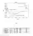
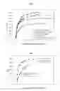

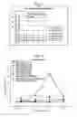
Sources:
- United States Patent and Trademark Office - verify current appl. status at the USPTO↗
Similar patent applications:
- » 20080112926
GAG binding proteins - » 20090005541
GAG binding protein - » 20110144305
GAG BINDING PROTEIN - » 20090023161
Antiviral screening method to identify inhibitors of phosphatidylinositol 4,5-bisphosphate [PI(4,5)P] binding to the HIV gag matrix (MA) protein
Recent applications in this class:
- » 20230303647 2023-09-28
CYCLIC CHEMERIN-9 DERIVATIVES - » 20230141043 2023-05-11
NOVEL IMMUNOMODULATOR - » 20220112257 2022-04-14
MUL TISPECIFIC PROCHEMOKINE THERAPEUTIC PROTEINS (PARK) AND METHOD OF MAKING AND USING THEREOF - » 20220064245 2022-03-03
FUSION PROTEINS COMPRISING A CYTOKINE AND SCAFFOLD PROTEIN - » 20190153055 2019-05-23
Fusion protein comprising CCL3 variant and use thereof - » 20140017790 2014-01-16
Control agent for controlling undifferentiated state and use thereof - » 20140005096 2014-01-02
METHODS OF TREATING INFLAMMATION - » 20130330305 2013-12-12
Cytokine derivatives - » 20130150303 2013-06-13
GLYCOSAMINOGLYCAN-ANTAGONISING MCP-1 MUTANTS AND METHODS OF USING SAME - » 20120082622 2012-04-05
CCL17
Recent applications for this Assignee:
- » 20140073557 2014-03-13
FGFRI-BASED ANTAGONISTS WITH IMPROVED GLYCOSAMINOGLYCAN AFFINITY AND METHODS OF USING SAME - » 20130150303 2013-06-13
GLYCOSAMINOGLYCAN-ANTAGONISING MCP-1 MUTANTS AND METHODS OF USING SAME - » 20120288474 2012-11-15
COMPOSITION FOR TREATMENT OF CXCL8-MEDIATED LUNG INFLAMMATION - » 20120046218 2012-02-23
GLYCOSAMINOGLYCAN-ANTAGONISING MCP-I MUTANTS AND METHODS OF USING SAME - » 20110144305 2011-06-16
GAG BINDING PROTEIN - » 20100298203 2010-11-25
SDF-I-BASED GLYCOSAMINOGLYCAN ANTAGONISTS AND METHODS OF USING SAME - » 20100197583 2010-08-05
Glycosaminoglycan-antagonising MCP-1 mutants - » 20100035294 2010-02-11
SYSTEM TO ASSESS THE BIOLOGICAL ACTIVITY OF CHEMOATTRACTANTS - » 20090005541 2009-01-01
GAG binding protein - » 20080112926 2008-05-15
GAG binding proteins