Somatic mutations in ATRX in brain cancer
US20140227271A1
2014-08-14
14/129,850
2012-06-28
✅ Patent granted
US 9,982,306 B2
2018-05-29
WO; PCT/US2012/044631; 20120628
WO; WO2013/003583; 20130103
Lei Yao
Fish & Richardson P.C.
2033-08-21
Abstract:
We determined the sequence of ATRX and DAXX in 447 cancers from various sites. We found mutations most commonly in pediatric glioblastoma multiformae (GBM) (11.1%), adult GBM (6.5%), oligodendrogliomas (7.7%) and medulloblastomas (1.5%); and showed that Alternative Lengthening of Telomeres (ALT), a telomerase-independent telomere maintenance mechanism found in cancers that have not activated telomerase, perfectly correlated with somatic mutations of either gene. In contrast, neuroblastomas, and adenocarcinomas of the ovary, breast, and pancreas were negative for mutations in ATRX and DAXX. Alterations in ATRX or DAXX define a specific molecular pathway that is closely associated with an alternative telomere maintenance function in human cancers.
Inventors:
- Bert VOGELSTEIN 225 🇺🇸 Baltimore, MD, United States
- Kenneth W. KINZLER 126 🇺🇸 Baltimore, MD, United States
- Ralph Hruban 22 🇺🇸 Baltimore, MD, United States
- Luis Diaz 23 🇺🇸 Ellicott City, MD, United States
- Nickolas Papadopoulos 97 🇺🇸 Towson, MD, United States
- Hai Yan 21 🇺🇸 Chapel Hill, NC, United States
- Darell Bigner 31 🇺🇸 Mebane, NC, United States
- Alan Meeker 2 🇺🇸 Pikesville, MD, United States
- Yuchen Jiao 4 🇺🇸 Baltimore, MD, United States
Assignee:
- DUKE UNIVERSITY 1,858 🇺🇸 Durham, NC, United States
- THE JOHNS HOPKINS UNIVERSITY 3,028 🇺🇸 Baltimore, MD, United States
Applicant:
Interested in similar patents?
Get notified when new applications in this technology area are published.
Classification:
C12Q1/6886 » CPC main
Measuring or testing processes involving enzymes, nucleic acids or microorganisms ; Compositions therefor; Processes of preparing such compositions involving nucleic acids; Nucleic acid products used in the analysis of nucleic acids, e.g. primers or probes for diseases caused by alterations of genetic material for cancer
C07K16/40 » CPC further
Immunoglobulins [IGs], e.g. monoclonal or polyclonal antibodies against enzymes
C12N15/1137 » CPC further
Mutation or genetic engineering; DNA or RNA concerning genetic engineering, vectors, e.g. plasmids, or their isolation, preparation or purification; Use of hosts therefor; Recombinant DNA-technology; DNA or RNA fragments; Modified forms thereof; Non-coding nucleic acids modulating the expression of genes, e.g. antisense oligonucleotides against enzymes
G01N33/57407 » CPC further
Investigating or analysing materials by specific methods not covered by groups -; Biological material, e.g. blood, urine ; Haemocytometers; Chemical analysis of biological material, e.g. blood, urine; Testing involving biospecific ligand binding methods; Immunological testing; Immunoassay; Biospecific binding assay; Materials therefor for cancer Specifically defined cancers
C12Q2600/118 » CPC further
Oligonucleotides characterized by their use Prognosis of disease development
C12Q2600/156 » CPC further
Oligonucleotides characterized by their use Polymorphic or mutational markers
G01N33/68 IPC
Investigating or analysing materials by specific methods not covered by groups -; Biological material, e.g. blood, urine ; Haemocytometers; Chemical analysis of biological material, e.g. blood, urine; Testing involving biospecific ligand binding methods; Immunological testing involving proteins, peptides or amino acids
C12Q1/68 IPC
Measuring or testing processes involving enzymes, nucleic acids or microorganisms ; Compositions therefor; Processes of preparing such compositions involving nucleic acids
G01N33/574 IPC
Investigating or analysing materials by specific methods not covered by groups -; Biological material, e.g. blood, urine ; Haemocytometers; Chemical analysis of biological material, e.g. blood, urine; Testing involving biospecific ligand binding methods; Immunological testing; Immunoassay; Biospecific binding assay; Materials therefor for cancer
C12N15/113 IPC
Mutation or genetic engineering; DNA or RNA concerning genetic engineering, vectors, e.g. plasmids, or their isolation, preparation or purification; Use of hosts therefor; Recombinant DNA-technology; DNA or RNA fragments; Modified forms thereof Non-coding nucleic acids modulating the expression of genes, e.g. antisense oligonucleotides
Description
This invention was made using funds from the U.S. governments. The U.S. retains certain rights to the invention under the terms of National institutes of Health grants CA121113, P50CA062924, P01CA134292, R01CA113669, RO1CA 43460 and CA57345, CA1403160, 5P50-N5020023-28 (SRC5R37), and CA011898-41.
TECHNICAL FIELD OF THE INVENTION
This invention is related to the area of cancer. In particular, it relates to brain cancers.
BACKGROUND OF THE INVENTION
Telomeric DNA functions to stabilize chromosomal ends and is progressively lost during cell division (the end replication problem), thus limiting cellular proliferative capacity.(1-3) The majority of cancers solve the end replication problem by expressing the telomere-synthesizing enzyme telomerase. A subset of the others utilizes a genetic recombination-based telomerase-independent telomere maintenance mechanism termed alternative lengthening of telomeres (ALT).(4-7) The prevalence of ALT varies widely, but is found more often in cancers of the central nervous system (CNS) and of mesenchymal tissues than in common epithelial tumors.(8, 9)
A recent study of pancreatic neuroendocrine tumors (PanNETs) revealed that 43% harbored inactivating mutations in the ATRX or DAXX genes.(10) Notably, these mutations were mutually exclusive, indicating that they functioned in the same pathway. This mutual exclusivity was intriguing, as independent studies had shown that the proteins encoded by ATRX and DAXX interact with one another.(11) The ATRX gene encodes a large protein possessing a C-terminal helicase/ATPase domain placing it in the SNF2 family of chromatin remodeling enzymes.(12) Inherited mutations in ATRX cause X-linked alpha thalassemia/mental retardation syndrome, characterized by multiple developmental abnormalities in affected males.(13, 14) DAXX is a nuclear protein that interacts with numerous SUMO-modified proteins and plays a role in transcriptional repression.(15) The ATRX and DAXX protein complex has been suggested to play multiple cellular roles, including functioning in chromatin remodeling.(11, 76) Notably, the ATRX/DAXX complex was recently found to be required for the incorporation of the histone variant H3.3 at telomeres.(17-19) This histone chaperone activity may play a role in establishing or maintaining telomere stability, at least in mouse embryonic stem cells.(20, 21) Reducing either ATRX or H3.3 levels in these cells decreased the amount of heterochromatic protein HPI-alpha at telomeres and increased markers of telomere dysfunction.(20, 21) Epigenetic changes in telomeric and subtelomeric chromatin have also been shown to affect telomere length, as well as recombination at telomeres.(22-24)
There is a continuing need in the art to identify markers for diagnosis, prognosis, stratifying, and targeting of brain tumors.
SUMMARY OF THE INVENTION
According to one aspect of the invention a method predicts outcome of a central nervous system (CNS) tumor in a patient. The CNS tumor is tested, or cells or nucleic acids shed from the tumor are tested, for the presence of an inactivating mutation in ATRX. The mutation is a positive prognostic indicator.
According to another aspect of the invention a method identifies a CNS tumor. The presence of an inactivating mutation in ATRX is tested for and identified in a tissue suspected of being a CNS tumor, or in cells or nucleic acids shed from the tumor. The presence of an inactivating mutation indicates a CNS tumor.
According to still another aspect, a method inhibits ATRX in a CNS tumor. An ATRX inhibitory agent is administered to the CNS tumor. The agent inhibits ATRX function or expression.
These and other embodiments which will be apparent to those of skill in the art upon reading the specification provide the art with methods for assessing, identifying, diagnosing, prognosticating, stratifying, and treating tumors of the central nervous system.
BRIEF DESCRIPTION OF THE DRAWINGS
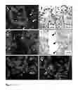
FIG. 1A-1G. Representative images of ALT-negative and ALT-positive tumors. (FIG. 1A) ALT-negative PanNET. Telomere FISH signals are markedly dimmer in PanNET cells (*) than in the surrounding stromal cells (arrowheads). Centromere-specific FISH probe serves as positive control for hybridization. (FIG. 1B and FIG. 1C) Immunolabeling of same PanNET as in A, shows nuclear positivity for ATRX and DAXX proteins, respectively. (FIG. 1D) Example of ALT-positive PanNET. Large, ultra-bright telomere FISH signals indicative of ALT are indicated (arrows). (FIG. 1E) Immunolabeling of same PanNET as in D, shows loss of nuclear DAXX protein in tumor cells. Benign endothelial cells (arrowheads) serve as positive staining controls. (FIG. 1F and FIG. 1G) Examples of ALT-positive GBM and medulloblastoma, respectively. Original magnification=400× for all images.
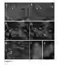
FIG. 2A-2G. Telomere-FISH and immunofluorescence co-staining in ALT-positive tumors. (FIG. 2A) ALT-positive PanNET telomere FISH and ATRX protein. (FIG. 2B) same image as in A, omitting telomere and DAPI channels, highlighting loss of nuclear ATRX. Benign stromal cells positive for nuclear ATRX protein are indicated by arrows. (FIG. 2C) ALT-positive PanNET co-stained with telomere FISH and DAXX protein. (FIG. 2D) same image as in FIG. 2C, omitting telomere and DAPI channels. Punctate nuclear DAXX staining in benign stromal cells is indicated by arrow heads. E, ALT-positive medulloblastoma stained for telomere FISH and PML protein. Arrows show co-localization of PML protein and ALT-associated telomere foci. F&G, high magnification images of telomere and PML protein co-staining showing typical targetoid appearance of ALT-associated PLM bodies (APB). Original magnification=400× for images FIG. 2A-FIG. 2AE, 1000× for images FIG. 2F and FIG. 2G.
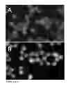
FIG. 3A-3B (S1). Telomere-FISH and immunofluorescence co-staining for ATRX protein. (FIG. 3A) ALT-positive osteosarcoma cell line U2-OS showing lack of nuclear ATRX protein. (FIG. 3B) ALT-negative PanNET cell line BON-1 showing ATRX nuclear-positivity, in both cases the nuclear DNA was counter stained with DAPI. Original magnification=400×.
DETAILED DESCRIPTION OF THE INVENTION
The inventors have found that ATRX mutations are frequently found in tumors of the central nervous system. Moreover, they are mutated together with IDH1 or IDH2 and TP53. ATRX mutations appear to be a later event in the progression of the brain tumors than the other mutations. Thus somatic mutations in ATRX can be used as a diagnostic, prognostic, or stratifying factor for such tumors.
Tumors of the CNS which may be assessed and treated include without limitation medulloblastoma, oligodendroglioma, pediatric glioblastoma multiforme, adult glioblastoma multiforme, oligoglioma, anaplastic oligodendroglioma, oligoastroglioma, anaplastic oligoastrocytoma, astrocytoma, anaplastic astrocytoma, ependymoma, anaplastic ependymoma, myxopapillary ependymoma, subependymoma, mixed glomas, polar spongioblastomas, astroblastoma, gliomatosis cerebri, medulloepithelioma, neuroblastoma, retinoblastoma, and ependymoblastoma. Glial tumors of any type may be assessed and treated.
Tests for ATRX mutations can be performed using protein based or nucleic based assays. Sequence determination of the nucleic acid can be used to identify mutations. Probes or primers, and kits and techniques employing both can be used. PCR or other specific or global amplification can be used. Mutations can be identified in any available genetic material including or example genomic DNA, cDNA, and RNA. Nucleic acids can be amplified, enriched, and/or purified prior to assessment. Protein based assays may involve specific antibodies and/or ATRX binding partner DAXX. The antibodies may be polyclonal or monoclonal, fragments (Fab, Fab′), single chain constructs (scFv), etc. Nucleic acid based assays include without limitation, hybridization to probes, amplification using specific primers, primer extension, ligation assay, etc. Any of these techniques can also be combined. Assays can be performed together with tests for other gene mutations or alterations of the genome. Results can be integrated and used to accurately and comprehensively characterize and/or identify a tumor or the patient.
Results of assays can be recorded in a written medium, an electronic medium, or transmitted orally or electronically to a health care provider, a patient, a family member, a hospital, etc. Testing requires physical steps, and typically involves chemical changes to occur to a test sample. Typically the test sample is a sample that is removed from the patient body, so that the test is performed outside of a patient body.
Samples which may be tested include without limitation brain tissue, tumor tissue, CNS fluid, neuronal tissue, blood, urine, saliva, tears, sputum, etc. These samples may be collected and processed and/or stored prior to testing. The samples may be frozen or fixed. They may be archival or freshly collected. Typically the tissue or body fluid will be isolated from the body and the assay will be performed ex vivo on the isolated sample.
ATRX inhibitory agents as used in this specification inhibit either ATRX function or expression. Such agents may be an antibody, an antibody fragment, or a single chain antibody construct. Alternatively it can be an inhibitory RNA or other inhibitory nucleic acid molecule, including but not limited to antisense oligonucleotides, antisense expression constructs, siRNA, and RNAi.
Any type of mutation may be identified. Inactivating mutations include without limitation R2079X, Q1874X, Q1788H, E2277K, Q2156H, K455X, W263X, R2153C, and R1803H. The mutation may be, for example, a frameshift mutation, a splice-site mutation, an indel (insertion or deletion) mutation, a large genomic rearrangement, or a missense mutation. Typically an indel may involve a small portion of a gene, such as 1-10 nt. A large rearrangement may involve large portions or all of a gene, such as greater than 10%, greater than 25%, greater than 50%, greater than 75% or greater than 100% of a gene. Particular mutations which may be identified include g.chrX:76778161—76778162insA; g.chrX:76824745—76824748delTCTC; g.chrX:76741670—76741673delCTAT; g.chrX:76798738—76798741delACTA; g.chrX:76665385C>A; g.chrX:76825188—76825194delTTGAGGA; g.chrX:76831065delG(hom); g.chrX:76806828—76806829insT; g.chrX:76825743—76825744delTG; g.chrX:76798774—76798775delAG(hom); g.chrX:76826615C>T(hom); g.chrX:76700843G>A(hom); and g.chrX:76760970C>T.
Stratification of patients can be used to assign a treatment regimen. It may be used in prospective or retrospective clinical studies. It can be used to assign a prognosis. Stratification typically assigns a patient to a group based on a shared mutation pattern or other observed characteristic or set of characteristics.
The above disclosure generally describes the present invention. All references disclosed herein are expressly incorporated by reference. A more complete understanding can be obtained by reference to the following specific examples which are provided herein for purposes of illustration only, and are not intended to limit the scope of the invention.
EXAMPLE 1
ATRX and DAXX Gene Mutations Correlate with ALT-Positivity
Given the potential role of ATRX and DAXX in modulating telomeric chromatin, we evaluated telomere status in pancreatic neuroendocrine tumors (PanNETs) with known ATRX and DAXX mutational status. Telomere-specific fluorescence in situ hybridization (FISH) was used to directly assess the telomeres in PanNETs. Neoplasms with ALT are readily distinguishable by large ultra-bright telomere FISH signals—a nearly universal feature of ALT-positive cell populations.(25) Although telomere FISH signals from these individual bright foci have often been shown to co-localize with PML protein, this localization is not as reliable as the strength of the FISH signals and was not used for classification in our study.(26-28)
Twenty-five of the 41 PanNETs (61%) examined by telomere FISH displayed evidence of ALT (Table 1, FIG. 1). Importantly, ALT was not observed in any of the surrounding non-neoplastic cells, including stromal fibroblasts, pancreatic acini, pancreatic ducts and islets of Langerhans (FIGS. 1&2).(29) ATRX and DAXX gene mutations both were significantly correlated with ALT-positivity (p<0.008 for either gene). In particular, all 21 (100%) PanNETs with ATRX or DAX gene mutations were ALT-positive by telomere FISH (Table 1).
| TABLE 1 |
| PanNET cases grouped according to mutation status |
| for ATRX and DAXX and displaying ALT-status |
| and impaunohistochemistry of ATRX and DAXX |
| ATRX | DAXX | |||
| Gene Status | Case | ALT-status* | IHC† | IHC† |
| ATRX Point mutations | PanNET5 | Pos | Neg | Pos |
| & indels | PanNET13 | Pos | Neg | Pos |
| PanNET27 | Pos | Neg | Pos | |
| PanNET35 | Pos | Neg | Pos | |
| PanNET52 | Pos | Neg | Pos | |
| PanNFT59 | Pos | Pos | Pos | |
| PanNET61 | Pos | Neg | Pos | |
| PanNET78 | Pos | Pos | Pos | |
| PanNET85 | Pos | Neg | Pos | |
| PanNET112‡ | Pos | Neg | Pos | |
| DAXX Point | PanNET25 | Pos | Pos | Het |
| mutations | PanNET31 | Pos | Pos | Het |
| & indels | PanNET44 | Pos | Pos | Neg |
| PanNET56 | Pos | Pos | Neg | |
| PanNET77 | Pos | Pos | Neg | |
| PanNET80 | Pos | Pos | Neg | |
| PanNET84 | Pos | Pos | Het | |
| PanNET87 | Pos | Pos | Het | |
| PanNET93 | Pos | Pos | Neg | |
| PanNET104 | Pos | Pos | Neg | |
| PanNET133 | Pos | Pos | Neg | |
| ATRX/DAXX WT for | PanNET6 | Neg | Pos | Pos |
| Point mutations & | PanNET10 | Neg | Pos | Pos |
| indels | PanNET21 | Neg | Pos | Pos |
| PanNET24 | Neg | Pos | Pos | |
| PanNET29 | Pos | Pos | Het | |
| PanNET36 | Neg | Pos | Pos | |
| PanNET39 | Pos | Neg | Pos | |
| PanNET45 | Pos | Pos | Neg | |
| PanNET57 | Neg | Pos | Pos | |
| PanNET63 | Neg | Pos | Pos | |
| PanNET64 | Pos | Het | Pos | |
| PanNET66 | Neg | Pos | Pos | |
| PanNET69 | Neg | Pos | Pos | |
| PanNET79 | Neg | Pos | Pos | |
| PanNET83 | Neg | Pos | Pos | |
| PanNET91 | Neg | Pos | Pos | |
| PanNET121 | Neg | Pos | Pos | |
| PanNET126 | Neg | Pos | Pos | |
| PartNET128 | Neg | Pos | Pos | |
| PanNET129 | Neg | Pos | Pos | |
| *The intensity of telomere FISH signals was assessed to be either negative or positive for the ALT phenotype. | ||||
| †Immunohistochemistry was scored as uniformly positive, negative or heterogeneous for nuclear labeling. | ||||
| ‡Multifocal tumor, featuring ALT and negative nuclear immunolabeling in the majority of the tumor. | ||||
| ALT, alternative lengthening of telomeres; | ||||
| Het, heterogeneous; | ||||
| IHC, immunohistochemistry; | ||||
| Neg, negative; | ||||
| PanNET, pancreatic neuroendocrine tumor; | ||||
| Pos, positive; | ||||
| WT, wild type. |
Four of the 20 cases without detectable mutations in ATRX or DAXX were ALT-positive. To determine whether ATRX and DAXX expression were also normal in these tumors, serial sections of the same tumors were immunolabeled with antibodies against the ATRX and DAXX proteins. Each of the four tumors demonstrated loss of nuclear expression of either ATRX (2 cases) or DAXX (2 cases) in either the entire tumor, or confluent parts of the tumor (FIGS. 1 and 2). Of note, the nuclear expression of the corresponding wild type partner (either DAXX or ATRX, respectively) was retained in each of these four cases. Though the immunohistochemical results on tumors with ATRX or DAXX mutations were expected, they provide excellent controls for the specificities of the antibodies used for immunolabeling. Notably, each of the 16 tumors without ALT showed robust nuclear labeling for both ATRX and DAXX (Table 1). The relationship between ALT-positivity and abnormal immunolabeling for either the ATRX or DAXX proteins was statistically significant (p=0.012 and p=0.003, respectively).
In sum, there was a perfect correlation between the absence of nuclear ATRX or DAXX expression and the ALT phenotype and >80% of the 23 tumors without ATRX or DAXX expression could be accounted for by point mutations and small indels, i.e., inactivating mutations of the ATRX or DAXX genes.
EXAMPLE 2
ATRX Mutations in Cancers of the CNS
To ascertain whether ATRX and DAXX gene mutations might be more generally associated with the ALT-phenotype, we examined 447 tumors of other types. We identified a low to moderate frequency of ATRX mutations in cancers of the CNS. Specifically, mutations were detected in 2 of 18 (11.1%) pediatric GBM, 8 of 123 (6.5%) adult GBM, 1 of 13 (7.7%) oligodendrogliomas, and 1 of 65 (1.5%) medulloblastomas. No mutations in either ATRX or DAXX were identified in 11 adult neuroblastomas, or in 25 ovarian adenocarcinomas, 96 breast adenocarcinomas and 96 pancreatic adenocarcinomas. (Table 2).
Further, ATRX mutations were found in anaplastic astrocytomas (41%), astrocytomas (29%), anaplastic oligoastrocytomas (20%), oliogoastrocytomas (33%), anaplastic oligogliomas (7%), and oligogliornas (14%). These mutations were highly associated with mutations in IDH1/2 and TP53.
EXAMPLE 3
To determine if the ALT status of these additional tumor types also correlated with the presence of somatic ATRX mutations, we performed telomere FISH as described above on 8 ATRX mutant cases in which tumor material was available. In each of the eight cases, extremely bright telomeric foci indicating ALT were identified in the neoplastic cells but not in the non-neoplastic cells surrounding them (examples in FIGS. 1 and 2). Immunolabeling with antibodies to DAXX or ATRX showed that these tumors had lost nuclear expression of ATRX. As controls, we studied 23 ATRX and DAXX-wild type tumors of the same types with the identical techniques. None of the tumors without ATRX or DAXX mutations demonstrated the ALT phenotype by telomere FISH and all 23 retained robust nuclear labeling for both ATRX and DAXX.
EXAMPLE 4
ATRX Mutated in Established Cancer Cell Line
For future mechanistic studies, we considered it important to determine whether a human cancer cell line with a mutation ATRX or DAXX could be identified. We chose to study U-2 OS, derived from an osteosarcoma, as this line was a prototype for delineating the ALT phenotype.(7) We found that exons 2 to 19 of ATRX were homozygously deleted in these cells, unequivocally inactivating the gene product, causing a lack of ATRX immunolabeling in these cells (Suppl. FIG. S1).
EXAMPLE 5
Discussion
The results described above show a remarkable correlation between inactivation of ATRX or DAXX and the ALT phenotype in unrelated tumor types. Though we cannot infer from these data that mutations in ATRX or DAXX actually cause this phenotype, prior studies provide a mechanism through which this might occur. It has been proposed that ATRX-DAXX function in replication-independent heterochromatin assembly at specific repetitive G-rich regions, such as the telomeres.(16, 18, 19) In patients with the ATRX syndrome, mutations in the ATRX gene lead to changes in the DNA methylation status at subtelomeric regions which, as described below, may impact telomere stability.(30) Furthermore, experimentally decreasing ATRX or H3.3 in mouse ES cells results in telomere destabilization and an up-regulation of telomere repeat-containing RNA (TERRA).(19-21, 31)
Current evidence indicates that the ALT telomere maintenance mechanism is dependent upon homologous recombination (HR).(8, 32) Telomeres are composed of long tracts of highly repetitive DNA terminating, in single stranded 3′ ends, which should provide an ideal substrate for HR; thus, it has been proposed that HR at telomeres is actively suppressed in normal cells.(29) In agreement with this, somatic cell telomeric and proximal subtelomeric DNA feature tightly packed nucleosomes organized in a repressive heterochromatic state enriched for repressive epigenetic chromatin marks, such as DNA methylation and trimethylation of lysine 9 on histone H3 and of lysine 20 on histone H4.(22-24, 33) Mice engineered to contain reduced levels of such repressive epigenetic marks possess abnormally long and heterogeneous telomeres, as well as increased levels of inter-chromatid telomere recombination all of which are hallmarks of the ALT phenotype.(22, 23) Our results are consistent with a model in which loss of ATRX-DAXX function impairs the heterochromatic state of the telomeres, perhaps due to reduced levels of H3.3 incorporation, leading to telomere destabilization and increased HR at the telomeres; thus, facilitating the development of ALT.
EXAMPLE 6
Methods
Tissue Samples and Mutational Analysis
In order to assess the potential consequences of ATRX and DAXX gene mutations on telomeres, 41 sporadic, nonfunctional PanNETs were chosen from a series of PanNETs whose ATRX and DAXX gene mutational status was previously determined by exomic sequencing.(1) Of the 41 PanNETs examined, ten had ATRX gene mutations, eleven had DAXX gene mutations, and the remaining 20 had wild type ATRX and DAXX genes. Details on the specific gene mutations as well as clinicopathologic variables for this PanNET series are presented in Supplementary Table S2. Clinical information on the patients evaluated in this study was obtained from the Johns Hopkins Hospital in the context of approved IRB protocols. The ATRX and DAXX genes were also sequenced in 65 cases of medulloblastoma, 13 cases of oligodendroglioma, 141 cases of glioblastoma multiforma (18 pediatric GBM and 123 adult GBM), 11 neuroblastomas, 25 ovarian carcinomas, 96 breast carcinomas and 96 pancreatic ductal adenocarcinomas.
Immunohistochemistry
Immunolabeling for the ATRX and DAXX proteins was performed on formalin-fixed, paraffin embedded sections as previously described.(1) Briefly, heat-induced antigen retrieval was performed in a steamer using citrate buffer (catalog #H-3300, Vector Laboratories) for 30 minutes. Endogenous peroxidase was blocked (catalog #S2003, Dako) and serial sections were then incubated with primary antibody; anti-ATRX (1:400 dilution; catalog #HPA001906, Sigma-Aldrich, lot R00473) or anti-DAXX (1:150 dilution; catalog #HPA008736, Sigma-Aldrich, lot A39105) for 1 hour at room temperature. The primary antibodies were detected by 30 minute incubation with HRP-labeled secondary antibody (catalog #PV6119, Leica Microsystems) followed by detection with 3,3′-Diaminobenzidine (Sigma-Aldrich), counterstaining with Harris hematoxylin, rehydration and mounting. Only nuclear labeling of either protein was evaluated. The immunolabeled PanNET slides were assessed and scored by 2 authors (R.H. and A.M.); the immunolabeled CNS tumor slides were assessed by F.R. Internal controls included islets of Langerhans in PanNETs and endothelial cells (including within intra-tumoral vessels) which demonstrated strong nuclear immunolabeling for both ATRX and DAXX.
Telomere-Specific FISH and Microscopy
Combined telomere-specific FISH and immunofluorescence labeling for ATRX, DAXX and PML was conducted as previously described.(2, 3) Briefly, deparaffinized slides were hydrated, steamed for 20 minutes in citrate buffer (catalog #H-3300; Vector Laboratories), dehydrated and hybridized with a Cy3-labeled peptide nucleic acid (PNA) probe complementary to the mammalian telomere repeat sequence ([N-terminus to C-terminus] CCCTAACCCTAACCCTAA; SEQ ID NO: 1). As a positive control for hybridization efficiency, a FITC-labeled PNA probe having specificity for human centromeric DNA repeats (ATTCGTTGGAAACGGGA(SEQ ID NO: 2); CENP-B binding sequence) was also included in the hybridization solution.(4) Following post-hybridization washes, the desired primary antibody was applied (anti-ATRX, as described above; anti-DAXX, as described above; anti-PML antibody, 45 minute incubation at 1:100 dilution; catalog #PG-M3, DAKO), followed by application of species-appropriate Alexa 488 fluorescent secondary antibody (Molecular Probes Cat #A-11034 and A-11001) and nuclear counterstaining with DAPI. Slides were imaged with a Nikon 50i epifluorescence microscope equipped with X-Cite series 120 illuminator (DUO Photonics Solutions Inc., Ontario, CA) and appropriate fluorescence excitation/emission filters. Grayscale images were captured for using Nikon NIS-Elements software and an attached Photometrics CoolsnapEZ digital camera, pseudo-colored and merged. Quantification from the digital images was conducted using Telometer, a custom software plugin created for the open source image analysis program imageJ, freely available for download (http://bui2.win.ad.jhu.edu/telometer/).
The FISH and immunolabeled slides were assessed and scored independently by 2 authors (C. H. and A. K. M.). The gold standard for determining the presence of ALT is the demonstration of telomere length maintenance over time in a continually growing cell population lacking any evidence of telomerase activity.(5) This standard, however, cannot be applied to fixed clinical specimens. Previously, it was shown that subsets of cells in ALT-positive cell lines harbor unusually large promyelocytic leukemia (PML) nuclear bodies which contain large amounts of telomeric DNA (ALT-associated PML bodies; APBs).(6) APBs are not observed in normal cells, nor are they observed in ALT-negative cancer cells, and thus APBs serve as specific biomarkers of ALT. APBs can be readily visualized in fixed tissues by combining telomere-specific FISH with fluorescent immunostaining for PML protein.(3) This method has been extensively validated and allows for the straightforward identification of ALT-positive cancers in fixed human tissue specimens.(7) APB-associated telomeric DNA aggregates are unique to ALT-positive cell populations and are significantly larger and brighter than the FISH signals emanating from individual telomeres in the same cell population.
In our study, PanNETs were classified as ALT-positive if they met the following criteria: (1) the presence of ultra-bright, intra-nuclear foci of telomere FISH signals, with integrated total signal intensities for individual foci being >10 fold that of the per cell mean integrated signal intensities for all telomeric signals in individual benign stromal cells within the same case (Quantitative data on representative individual telomere aggregates in 10 randomly chosen PanNET cases is presented in Supplementary Table S3); (2) ≧1% of neoplastic cells displaying ALT-associated telomeric DNA foci. Tumor samples lacking ALT-associated telomeric foci in which at least 5000 cells were assessed were considered ALT-negative. In all cases, areas exhibiting necrosis were excluded from consideration.
REFERENCES FOR EXAMPLE 6 ONLY
The contents of each are expressly incorporated herein.
- 1. Y. Jiao et al., Science (January 20).
- 2. A. K. Meeker et al., Am J Pathol 160, 1259 (April 2002).
- 3. E. Montgomery, P. Argani, J. L. Hicks, A. M. DeMarzo, A. K. Meeker, Am J Pathol 164, 1523 (May 2004).
- 4. C. Chen, Y. K. Hong, S. D. Ontiveros, M. Egholm, W. M. Strauss, Mamm Genome 10, 13 (January 1999).
- 5. J. D. Henson, R. R. Reddel, FEBS Lett 584, 3800 (September 10).
- 6. T. R. Yeager et al., Cancer Res 59, 4175 (Sep. 1, 1999).
- 7. J. D. Henson et al., Clin Cancer Res 11, 217 (Jan. 1, 2005).
REFERENCES (FOR ALL BUT EXAMPLE 6)
The disclosure of each reference cited is expressly incorporated herein.
- 1. E. H. Blackburn, Nature 350, 569 (Apr. 18, 1991).
- 2. M. Z. Levy, R. C. Allsopp, A. B. Futcher, C. W. Greider, C. B. Harley, J Mol Biol 225, 951 (Jun. 20, 1992).
- 3. A. M. Olovnikov, J Theor Biol 41, 181 (Sep. 14, 1973).
- 4. J. W. Shay, S. Bacchetti, Eur J Cancer 33, 787 (April 1997).
- 5. A. K. Meeker, D. S. Coffey, Biochemistry (Mosc) 62, 1323 (November 1997).
- 6. R. R. Reddel, T. M. Bryan, L. M. Colgin, K. T. Perrem, T. R. Yeager, Radiat Res 155, 194 (January 2001).
- 7. T. M. Bryan, A. Englezou, L. Dalla-Pozza, M. A. Dunham, R. R. Reddel, Nat Med 3, 1271 (November 1997).
- 8. J. D. Henson, R. R. Reddel, FEBS Lett 584, 3800 (September 10),
- 9. C. M. H. A. P. Subhawong, S. M Hong, M. Goggins, E. A. Montgomery, E. Gabrielson, G. J. Netto, W. H. Westra, P. Argani, C. A. Iacobuzio-Donahue, I-M Shih, M. Torbenson, and A. K. Meeker, in 100th Annual Meeting of the United States and Canadian Academy of Pathology. (Nature Publishing Group, Modern Pathology, Volume 24, Supplement 1, February 2011., San Antonio, Tex. U.S.A., 2011).
- 10. Y. Jiao et al., Science (January 20).
- 11. Y. Xue et al., Proc Natl Acad Sci USA 100, 10635 (Sep. 16, 2003).
- 12. R. J. Gibbons, D. J. Pickens, L. Villard, D. R. Higgs, Cell 80, 837 (Mar. 24, 1995).
- 13. R. Gibbons, Orphanet J Rare Dis 1, 15 (2006).
- 14. R. J. Gibbons et al., Hum Mutat 29, 796 (June 2008).
- 15. H. M. Shih, C. C. Chang, H. Y. Kuo, D. Y. Lin, Biochem Soc Trans 35, 1397 (December 2007).
- 16. S. J. Elsaesser, C. D. Allis, Cold Spring Barb Symp Quant Biol (December 7).
- 17. P. W. Lewis, S. J. Elsaesser, K. M. Noh, S. C. Stadler, C. D. Allis, Proc Natl Acad Sci USA 107, 14075 (August 10).
- 18. M. J. Law et al., Cell 143, 367 (October 29).
- 19. A. D. Goldberg et al., Cell 140, 678 (March 5).
- 20. L. H. Wong et al., Genome Res 20, 351 (March).
- 21. L. H. Wong et al., Genome Res 19, 404 (March 2009).
- 22. M. Garcia-Cao, R. O'Sullivan, A. H. Peters, T. Jenuwein, M. A. Blasco, Nat Genet 36, 94 (January 2004).
- 23. S. Gonzalo et al., Nat Cell Biol. 8, 416 (April 2006).
- 24. G. Grafi et al., Dev Biol 306, 838 (Jun. 15, 2007).
- 25. T. R. Yeager et al., Cancer Res 59, 4175 (Sep. 1, 1999).
- 26. M. A. Cerone, C. Autexier, J. A. Londono-Vallejo, S. Bacchetti, Oncogene 24, 7893 (Nov. 24, 2005).
- 27. C. L. Fasching, K. Bower, R. R. Reddel, Cancer Res 65, 2722 (Apr. 1, 2005).
- 28. R. A. Marciniak et al., Cancer Res 65, 2730 (Apr. 1, 2005).
- 29. A. J. Cesare, R. R. Reddel, Nat Rev Genet 11, 319 (May).
- 30. R. J. Gibbons et al., Nat Genet 24, 368 (April 2000).
- 31. C. M. Azzalin, P. Reichenbach, L. Khoriauli, E. Giulotto, J. Lingner, Science 318, 798 (Nov. 2, 2007).
- 32. A. Muntoni, A. A. Neumann, M. Hills, R. R. Reddel, Hum Mol Genet 18, 1017 (Mar. 15, 2009).
- 33. V. L. Makarov, S. Lejnine, J. Bedoyan, J. P. Langtnore, Cell 73, 775 (May 21, 1993).
Claims
1. A method for predicting outcome of a Central Nervous System (CNS) tumor in a patient, comprising:
testing the CNS tumor, or cells or nucleic acids shed from the tumor, for the presence of an inactivating mutation in ATRX, wherein the mutation is a positive prognostic indicator.
2. The method of claim 1 wherein the step of testing comprises an immunohistochemical analysis.
3. The method of claim 1 wherein the step of testing comprises a nucleic acid analysis.
4. A method of identifying a CNS tumor, comprising:
testing for and identifying presence of an inactivating mutation in ATRX in a tissue suspected of being a CNS tumor, or in cells or nucleic acids shed from the tumor, wherein the presence of an inactivating mutation indicates a CNS tumor.
5. A method for classifying a tumor of the Central Nervous System (CNS) of a patient comprising:
testing the CNS tumor, or cells or nucleic acids shed from the tumor, for the presence of an inactivating mutation in ATRX;
assigning the CNS tumor to a class based on the presence of the mutation or the identity of the mutation.
6. The method of claim 5 wherein the patient is assigned to a clinical trial group according to the class.
7. The method of claim 5 wherein a treatment is prescribed based on the class.
8. A method of inhibiting ATRX in a CNS tumor, comprising:
administering an ATRX inhibitory agent to the CNS tumor, wherein the agent inhibits ATRX function or expression.
9. The method of claim 8 wherein the inhibitory agent is an antibody, an antibody fragment, or a single chain antibody construct.
10. The method of claim 8 wherein the inhibitory agent is an inhibitory RNA molecule,
11. The method of claim 1, 4, 5, or 8 wherein the CNS tumor is a glial tumor.
12. The method of claim 1, 4, 5, or 8 wherein the CNS tumor is selected from the group consisting of: pediatric glioblastoma, adult glioblastoma, oligodendroglioma, medulloblastoma, anaplastic oligodendroglioma, oligoastrocytoma, anaplastic oligoastrocytoma, astrocytoma, and anaplastic astrocytoma.
13. The method of claim 1, 2, 4, or 5 wherein the mutation is selected from the group consisting of R2079X, Q1874X, Q1788H, E2277K, Q2156H, K455X, W263X, R2153C, and R1803H.
14. The method of claim 1, 4, or 5 wherein the mutation is a frameshift mutation.
15. The method of claim 1, 4, or 5 wherein the mutation is selected from the group consisting of a splice-site mutation or an indel mutation.
16. The method of claim 1, 4, or 5 wherein the mutation is a missense mutation.
17. The method of claim 1, 4, or 5 wherein the mutation is selected from the group consisting of: g.chrX:76778161—76778162insA; g.chrX:76824745—76824748delTCTC; g.chrX:76741670—76741673delCTAT; g.chrX:76798738—76798741delACTA; g.chrX:76665385C>A; g.chrX:76825188—76825194delTTGAGGA; g.chrX:76831065delG(hom); g.chrX:76806828—76806829insT; g.chrX:76825743—76825744delTG; g.chrX:76798774—76798775delAG(hom); g.chrX:76826615C>T(hom); g.chrX:76700843G>A(hom); and g.chrX:76760970C>T.
18. The method of claim 1, 4, or 5 wherein the mutation is a large genomic rearrangement.
Images & Drawings included:
Sources:
- United States Patent and Trademark Office - verify current appl. status at the USPTO↗
Similar patent applications:
- » 20180334726
Somatic mutations in ATRX in brain cancer
Recent applications in this class:
- » 20250290156 2025-09-18
EVALUATING OVARIAN CANCER CHEMOTHERAPY RESPONSE USING GENE EXPRESSION DATA AND MACHINE LEARNING - » 20250290155 2025-09-18
CLINICAL WORKFLOW FOR TREATMENT OF BREAST CANCER - » 20250290154 2025-09-18
PREDICTIVE MARKERS FOR IMMUNOTHERAPY - » 20250290153 2025-09-18
METHODS OF DIAGNOSING, DETERMINING THE PROGRESSION OF, AND TREATING A PROSTATE CANCER - » 20250290152 2025-09-18
GENETIC TRIO OF BRAF AND TERT MUTATIONS AND RS2853669TT IN PAPILLARY THYROID CANCER AGGRESSIVENESS - » 20250290151 2025-09-18
Differentially-Methylated Regions of the Genome Useful as Markers of Embryo-Adult Transitions - » 20250290150 2025-09-18
METHODS AND SYSTEMS FOR CHARACTERIZATION, DIAGNOSIS, AND TREATMENT OF CANCER - » 20250290149 2025-09-18
SYSTEMS AND METHODS FOR ENRICHING CELL-FREE MICROBIAL NUCLEIC ACID MOLECULES - » 20250290148 2025-09-18
DIFFERENTIAL ALTERNATIVE SPLICING IN RELAPSED AND REFRACTORY DIFFUSE LARGE-B CELL LYMPHOMA PATIENTS RECEIVING CAR-T THERAPY - » 20250290147 2025-09-18
PROSTATE CANCER MARKERS
Recent applications for this Assignee:
- » 20250281762 2025-09-11
MRI-COMPATIBLE CARDIAC DEFRIBRILLATOR - » 20250277078 2025-09-04
TRI-ARM STAR BOTTLEBRUSH POLYMER WITH DEFINED VISCOSITY AND OPTICAL PROPERTIES FOR USE IN A NOVEL INTRAOCULAR LENS - » 20250268937 2025-08-28
ENGINEERED IMMUNE CELLS TO TARGET SARS-COV2 - » 20250255823 2025-08-14
COMPOSITION AND METHOD FOR TREATMENT OF NEUROPSYCHIATRIC DISORDERS - » 20250255470 2025-08-14
FLUOROPHORE IMAGING DEVICES, SYSTEMS, AND METHODS FOR AN ENDOSCOPIC PROCEDURE - » 20250235109 2025-07-24
NEUROMODULATION BASED NERVE IDENTIFICATION - » 20250224350 2025-07-10
Method for Capture of Small-Angle Scatter Over Wide Fields of View - » 20250205784 2025-06-26
Real Time Control Of Laser Additive Manufacturing With High Speed Optically Calibrated On Axis Sensing - » 20250195088 2025-06-19
AUTOMATED CRANIAL BURR HOLE DEVICE AND METHOD - » 20250191780 2025-06-12
ANOMALY DETECTION FOR IDENTIFYING EXPOSURE EVENTS FROM BASELINE MOLECULAR MEASUREMENTS IN HUMAN HEALTH